1
Vol.59: e16160280, January-December 2016 http://dx.doi.org/10.1590/1678-4324-2016160280
ISSN 1678-4324 Online Edition
BRAZILIAN ARCHIVES OF BIOLOGY AND TECHNOLOGY
A N I N T E R N A T I O N A L J O U R N A L
Identification and characterization of Fusarium mangiferae
as pathogen of mango malformation in India
Pradeep Kumar
1*;
Madhu Kamle
2; Asok Kumar Misra
3; Anthonia O’Donovan
4; Marcela
Pagano
5; Dinesh Raj Modi
6.
1Yeungnam University, School of Biotechnology, Gyeongsan, Korea; 2Ben-Gurion University of the Negev, Dryland
Agriculture and Biotechnology, Beer-Sheva, Israel; 3Central Institute for Subtropical Horticulture Lucknow, Uttar
Pradesh, India; 4National University of Ireland, Discipline of Biochemistry, Galway, Ireland; 5Lunds Universitet,
Department of Biology, Lund, Sweden; 6Babasaheb Bhimrao Ambedkar University, Department of Biotechnology,
Lucknow, Uttar Pradesh, India;
ABSTRACT
Fusarium mangiferae (=F. subglutinans) isolates collect from malformed samples from major mango-growing area of North India. Molecular identification and characterization of eleven most virulent isolates of F. mangiferae, based on pathogenicity tests used for the present study. Species-specific, genus specific ITS-PCR and PCR-RFLP performed for the accurate and easy detection of F. mangiferae. The rDNA-ITS 28S region sequences used for phylogenetic analysis of Fusarium isolates from India and other countries for homology search between them. The phylogenetic tree divided the isolates into three clades (i.e., American, Asian and African) and showed the high level of sequence based similarity (69-99%) among all Fusarium sequences from Asia. Thus, claimed Fusarium mangiferae as dominant pathogen of mango malformation. Furthermore, we conclude that exploiting the nested PCR coupled with PCR-RFLP will help in rapid and accurate detection of F. mangiferae pathogen of mango malformation
.
Key words: Fusarium mangiferae, detection, phylogenetic analysis, vegetative malformation, floral malformation.
1Authors for correspondence: [email protected]
INTRODUCTION
The Mango ‘King of Fruits’ is the most delicious fruit commonly grown in Southern Asia. Mango malformation emerge as most threatening constraint that limits Mango production and cause severe economic losses (10-90%) to stakeholders of tropical and subtropical countries [1-2]. The most
visible symptoms of mango malformation were malformed flower and bunchy appearance of vegetative tissues. Panicles affected become small, do not bear fruits and aborted prematurely. The variability of malformation pathogen influenced by environmental conditions and agro-climatic regions. Disease spread severely in new areas by malformation of mature trees and infected seedlings
[3]. In spite of several decades of never-ending
research, the etiology of the disease becomes a contentious issue and no effective control measure yet known [4]. However, various fungicides assessed
for their ability to control the pathogen under laboratory, greenhouse and field conditions [5], but
still unable to cure completely in the field conditions. Several biotic and abiotic factors reported to affect mango malformation disease. Fungi is the most counted factor reported [6].
Summanwar [7] first reported the malformation
caused by F. moniliforme Shield and proved its pathogenicity in India. F. mangiferae Britz, Wingfield & Marasas is universal spread pathogen of mango and isolated from Egypt, Florida (USA) and Israel. F. mangiferae (earlier reported=F. subglutinans) isolated from floral and vegetative malformed tissues [8] as evidenced by cultural and
morphological data. Histopathological studies of floral and vegetative tissues confirmed the association of F. mangiferae [9]. According to recent
classification, F. subglutinans now registered as F. mangiferae causing mango malformation disease [6, 10-13].
Disease management strategies required for detection and identification of damaging pathogens but also understand the pathogen threshold density, interaction between the pathogens and environments [14]. In associate with continuous
monitoring and basic pathological techniques that are tedious, it is complementary to perform molecular characterization at genus and species level for significant management of disease incidence. Internal transcribed spacer (ITS) regions are highly conserved within fungal species (with intra-specific similarities >99%) but are variable between species and accepted by fungal
taxonomists [15-16]. The rDNA ITS sequences have
been successfully used in resolving species-level and phylogenetic relationships in Fusarium [17-18].
The ITS along with 5.8S rDNA region amplified using ITS primer-pairs [19] which discern genetic
variation in the population studied. Earlier, molecular characterization confirmed the association of F. mangiferae with mango malformation [20-21]. In this study, we developed
species-specific and β-tubulin gene specific markers for identification of F.mangiferae isolates from mango malformed samples and their sequence based phylogenetic analysis to understand the diversity relationship among the Indian isolates compared with other countries geographical location.
MATERIAL AND METHODS
Isolation of Pathogen and Pathogenicity Test The vegetative and floral malformed samples collected from different orchards of mango (Table 1), cultured on Potato Dextrose Agar (PDA, HiMedia)and incubate at 28 ± 2ºC for 6 days. Cultural and microscopic studies confirmed pathogens as F. mangiferae [22]. Pure culture using
single spore and hyphal tip isolation techniques by
following Koch’s postulates [23]. Pathogenicity tests
for all isolates performed on the mango seedlings of Dashehari cultivar (sensitive to disease) under field conditions as well as in greenhouse conditions with controlled humidity at 85% and temperature 10-25 ºC. Pure cultures from each representative isolate used for inoculation in November to the host tissue covered with sterile moist cotton. Prior to inoculation, buds treated with 20% hydrogen peroxide (H2O2) which detoxified mangiferin (the
host defense anti Fusarium compound) as described by Chand and Chakrabarti [24].
DNA Isolation
Fresh mycelium from all 11 representative isolates collected for genomic DNA extraction according to the protocol of Abd-Elsalam et al [25]. 100 mg
Molecular Characterization of Fusarium Mangiferae 3
Centrifuge samples at 14,000 rpm for 20 min at 4ºC and collect DNA pellet, washed pellet twice with 70% ethanol to remove excess salt. Finally, the isolated DNA dissolved in a minimum quantity of Tris-EDTA (10 mM) buffer and stored at -20ºC until use. Quantification and purity of DNA recorded using standard spectroscopic methods at an absorbance wavelength of 260 nm [26].
ITS-PCR
ITS-PCR for isolates performed by adopting the protocol described by Abd- Elsalam et al. [25]. PCR
reactions for amplification of ITS regions were
performed using the ITS1
5’TCCGTAGGTGAACCTGCGG 3’ and ITS4 3’
TCCTCCGCTTATTGATATGC 5’ primer pair[19]
in reaction volumes of 50 µl containing 25 ng of genomic DNA, 1X PCR buffer (Invitrogen), 0.20 mM of each dNTP (Invitrogen), 1.5 mM MgCl2, 1
µM primers and 1U Taq polymerase (Invitrogen). DNA amplification was performed in a Thermal Cycler (Eppendorf) and the program consisted of an initial denaturing step at 94ºC for 1 min, followed by 30 cycles of 60 s at 94ºC, 2 min at 58ºC and 60 s at 72ºC and a final extension step of 5 min at 72ºC.
Genus and β -Tubulin Gene-Specific PCR
PCR for Fusarium isolates using primers ITS-Fu-F and ITS-Fu-R for genus-specific PCR performed as given description by Kamel et al [27]. The primer
pair specific to Fusarium spp. ITS-Fu-F 5' CAACTCCCAAACCCCTGTGA 3' and ITS-Fu-R 5' GCGACGATTACCAGTAACGA 3'. PCR reaction conditions described using ITS1 and ITS4 followed. Fusarium species reported to characterize
at the molecular level using β-tubulin gene specific
primers. Highly specific β-tub F 5'TGCTTCGGCGGGTAGGGTC 3' and β-tub R 5' ACGCAAAGGAGGCTCCGGGA 3' primers used. PCR reactions for gene specific amplification were performed in reaction volumes of 50 µl containing 25 ng of genomic DNA, 1X PCR buffer (Invitrogen), 0.20 mM of each dNTP (Invitrogen), 1.5 mM MgCl2, 1 µM primers, and 1U Taq
polymerase (Invitrogen). PCR amplification was performed in a Thermal Cycler (Eppendorf) and the program consisted of an initial denaturing step at 94ºC for 1 min, followed by 30 cycles of 60 s at 94ºC, 2 min at 58ºC and 60 s at 72ºC; and a final extension step of 5 min at 72ºC. PCR products were analyzed using 2.0% Agarose gel electrophoresis.
PCR-RFLP
PCR-RFLP performed as per the protocol described by Bogale et al. [28] with minor modifications and
the reaction conditions were standardized. For ITS-RFLP, restriction digestion of PCR products of all isolates carried out by a single digestion using restriction enzymes, viz., Alu I, Msp I, Rsa I and
Taq I (Fermantas). The digestion reaction conditions include 1X buffer, 10U/ µl of enzyme and 100 µg of PCR product for a total reaction volume of 25 µl. The restriction digestion reactions carried out by incubation at 37⁰C for 2-3 hrs. The digested samples analyzed on 2.5% Agarose gel electrophoresis with control (undigested PCR product) and molecular size standard markers (100 bp DNA ladder).
Sequencing and Phylogenetic Analyses
The PCR products of 11 isolates purified using a QIA quick PCR purification Kit (Qiagen GmbH, Germany). Representative Fusarium isolates sequenced in both directions with primers ITS1 and ITS4. Reactions were performed on an ABI PRISM 377 automated DNA sequencer with an ABI PRISM Dye Terminator Cycle ready reaction Kit (Perkin-Elmer, Warrington, UK). Sequence-based comparative analyses of Fusarium performed using reference sequences retrieved from NCBI-Genbank and search algorithm BLAST [29]. Multiple
sequence alignments performed using ClustalW algorithm [30]. For phylogenetic analyses,
ITS-rDNA sequences aligned manually by inserting gaps and checked for accuracy. Fusarium and
Giberrella isolates selected from GenBank were at least 90% identical, determined using the molecular evolutionary genetics analysis (MEGA) version 5.05 software [31]. One thousand Bootstrap
replicates employed to determine confidence in the branches order.
RESULTS
Cultural and Pathogenicity Bioassays
The morphological and cultural characterization of the 11 isolates grow on PDA medium studied and compared with those mentioned by Booth [22]. The
had aseptate hyaline, mostly oval and typically borne on polyphialides and the size range of 7.8 X 2.8µm. Macroconidia thin walled, falcate with mostly 2-3 septate and the size range of 25 X 42 µm. All isolates showed positive pathogenicity test in three replicates performed on seedlings of Dashehari in controlled greenhouse conditions and in field. Bud swelling and scale like leaf disease
symptoms appeared approximately 2-3 months after the inoculation. Complete symptom appearance was achieved after 6-7 months and recovery of the pathogen performed to prove the
‘Koch’s Postulates’. However, no malformation
symptoms noticed in the control sets.
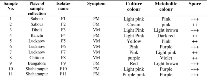
Table 1. Cultural and morphological characters of 11 F. mangiferae isolates collected from different agro-ecological regions of India.
Sample No.
Place of sample collection
Isolates name
Symptom Culture
colour
Metabolite colour
Spore
1 Sabour F1 FM Light pink Pink +++
2 Sabour F2 FM Cream pink ++
3 Dholi F3 VM Light Pink Light brown +++
4 Ranchi F4 FM Light Pink Dark red ++
5 Lucknow F5 VM Yellow Pink ++
6 Lucknow F6 VM Pink Purple +++
7 Lucknow F7 VM Pink Light pink ++
8 Chittour F8 VM purple Violet ++
9 Bangalore F9 FM Red Light brown +++
10 11
Shaharanpur Shaharanpur
F10 F11
FM FM
Light pink Purple pink
Purple Purple
+++ +++
VM: Vegetative malformation; FM: Floral malformation; ++ Moderate spore; +++ High Spores
Molecular Characterization
Genomic DNA of all 11 isolates of F. mangiferae
and control sample successfully amplified using ITS1 (5’TCCGTAGGTGAACCTGCGG3’) and ITS4 (5’ TCCTCCGCTTATTGATATGC 3’) primers [19] obtained 570 bp band (Fig. 1a). Fusarium genus-specific primer Fu-f and ITS-Fu-r [27] specifically amplified a 398 bp band among
all the isolates (Fig. 1b). No cross-reaction amplification detected in negative control
Colletotrichum gloeosporioides tested for non-specific amplification. The PCR-RFLP restriction
digestions of Fusarium isolates performed using
AluI, HindIII and RsaI restriction enzymes. Hind III and Rsa I show no restriction sites. However, AluI restriction gives two clear restriction bands of 410 bp and 120 bp. The gene specific PCR using β -tubulin primers resulted in a single band of 235 bp and no amplification in negative control. Hence, β -tubulin primer-pair can potentially employed as marker to discriminate F. mangiferae (Fig. 1c & d).
Molecular Characterization of Fusarium Mangiferae 5
Fig1 (b): PCR amplification using Genus-specific primer of F. mangiferae isolates using ITS-Fu F&R primers (Abd-Elsalam, 2003).
Fig1(c): PCR-Restriction digestion with AluI. (Lane 1-11 F. mangiferae; N- F. oxysporum
(Negative control).
Fig 1(d). Nested-PCR using beta-tubulin (β-tub) gene-specific primer (Lane 1-11) F. mangiferae
and lane 12- Negative control (C. gloeosporioides).
Phylogenetic Analysis
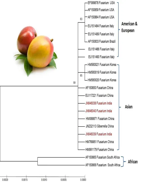
Twenty-two sequences of Fusarium spp. reported to cause mango malformation from different countries (China, Italy, Brazil, Korea, South Africa and USA) selected and retrieved from NCBI. Three
Fusarium isolates from present study submitted to NCBI (Accession id: JN646038, JN646039,
phylogenetic tree constructed using 22 Fusarium
spp. isolates from other countries along with the three representative isolates from the present study showed more than 90% nucleotide sequence similarity and grouped the isolates into two major clusters I and II. Cluster I further divided into two major subgroups covering the present study
Fusarium isolates (India) and isolates from Brazil, USA, Italy, China and Korea. However, cluster II divided into two sub clades covering the two
Fusarium spp. of South Africa and depicts genetic variability at the species level (Fig. 2).
Molecular Characterization of Fusarium Mangiferae 7
DISCUSSION
Normal apical growth observed on the apex of healthy seedlings while seedlings inoculated with
F. mangiferae showed vegetative malformed growth followed by excessive proliferation of leaves. The gathering of stubby leaves into close folds similar to incipient bunchy top met under natural normal conditions, which is in accordance with previous workers [9, 32] on pathogenicity. Early
and easy disease detection of F. mangiferae is necessary for effective and timely management of mango malformation. PCR based molecular disease diagnostic techniques provide a rapid, cost-effective specific and sensitive detection system in comparison to conventional methods [33]. Globally,
three Fusarium species under the Giberrella fuzikoroi species complex reported as the causal agents of mango malformation disease. The species are F. mangiferae from Egypt [3], Israel [34], South
Africa [10]
, Pakistan [35], F. proliferatum from
Malaysia [36], and F. subglutinans reported from
South Africa, India and USA [37]. PCR products of
570 bp, 398 bp and 235 bp amplified in F. mangiferae using Fusarium species-specific [25] ITS
primers and β-tubulin gene-specific primers, respectively. By contrast, the nested PCR approach presented here produced consistent and
reproducible results using β-tubulin gene-specific
designed primers. Nested PCR increased the sensitivity of F. mangiferae (=F. subglutinans) β -tubulin gene-specific primers by 1000 fold [38]. The
nested PCR assay potentially employed as a diagnostic tool for effective detection of F. mangiferae in mango malformation affected tissues. Moreover, nested PCR permits rapid identification of the fragmented samples and does not require sequencing. This technique is advantageous as it is rapid, reliable and robust. However ITS-PCR-RFLP using Alu I restriction enzyme proficiently identifies the pathogen by giving two clear bands of 410 and 120 bp sizes and making it an effective marker for F. mangiferae. The phylogenetic tree constructed using ITS-gene sequences comparing Fusarium spp. associated with mango malformation disease from different countries (Brazil, China, USA, Korea, Italy and South Africa) indicate a wide diversity among F. mangiferae isolates. This range is consistent with the previous description of isolates reported to cause malformation by F. subglutinans belongs to the same species within the G. fuzikoroi complex
[39]. Twenty-two Fusarium species associated with
mango malformation in the G. fuzikoroi complex along with the present study isolates (JN646038, JN646039, and JN646040) divided into three distinct clades Asian clade, American clade and African clade. The phylogenetic tree showed 65-99% as overall genetic similarity between the isolates in two clusters. Cluster I is a major cluster and is further divided into group A comprising 8
Fusarium species (American and European clade) and group B comprising 12 Fusarium species (Asian clade). Cluster II is a minor cluster and is divide into two groups (South African clade). Based on sequence similarity of Fusarium spp. we concluded that the F. mangiferae is a dominant pathogen of mango malformation disease in India. Our findings are consistent and supports the findings of several research groups that identified and reported F. mangiferae as a pathogen causing mango malformation disease in Asia and America
[37,40]. Similarly, American isolates sequences also
supports that F. mangiferae is a mango malformation pathogen. However, the South African Fusarium species showed some level of distinctness as the clade represented by two
Fusarium spp. viz. F. sterilihyphosum and F. subglutinans. Therefore, in the present study attempted, we develop a nested PCR based detection assay and sequence based phylogenetic analysis thus, claimed F. mangiferae as the dominant pathogen of mango malformation in India.
CONCLUSION
The ITS PCR based species-specific and gene-specific detection and identification of Fusarium
species isolates will be helpful in understanding the pathogenic relationship among the isolates from other geographical location. Identification and confirmation of F. mangiferae as the dominant pathogen of mango malformation disease in India. This PCR based technique would be a valuable component in large-scale pathogen diagnosis and confirmation. This study further explored beside effective crop management strategies.
REFERENCES
2. Kumar P, Misra AK, Modi DR. Current status of mango malformation in India. Asian JPlant Sci.
2011; 10: 1-23.
3. Ploetz R, Zheng QI, Vazquez A, Sattar A MA. Current status and impact of mango malformation in Egypt. Intern J Pest Manag. 2002; 48: 279-285. 4. Bains G and Pant RC. Mango malformation: Etiology and preventive measures. Physiol Mol Biol Plant. 2003; 9: 41-61.
5. Freeman S, Maymon M, Biton A, Levin AG, Shtienberg D. Management of mango malformation disease based on a novel strategy of timing of fungicide applications combined with sanitation. Crop Protection. 2014; 61:84–91. 6. Steenkamp ET, Britz H, Coutinho TA, Wingfield
BD, Marasas WFO, Wingfield MJ. Molecular characterization of Fusarium subglutinans
associated with mango malformation. Mol Plant Pathol. 2000; 1: 187-193.
7. Summanwar AS, Raychaudhuri SP, Pathak SC. Association of fungus Fusarium moniliforme Sheld with the malformation in mango. Ind Phytopathol.
1966; 19: 227-228.
8. Kumar P, Misra AK, Srivastava AK, Modi DR. Mapping of Fusarium moniliforme var.
subglutinans from normal and malformed panicles and seedlings of mango by recovery method. Plant Arch. 2011; 11: 567–569.
9. Kumar P, Kamle M, Gupta VK, Pandey BK, Misra AK, Modi DR. Host-pathogen interaction studies in malformed affected tissues of Mangifera indica L.
Amer J Agri Biol Sci. 2013; 8 (3): 199-203. 10. Britz H, Steenkamp ET, Coutinho TA, Wingfield
BD, Marasas WFO, Wingfield MJ. Two new species of Fusarium section Liseola associated with mango malformation. Mycologia. 2002; 94: 722-730
11. Leslie JF, Summerell BA. The Fusarium Laboratory Manual. Ames, IO, USA: Blackwell Professional. 2006.
12. Chakrabarti DK. Mango Malformation. Springer-Netherlands. 2011; 148. ISBN-978-94-007-0362-9. 13. Newman Z, Freeman S, Biton I, Saada D, Paz T. Molecular diagnosis of mango malformation disease and phylogeny of Fusarium mangiferae.
Phytoparasitica. 2012; 40: 287-297.
14. Li W, Li D, Twieg E, Hartung JS, Levy L. Optimized quantification of unculturable
Candidatus Liberibacter spp. in host plants using real-time PCR. Plant Dis. 2008; 92: 854–861. 15. Gomes EA, Kasaya MC, de Barros EG, Borgs AC,
Araujo EF. Polymorphism in the internal transcribed spacer (ITS) of the ribosomal DNA of 26 isolates of ectomycorrhizal fungi. Genet Mol Biol. 2002;25(4): 477-483.
16. O’Brien HE, Parrent JL, Jackson JA, Moncalvo
JM, Vilgalys R. Fungal communities’ analysis by
large-scale sequencing of environmental samples.
Environ Microb. 2005; 71(1): 5544-5550.
17. O’Donnell K, Cigelnik E, Nirenberg HI. Molecular
systematics and phylogeography of the Gibberella fujikuroi species complex. Mycologia. 1998; 90: 465-493.
18. Kvas M, Marasas WFO, Wingfield BD, Wingfield MJ, Steenkamp ET. Diversity and evolution of
Fusarium species in the Gibberella fujikuroi
complex. Fungal Diversity. 2009;1: 1–21.
19. White TJ, Bruns T, Lee S, Taylor J. Amplification and direct sequencing of fungal ribosomal RNA genes for phylogenetics. In: PCR Protocols: a guide to methods and applications. (Innis MA, Gelfand DH, Sninsky JJ, White TJ, eds). Academic Press, New York, USA. 1990; 315–322.
20. Arif M, Pani DR, Zaidi NW, Singh US. PCR-based identification and characterization of Fusarium sp. associated with mango malformation. Biotech Res Int. 2011, Article ID 141649, 6 pages.
21. Kumar P, Gupta VK, Misra AK, Modi DR. Molecular characterization of Fusarium moniliforme var. subglutinans isolates. J Environ Biol. 2014; 35(1): 211–216.
22. Booth C. The genus Fusarium. Commonwealth Mycological Institute, Kew, Surrey, UK. 1971; pp237.
23. Ali NA, Jackson RM. Stimulation of germination of spores of some ectomycorrhizal fungi by other microorganisms. Mycol Res. 1989; 93:182-186. 24. Chand D, Chakrabarti DK. Techniques to produce
malformation in mango (Mangifera indica L.).
Mycol Plant Pathol. 2003; 33: 431-438.
25. Abd-Elsalam KA, Schneider F, Guo JR. A modified DNA extraction mini-preparation protocol for Fusarium isolates. J Rap Meth Autom Microb. 2003; 11: 75-79.
26. Sambrook J, Fritsch EF, Maniatis T. Molecular Cloning: A Laboratory Manual, (2nd ed.) Cold Spring Harbor: Cold Spring Harbor Laboratory Press. 1989; 1051–1067.
27. Kamel AA. Bioinformatics tools and guideline for PCR primer design. Afr J Biotech. 2003; 2: 91-95. 28. Bogale M, Wingfield BD, Wingfield MJ,
Steenkamp ET. Species-specific primers for
Fusarium redolens and a PCR-RFLP diagnostic technique for distinguishing among three clades of
Fusarium oxysporum. FEMS Micro Letter. 2007; 271: 27-32.
29. Altschul SF, Gish W, Miller W, Myers EW, Lipman DJ. Basic local alignment search tool. J Mol Biol. 1990; 215: 403–410.
Molecular Characterization of Fusarium Mangiferae 9
31. Tamura K, Dudley J, Nei M, Kumar S. MEGA4: molecular evolutionary genetic analysis (MEGA) software version 4.0. Mol Biol Evol. 2007; 24: 1596-1599.
32. Iqbal Z, Rehman M, Dasti AA, Saleem A, Zafar Y. 2006 - RAPD analysis of Fusarium isolates causing mango malformation disease in Pakistan. World J Microb Biot. 22; 1161-1167.
33. Martin C, Roberts D, Weide MVD, Rossau R, Jannes G, Smith T. Development of a PCR-based line probe assay for identification of fungal pathogens. J Clin Microb. 2000; 38: 3735–3742. 34. Freeman S, Klein-Gueta D, Korolev N, Sztejnberg
A. Epidemiology and survival of Fusarium mangiferae, the causal agent of mango malformation disease. Acta Hort. 2004;645: 487– 491.
35. Iqbal Z, Pervez MA, Saleem BA, Salman A, Altaf AD, Saleem A. Potential of Fusarium mangiferae
as an etiological agent of mango malformation. Pak J Bot. 2010; 42: 409–415.
36. Marasas WFO, Ploetz RC, Wingfield MJ, Steenkamp ET. Mango malformation disease and the associated Fusarium species. Phytopathol.
2006; 96(6):667–672.
37. Steenkamp ET, Wingfield BD, Coutinho TA, Wingfield MJ, Marasas WFO. Differentiation of
Fusarium subglutinans f. sp. pini by histone gene sequence data. Appl Env Microb.1999; 65: 3401– 3406.
38. Judelson HS, Tooley PW. Enhanced polymerase chain reaction methods for detecting and quantifying Phytophthora infestans in plants. Phytopathol. 2000; 90:1112–1119.
39. Leslie JF, Zeller KA, Logrieco A, Mulè G, Moretti A, Ritieni A. Species Diversity of and Toxin Production by Gibberella fujikuroi Species Complex Strains Isolated from Native Prairie Grasses in Kansas. Appl Env Micro. 2004; 70(4): 2254–2262.
40. Freeman S, Maimon M, Pinkas Y. Use of GUS transformants of Fusarium subglutinans for determining etiology of mango malformation disease. Phytopathol. 1999; 89: 456–61.