Research Article
Antileishmanial Activity of
Handroanthus serratifolius
(Vahl)
S. Grose (Bignoniaceae)
Erica Vanessa Souza Costa,
1Heliton Patrick Cordovil Brígido,
1João Victor da Silva e Silva,
1Marlia Regina Coelho-Ferreira,
2Geraldo Célio Brandão,
3and Maria Fâni Dolabela
11Programa de P´os-Graduac¸˜ao em Ciˆencias Farmacˆeuticas, ICS, Universidade Federal do Par´a, Bel´em, PA, Brazil 2Departamento de Botˆanica, Museu Paraense Em´ılio Goeldi, Bel´em, PA, Brazil
3Escola de Farm´acia, Universidade Federal de Ouro Preto, Ouro Preto, MG, Brazil
Correspondence should be addressed to Maria Fˆani Dolabela; fanidolabela20@gmail.com
Received 20 October 2016; Revised 22 December 2016; Accepted 18 January 2017; Published 12 February 2017
Academic Editor: V´ıctor L´opez
Copyright © 2017 Erica Vanessa Souza Costa et al. This is an open access article distributed under the Creative Commons Attribution License, which permits unrestricted use, distribution, and reproduction in any medium, provided the original work is properly cited.
This study aimed to evaluate the leishmanicidal activity of ethanol extract, fractions, and isolated substance fromHandroanthus
serratifoliusagainstLeishmania amazonensis. Furthermore, this activity was related to cytotoxicity, and the selectivity index was determined. The ethanol extract was obtained by maceration of the stem powder, and the extract was subjected to fractionation on chromatographic column. The lapachol was obtained by acid base extraction followed by purification in chromatographic column. The antipromastigote activity and cytotoxicity tests were carried out by the cell viability method (MTT). Modified THP-1 cells were
infected withL. amazonensispromastigotes and treated for 24 h with different concentrations of the extract, fractions, and lapachol.
The ethanol extract, dichloromethane, and ethyl acetate fractions were not active against promastigotes (IC50>200𝜇g/mL) or
cyto-toxic (CC50>500𝜇g/mL), and the selectivity index (SI) was greater than 2.5. The ethyl acetate fraction was active only in
promastig-otes; it is not cytotoxic (CC50 >500𝜇g/mL, SI>5). The lapachol was selectively active only against amastigote (IS>2.5, CC50 >
500𝜇g/mL). In summary, lapachol and ethyl acetate fraction are promising against amastigote and promastigote forms, respectively.
1. Introduction
Leishmaniasis is caused by over 20Leishmaniaspecies and
it is transmitted to humans by the infected phlebotomine female sandflies. There are three main forms of the disease: visceral leishmaniasis (VL), cutaneous leishmaniasis (CL), and mucocutaneous leishmaniasis. It is estimated that about 200,000 to 400,000 new cases of VL occur worldwide each year. Over 90% of new cases occur in 6 countries: Bangladesh, Brazil, Ethiopia, India, South Sudan, and Sudan. The cuta-neous leishmaniasis is the most common form of leishma-niasis. About 95% of CL cases occur in the Americas, the Mediterranean basin, the Middle East, and Central Asia. Almost 90% of mucocutaneous leishmaniasis cases occur in Bolivia, Brazil, and Peru [1].
The leishmaniasis control is based on vector combat, extermination of infected dogs, and treatment of infected
individuals [2]. The amphotericin andN-methyl glucamine
antimoniate (Glucantime) [3] are drugs currently used in
the treatment of leishmaniasis. However, they have problems as severe adverse effects, and some strains have already pre-sented increased parasitic resistance [2, 4, 5]. In addition, all drugs are currently available for parenteral administration [4]. As a result, many patients abandon treatment; this fact favors the appearance of resistant strains [6].
In this context, plant species are the best and greatest source of drugs for mankind. Ethnobotanical studies have demonstrated the popular use of plants in the treatment of leishmaniasis both orally and in the topical application on lesions [7, 8]. Many plants present in their composition sub-stances of the classes of alkaloids, terpenes, naphthoquinones, lignans, chalcones, flavonoids, and sesquiterpene lactones, compounds described in the literature as effective in leish-manicidal activity [9–11].
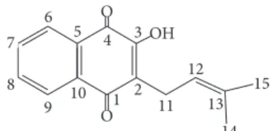
O OH O1 2 3 4 5 6 7 8
9 10 11
12
13 14
15
Figure 1: Chemical structure lapachol.
The search for alternative therapies for leishmaniasis is very important. Many species of the Bignoniaceae family are used in folk medicine to treat external ulcers, skin diseases, and skin disorders [7]. However, the antileishmanial activity of these species has not been tested yet.
Handroanthus serratifolius(Bignoniaceae) is used in tra-ditional medicine as antitumor, antiparasitic, and antimalar-ial agent [12–14]. Originally the following substances were
isolated from species of the Bignoniaceae family as
Han-droanthus serratifolius, (Figure 1), 𝛼
-1,4-naphthoquinone-methylfuran, dehydro-𝛼-lapachone, 𝛼-lapachone,
tecoma-quinone I, and dehydroiso-𝛼-lapachone [15].
The antipromastigote activity of lapachol, isolapachol, and dihydrolapachol, with soluble derivatives (potassium salt), was evaluated. All substances inhibited the growth of Leishmania amazonensis and L. brasiliensis promastigotes,
with a greater effect inL. amazonensis. The lapachol showed
activity in L. amazonensis (IC50 = 5.2𝜇g/mL) than L.
braziliensis(IC50= 11.9𝜇g/mL) [16].
Other studies evaluated the leishmanicidal activity of lapachol and compared its efficacy with a reference drug,
sodium stibogluconate (Pentostam). These compounds
were evaluated against amastigotes ofLeishmania(Viannia)
braziliensis(LVb). In vitro, lapachol exhibited antiamastigote effect, whereas in vivo it did not prevent the development of LVb induced lesions at an oral dose of 300 mg/kg/day for 42
days. Pentostamdemonstrated a significant antiamastigote
effect in vitro and in vivo (60 mg/kg/day). Perhaps the lapachol inhibits the microbicide function of macrophages in vivo. Alternatively, it might be transformed into an inactive metabolite(s) or neutralized, losing its leishmanicidal activity [17].
This study aimed to evaluate the leishmanicidal activity of ethanol extract, fractions, and isolated substance obtained fromHandroanthus serratifoliusagainstLeishmania amazo-nensis. Furthermore, this activity is related to cytotoxicity determining the selectivity index.
2. Material and Methods
2.1. Plant Material and Extraction. Plants were collected on 10 March 2014 in Em´ılio Goeldi Museum, Par´a, Brazil (S
01∘273.031, W 48∘2640.2). The voucher specimen (MG
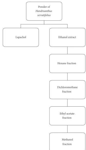
206637) was deposited in the Jo˜ao Murc¸a Pires Herbarium. Plants were dried at room temperature for seven days. The material was powdered and extracted with ethanol by cold maceration. The resultant solution was concentrated in a rotary evaporator to obtain the ethanol extract. The extract was fractioned in chromatographic column (CC) with silica gel as stationary phase and increasing polarity solvents
(hexane, dichloromethane, ethyl acetate, and methanol) as mobile phase (Figure 2).
The powder of H. serratifolius was treated with 2.5%
sodium carbonate solution for 24 h for lapachol isolation. The solution was filtered, and the precipitate was solubilized in aqueous hydrochloric acid. After 30 minutes, it was cen-trifuged (3,000 rpm/10 minutes) and a yellow solid precipitate was separated (Figure 2). The precipitate was dried and sub-mitted to fractionation on chromatographic column. Nuclear magnetic resonance was used to identify the isolated com-pounds.
Lapachol.NMR1H (200 Hz, CDCl3): 8,13 (dd,𝐽 = 6.2and
1.4 Hz); 8,05 (dd,𝐽 = 7.6and 1.4 Hz); 7,78 (dt,𝐽 = 6.2and
1.4 Hz); 7,63 (dt,𝐽 = 7.3and 1.4 Hz); 7,34 (dt,𝐽 = 7.3and
1.4 Hz); 5,21 (m); 3,30 (d,𝐽= 7,3 Hz); 1,79 (s); 1,68 (s). NMR
13C (50 Hz, CDCl
3): 184,4 (C-4); 181,71 (C-1); 152,72 (C-2);
134,77 (C-7); 133,73 (C-13); 133,04 (C-8); 132,78 (C- 5); 129,54 (C-10); 126,78 (C-6); 126,02 (C-9); 123,57 (C-3); 119,73 (C-12); 25,67 (C-14); 22,65 (C-11); and 17,85 (C-15).
2.2. Antileishmanial Activity of Leishmania amazonensis
2.2.1. Antipromastigotes Assay. Strains isolated from
leishma-niasis (Leishmania amazonensisMHOM/BR/2009/M26361)
were obtained from the Evandro Chagas Institute, Ananin-deua, Brazil.
TheL. amazonensispromastigotes were cultivated at 26∘C in RPMI 1640 medium supplemented with 10%
heat-inacti-vated fetal bovine serum (Gibco, Grand Island, NY, USA),
penicillin (100 U/mL), and streptomycin (100𝜇g/mL) [18].
Culture of promastigote forms in logarithm phase was
adjusted to 5×10⁶parasites/100𝜇L. The susceptibility testing
was performed in 96-well plates. The extract, fraction, and lapachol were tested in triplicate in a concentration gradient
(200 to 3.125𝜇g/mL). Negative control was performed with
parasites and incubation medium. The positive control was
made with amphotericin B (25–0.3906𝜇g/mL). After 24 h of
incubation at 26∘C in 5% de CO2, 10𝜇L of tetrazolium salt
(5 mg/mL) was added to each well, and the parasites were quantified in enzyme-linked immunosorbent-assay plate
reader. The IC50was determined by linear regression (Graph
Pad Prism version 5.04). The results were classified as follows:
IC50 ≤100𝜇g/mL were considered active, IC50between 101
and 200𝜇g/mL were considered moderate active, and IC50≥
200𝜇g/mL were considered to be inactive [18].
2.2.2. Antiamastigote Assay. Modified THP-1 cell (4 × 10⁵ cells/0.1 mL) was cultured in RPMI-1640 (Roswell Park
Memorial Institute 1640) medium (Sigma Aldrich, USA),
supplemented with 5% of fetal calf serum, kept in a 5% CO2
atmosphere at 37∘C with phorbol ester as inducing agent. The
cells were added the circular coverslips (2 × 10⁵); then L.
amazonensispromastigotes were added (5×10⁶). The samples treatment was performed with concentrations of 250, 125, and
62,5𝜇g/mL/24 h. The coverslips were removed and stained
Powder of
Handroanthus serratifolius
Lapachol Ethanol extract
Hexane fraction
Dichloromethane fraction
Ethyl acetate fraction
Methanol fraction
Figure 2: Extract, fractions, and isolated substance obtained ofHandroanthus serratifolius.
2.3. Viability Assay and Selective Index. Cell viability was determined by the MTT [3-(4,5-dimethylthiazol-2-yl)-2,5-diphenyl tetrazolium bromide] [19]. Modified THP-1 cell (4
×105cells/0.1 mL) was cultured in RPMI-1640 (Roswell Park
Memorial Institute 1640) medium (Sigma Aldrich, USA),
supplemented with 5% of fetal calf serum, kept in a 5% CO2
atmosphere at 37∘C. The cells were treated with extracts,
fractions, or lapachol in different concentrations (between
500 and 25𝜇g/mL). MTT was added (5.0 mg/mL) after 24 h
of further incubation. The plate was incubated at 37 C in an
atmosphere of 5% CO2for 4 h. Dimethyl sulfoxide was added
to each well to solubilize the formazan crystals. The optical density was determined at 490 nm (Stat Fax 2100 microplate reader, Awareness Technology, Inc., USA). The cell viability was expressed as percentage of the control absorbance (absorbance of control group) in the untreated cells after sub-tracting the appropriate background. The cytotoxic
concen-tration (CC50) was determined by linear regression. Samples
with CC50 >500𝜇g/mL were considered of low cytotoxicity.
Selectivity index (SI) for the antipromastigote activity was
calculated based on the rate between CC50 and IC50for the
in vitro activity againstL. amazonensis[20].
3. Results and Discussion
In this study, lapachol (C15H14O3) was isolated from stem
powder of H. serratifolius. However, other studies isolated
ethanol extract ofH. serratifoliuslapachol (2.9% yield) [21].
The antipromastigote activity of lapachol has been described in posterior study [16].
To verify if H. serratifolius has other substances with
Yield of extract, fractions, and lapachol
58,02%
28,64%
13%
3,68% 8,02% 2,90%
EE FrHex FrDicl FrAcOEt FrMet Lapachol 20
30 40 50 60 70
(%)
Figure 3: Yield of extract, fractions, and isolated substance obtained ofHandroanthus serratifolius. EE: ethanol extract; FrHex: hexane fraction; FrDcl: dichloromethane fraction; FrAcOEt: ethyl acetate fraction; FrMet: methanol fraction.
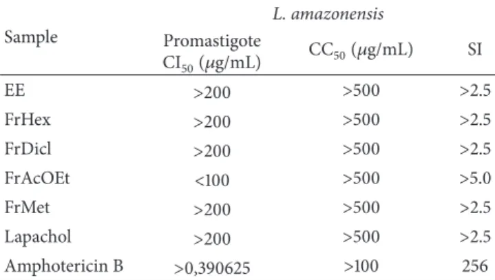
Table 1: Antipromastigote activity and cytotoxicity ofHandroanthus
serratifolius.
Sample
L. amazonensis
Promastigote
CI50(𝜇g/mL) CC50(𝜇g/mL) SI
EE >200 >500 >2.5
FrHex >200 >500 >2.5
FrDicl >200 >500 >2.5
FrAcOEt <100 >500 >5.0
FrMet >200 >500 >2.5
Lapachol >200 >500 >2.5
Amphotericin B >0,390625 >100 256
IC50: inhibitory concentration 50%; CC50: concentration cytotoxic 50%;
IS: selectivity index; EE: ethanol extract; FrHex: hexane fraction; FrDcl: dichloromethane fraction; FrAcOEt: ethyl acetate fraction; FrMet: methanol fraction.
Lapachol, ethanol extract, and fractions were tested
against L. amazonensis promastigotes. Unlike a previous
study [16], lapachol was not active inL. amazonensis
pro-mastigotes (IC50 >200𝜇g/mL; Table 1). TheL. amazonensis
strain used in this assay was isolated from a patient who had previously not responded to conventional therapy. This may explain the divergent response.
The ethanol extract, dichloromethane, and methanol
fractions did not show activity against promastigotes (IC50>
200𝜇g/mL; Table 1). The ethyl acetate fraction was promising
(IC50 <100𝜇g/mL; Table 1). Study on thin-layer
chromatog-raphy (results not shown) suggests coumarins in ethyl acetate
fraction. Coumarins were isolated fromH. impetiginosa[22].
The 7-{[(2R∗
)-3,3-dimethoxyloxiran-2-2-yl]methylorix-an}-8-[(2R∗,3R∗
)-3-isopropenyloxira-2-yl]-2H-chromen-2-one, phebalosin, and
7-methoxy-8-8(4-methyl-3-3-furyl)-2H-chromen-2-one were tested againstLeishmania
panamen-sisamastigotes. The coumarins were active (IC509.9, 10.5 and
14.1 mg/mL, resp.) and cytotoxic in human promonocytic
U-937 cells (CC509.7, 33.0 and 20.7, resp.; [23]). The
fractiona-tion of the ethyl acetate fracfractiona-tion may contribute to antipro-mastigote activity.
We assessed the cytotoxicity of all samples for modified THP-1 cell line. Extract, fractions, and lapachol showed no
Table 2: Antiamastigote activity ofHandroanthus serratifolius.
(a)
Sample
% reduction
concentration (𝜇g/mL)
250 125 62,5
EE 0 0 0
FrHex 0 0 0
FrDicl 0 0 0
FrAcOEt 0 0 0
FrMet 0 0 0
Lapachol 53.97 44.45 22.49
(b)
Sample % concentration (𝜇g/mL)
50 25 12,5
Amphotericin B 86.7 81.5 79.9
EE: ethanol extract; FrHex: hexane fraction; FrDcl: dichloromethane frac-tion; FrAcOEt: ethyl acetate fracfrac-tion; FrMet: methanol fraction.
toxicity for this cell (CC50 >500𝜇g/mL; Table 1). Similarly,
another study showed that the ethanol extracts of leaves and
flowers fromH. aureuswere not cytotoxic for macrophages
rats (CC50 > 1000𝜇g/mL) [24]. Unlike this study, several
studies describe the cytotoxicity of lapachol [25–27]. The most active fraction against promastigotes showed higher
selectivity index (SI>5). Lapachol showed selectivity index
greater than 2.5 (Table 1).
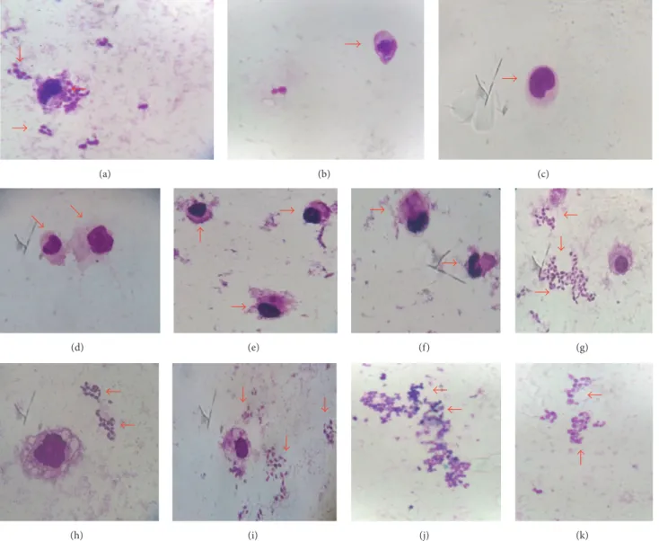
Lapachol reduced the infection of macrophages, with
greater effect observed at 250𝜇g/mL (Figure 4; Table 2).
Antiamastigote activity of lapachol againstLeishmania(
Vian-nia)braziliensiswas described [17]. This effect has been linked to stabilization of the complex and DNA topoisomerase [28]. Some have antiparasitic effect as time-dependent [29, 30]. Thus, increased exposure time can contribute to the inhibi-tory effect.
4. Conclusion
The ethanol extract, hexane, dichloromethane, and methanol
fractions fromH. serratifoliusshowed no antipromastigote
and antiamastigote activities. It was also not cytotoxic. The ethyl acetate fraction showed selective effect for promastig-otes, while lapachol was active for amastigotes.
Competing Interests
The authors declare that there is no conflict of interests.
Acknowledgments
(a) (b) (c)
(d) (e) (f) (g)
(h) (i) (j) (k)
Figure 4: Antiamastigote activity ofHandroanthus serratifolius. (a) Negative control; (b) macrophage without infection; (c) amphotericin
B (50𝜇g/mL); (d, e, and f) lapachol (250𝜇g/mL, 125𝜇g/mL, and 62.5, resp.); (g) ethanol extract; (h) hexane fraction: (i) dichloromethane
fraction; (j) ethyl acetate fraction; and (k) methanol fraction (250𝜇g/mL); increase of 100x.
References
[1] WHO,Leishmaniasis—A Brief History of the Disease, World
Health Organization, Geneva, Switzerland, 2014, http://apps .who.int/iris/bitstream/10665/111008/1/WHO DCO WHD 2014 .1 eng.pdf.
[2] R. J. Soares-Bezerra, L. Leon, and M. Genestra, “Recentes avanc¸os da quimioterapia das leishmanioses: mol´eculas
intracelulares como alvo de f´armacos,” Revista Brasileira de
Ciˆencias Farmacˆeuticas, vol. 40, no. 2, pp. 139–149, 2004.
[3] M. T. G. Casavechia, T. G. V. Silveira, U. Teodoro, V. Janeiro, M. Udo, and M. V. C. Lonardoni, “Variables associated with the post-treatment healing of lesions in patients with American
cutaneous leishmaniasis in Paran´a State, Brazil,” Brazilian
Journal of Pharmaceutical Sciences, vol. 45, no. 4, pp. 841–847, 2009.
[4] B. Ullman, E. Carrero-Valenzuela, and T. Coons, “
Leish-mania donovani: isolation and characterization of sodium
stibogluconate (Pentostam)-resistant cell lines,”Experimental
Parasitology, vol. 69, no. 1, pp. 157–163, 1989.
[5] R. Lira, S. Sundar, A. Makharia et al., “Evidence that the high incidence of treatment failures in Indian Kala-Azar is due
to the emergence of antimony-resistant strains ofLeishmania
donovani,”Journal of Infectious Diseases, vol. 180, no. 2, pp. 564– 567, 1999.
[6] V. S. Amato, “Tratamento da Leishmaniose tegumentar
amer-icana. T´opicos em terapˆeutica,”Revista Brasileira de Medicina,
vol. 53, pp. 202–212, 1998.
[7] S. A. G. Da Silva, S. S. Da Costa, S. C. F. Mendonc¸a, E. M. Silva, V. L. G. Moraes, and B. Rossi-Bergmann, “Therapeutic effect
of oralKalanchoe pinnataleaf extract in murine leishmaniasis,”
Acta Tropica, vol. 60, no. 3, pp. 201–210, 1995.
´area endˆemica da Amazˆonia do Maranh˜ao, Brasil,”Cadernos de Sa´ude P´ublica, vol. 18, no. 1, pp. 187–195, 2002.
[9] E. F. Queiroz, F. Roblot, A. Cav´e, M. D. Q. Paulo, and A. Fournet,
“Pessoine and spinosine, two catecholic berbines fromAnnona
spinescens,”Journal of Natural Products, vol. 59, no. 4, pp. 438– 440, 1996.
[10] E. C. Torres-Santos, D. L. Moreira, M. A. C. Kaplan, M. N. Meirelles, and B. R. Bergmann, “Selective effect of
2,6-dihy-droxy-4-methoxychalcone isolated from Piper aduncum on
Leishmania amazonensis,” Antimicrobial Agents and Chemo-therapy, vol. 43, pp. 1234–1241, 1999.
[11] L. G. Rocha, J. R. G. S. Almeida, R. O. Macˆedo, and J. M. Barbosa-Filho, “A review of natural products with
antileishma-nial activity,”Phytomedicine, vol. 12, no. 6-7, pp. 514–535, 2005.
[12] A. B. Oliveira, D. S. Raslan, M. C. M. E. Miraglia, and A. A. L. Mesquita, “Estrutura qu´ımica e atividade biol´ogica de
naftoquinonas de Bignoniaceas brasileiras,”Qu´ımica Nova, vol.
13, no. 4, pp. 302–307, 1990.
[13] T.-S. Wu, H.-C. Hsu, P.-L. Wu et al., “Naphthoquinone esters
from the root ofRhinacanthus nasutus,”Chemical and
Pharma-ceutical Bulletin, vol. 46, no. 3, pp. 413–418, 1998.
[14] F. J. Jim´enez-Gonz´alez, L. A. Veloza, and J. C. Sep´ulveda-Arias, “Anti-infectious activity in plants of the genus Tabebuia,”
Universitas Scientiarum, vol. 18, no. 3, pp. 257–267, 2013. [15] A. M. P. Silva, S. R. Paiva, M. R. Figueiredo, and M. A. C.
Kaplan, “Atividade Biol´ogica de Naftoquinonas de Esp´ecies de
Bignoniaceae,”Revista Fitos, vol. 7, no. 4, pp. 207–215, 2012.
[16] N. M. F. Lima, C. S. Correia, L. L. Leon et al., “Antileishmanial
activity of lapachol analogues,”Memorias do Instituto Oswaldo
Cruz, vol. 99, no. 7, pp. 757–761, 2004.
[17] M. J. Teixeira, Y. M. De Almeida, J. R. Viana et al., “In vitro and in vivo leishmanicidal activity of
2-hydroxy-3-(3-methyl-2-butenyl)-1,4-naphthoquinone (lapachol),”
Phytother-apy Research, vol. 15, no. 1, pp. 44–48, 2001.
[18] E. F. Mota, D. M. Rosario, A. S. S. Veiga, D. Do Socorro Barros Brasil, F. T. Silveira, and M. F. Dolabela, “Biological activities of
Croton palanostigmaKlotzsch,”Pharmacognosy Magazine, vol. 11, no. 43, pp. 601–606, 2015.
[19] T. Mosmann, “Rapid colorimetric assay for cellular growth and survival: application to proliferation and cytotoxicity assays,”
Journal of Immunological Methods, vol. 65, no. 1-2, pp. 55–63, 1983.
[20] C. V. Nakamura, A. O. Santos, M. C. Vendrametto et al., “Atividade antileishmania do extrato hidroalco´olico e de frac¸˜oes
obtidas de folhas dePiper regnellii(Miq.) C. DC. var. pallescens
(C. DC.) Yunck,”Revista Brasileira de Farmacognosia, vol. 16,
no. 1, pp. 61–66, 2006.
[21] M. Fernandes-de-Oliveira, Contribuic¸˜ao ao conhecimento
qu´ımico das esp´ecies Tabebuia serratifolia Nichols e Tabebuia rosa Bertol [Ph.D. thesis], Universidade Federal do Cear´a, 2000. [22] S. Panizza, “Contribuic¸˜ao ao estudo morfol´ogico e anatˆomico deJacaranda caroba(Velloso) DC. Bignoniaceae,”Revista de Farm´acia e Bioqu´ımica da Universidade de S˜ao Paulo, vol. 5, pp. 93–106, 1967.
[23] V. Arango, S. Robledo, B. S´eon-M´eniel et al., “Coumarins fromGalipea panamensisand their activity againstLeishmania panamensis,”Journal of Natural Products, vol. 73, no. 5, pp. 1012– 1014, 2010.
[24] R. Santos, L. Conserva, M. Bastos, and E. Campesatto,
“Avaliac¸˜ao do potencial biol´ogico da Tabebuia aurea (Silva
Manso) como fonte de mol´eculas bioativas para atividade
antimicrobiana, antiedematogˆenica e antirradicalar,” Revista
Brasileira de Plantas Medicinais, vol. 17, no. 4, pp. 1159–1168, 2015.
[25] M. Maeda, M. Murakami, T. Takegami, and T. Ota, “Promotion or suppression of experimental metastasis of B16 melanoma
cells after oral administration of lapachol,” Toxicology and
Applied Pharmacology, vol. 229, no. 2, pp. 232–238, 2008. [26] M. F. Oliveira, T. L. G. Lemos, M. C. De Mattos, T. A. Segundo,
G. M. P. Santiago, and R. Braz-Filho, “New enamine
deriva-tives of lapachol and biological activity,”Anais da Academia
Brasileira de Ciencias, vol. 74, no. 2, pp. 211–221, 2002. [27] E. O. Silva, T. C. de Carvalho, I. A. Parshikov, R. A. dos
Santos, F. S. Emery, and N. A. J. C. Furtado, “Cytotoxicity of
lapachol metabolites produced by probiotics,”Letters in Applied
Microbiology, vol. 59, no. 1, pp. 108–114, 2014.
[28] M. T. Muray and J. E. Pizzorno, Encyclopedia of Natural
Medicine, PA4, Prima Pub, Rocklin, Calif, USA, 2nd edition, 1998.
[29] A. Dagan, L. Efron, L. Gaidukov, A. Mor, and H. Ginsburg, “In vitro antiplasmodium effects of dermaseptin S4 derivatives,”
Antimicrobial Agents and Chemotherapy, vol. 46, no. 4, pp. 1059– 1066, 2002.
[30] O. Oriakpono, H. Aduabobo, G. D. B. Awi-Waadu, and S. Nzeako, “Anti-parasitic effects of methanolic extracts of
Submit your manuscripts at
https://www.hindawi.com
Stem Cells
International
Hindawi Publishing Corporationhttp://www.hindawi.com Volume 2014
Hindawi Publishing Corporation
http://www.hindawi.com Volume 2014
INFLAMMATION
Hindawi Publishing Corporation
http://www.hindawi.com Volume 2014
Behavioural
Neurology
Endocrinology
International Journal ofHindawi Publishing Corporation
http://www.hindawi.com Volume 2014
Hindawi Publishing Corporation
http://www.hindawi.com Volume 2014
Disease Markers
Hindawi Publishing Corporation
http://www.hindawi.com Volume 2014
BioMed
Research International
Oncology
Journal ofHindawi Publishing Corporation
http://www.hindawi.com Volume 2014
Hindawi Publishing Corporation
http://www.hindawi.com Volume 2014 Oxidative Medicine and Cellular Longevity Hindawi Publishing Corporation
http://www.hindawi.com Volume 2014
PPAR Research
The Scientific
World Journal
Hindawi Publishing Corporation
http://www.hindawi.com Volume 2014
Immunology Research
Hindawi Publishing Corporation
http://www.hindawi.com Volume 2014
Journal of
Obesity
Journal ofHindawi Publishing Corporation
http://www.hindawi.com Volume 2014
Hindawi Publishing Corporation
http://www.hindawi.com Volume 2014
Computational and Mathematical Methods in Medicine
Ophthalmology
Journal ofHindawi Publishing Corporation
http://www.hindawi.com Volume 2014
Diabetes Research
Journal ofHindawi Publishing Corporation
http://www.hindawi.com Volume 2014
Hindawi Publishing Corporation
http://www.hindawi.com Volume 2014
Research and Treatment
AIDS
Hindawi Publishing Corporation
http://www.hindawi.com Volume 2014
Gastroenterology Research and Practice
Hindawi Publishing Corporation
http://www.hindawi.com Volume 2014
Parkinson’s
Disease
Evidence-Based Complementary and Alternative Medicine
Volume 2014