(Annals of the Brazilian Academy of Sciences) ISSN 0001-3765
www.scielo.br/aabc
Endophytic colonization of rice (
Oryza sativa
L.) by the diazotrophic
bacterium
Burkholderia kururiensis
and its ability
to enhance plant growth
KATHERINE A. MATTOS1†, VANIA L.M. PÁDUA2†‡, ALEXANDRE ROMEIRO1†, LETICIA F. HALLACK1†, BIANCA C. NEVES1, TECIA M.U. ULISSES1, CLAUDIA F. BARROS3, ADRIANE R. TODESCHINI1,
JOSÉ O. PREVIATO1 and LUCIA MENDONÇA-PREVIATO1
1Instituto de Biofísica Carlos Chagas Filho, Universidade Federal do Rio de Janeiro, Centro de Ciências da Saúde, Cidade Universitária, Ilha do Fundão, 21944-970 Rio de Janeiro, RJ, Brasil
2Instituto de Bioquímica Médica, Centro de Ciências da Saúde, Universidade Federal do Rio de Janeiro, Cidade Universitária, Ilha do Fundão, 21944-970 Rio de Janeiro, RJ, Brasil
3Laboratório de Botânica Estrutural, Instituto de Pesquisas Jardim Botânico do Rio de Janeiro, Rua Jardim Botânico, 1008, Jardim Botânico, 22460-000 Rio de Janeiro, RJ, Brasil
Manuscript received on April 14, 2008; accepted for publication on May 27, 2008; contributed byJOSÉO. PREVIATO*and LUCIAMENDONÇAPREVIATO*
ABSTRACT
Burkholderia kururiensisis a diazotrophic bacterium originally isolated from a polluted aquifer environment and
presents a high level of similarity with the rice endophyte “B. brasilensis” species. This work assessed the ability of B. kururiensisto endophytically colonize rice plantlets by monitoring different tissues of root-inoculated plants for the presence of bacterial growth in different media, electron microscopy and by 16S rDNA analysis. Observations of roots, stems and leaves of inoculated rice plantlets by electron microscopy revealed B. kururiensiscolonization predominantly on root hair zones, demonstrating endophytic colonization primarily through the endodermis, followed by spreading into xylem vessels, a possible pathway leading to aerial parts. Although indifferent for the bacterial growth itself, addition of a nitrogen source was a limiting factor for endophytic colonization. As endophytic colonization was directly associated to an enhanced plant development, production of phytohormone auxin/indole-3-acetic acid by
B. kururiensiswas assayed with transgenic rice plantlets containing an auxin-responsive reporter (DR5-GUS). Our findings suggest the ability of auxin production by plant-associatedB. kururiensiswhich may have a stimulatory effect on plant development, as evidenced by activation of DR5-GUS. We hereby demonstrate, for the first time, the ability ofB. kururiensisto endophytically colonize rice, promoting both plant growth and rice grain yield.
Key words:Burkholderia kururiensis, colonization, rice, diazotrophic bacterium, auxin.
INTRODUCTION
Burkholderia a phylogenetically well-defined genus which is remarkably diverse in view of its wide envi-ronmental distribution and its capabilities for
biodegra-*Member Academia Brasileira de Ciências †These authors contributed equally to this study.
‡Present address: Departamento de Pesquisa e Extensão, Centro Uni-versitário Estadual da Zona Oeste, 21070-200 Rio de Janeiro, RJ, Brasil Correspondence to: Lucia Mendonça-Previato
E-mail: luciamp@biof.ufrj.br
asso-ciations (Barraquio et al. 1997, Sevilla et al. 2001, Caballero-Mellado et al. 2004, Chen et al. 2006, Perin et al. 2006). Unlike legume-nodulating symbionts, en-dophytically associated species have developed intimate and mutually beneficial interactions with different parts of the plants, colonizing roots, stems and leaves. Those includeB. vietnamiensis(Gillis et al. 1995, Govindara-jan et al. 2008), B. unamae(Caballero-Mellado et al. 2004), B. tropica (Reis et al. 2004), “B. brasilensis” (Baldani et al. 1997), and B. phytofirmans(Sessitsch et al. 2005). Plant-growth capabilities so far observed during endophytic associations suggest that the produc-tion of phytohormones and/or fixaproduc-tion of atmospheric nitrogen could act as positive regulators of mechanisms that lead to the plant development (Muñoz-Rojas and Caballero-Mellado 2003, Pedraza et al. 2004).
B. kururiensis, a species isolated from a trichloro-ethylene-polluted aquifer environment (Zhang et al. 2000), and which association to plants has not been de-scribed to date, was shown capable of fixing atmospheric nitrogen comparable to “B. brasilensis” (Estrada-de los Santos et al. 2001), a species isolated from roots of rice plants growing in Brazil (Baldani et al. 1997). Fur-ther studies on sequence comparisons of amplified poly-merase chain reaction glnB,nifH, and 16S rDNA gene fragments of nitrogen-fixingBurkholderiaspecies sug-gested a close phylogenetic and functional relationship betweenB. kururiensisand “B. brasilensis” (Marin et al. 2003); nevertheless, “B. brasilensis” is a species not offi-cially validated (Caballero-Mellado et al. 2007). There-fore, a detailed investigation of a putativeB. kururiensis -plant association is of great interest, specially with rice, which is one the most important staple crop in the devel-oping world (Barraquio et al. 1997, Kuklinsky-Sobral et al. 2004), since many studies have been focused on the search for natural diazotrophs that can possibly act as plant-growth promoting associative bacteria (Baldani et al. 2000, Biswas et al. 2000, Trân Van et al. 2000, Ciccillo et al. 2002, Compant et al. 2005, Muthukuma-rasamy et al. 2006, Rosenblueth and Martinez-Romero 2006, Govindarajan et al. 2008).
In the present study, we have investigated the im-portance of B. kururiensis during rice cultivation, by assessing its ability to establish an endophytic associ-ation with rice plants. Its potential as a
plant-growth-promoting bacterium was clearly demonstrated along with the ability to endophytically colonize rice, under laboratory and greenhouse conditions. We investigated the production of bacterial auxin/indole-3-acetic acid (IAA), a reciprocal signaling molecule in plant-bacteria interactions. Based on electron microscopy observa-tions we provided an insight about the route followed by B. kururiensis to colonize and spread through the rice plant tissues.
MATERIALS AND METHODS
BACTERIALSTRAIN ANDGROWTHCONDITIONS
B. kururiensis, strain KP23T( Zhang et al. 2000) isolated from an aquifer environment, was kindly provided by Dr. Yoichi Kamagata (National Institute of Bioscience and Human Technology, Agency of Industrial Science and Technology, Japan). “B. brasilensis” strain 130 iso-lated from the roots of rice plants growing in Brazil (Baldani et al. 1997) was obtained from the Culture Col-lection of Empresa Brasileira de Pesquisa Agropecuária (EMBRAPA, Rio de Janeiro, Brasil). Escherichia coli strain E 2348/69 (Levine et al. 1985) was from our cul-ture collection. Bacteria were grown in liquid DYGS medium (Rodrigues Neto et al. 1986) at 28◦C and shaken
at 200 rpm for 24 h. Quantification of bacterial popula-tion in plant macerates was performed by colony forming units (CFU) on agar plates, using either DYGS medium or JMV medium (Reis et al. 2004). To investigate IAA production: B. kururiensis KP23T was grown on nitrogen-free MAZ modified medium (g/liter): K2HPO4.3H2O, 6.0; KH2PO4, 4.0; MgSO4.7H2O, 0.2; NaCl, 0.1; CaCl2, 0.02; FeCl3.6H2O, 0.01; H3BO3, 0.0028; MnSO4.H2O, 0.0021; NaMoO4.2H2O, 0.002; ZnSO4.7H2O, 0.0024; CuSO4.5H2O, 0.0016 supple-mented with 100µg/ml tryptophan (Albrecht and Okon
1980).
CULTIVATION OFPLANTS
DYGS medium. Surface sterilized seeds were then asep-tically transferred to water-based 1% agar plates for seed pre-germination. Following incubation for three days at 28◦C, in absence of light, the pre-germinated rice seed
were aseptically transferred to glass tubes (4 cm in diam-eter, 29 cm in height) containing 20 ml of a nitrogen-free Hoagland’s nutrient solution (Hoagland 1975).
BACTERIACOLONIZATIONASSAYUSINGRICE AS THE in plantaMODEL INGNOTOBIOTIC ANDHYDROPONIC
CONDITIONS
Plantlets were collected for detection and quantifica-tionB. kururiensis, “B. brasilensis” orE. colipopulation within internal plant tissues. Seven day-old plantlets were cut below the cotyledon. Surface sterilization of plant segments was optimized in order to successfully inactivate surface-colonizing bacteria, but not bacteria present within internal plant tissues (endophytic colo-nization). Sterilizing conditions tested for the plantlets were (i) 1% sodium hypochlorite for 5 min (Barac et al. 2004), or (ii) 5% sodium hypochlorite for 1 h (Reinhold et al. 1986), followed by several washes with sterile water. Plant segments were subsequently immersed in 20 ml of sterile wash solution (g/liter): KH2PO4, 4.23; K2HPO4, 5.41; NaCl, 5.0; MgSO4.7H2O, 0.2; CaCl2, 0.02; pH 7.0) in a 125 ml Erlenmeyer, added with 3.0 g of glass beads and shaken at 200 rpm for 30 min, at 28◦C. The resulting supernatants were cultured on
JMV agar, and any bacterial growth observed after three-day incubation at 28◦C should be regarded as
rhizo-plane population that had resisted the surface steriliza-tion. Surface-sterilized plants were mechanically mac-erated with 1 ml sterile wash solution and ten-fold di-luted in 0.8% saline. 100µl aliquots were plated onto
JMV nutrient agar and incubated for 3 days at 28◦C.
Re-covery of viable bacteria from internal plant tissues pro-vided evidence that they would endophytic colonization of rice plantlets. Surface sterilization with 1% sodium hypochlorite for 5 min (Barac et al. 2004) was regarded as optimum, allowing recovery of endophytic bacteria, but not from plant surfaces.
For rice seedlings inoculation bacteria obtained af-ter 24 h of growth in liquid DYGS medium was har-vested by centrifugation (4,500×g, 15 min, 4◦C) and
the pellet resuspended in a nutrient solution containing
(g/liter): KH2PO4, 3.4; MgSO4.7H2O, 0.2; NaCl, 0.1; CaCl2, 0.02; ZnSO4.7 H2O, 0.0024; Na2MoO4.2H2O; H3BO3, 2.8; CuSO4.5H2O, 0.0008; MnSO4.H2O, 0.0235; KOH, 4.5; FeCl3.6H2O, 0.0049 and EDTA, 0.015mg; pH 7.0 to a final optical density of 1.0 at 600 nm (approximately of 109cells/ml). Plantlets inva-sion assays were carried out by inoculation with 100µl
of the above bacterial suspension into the rhizosphere of each rich seedling in the glass tube. Non-inoculated control plantlets received only nutrient solution. Af-ter incubation for 7 days with a 12 h photoperiod at 28◦C, plantlets were collected and cut below the
cotyle-don to excise the roots as well as above it to recover the aerial parts of the plant (stems and leaves). The excised plant segments were subjected to surface ster-ilization as described above. Plant segments were then weighed and transferred to microfuge tubes containing 1 ml of sterile nutrient solution and macerated with a pestle. From each of the obtained suspension, a series of 10-fold dilutions were prepared using sterile saline, and aliquots of 100µl were spread-plated onto DYGS
medium and incubated for 4 days at 28◦C. The presence
of viable bacteria was the initial indication of their abil-ity to endophytically colonize rice plantlets. Bacterial quantification was expressed as CFU/g of fresh weight plant tissue, and three replicates, from three independent colonizing assays were used to determine the average CFU values.
B. kururiensisCOLONIZATIONASSAYUSINGRICE AS THEin plantaMODEL INGREENHOUSECONDITION
Experiments using greenhouse condition were carried out using plantlets obtained under the same conditions described above. Inoculated and non-inoculated plant-lets were maintained in 120 ml test tubes for 15 days post-germination and then transplanted into soil-filled pots containing sterilized vermiculite and plantlets main-tained under greenhouse conditions with temperature be-tween 28◦C and 30◦C, in the natural photoperiod from
re-moved from the pots, washed with tap water, disinfected with 1% sodium hypochlorite solution for 10 min and 70% ethanol for 1 min, followed by a series of wash-ing with sterile water. After surface disinfection, leaf, stem and root tissues were cut, weighed and ground in a mortar and pestle in 3 ml of sterile saline. Homogenates were maintained at 28◦C for 1 h. Dilutions appropriated
were plated onto JMV and DYGS media for 2-4 days at 28◦C and CFU counting was used to confirm and
quan-tify bacterial colonization within internal plant tissues. Observation of colony morphology and the sequencing of PCR-amplified 16S rDNA fragments were additional parameters used to identify the colonizing bacteria as beingB. kururiensis.
EVALUATION OFPLANTDEVELOPMENT IN THE
PRESENCE OFB. kururiensis
Effects of theB. kururiensiscolonization on rice growth were assessed in greenhouse conditions. Inoculated and non-inoculated plants were incubated and maintained under the same conditions as described above, harvested 120 dpi after planting for measurements of height, wet weight, determination of the number of stems and flow-ering and amount of seed production. The same analyses described before were also performed to verify the bac-terial establishment in a later stage of plant development. Fifteen replicates were used for each treatment, contain-ing inoculated and non-inoculated control plants.
B. kururiensisINFECTIONASSAY INPRESENCE OR
ABSENCE OFNITROGENCOMPOUNDS
Influence of nitrogen sources on the endophytic estab-lishment of B. kururiensiswas evaluated in the condi-tions described above, for the gnotobiotic and hydro-ponic experimental conditions, with addition of a nitro-gen compound. Rice plantlets were cultivated in 20 ml mannitol-containing Hoagland’s medium in absence or presence of 4 mM or 8 mM (NH4)2SO4. Plantlets were inoculated with 100µl of a bacterial suspension
contain-ing 108 CFU/ml and maintained in a greenhouse for 7 days at 28◦C. Plantlets were surface-sterilized to
esti-mate the endophytic population. Roots were excised, weighed, macerated in saline, and plated onto DYGS solid medium. Counting of CFU/g in plantlet tissue after 7 dpi was used as an indicator of bacterial invasion
capacity in the presence or absence of nitrogen source. Numbers of CFU/ml in the external rooting medium were also analyzed to determine bacterial viability in the presence of the nitrogen source.
DNA MANIPULATION ANDPCR AMPLIFICATION
DNA template used for PCR amplification of the 16S rDNA was obtained either directly, from extracts of roots and shoots (from 7-dpi plantlets) or indirectly by genomic DNA extraction from bacteria isolated from 120-dpi plants. Axenically grown rice surfaces were disinfected with 70% ethanol and plantlet tissues were ground with a mortar and pestle in 1 ml of extraction buffer consisting of 0.01 M phosphate-buffer pH 7.2; 0.14 M NaCl; 0.1% Tween 20; 0.5% polyvinyl-polypyr-rolidone powder (PVPP, Sigma, St Louis, MO, USA); and 0.5% polyvinyl-pyrrolidone (PVP, Sigma, St Louis, MO, USA). The insoluble PVPP was removed by low speed centrifugation at 110×g for 10 min and a 2µl aliquot of supernatant was directly subjected to PCR. In the case of the greenhouse experiment (120 dpi) the DNA template was obtained from greenish isolated colonies grown on DYGS plates recovered from plant tissue and subsequently grown in liquid DYGS medium for 48 h at 28◦C. Genomic DNA was then extracted from bacterial
culture. A 16S rDNA fragment was amplified with 5µl
of 20µM solutions of each universal eubacterial primers,
907R (5’CCGTCAATTCMTTTGAGTTT3’) and 27F (5’AGAGTTTGATCCTGGCTCAG3’). Universal pri-mers were designed from conserved bacterial sequences at the 5’ and 3’ ends of the 16S rRNA gene (Marin et al. 2003). Amplification was carried out in a 25µl reaction,
containing either 50 ng of bacterial genomic DNA or 1-3µl of plant extract as template, 200µM of each dNTP,
3.75 mM MgCl2 and 1U of Taq DNA polymerase (In-vitrogen). PCR cycles consisted of denaturation at 95◦C
for 10 min, 40 cycles of denaturation at 95◦C for 45 s,
annealing at 54◦C for 1 min and primer extension at 72◦C
for 1 min; followed by a final extension at 72◦C for 10
Pharmacia Biotech), as recommended by the manufac-turer. Nucleotide sequence of the amplified 16S rDNA fragment was determined with primers 907R and 27F, in a Perkin Elmer ABI/Prism 377 automated DNA se-quencer according to manufacturer´s instructions. DNA sequences were analyzed for identity with the BLAST program (http://www.ncbi.nlm.nih.gov/BLAST).
SCANNINGELECTRONMICROSCOPY(SEM)
Plant tissues collected 7 dpi were washed twice with sterile water, fixed with 2.5% (v/v) glutaraldehyde and 4% (v/v) paraformaldehyde in 0.1 M sodium cacodylate buffer, pH 7.2-7.4, for 2 h at 28◦C. Fixed tissues were
washed three times with phosphate buffered saline (PBS, pH 7.2) and cut to separate roots and aerial parts. Plant segments were post-fixed in 1% (w/v) osmium tetrox-ide in PBS, for 2 h at 4◦C, dehydrated in a 50-100%
(v/v) gradient ethanol series, and dried by the CO2 criti-cal point method in a Balzers apparatus, model CDP-20. Samples were subsequently mounted on aluminum stubs with double coated carbon conductive tape (Pelco Int.) and sputtered with gold in a Balzers apparatus, model FL-964. Observations and micrographies were made in a Jeol JSM-5310 scanning electron microscope.
TRANSMISSIONELECTRONMICROSCOPY(TEM)
Intact roots of plants 7 dpi were washed, fixed, sectioned, post-fixed, and dehydrated as described above for SEM. The sections obtained from the upper pillow region of the roots were embedded in Spurrr(Polysciences, Inc. War-rington, PA, USA). Then, ultrathin sections (50-70 nm thick) stained in uranyl acetate 5% (w/v) and in lead cit-rate were observed and photographed in a transmission electron microscope (FEI - Morgagni).
DETECTION OFBACTERIALAUXINPRODUCTION BY B. kururiensisUSINGAUXININDUCIBLEREPORTER
DR5-GUS
O. sativa(L.) Japonica cultivar Taichung 65, transgenic to the auxin-inducible reporter DR5-GUS, has been de-scribed previously (Scarpella et al. 2003) and was kindly provided by Dr. Annemarie Meijer (Institute of Biol-ogy, Leiden University, The Netherlands). Seeds from, at least, the third generation of the progeny, homozygous for the transgene, were used. Initially, seeds had their
surface sterilized with 0.2% HgCl2for 5 min and germi-nated in a 12 h light at 28◦C for seven days on
Mura-shige and Skoog medium (MS medium) (MuraMura-shige and Skoog 1962) supplemented with (g/liter): 10.0 sucrose and 7.0 agar. An aliquot ofB. kururiensis containing 108CFU/ml of a 48 h culture in DYGS medium (500µl) was used to inoculate seven-day seedlings of transgenic plantlets. Control DR5-GUS plantlets were inoculated with sterile water instead of bacterial suspension. DR5-GUS expression was assessed by histochemical staining for GUS, 12 h postinoculation.
HISTOCHEMICALSTAINING FORGUS ACTIVITY
The basal part of inoculated rice plantlets, mainly the radicular system, was immersed in a solution consisting of 1 mg/ml 5-bromo-4-chloro-3-indolyl β
-D-glucuro-nide (X-Gluc, Sigma) solution in 100 mM sodium phos-phate buffer pH 7.2; 0.1% Triton X-100; 10 mM EDTA; 5 mM K4Fe(CN)6; 5 mM K3Fe(CN)6at 37◦C in dark-ness for 30 min and subsequently cleared in 70% ethanol, before visualization. GUS expression was examined with an optical microscope (BX50; Olympus) and im-ages recorded with a digital camera (CoolSnap-Pro; Olympus). The image was digitally controlled and an-alyzed using the Image-pro-Plus, version 4.1, software package.
IAA BIOLOGICALASSAY
O. sativa(L.) seeds were surface sterilized as described above. The plantlets were grown in hydroponic medium supplemented withB. kururiensis(108cells) cultured in MAZ culture medium or filter-sterilized B. kururiensis culture supernatant (corresponding to 5µg of indole
com-pounds estimated by Salkowski reagent (Elmann 1977)). MAZ culture medium (500µl) or addition of 50µM IAA
(Sigma) was used as a negative or positive controls, re-spectively. Root hair production was assessed after 24 h and viewed under optical microscopy.
ANALYSIS OFBACTERIALIAA
ex-traction was performed in three stages with the addition of equal volume of ethyl acetate (Crozier et al. 1988). The extracts were dried under vacuum and the resulting concentrates were dissolved in 2 ml of methanol, applied to a reverse-phase LC-18 SPE column (Supelco) (Tomita et al. 1987) and eluted with a solution containing 40% isopropanol (v/v) and 5% acetic acid (v/v). Trimethyl-silylated derivative (TMS) samples and standard IAA were prepared by dissolving in 15µl acetonitrile and
15µl of bis-(trimethysily)trifluoroacetamide, heated at
70◦C for 10 min. GC-MS analyses were performed
in a Shimadzu GC 17 A gas chromatograph, equipped with a DB-1 capillary column, interfaced with a GC-MS-QP5050 quadruple mass spectrometer (Shimadzu). Electron ionization (EI) was performed using an ion-ization potential of 70 eV and an ionion-ization current of 0.2 mA (Mattos et al. 2005). The temperature program for the analysis TMS derivatives ranged from 155◦C to
200◦C at 10◦C min-1.
STATISTICALMETHODS
Statistical analyses were performed with Excel (Micro-soft, version 5.0) software package; and statistical differ-ences were analyzed for significance with the two-tailed Student’st-test. A P-value<0.01 was considered
sig-nificant.
RESULTS
B. kururiensisESTABLISHESTYPICALENDOPHYTIC
ASSOCIATION WITHRICE
Efficient establishment of endophytic colonization of rice plants by B. kururiensiswas demonstrated with a gnotobiotic system, using axenic rice plantlets. B. kuru-riensisand “B. brasilensis” (data not shown) were suc-cessfully isolated from both roots and aerial parts of the host plants and a markedly high recovery were recorded from the mixture of leaves and stems of 7 dpi plantlets. The average number of B. kururiensis endophytically colonizing roots and shoots of rice was respectively 8.5×10⁹and 2.95×10⁶CFU/g of fresh weight plant-let tissue. Colonization extent was considerably higher in roots, compared to stems and leaves. No bacteria could be isolated from non-inoculated plants or plants inoculated withE. coliE 2348/69.
Assessment at 120 dpi reflected the number of CFU/g of wet weight of roots or stems or leaves.
Tempo-ral changes in bacterial densities according to the plant age were observed. In this case, bacterial counting was in the order of 8.0×10²and 7.5×10²CFU/g of fresh weight in roots and aerial parts (stems and leaves), respectively.
B. kururiensisINFECTIONROUTEINVOLVESVASCULAR
COLONIZATION
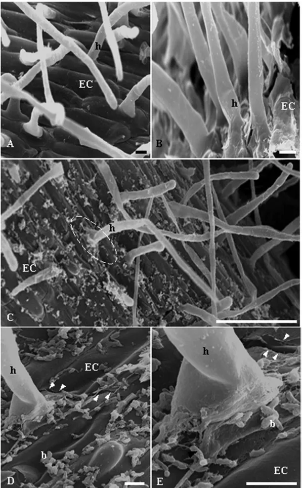
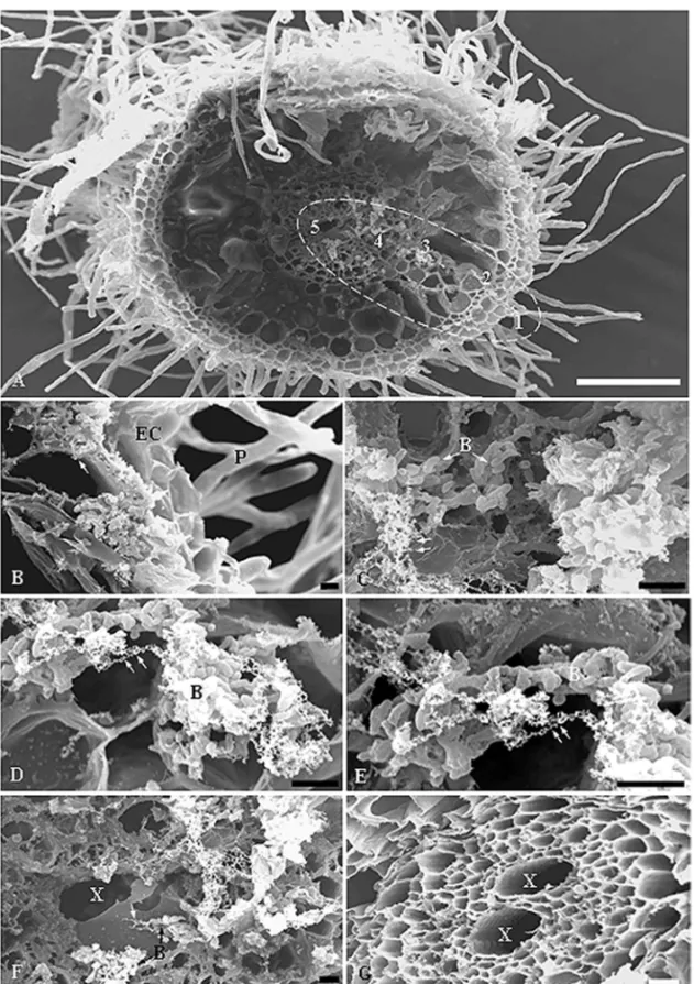
Interaction between rice roots from 7 dpi plantlets andB. kururiensisobtained under gnotobiotic conditions was observed by SEM. Rod-shaped bacteria were observed, mostly in pairs or in small tridimensional microcolonies covering the root surface (Fig. 1C-E). A major presence of bacteria was observed mainly in the root hair zone (Fig. 1C) where bacteria were apparently adhered to the root epidermal cells. Bacteria were particularly accu-mulated in the basal portion of the root hair, forming microaggregates on disrupted areas of the epidermal sur-face (Fig. 1D-E). Such damage of the epidermal sursur-face on heavily colonized areas suggests an active invasion mechanism, probably associated to a high density of bac-terial population as compared to the non-infected control plant (Fig. 1A-B). Figure 2A shows consecutive stages ofB. kururiensisendophytic colonization, characterizing the infection route in rice roots. Following colonization and disruption of epidermis, bacteria appear invading the apoplast (Fig. 2B).
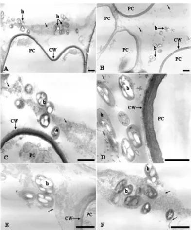
Further analysis of the interaction using TEM showed that B. kururiensis entered the primary roots after 7 dpi. The invasion process followed a pattern with fewer bacteria observed entering the roots and the Burkholderia mainly localized within intercellular spaces of the parenchyma cells (Fig. 3A-F). At this stage, B. kururiensis was observed entering the inner cortex along the vascular cylinder where it was visual-ized by SEM as strings and large microcolonies inside xylem vessels (Fig. 2D-E). No bacteria were observed by SEM on the surface (Fig. 1A-B) or within the tissues (Fig. 2G) of non-infected control plants.
Fig. 2 – SEM showing the infection route byB. kururiensis. (A) General view of a transversal section of an infected root where five zones (1-5) are distinguished characterizing the pathway taken by bacteria from the epidermal surface towards the xylem vessels. (B) Detailed view of zone
Fig. 3 – TEM of the cortex region of the root 7 days incubated withB. kururiensis. In (A) and (B) are shown a general view of the bacteria invasion. Note that bacteria can be observed close to the parenchyma cells (PC) surface (A) or in the intercellular space among these cells (B). In (C) and (D) these bacteria are seen close (*) to the parenchyma cell wall (CW) or associated to a fibrilar material (thin arrows) commonly presented in the intercellular space, that seems to be involved in the formation of bacteria clusters during their interaction with the host plant (EandF). Bar=1µm.
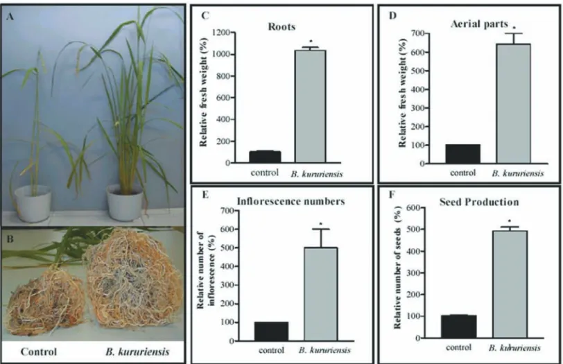
Significant increases in relative fresh weight of whole and isolated parts of the plants, as of flowering and seed production were observed in the analyses of mature plants (Fig. 4A-F). A noteworthy data was observed in the weight and thickness of the roots from inoculated plants, when compared with controls (Fig. 4B). The most remarkable difference was observed in the relative fresh weight of roots and aerial parts (Fig. 4C-D). Other scored yield-increasing traits were flowering and seed produc-tion per pot in inoculated plants as compared to control experiments (Fig. 4E-F).
B. kururiensisDETECTION BY16SRDNA PCR AMPLICATION ANDSEQUENCING
PCR amplification of aB. kururiensischromosomal se-quence was performed with universal bacterial 16S
rDNA primers, which yielded a product with around 900 bp. The template DNA was prepared either di-rectly from the roots and shoots of the infected plantlets (7 dpi) or from genomic DNA purified from bacterial cells isolated of mature plants (120 dpi) (data not shown). PCR products were obtained with universal eubacterial primers, which required additional approach for speci-ficity, as DNA sequencing. Both DNA strands were se-quenced and BLAST search was performed, confirming 100% identity to the correspondingB. kururiensis16S rDNA (GenBank accession number AY586520).
When plant tissues were ground without treatment with PVP and PVPP, amplification was not obtained, suggesting the presence of inhibitory substances in plant extract. PCR efficiency in plant samples was tested using extracts to whichB. kururiensishad been added. Samples taken from mature plants interfered with the PCR (Maes et al. 1996), blocking or decreasing the PCR amplification. Furthermore, the PCR efficiency depended upon the physiological condition of the as-sayed plant tissue as plant age, since PCR amplifica-tion of eubacterial 16S rDNA directly from total tissues DNA was successful from plantlets up to 7 dpi but nega-tive from mature plant tissues (data not shown). Neganega-tive results were probably due to the low number of bacteria inside those plants.
ESTABLISHMENT OFB. kururiensisENDOPHYTIC
COLONIZATION ISIMPAIRED BYNITROGEN
FERTILISATION IN ADOSE-DEPENDENTMANNER
gno-Fig. 4 – Enhanced rice development promoted byB. kururiensisassociation. Picture shows a comparative view of inoculated and non-inoculated plants at 120 dpi. (A) Aerial parts showing extensive differentiation. (B) Root system showing significant difference in weight and thickness. (C) Percentages of relative fresh weight of root systems and (D) aerial parts. (E) Percentage of relative flowering numbers, and seed production (F). Bars represent means, and error bars indicate standard deviations.
tobiotically cultured plantlets the number of viable bac-teria in the external rooting medium range in 1011CFU, regardless the amount of nitrogen supply.
INDUCTION OFINDOLE-3-ACETICACID(IAA) REPORTER
ACTIVITY BYB. kururiensis
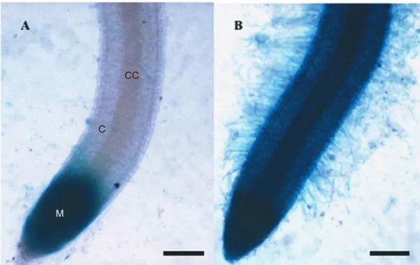
Transgenic seven-day-old rice plantlets, containing the auxin-inducible reporter DR5-GUS, were inoculated with B. kururiensis to assess its ability to induce the production of IAA. 24 h after inoculation, clear differ-ences were observed in the distribution of GUS activity within the plant as compared to non-inoculated plant-lets. Infected plantlets showed blue cells in the root cortex and vascular cylinder indicating that the observed staining was due to activity of the GUS enzyme, sug-gesting that DR5-GUS expression was up-regulated in the presence ofB. kururiensisdue to bacterial auxin pro-duction and secretion within plant tissues. Infected roots
showed intensive blue colour, which included root hairs, apical region of the roots and meristem tissues (Fig. 5B). In several places, a clear, more intense staining was ob-served in the vascular cylinder due to a higher reporter activity. Colonization byB. kururiensisinduced, in all inoculated plants, a large increase in root hair number and length (Fig. 5B). The non-inoculated plantlets ex-hibit a typical blue staining indicative for GUS basal ac-tivity in the root tip, which was higher than in the rest of the root, probably due a higher amount of endogenous auxin at this meristematic tissue (Fig. 5A). Aerial parts were not analyzed for GUS activity.
ANALYSIS OFIAA
Fig. 5 – Bright field microscopy of the GUS expression pattern in the root of transgenic seven-day-old rice plantlets. (A) DR5-GUS activity in control plantlets was detected only in the meristematic tissue,M. (B) Root system showing extensive blue stained due the DR5-GUS activity in response to
B. kururiensis. Radicular cortex,C; Central cylinder,CC; Bar=100µm.
compounds/ml of culture. Based on GC-MS analysis the acidic indole fraction of bacterial culture supernatant showed a retention profile and mass fragmentation pat-tern identical to that of IAA standard. Monitored ions were m/z 202 for the base peak (quinolinium ion) and m/z 319 for the molecular ion (IAA with a trimethylsilyl on the carboxyl and on the indolylic nitrogen) (Allen et al. 1979) (data not shown).
EFFECT OFIAA PRODUCED BYB. kururiensisON THE
DEVELOPMENT OFROOTS
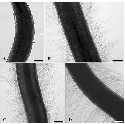
The effect of supernatant ofB. kururiensisculture on the rice roots was verified by microscopy. The presence of a higher number of lateral roots and more abundant root hairs were observed in response to the B. kururiensis supernatant when compared with either B. kururiensis or standard IAA-inoculated roots (Fig. 6). The present observation on bioassays in rice plantlets clearly demon-strates thatB. kururiensis culture supernatant displays a typical auxin activity, such as lateral root formation. This finding is strongly supported by the GC-MS anal-ysis profile mentioned above, which demonstrates the presence of a typical IAA molecule (data not shown).
DISCUSSION
The Gramineae rice (Oryza sativaL.) was chosen as a plant model in this study with the aim of investigating B. kururiensisability to colonize plant tissues. This bac-terium is a TCE-degrading bacbac-terium isolated from an aquifer sample collected at a TCE-polluted site in Japan (Zhang et al. 2000) and has the ability to fix nitrogen (Estrada-de los Santos et al. 2001). The main driving force to determine this putative plant-bacteria association was the phylogenetic closeness betweenB. kururiensis and “B. brasilensis” (Baldani and Baldani 2005, Estrada-de los Santos et al. 2001, Marin et al. 2003), an en-dophytic diazotrophic bacterium isolated from rice in Brazil (Baldani et al. 1997) which have a 100% simi-larity with thenifHgene sequence withB. kururiensis (Marin et al. 2003). In fact, “B. brasilensis” has very recently been reclassified asB. kururiensis (Caballero-Mellado et al. 2007). These data emphasizes the wide geographic and environmental distribution of the spe-ciesB. kururiensis.
Fig. 6 – Effect of IAA produced byB. kururiensison root hairs ofO. sativagrownin vitro. Treated plants were incubated for 24 h in hydroponic media supplemented with
(A) medium non-inoculated (500µl); (B) 50µM IAA standard; (C) 108 B. kururiensis
cells; (D) filter-sterilized culture supernatants ofB. kururiensis. Bar=200µm.
to a remarkably enhanced plant growth and seed pro-duction. Since analysis of auxin/IAA was unequivo-cally identified in culture supernadant ofB. kururiensis we proposed that IAA signaling is essential for guiding auxin-mediated those plant responses. Recently, the pro-duction of IAA inB. vietnamiensishas been described (Govindarajan et al. 2008). Herein, we demonstrateB. kururiensisability to endophytically colonize rice plants, from 7 days and to remain for, at least, 120 dpi within ma-ture and fertile rice plants. At 7 dpi all rice plantlets stud-ied were found endophytically colonized byB. kururien-siswith approximately 109CFU/g of root fresh weight. Although this bacterium had persisted inside rice tissues farther than the initial point of colonization, the number of cells among the tested 120 dpi plants was variable. Opposite pictures are observed in pathogenic plant in-fections, where the presence of characteristic symptoms is directly correlated with very high numbers of bacte-ria within the vascular bundles in later stages of
infec-tion (Purcell and Hopkins 1996, Sabaratnam and Beattie 2003). After 120 dpi the rice plants inoculated withB. kururiensisdid not show any pathologic symptoms.
growth pattern, in pure bacterial cultures. However, our data provide evidences that nitrogen can limit the bacte-rial invasion process, depending on its concentration.
In addition, our results distinguish consecutive stages ofB. kururiensis invasion in rice roots, charac-terizing a possible infection route. The initial step con-sists of the attachment of bacteria onto epidermal cells of the root surface, where root hair zone represent one of the major sites of primary colonization, mainly on the basal region of emerging hairs. At this stage, fil-amentous structures were observed in the microscopic analysis, apparently promoting bacteria-to-bacteria and bacteria-to-plant surface cross-linking. Similar obser-vations have been described in other endophytic inter-actions, resembling bacterial polysaccharide-like struc-tures that may point to crucial stages of ligand-receptor apparatuses with significant potential to modulate the plant-bacteria interactions (Bacilio-Jiménez et al. 2001, James et al. 1994, Ramey et al. 2004, Roncato-Maccari et al. 2003). However, the nature and origin of these fibrils present on inoculated rice are unknown yet. Next, a second stage was characterized by bacterial prolifer-ation through basal hair tissues, where superficial dam-ages were readily detected suggesting an active process, probably mediated by cell wall-degrading enzymes, al-lowing cortical infection by apoplastic dispersion of bacteria as previously described forH. seropedicaeand G. diazotrophicus(Dong et al. 1997, Roncato-Maccari et al. 2003). Although the superficial tissue damage re-sembles an aggressive mode of invasion, a similar picture has been already observed during the interaction of rice with beneficial bacteria likeH. seropedicae(James et al. 1994). A third stage was consistent with the intercel-lular invasion of the internal tissues and subsequent en-trance into the root vascular system, where bacteria could possibly be translocated to the lower stem in the xylem. The main localization ofB. kururiensis in xylem ves-sels suggests that systematic spreading into shoots may be mediated through the transport vessels. The system-atic spreading ofB. kururiensisinto young rice roots was supported by independent lines of evidence (microscopy, 16S rDNA PCR amplification and CFU counting) pre-pared from rice tissues. This study is clearly showing thatB. kururiensiscolonization of rice seedlings occurs in a similar fashion to that described for several
nitrogen-fixing grass endophytes, such asAzoarcussp.,H. sero-pedicae,G. diazotrophicus(Hurek et al. 1994, James et al. 1997).
Several authors showed that endophytic diazo-trophic bacteria as G. diazotrophicus (Sevilla et al. 2001), H. seropediceae (Baldani et al. 2000, Baldani and Baldani 2005, James and Olivares 1998), Azospi-rillum brasilense(Bacillo-Jiménez et al. 2001),B. viet-namiensis(Trân Van et al. 2000) and “B. brasilensis” (Baldani et al. 2000, Baldani and Baldani 2005, Olivei-ra et al. 2002) have the ability to increase the total dry matter of host plants to which they associate, suggest-ing an effect of these bacteria as plant-growth-promotsuggest-ing agents. We propose thatB. kururiensiscan be included in the associative category mentioned above, because it positively acts as a plant growth-promoting agent, also resulting in increased seed yields. The most remarkable plant benefit was observed as the increase in root biomass of plants at 30 to 120 dpi. However, our results demon-strate that, at least for the analyzed rice species, the com-munity of root-colonizing diazotrophs is not stable, as the environment (e.g. availability of nitrogen compounds) and especially plant physiology parameters, such as plant age, have strong influence on bacterial response. The ex-act mechanisms responsible for the plant benefit in dif-ferent plant-bacteria interactions are not clear, although some evidences point to resistance to diseases (Adhikari et al. 2001), increase in available nutrients (Caballero-Mellado et al. 2004), biological nitrogen fixation (Bal-dani et al. 2000, Bal(Bal-dani and Bal(Bal-dani 2005) and bac-terial auxin production (Costacurta and Vanderleyden 1995, Fuentes-Ramírez et al. 1993, James et al. 2002, Pedraza et al. 2004). To study the role of the multifac-torial phytohormone auxin/IAA produced by B. kuru-riensisin our system, the transcriptional regulation of the exogenous auxin-reponsive promoter element DR5 (Scarpella et al. 2003) was monitored in rice roots from plantlets infected with the B. kururiensis. A specific and stronger activation of DR5-GUS was observed in the inoculated plantlets, suggesting that local increase in auxin perception could be due to bacterial auxin/IAA accumulation.
beneficial effects on the growth and seed production of rice plants, possibly associated to differential auxin bal-ance which enhbal-ances the rice plant development. Our findings are based on laboratory cultivation, which may not reproduce exactly the conditions of natural environ-ment, but may indeed reflect the functional diversity of theBurkholderiaspecies. Therefore, B. kururiensis arises as a potential tool for future agricultural applica-tions, providing higher production yields, reduced input costs and negative environmental impact due to the use of nitrogen fertilizers.
ACKNOWLEDGMENTS
The authors are thankful to Prof. Thais Souto-Padron and Prof. Ulysses Garcia Casado Lins from the Elec-tron Microscopy Sector of the Instituto de Microbiologia, Universidade Federal do Rio de Janeiro, for the use of transmission electron microscope FEI-Morgagni. This work was supported by grants from Conselho Nacional de Desenvolvimento Científico e Tecnológico (CNPq) and Fundação Carlos Chagas Filho de Amparo à Pes-quisa do Estado do Rio de Janeiro (FAPERJ).
RESUMO
Burkholderia kururiensisé uma bactéria diazotrófica, original-mente isolada de um ambiente aquático poluído e apresenta alto nível de similaridade com a espécie endofítica “B. brasilensis” encontrada na planta de arroz. Este artigo demonstrou a habili-dade deB. kururiensiscolonizar endofiticamente plântulas de
arroz, após esta bactéria ter sido inoculada na raiz das plantas. Esta capacidade foi confirmada pelo crescimento bacteriano em diferentes tecidos da planta, por microscopia eletrônica e pela análise do 16S rADN. Observação por microscopia eletrônica das raízes, caule e folhas das plântulas de arroz inoculadas, revelou predominância da colonização de B. kururiensisna zona pilífera da raiz, demonstrando que a colonização endo-fítica inicia-se na endoderme, espalha-se pelo xilema, sendo esta a possível via para a bactéria alcançar as partes aéreas. A adição de uma fonte de nitrogênio, embora não tenha in-fluenciado no crescimento bacteriano, foi um fator limitante para a colonização endofítica. Como a colonização endofítica mostrou-se diretamente associada ao aumento no desenvolvi-mento da planta, a produção do fitohormônio auxina/ácido 3-indolacético pelaB. kururiensisfoi verificada utilizando uma plântula de arroz transgênica, contendo o repórter responsivo
para auxina (DR5-GUS). Nossos resultados sugerem que a pro-dução de auxina pelaB. kururiensisé responsável pelo estímulo
no desenvolvimento da planta verificado pela ativação do DR5-GUS. Neste trabalho demonstramos, pela primeira vez, a habi-lidade deB. kururiensiscolonizar endofiticamente a planta de arroz, promovendo tanto o aumento no crescimento da planta como a produção de sementes de arroz.
Palavras-chave: Burkholderia kururiensis, colonização,
ar-roz, bactéria diazotrófica, auxina.
REFERENCES
ADHIKARITB, JOSEPHCB, YANGG, PHILLIPSDAAND
NELSONLM. 2001. Evaluation of bacteria isolated from rice for plant growth promotion and biological control of seedling disease of rice. Can J Microbiol 47: 916–924. ALBRECHTSLANDOKONY. 1980. Cultures of
Azospiril-lum. Methods Enzymol 69: 740–749.
ALLENJRF, GREENWAYAMANDBAKERDA. 1979.
De-terminations of indole-3-acetic acid in xylem sap of
Ricinus communisL. using mass fragmentography. Planta 144: 299–303.
BACILIO-JIMÉNEZ M, AGUILAR-FLORES S, DEL VALLE
MV, PÉREZA, ZEPEDAAANDZENTENOE. 2001. En-dophytic bacteria in rice seeds inhibit early colonization of roots byAzospirillum brasilense. Soil Biol Biochem
33: 167–172.
BALDANIJIANDBALDANIVLD. 2005. History on the
bio-logical nitrogen fixation research in graminaceous plants: special emphasis on the Brazilian experience. An Acad Bras Cienc 77: 549–579.
BALDANI VLD, OLIVEIRA E, BALOTA E, BALDANI JI, KIRCHHOFG ANDDOBEREINERJ. 1997. Burkholde-ria brasilensis sp. nov., uma nova espécie de bactéria
diazotrófica endofítica. An Acad Bras Cienc 69: 116. BALDANI VLD, BALDANI JI ANDDOBEREINER J. 2000.
Inoculation of rice plants with the endophytic diazotrophs
Herbaspirillum seropedicaeandBurkholderiaspp. Biol Fertil Soils 30: 485–491.
BARAC T, TAGHAVI S, BORREMANS B, PROVOOST A, OEYENL, COLPAERTJV, VANGRONSVELDJAND VAN DER LELIE D. 2004. Engineered endophytic bacteria improve phytoremediation of water-soluble, volatile, or-ganic pollutants. Nat Biotechnol 22: 583–588.
BARRAQUIOWL, REVILLALANDLADHAJK. 1997.
BISWASJC, LADHAJKANDDAZZOFB. 2000. Rhizobia in-oculation improves nutrient uptake and growth of lowland rice. Soil Sci Soc Am J 64: 1644–1650.
CABALLERO-MELLADOJ, MARTINEZ-AGUILARL, PARE
-DES-VALDEZ G AND ESTRADA-DE-LOS SANTOS P. 2004.Burkholderia unamaesp. nov., an N2-fixing
rhizo-spheric and endophytic species. Int J Syst Evol Microbiol 54: 1165–1172.
CABALLERO-MELLADOJ, ONOFRE-LEMUSJ, ESTRADA -DE-LOSSANTOSJANDMARTINEZ-AGUILARL. 2007. The tomato rhizosphere, na environment rich in nitrogen-fixingBurkholderiaspecies with capabilities of interest for agriculture and bioremediation. Appl Environ Microbiol 73: 5308–5319.
CHEN WM, JAMESEK, COENYE T, CHOUJH, BARRIOS
E, DE FARIA SM, ELLIOT GN, SHEU SY, SPRENT
JIANDVANDAMMEP. 2006. Burkholderia mimosarum
sp.nov., isolated from root nodules ofMimosaspp. from Taiwan and South America. Int J Syst Evol Microbiol 56: 1847–1851.
CICCILLO F, FIORE A, BEVIVINO A, DALMASTRI C, TABACCHIONISANDCHIARINIL. 2002. Effects of two different application methods ofBurkholderia ambifaria
MCI 7 on plant growth and rhizospheric bacterial diversity. Environ Microbiol 4: 238–245.
COMPANTS, REITER B, SESSITSCH A, NOWAK J, CLÉ -MENT CANDBARKAEA. 2005. Endophytic coloniza-tion ofVitis viniferaL. by plant growth-promoting bac-teriumBurkholderiasp. strain PsJN. Appl Environ Mi-crobiol 71: 1685–1693.
COSTACURTAAANDVANDERLEYDENJ. 1995. Synthesis of phytohormones by plant-associated bacteria. Crit Rev Microbiol 21: 1–18.
CROZIERA, ARRUDAP, JASMINJM, MONTEIROAMAND
SANDBERGG. 1988. Analysis of indole-3-acetic acid and related indoles in culture medium from Azospiril-lum lipoferumandAzospirillum brasilense. Appl Environ Microbiol 54: 2833–2837.
DONGZ, MCCULLYME AND CANNY MJ. 1997. Does
Acetobacter diazotrophicuslive and move in the xylem of
sugarcane stems anatomical and physiological data. Ann Bot 80: 147–158.
EL-BANNAN ANDWINKELMANN G. 1998. Pyrrolnitrin
from Burkholderia cepacia: antibiotic activity against fungi and novel activities against streptomycetes. J Appl Microbiol 85: 69–78.
ELMANNA. 1977. The Van Urk-Salkowski reagent-a
sen-sitive and specific chromatogenic reagent for silica gel thin-layer chromatographic detection and identification of indole derivatives. J Chromatogr 132: 267–276. ESTRADA-DE LOS SANTOS P, BUSTILLOS-CRISTALES R
ANDCABALLERO-MELLADOJ. 2001. Burkholderia, a genus rich in plant-associated nitrogen fixers with wide environmental and geographic distribution. Appl Environ Microbiol 67: 2790–2798.
FUENTES-RAMÍREZLE, JIMENEZ-SALGADOT, ABARCA
-OCAMPO IR AND CABALLERO-MELLADO J. 1993.
Acetobacter diazotrophicus, an indoleacetic acid pro-ducing bacterium isolated from sugarcane cultivars of Mexico. Plant Soil 154: 145–150.
GILLISM, TRÂN VANV, BARDINR, GOOR M, HEBBAR
P, WILLEMS A, SEGERS P, KERSTERS K, HEULINT ANDFERNANDEZ MP. 1995. Polyphasic taxonomy in
the genusBurkholderialeading to an emended description
of the genus and proposition ofBurkholderia vietnamien-sissp. nov. for N2-fixing isolates from rice in Vietnam. Int J Syst Evol Microbiol 45: 274–289.
GORIS J, DE VOS P, CABALLERO-MELLADO J, PARK J, FALSEN E, QUENSEN III JF, TIEDJE JM AND VAN
-DAMMEP. 2004. Classification of the biphenyl- and
poly-chlorinated biphenyl-degrading strain LB400T and rela-tives asBurkholderia xenovoranssp. nov. Int J Syst Evol Microbiol 54: 1677–1681.
GOVINDARAJANM, BALANDREAUJ, KWONS-W, WEON
H-YANDLAKSHMINARASIMHANC. 2008. Effects of the inoculation ofBurkholderia vietnamensisand related endophytic diazotrophic bacteria on grain yield of rice. Microb Ecol 55: 21–37.
HOAGLANDDR. 1975. Mineral nutrition. In: LABOVITCH
A, ANDERSON-PROUTY N AND GHOSHEH S (Eds),
Laboratory Experiments in Plant Physiology. Macmillan Publishing Co. Inc, New York, NY, p. 129–134. HUREKT, REINHOLD-HUREKB,VANMONTAGUMAND
KELLENBERGER E. 1994. Root colonization and sys-temic spreading ofAzoarcussp. strain BH72 in grasses. J Bacteriol 176: 1913–1923.
JAMESEKANDOLIVARESFL. 1998. Infection and colo-nization of sugarcane and other graminaceous plants by endophytic diazotrophs. Crit Rev Plant Sci 17: 77–119. JAMES EK, REIS VM, OLIVARES FL, BALDANI JI AND
DOBEREINER J. 1994. Infection of sugarcane by the nitrogen-fixing bacteriumAcetobacter diazotrophicus. J Exp Bot 45: 757–766.
J. 1997. Herbaspirillum, an endophytic diazotroph col-onizing vascular tissue in leaves ofSorghum bicolorL. Moench. J Exp Bot 48: 785–797.
JAMES EK, GYANESHWAR P, MATHAN N, BARRAQUIO
WL, REDDY PM, IANETTAPPM, OLIVARESFLAND
LADHA JK. 2002. Infection and colonization of rice
seedlings by the plant growth-promoting bacterium
Herbaspirillum seropedicae Z67. Mol Plant Microbe
Interact 15: 894–906.
KUKLINSKY-SOBRAL J, ARAÚJO WL, MENDES R, GE
-RALDI IO, PIZZIRANI-KLEINER AA AND AZEVEDO
JL. 2004. Isolation and characterization of soybean-as-sociated bacteria and their potential for plant growth pro-motion. Environ Microbiol 6: 1244–1251.
LEVINE MM, NATARO JP, KARCH H, BALDANI MM,
KAPER JBAND BLACK RE. 1985. The diarrhead
re-sponse of humans to some classic serotypes of entero-pathogenicEscherichia coli is dependent on a plasmid encoding an enteroadhesiveness factor. J Infection Dis 152: 550–559.
MAESM, GABERNA P ANDCREPEL C. 1996. Identifica-tion and sensitive endophytic detecIdentifica-tion of the fire blight pathogenErwinia amylovorawith 23S ribosomal DNA
sequences and the polymerase chain reaction. Plant Pathol 45: 1139–1149.
MARINVA, TEIXEIRAKRSANDBALDANIJI. 2003.
Char-acterization of amplified polymerase chain reactionglnB
andnifHgene fragments of nitrogen-fixingBurkholderia
species. Lett Appl Microbiol 36: 77–82.
MATTOS KA, TODESCHINI AR, HEISE N, JONES C, PREVIATO JO AND MENDONÇA-PREVIATO L. 2005. Nitrogen-fixing bacteriumBurkholderia brasilensis
pro-duces a novel yersiniose A containing O-polysaccharide. Glycobiology 15: 313–321.
MUÑOZ-ROJAS J AND CABALLERO-MELLADO J. 2003. Population dynamics ofGluconacetobacter diazotrophi-cusin sugarcane cultivars and its effect on plant growth. Microbiol Ecol 46: 454–464.
MURASHIGETANDSKOOGF. 1962. A revised medium for rapid growth and bioassays with tobacco tissue culture. Physiol Plant 15: 473–497.
MUTHUKUMARASAMYR, REVATHIGANDLOGANATHAM
P. 2002. Effect of inorganic N on the population,in vitro
colonization and morphology ofAcetobacter diazotroph-icus(syn.Gluconacetobacter diazotrophicus). Plant Soil 243: 91–102.
MUTHUKUMARASAMYR, GOVINDARAJANM, VADIVELU
M AND REVATHI G. 2006. N-fertilizer saving by the inoculation of Gluconacetobacter diazotrophicus and
Herbaspirillumsp. in micropropagated sugarcane plants. Microbiol Res 161: 238–245.
OLIVEIRAALM, URQUIAGAS, DOBEREINERJANDBAL
-DANIJI. 2002. The effect of inoculating endophytic N2
-fixing bacteria on micropropagated sugarcane-plants. Plant Soil 242: 205–215.
PEDRAZARO, RAMIREZ-MATAA, XIQUIMLANDBAÇA
BE. 2004. Aromatic amino acid aminotransferase activity and indole-3-acetic acid production by associative nitro-gen-fixing bacteria. FEMS Microbiol Lett 233: 15–21. PERINL, MARTINEZ-AGUILARL, CASTRO-GONZALEZR,
ESTRADA-DE LOS SANTOS P, CABELLOS-AVELAR T, GUEDES HV, REIS VM ANDCABALLERO-MELLADO
J. 2006. Diazotrophic Burkholderiaspecies associated
with field-grown maize and sugarcane. Appl Environ Microbiol 72: 3103–3110.
PURCELLAHANDHOPKINSDL. 1996. Fastidious xylem-limited bacterial plant pathogens. Annu Rev Phytopathol 34: 131–151.
RAMEY BE, KOUTSOUDIS M, VON BODMAN SB AND
FUQUAC. 2004. Biofilm formation in plant microbe as-sociations. Curr Op Microbiol 7: 602–609.
REINHOLDB, HUREK T, NIEMANNEGANDFENDRIKI.
1986. Close association ofAzospirillumand diazotrophic
rods with different root zones of kallar grass. Appl Envi-ron Microbiol 52: 520–526.
REISVMET AL. 2004.Burkholderia tropicasp.nov., a novel nitrogen-fixing, plant-associated bacterium. Int J Syst Evolut Microbiol 54: 2155–2162.
RODRIGUESNETOJ, MALAVOLTAJRVAANDVICTORO. 1986. Meio simples para isolamento e cultivo de Xan-thomonas campestrispv. citri Tipo B. Sum Phytol 2: 16.
RONCATO-MACCARI LDB, RAMOS HJO, PEDROSA FO,
ALQUINI Y, CHUBATSU LS, YATES MG, RIGO LU,
STEFFENS MBR AND SOUZA ES. 2003. Endophytic Herbaspirillum seropedicaeexpressesnifgenes in grami-nous plants. FEMS Microbiol Ecol 1519: 1–9.
ROSENBLUETHMANDMARTINEZ-ROMEROE. 2006. Bac-terial endophytes and their interactions with hosts. Mol Plant Microbe Interact 19: 827–837.
SABARATNAMSANDBEATTIEGA. 2003. Differences be-tween Pseudomonas syringae pv. syringaeB728a and Pantoea agglomeransBRT98 in epiphytic and endophytic
SANTOS AV, DILLON RJ, DILLON VM, REYNOLDS SE
ANDSAMUELSRI. 2004. Occurrence of the antibiotic producing bacteriumBurkholderiasp. in colonies of the leaf-cutting antAtta sexdens rubropilosa. FEMS Micro-biol Lett 239: 319–323.
SCARPELLAE, RUEBSANDMEIJERAH. 2003. The
RADI-CLELESS1 gene is required for vascular pattern forma-tion in rice. Development 130: 645–658.
SESSITSCH A ET AL. 2005. Burkholderia phytofirmans
sp.nov., a novel associated bacterium with plant-beneficial properties. Int J Syst Evol Microbiol 55: 1187–1192.
SEVILLAM, BURRISRH, GUNAPALANANDKENNEDYC. 2001. Comparison of benefit to sugarcane plant growth and 15N
2 incorporation following inoculation of sterile
plants withAcetobacter diazotrophicuswild-type and
Nif-mutant strains. Mol Plant Microbe Interact 14: 358–366. TANZ, HUREKT ANDREINHOLD-HUREK B. 2003.
Ef-fect of N-fertilization, plant genotype and environmental conditions onnifHgene pools in roots of rice. Environ Microbiol 5: 1009–1015.
TOMITAK, KITSUWAT, MURAYAMATANDNAKAMURA
T. 1987. Identification of indol-3-acetic acid in Neuros-pora crassa. Agric Biol Chem 59: 2633–2634.
TRÂN VANV, BERGEO, NGOKES, BALANDREAUJAND
HEULINT. 2000. Repeated beneficial effect of rice in-oculation with a strain ofBurkholderia vietnamiensison
early and late yield components in low fertility sulphate acid soils of Vietnam. Plant Soil 218: 273–284.
WANGJL, MAOZY, HANLPANDQIANY. 2004.
Bioreme-diation of quinoline-contaminated soil using bioaugmen-tation in slurry-phase reactor. Biomed Environ Sci 17: 187–195.
ZHANG H, HANADA S, SHIGEMATSU T, SHIBUYA K, KAMAGATAY, KANAGAWATANDKURAMER. 2000.
Burkholderia kururiensis sp.nov., a trichloroethylene