Printed version ISSN 0001-3765 / Online version ISSN 1678-2690
www.scielo.br/aabc
http://dx.doi.org/10.1590/0001-3765201420140167
Complete assignments of NMR data and assessment of trypanocidal activity of new eremantholide C derivatives
DÊNIA A. SAÚDE-GUIMARÃES1, DÉLIO S. RASLAN2, EGLER CHIARI3 and ALAÍDE B. DE OLIVEIRA4 1
LAPLAMED, DEFAR, Escola de Farmácia, Universidade Federal de Ouro Preto, Campus Morro do Cruzeiro, s/n, Bauxita, 35400-000 Ouro Preto, MG, Brasil
2Departamento de Química, Universidade Federal de Minas Gerais, Av. Antônio Carlos, 6627, Pampulha, 31207-901 Belo Horizonte, MG, Brasil 3
Departamento de Parasitologia, ICB, Universidade Federal de Minas Gerais, Av. Antônio Carlos, 6627, Pampulha, 31207-901 Belo Horizonte, MG, Brasil 4
Departamento de Produtos Farmacêuticos, Faculdade de Farmácia, Universidade Federal de Minas Gerais, Av. Antônio Carlos, 6627, Pampulha, 31270-901 Belo Horizonte, MG, Brasil
Manuscript received on April 4, 2014; accepted for publication on July 8, 2014
ABSTRACT
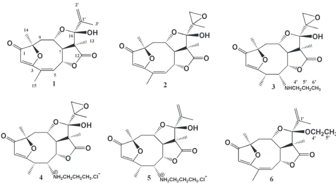
Chemical transformations of eremantholide C (1), a sesquiterpene lactone that was isolated from
Lychnophora trichocarpha Spreng. led to five new derivatives: 1’,2’- epoxyeremantholide C (2), 5-n-propylamine-4,5-dihydro-1’,2’-epoxyeremantholide C (3), 5-n-propylammonium-4,5-dihydro-1’,2’-epoxyeremantholide C chloride (4), 5-n-propylammonium-4,5-dihydroeremantolide C chloride (5) and 16-O-ethyleremantholide C(6). The structures of all these derivatives were assigned on the basis of IR, MS, 1H and 13C NMR data by 1D and 2D techniques. Eremantholide C and the derivatives 2, 4 and 5 were
evaluated against trypomastigotes Y and CL strains of Trypanosoma cruzi. Eremantholide C completely
inhibited the growth of both the parasites strains while all derivatives were partially active against the CL strain and inactive against the Y strain.
Key words: Eremantholide C derivatives, Lychnophora trichocarpha, NMR, sesquiterpene lactones, trypanocidal activity.
Correspondence to: Dênia Antunes Saúde-Guimarães E-mails: saude@ef.ufop.br / saudeguima@gmail.com
INTRODUCTION
Sesquiterpene lactones are chemical markers of certain plant families such as Asteraceae. The large
number of biological activities experimentally
described up to now, raised great interest on this group of substances. The sesquiterpene lactones
of the furanheliangolide type are biogenetically
derived from heliangolides and are often found in
species of the genus Lychnophora, that is native from Brazil (Bohlmann and Jakupovic 1990). In previous studies, eremantholide C and its oxidized
derivatives showed activity against Trypanosoma
cruzi (Oliveira et al. 1996, Saúde-Guimarães
et al. 2007). Other biological/pharmacological activities were reported for eremantholide C, such
as antibacterial, anti-hyperuricemic, anti-gouty arthritis, anti-inflammatory and antitumor (Barrero
Ferrari et al. 2013, Saúde-Guimarães et al. 2014). Aiming to obtain new bioactive derivatives of eremantholide C (Figure 1), this sesquiterpene
lactone was submitted to chemical modifications.
MATERIALS AND METHODS
GENERAL PROCEDURES
Mass spectra were obtained with VG Autospec (electron impact) and HP 5988A (chemical
ionization with methane) spectrometers by direct
injection (ionization chamber at 200 °C). IR spectra
were taken at Galaxy 3000-FTIR spectrophotometer
(Mattson Instruments). NMR spectra were taken at Bruker Avance DPX (4.7T), DRX (9.4T), equipped with a 5mm dual probe, at 300 K, with TMS as internal reference. One-dimensional 1H and 13C NMR spectra were acquired under standard
conditions, with 90° pulse widths of 8.00 µs and 8.50 µs for 1H and 13C, respectively. 1H NMR spectra were obtained using a sweep width of 3 kHz over 32k data points. 13C NMR spectra were obtained
using a sweep width of 31 kHz. DEPT, 1H,1H COSY
and 1H,13C HETCOR techniques were performed
using standard pulse sequences supplied by the
spectrometer manufacturer.
SYNTHESIS OF EREMANTHOLIDE C DERIVATIVES 2 TO 6
Eremantholide C (1) was obtained from aerial parts
of L. trichocarpha Spreng. that were collected
in Minas Gerais, Brazil, as previously described
(Saúde et al. 1998, Ferrari et al. 2013). Its structural characterization is described elsewhere (Le Quesne et al. 1978, Saúde et al. 1998, Saúde-Guimarães et al. 2007).
1',2'-Epoxyeremantholide C (2) was prepared
by reaction of 1 (2.9 mmol) with m-chloroperbenzoic
acid (5.3 mmol) in 50 mL of anhydrous CHCl3. The
mixture was stirred at room temperature for 2 h. The reaction mixture was worked out according to Carda et al. 1986. The resulting residue (1.14 g)
was purified by column chromatography (silica gel,
hexane: EtOAc 1:1), yielding 970 mg of 2 (93%
yield, m.p. 213-217 °C).
1’,2’-Epoxyeremantholide C (2):
6,9-Epoxy-2H-1,4-dioxacyclodeca[c,d]pentalene-2,7(4aH
)-
dioxane,2a,3,5,6,11a,11b-hexahydro-3-hydroxy-2a,6,10-trimethyl-3-(1’,2’-epoxy)-2aR*,3S*,4a
R*,6S*,10Z,11aS*,11bS*). White solid, m.p.
213-217°C. IR (KBr) ν max (cm-1): 3400 (OH), 1770
(C=O, γ-lactone), 1700 (ketone C=O), 1650 (C=C),
1590 (C=COR, furanone), 1450, 1350, 1300, 1275, 1150, 1100, 1050, 1000, 900. MS (EI), m/z (rel. int.):
362 [M+, C
19H22O7] (20), 344 (M - H2O, 17), 234
(19), 205 (19), 189 (20), 177 (29), 165 (32), 149 (25), 138 (55), 122 (53), 95 (100), 69 (58), 57 (22).
5-n- Propylamine
-4,5-dihydro-1’,2’-epoxye-remantholide C (3) was obtained by reaction of 2 (0.55 mmol) with n-propylamine (5.94 mmol), at
-18 °C for 16 h (Kirk 1973). After this period, the
excess of the amine was removed by evaporation
under reduced pressure, at room temperature. The
residue obtained was purified by PTLC on silica
gel (0.5 mm thickness, eluent - hexane: EtOAc
1:1) resulting in 82 mg (35% yield) of 3 as a white
solid. This compound was dissolved in dry THF and
treated with gaseous HCl leading to the formation of
4 as a white hydrosoluble solid (quantitative yield).
5 n P ro p y l a m i n e 4 , 5 d i h y d ro 1 ’ , 2 ’ -epoxyeremantholide C (3): White solid, m.p. 183-189 0C. IR (KBr) ν
max (cm-1): 3400 (OH),
1770 (C=O, γ-lactone), 1700 (C=O, ketone), 1590
(C=COR, furanone), 1450, 1380, 1300, 1200, 1150, 1100, 1050, 1000, 900, 850. MS (EI), m/z (rel. int.): 421 (M+, C
22H31O7N, 99), 403 (M - H2O, 28), 238 (25), 195 (64), 168 (31), 151 (25), 138 (33), 125 (100), 99 (23), 72 (35).
5-n-Propylammonium-4,5-dihydro-1’,2’-epoxyeremantholide C chloride (4): white solid, m.p. 176-179 0C. MS (EI), m/z (rel. int.): 457 (M+, C22H32O7NCl, 72), 439 (M - H2O, 11), 390 (18), 364 (31), 336 (21), 318 (59), 195 (79), 168 (44), 152 (19), 138 (45), 125 (100), 99 (45), 81 (18), 72 (46).
5-n-Propylammonium-4,5-dihydroereman-tolide Cchloride(5)was obtained from the reaction
of 1 (0.58 mmol) with n-propylamine (5.94 mmol),
at -18 °C for 10 h (Kirk 1973). After this period, the
excess of amine was removed by evaporation under
reduced pressure, at room temperature. The residue
obtained was purified by PTLC on silica gel (0.5 mm
thickness, eluent - hexane: EtOAc 1:1) affording
120 mg of a yellowish pasty material. Silica TLC of this material showed three spots when revealed by iodine. After development with ninhydrin, the TLC showed only a rosy spot, characteristic of amines. This material was then dissolved in dry THF and treated with gaseous HCl to give the hydrochloride
5 as a white hydrosoluble solid (70 mg).
5-n-Propylammonium-4,5-dihydroere-mantolide C chloride (5): White solid, m.p. 195-197 0C. MS (EI), m/z (rel. int.): 405 (60), 387 (M - H2O, 5), 195 (40), 168 (32), 135 (21), 125 (100), 69 (35).
16-O-Ethyleremantholide C (6) was obtained,
according to the methodology described by
Partwardhan et al. 1974, by reacting 1 (0.28 mmol)
with 0.08 mL of triethyl orthoformate and 25 mg of Amberlyst resin 15. The reaction mixture was stirred at room temperature for 3 days. Then it
was neutralized with aqueous K2CO3 solution
and filtered. The product was extracted with diethyl ether. The organic layer was washed with water, filtered on anhydrous sodium sulfate and concentrated under reduced pressure, yielding 72 mg
of ether 6 as a white solid (66% yield).
16-O-Ethyleremantholide C (6): White
solid. IR (KBr) νmax (cm-1): 1770 (C=O, γ-lactone),
1700 (C=O, ketone), 1660 (C=C), 1590 (C=COR, furanone), 1450, 1380, 1300, 1280, 1260, 1210, 1200, 1150, 1140, 1100, 1060, 1000, 870, 750. MS (EI), m/z (rel. int.): 375 (M+1)+, C
21H26O6, 3), 329 (54), 285 (17), 199 (9), 165 (49), 95 (85), 69 (100), 67 (18), 45 (23), 43 (39).
IN VITROASSAYS WITH TRYPANOSOMA CRUZI
TRYPOMASTIGOTES
Samples of eremantholide C and of the deriva-tives 2, 4 and 6 were dissolved or suspended in
dimethyl sulfoxide (DMSO) (0.2 mL) and plus
Krebs-Ringer-glucose (2.0 mL) and mixed with an equal volume of parasitized whole blood diluted
in bovine calf serum. A parasite density of 2 x 10⁵
trypomastigotes/0.1 mL was calculated for each
flat-bottomed test tube (4mL, 56 x 13 mm); control
tubes without the test extracts were included. After incubation at 4 °C for 24 h the suspensions were
examined microscopically. Only those samples
that killed 100% of the parasites were considered active. Samples that inhibited 50% of parasite growth compared to control were considered
partially active.
RESULTS AND DISCUSSION
Data of the 1H and 13C NMR spectra of derivatives
2-6 are given at on Tables I and II, respectively.
The chemical shifts were assigned by consideration
of known substituent effects of the groups concerned and with the aid of both 1H,1H COSY and 1H,13C
HETCOR contour maps. The stereochemistry of
the carbons were determined based on coupling constants and 1H,1H NOESY contour maps.
From the reaction of 1 with m-chloroperbenzoic acid, the main product 2 was obtained and it was
characterized by 1H NMR data. The presence of an
epoxy group in 2 was highlighted in the 1H NMR
spectrum by the two doublets at δ3.11 (J = 5.5 Hz) and δ2.70 (J = 5.5 Hz), attributed to H-2’a and
H-2’b, which appear in the 1H NMR spectrum of 1
at δ5.31 (br s) and δ5.07 (t, J = 1.6 Hz), respectively. The signals at δ130.00 and δ115.80, attributed to the olefinic carbons C-1’ and C-2’, respectively,
in the 13C NMR spectrum of 1, are replaced in the spectrum of 2 by the signals at δ59.50 and δ58.47, typical chemical shifts of oxygenated carbons with
hybridization sp3. In addition, the signal attributed
to C-3 in the spectrum of 1 (δ19.00) is shifted
to δ11.44 in the spectrum of 2, confirming the
epoxidation of the ∆1double bond of 1.
The mass spectrum of 2 showed the molecular ion peak at m/z 362, which represents an increment of 16 mass units to the molar mass of 1,
correspon-ding to the addition of an oxygen atom. This mass
is consistent with the molecular formula C19H22O7. Data of IR, MS, 1H NMR and 13C NMR of 2, are in agreement with the new derivative
1’,2’-epoxyeremantholide C.
Epoxide 2 was reacted with methylamine, cyclohexylamine, diethylamine and n-propylamine. Reactions with the first three amines led to mixtures of many products, in such a way that no derivative with suitable purity was obtained. The reaction
of 2 with n- propylamine afforded a less complex
mixture of products which, after separation by column chromatography on silica gel, led to the
amino product 3 with a 35% yield. The amine 3 was transformed into its hydrochloride salt by reaction with gaseous HCl in dry THF, leading to
the hydrosoluble compound 4.
The multiplets in δ5.02-4.98 and δ6.03-6.00
attributed, respectively, to H-6 and H-5, in the 1H
NMR spectrum of 2 appear in the spectrum of 3 at
δ4.26 (d, J = 6.5 Hz) and at δ2.99 (brs), respectively. The quartet at δ2.89 (J = 7.2 Hz), that appears in
the 1H NMR spectrum of 2, was assigned to H-4. A correlation of this signal with the doublets at
δ1.37 (J = 7.2 Hz), assigned to H-15, is indicated by the values of the coupling constants and by the
COSY 1H-1H contour map. The signals at δ134.27
and δ130.24 which are attributed to the olefinic carbons C-5 and C-4, respectively, as well as the
signal attributed to C-15 (δ20.39) in the 13C NMR
spectrum of 2, are shifted in the 13C NMR spectrum
of 3 to δ65.24, δ38.20 and δ16.70, respectively,
allowing the proposition that the amine group was linked to the C-5 of compound 2.
H 1*, 1 2**, 1 3**, 1 4**, 2 5**, 2 6**, 1
2 5.63 s 5.62 s 5.56 s 5.99 s 5.93 s 5.62 s
4 - - 2.89 q
J 4,15 = 7.2
3.56 q J4,15 = 7.2
3.45 q J 4,15 = 7.2
-5 6.04-6.03 m 6.03-6.00 m 2.99 brs 3.97 brs 3.88 brs 6.03-6.01 m
6 5.02-4.98 m 5.02-4.97 m 4.26 d J6,7 = 6.5 4.74 a 4.73 d J6,7 = 7.9 4.90-4.86 m
7
2.82 dd J6,7 = 7.1; J7,8 = 4.2
2.87 dd J6,7 = 7.3;
J7,8 = 4.2
2.95 dd J6,7 = 6.5;
J7,8 = 4.9
3.09 dd J6,7 = 7.8;
J7,8 = 4.2
3.04 dd J6,7 = 7.8;
J7,8 = 4.3
2.84 dd J6,7 = 7.2;
J7,8 = 4.4
8
4.09 ddd J7,8 = 4.2; J8,9a = 2.5; J8,9b = 12.0
4.07 ddd J7,8 = 4.2; J8,9a = 2.6; J8,9b = 11.9
3.91 ddd J7,8 = 4.9; J8,9a = 2.3; J8,9b = 11.9
3.92 m
3.96 ddd J7,8 = 4.3; J8,9a = 2.8; J8,9b = 113
3.85 ddd J7,8 = 4.4; J8,9a = 2.4; J8,9b = 12.0
9a
2.48 dd J8,9a = 2.5; J9a,9b = 13.5
2.39 dd J8,9a = 2.6; J9a,9b = 13.6
2.39 dd J8,9a = 2.3; J9a,9b = 13.5
2.32 dd J8,9a = 2.3; J9a,9b = 13.6
2.27 dd J8,9a = 2.8; J9a,9b = 13.8
2.41 dd J8,9a = 2.4; J9a,9b = 13.6
9b
2.00 dd J8,9b = 12.0; J9a,9b = 13.5
1.94 dd J8,9b = 11.9; J9a,9b = 13.6
1.90 dd J8,9b = 11.9; J9a,9b = 13.5
2.39 J8,9b = 11.5; J9a,9b= 13.6
2.34 dd J8,9b = 11.3; J9a,9b = 13.8
2.06 dd J8,9b = 12.0; J9a,9b = 13.6
13 1.18 s 1.33 s 1.38 s 1.39 s 1.21 s 1.16 s
14 1.45 s 1.48 s 1.43 s 1.50 s 1.45 s 1.50 s
15 2.05 t
J5.15 =6,15 = 1.9
2.05 t J5,15 =6,15 = 2.0
1.37 d J4,15 = 7.2
1.52 d J4,15 = 7.2
1.48 d J4,15 = 7.2
2.06 dd J5,15 = 2.0;
J6,15 = 1.6
2'a 5.31 brs 3.11 d J2’a,2´b = 5.5
3.12 d J2’a,2´b = 5.5
4.04 d
J2’a,2´b = 11.9 5.22 brs
5.28 d J2’a,2´b = 1.2
2'b 5.07 t
J2’a,2´b = 1.6
2.70 d J2’a,2´b = 5.5
2.71 d J2’a,2´b = 5.5
3.75 d J2’a,2´b = 11.9
5.13 t
J2’a,2´b = 1.4 5.13 brs
3' 1.91 s 1.55 s 1.57 s 1.50 s 1.77 s 1.79 s
4’a -
-2.8 dt J4’a,4´b = 11.2;
J4’a,5’ = 7.0
3.28 td J4’a,4´b = 11.2;
J4’a,5’ = 5.8;
3.18 dt J4’a,4´b = 10.5;
J4’a,5’ = 5.7
3.36 dq J4’a,4´b = 9.2;
J4’a,5’ =7.2
4’b -
-2.53 dt J4’a,4´b = 11.2;
J4’b,5’= 6.9;
3.17 td J4’a,4´b = 11.2;
J4’b,5’=5.8
3.09 dt J4’a,4´b = 10.5;
J4’b,5’ =5.7
3.20 m
5’ - - 1.50 m 1.80 m 1.75 m 1.07 t
J4’a,5’ =,4´b,5’ = 7.2
6’ - - 0.93 t
J5’,6´ = 7.3
0.99 t J5’,6´ = 7.4
0.94 t
J5’,6´ = 7.3
-OH 3.79 s 3.69 s
TABLE I
1H NMR data for compounds 1, 2, 3, 4, 5 and 6, δ, J (Hz).
indicated the presence of a propylamine group
at 3. This group was confirmed by the 13C NMR
signals at δ50.51, δ17.52 and δ11.65, attributed to carbons 4’, 5’ and 6’, respectively.
The mass spectrum of 3 presented the molecular ion peak at m/z 421 u, corresponding to 59 mass units higher than the molecular ion
of 2, and it corresponds to n-propylamine group
(C3H7NH2). The molar mass of 3 was compatible with the molecular formula C22H31O7N.
Based on data retrieved from IR, MS, 1H and 13C NMR it was concluded that the product obtained was that resulting from the amine addition to carbon-5 of the epoxide 2, through a
Michael’s reaction, generating the novel derivative
5-n-propylamine-1’,2’-epoxyeremantholide (3). This
might be com pared to the proposed mechanism
for the antitumor activity of the eremantholides by
reaction with biological nucleophiles (Mc Dougal et al. 1989).
The shifting of the signals due to the hydrogen and carbon atoms 4, 5, 15, 4’a, 4’b, 5’ e 6’ of
com-pounds 3 and 4 could be observed by comparing
their 1H and 13C NMR spectra.
The signals relative to H-4, H-5, H-15, H-4’a and H-4’b, H-5’ and H-6’ appear in the spectrum of
3, respectively, at δ2.89 (q, J = 7.2 Hz), δ2.99 (brs),
δ1.37 (d, J = 7.2 Hz), δ2.80 (dt, J = 7.0 and 11.2 Hz), δ2.53 (dt, J = 6.9 and 11.2 Hz), δ1.50 (m) and
δ0.93 (t, J = 7.3 Hz), while in the spectrum of 4
they are shown at δ3.56 (q, J = 7.2 Hz), δ3.97 (brs), δ1.52 (d J = 7.2 Hz), δ3.28 (td, J = 5.8 and 11.2 Hz), δ3.17 (td, J = 5.8 and 11.2 Hz), δ1.80 (m) and δ0.99 (t, J = 7.4 Ηz), respectively.
In the 13C NMR spectrum of 3, the signals at δ
38.20, δ 57.18, δ 16.70, δ 50.51, δ17.52 and δ11.65 were assigned to C-4, C-5, C-15, C-4’, C-5’ and C-6’, respectively. These signals were observed at δ33.44, δ58.65, δ15.06, δ49.11, δ19.04 and δ10.52,
respectively, in the spectrum of 4.
The signal corresponding to H-6 in the 1H NMR spectra of 4 overlapped that of the solvent
(D2O) at δ4.74. This was confirmed by the 1H-1H
COSY contour map of 4, evidencing correlations
between the signal at δ4.74 (brs), and the signals attributed to H-7 (δ3.09, dd, J = 4.2 and 7.8 Hz),
C 1 *, 1 2**, 1 3**, 1 4**, 2 5**, 2 6**, 1
C-1 205.89 205.23 204.53 209.06 209.34 205.24
C-2 104.54 104.50 104.03 105.93 106.08 104.45
C-3 187.27 186.64 192.93 192.39 192.53 186.91
C-4 130.00 130.15 38.14 33.44 33.68 139.10
C-5 134.77 134.28 57.18 58.65 57.30 134.75
C-6 81.46 81.34 82.42 80.77 81.00 81.75
C-7 62.53 63.06 65.24 63.79 63.99 63.10
C-8 78.37 78.38 77.45 76.47 77.05 78.43
C-9 43.46 43.25 43.84 42.38 42.60 43.89
C-10 90.24 89.95 89.88 91.88 91.99 89.98
C-11 59.88 58.43 58.51 61.65 60.61 60.00
C-12 175.72 174.70 175.09 177.21 178.19 175.76
C-13 21.94 20.64 21.16 21.52 20.83 21.66
C-14 20.48 20.39 20.51 20.16 20.22 20.64
C-15 20.30 20.33 16.70 15.06 15.17 20.49
C-16 106.09 104.57 104.94 107.06 107.27 109.59
C-1’ 142.22 59.60 59.71 76.67 141.39 139.28
C-2’ 115.80 53.59 53.80 50.06 116.63 117.61
C-3’ 19.00 11.44 17.55 20.07 19.19 18.46
C-4’ - - 50.51 49.11 49.22 57.26
C-5’ - - 17.52 19.04 18.70 15.29
C-6’ - - 11.65 10.52 10.62 -* - 75 MHz; -*-* - 100 MHz; 1 - CDCl3; 2 - D2O.
TABLE II
13C NMR data (δ) for 1, 2, 3, 4, 5 and 6.
The two doublets at δ2.71 (J = 5.5 Hz) and δ3.12 (J = 5.5 Hz) assigned to H-2’a and b, respectively, and the signals at δ59.71 and δ53.80
on the 13C NMR spectrum, due to carbons 1’ and 2’,
respectively, showed that the epoxy group remained
unchanged in the molecule of 3.
The assignments made to the signals in the 1H NMR spectrum of 3 were confirmed by the
values of the coupling constants and by the 1H-1H
COSY contour map which showed the following correlations: H-4 and H-15; H-4’a, H-4’b and H-5’; H-5’ and H-6’; H-6 and H-7, and H-2’a and
b. Chemical shifts for 13C NMR spectrum of 3 were
assigned by comparison with the spectrum of 2 and
and to H-5 (δ3.97, brs). The 1H-1H COSY also evidenced correlations between the following
hydrogen atoms: H-4 and H-15; H-8, H-9a and H-9b; H-9a and H-9b; H-4’a, H-4’b and H-5’; H-5’and H-6’; H-2’a, H-2’b and H-3’.
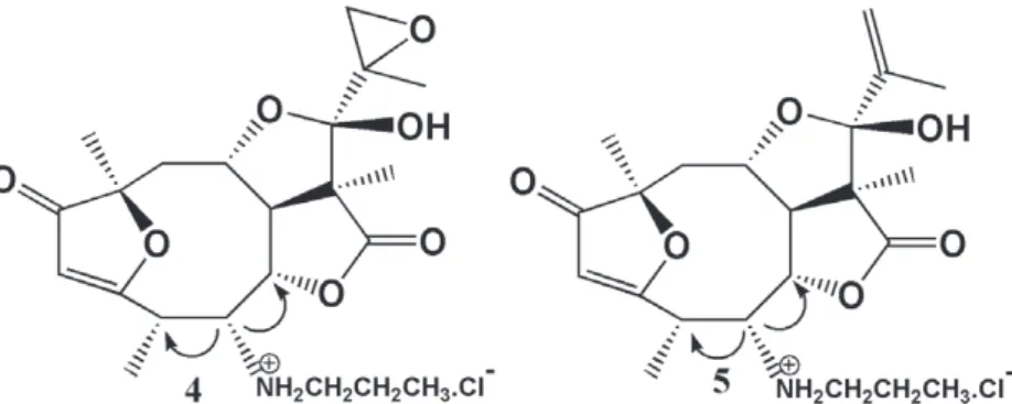
The relative configuration of the n-propylamine and the C-15-methyl group was defined on
the basis of the 1H,1H NOESY (Figure 2), that
indicated correlation between the H-5 signal
at δ3.97 (brs) , with the signals at δ4.74 and δ3.56 (q, J = 7.2 Hz), attributed to H-6 and H-4, respectively. This correlation showed a –cis
relationship between the H-4 and H-5 atoms with
a β configuration. Consequently, the C-15-methyl group and the n-propylamine group should have an α configuration.
Figure 2 - Arrows represent NOESY correlations.
The mass spectrum of 4 presented a molecular ion peak m/z 457 u, compatible with a molecular formula C22H32O7NCl. Such a molecular mass represents an increment of 36 units compared with the mass of 3, compatible with the addition of one HCl per molecule of 4.
Based on data from IR, MS, 1H and 13C
NMR, COSY and NOESY it was concluded that
compound 4 is 5-n-propylammonium-4,5-dihydro-1’,2’-epoxyeremantholide C chloride, a novel com-pound that is firstly described in this paper.
In order to confirm the positioning of the
n-propylamine group at the C-5 of compound 2,
the reaction of 1 with n-propylamine was carried
out. Three spots were observed when a silica gel
TLC plate of the reaction mixture was sprayed with iodine. Under ninhydrin, only a rosy spot typical
of amine, was observed. Then, the mixture was
treated with gaseous HCl in dry THF yielding 5 as
a white hydrosoluble solid.
In the 1H NMR spectrum of 5, the signals at
δ3.18 (td, J = 5.7 and 10.5 Hz), δ3.09 (td, J = 5.7 and 10.5 Hz), δ1.75 (m) and δ0.94 (t, J = 7.3 Hz) were
attributed to H-4’a, H-4’b, H-5’and H-6’ of the n-propylammonium group, respectively. These data were further confirmed by the signals at δ49.22 and
δ18.70, together with the signal at δ10.62 in the 13C
NMR of 5, which were attributed, respectively, to
C-4’, C-5’ and C-6’ of the n- propylammonium group.
The 1H NMR spectrum of 5 presented a quartet
at δ3.45 (1H, J = 7.2 Hz) that was attributed to H-4.
The signals for H-5 and H-15 in this spectrum
appeared as a broad singlet at δ3.88 and a doublet
at δ1.48 (J=7.2 Hz), respectively. In the 1H NMR
spectrum of 1 these atoms are represented by signals at δ6.04−6.03 (m) and δ2.05 (t, J = 1.9 Hz), respectively.
Values of coupling constants for H-4 and H-15
indicate that these hydrogens are in vicinal positions.
The signal attributed to H-6 in the 1H NMR spectra of 5 is probably superimposed to that of
H2O in the solvent (D2O), which appear at δ4.74. In order to shift the signal of H2O and observe the one from H-6, the 1H NMR spectra of 5 in D
2O was run at 330 K, when the signal due to the solvent was
The stereochemistry of the n-propylammonium and of the C-15 methyl groups were inferred based on the NOESY technique (Figure 2), which indicated a correlation between the signal at δ3.88 (brs), attributed to H-5, and the signals at δ4.73 (d, J = 7.9 Hz) and δ 3.45 (q, J = 7.2 Hz), corresponding to H-6 and H-4, respectively. These data suggest
that H-4 and H-5 have a cis relationship and a β
configuration. Consequently, the C-15 methyl and the n- propylammonium groups have a α configuration.
The molecular ion peak m/z 441 u, corresponding to the molecular formula C22H32O6NCl, was not observed at the mass spectrum of 5, but it showed a peak at m/z 405 u that was compatible to the loss of a molecule of HCl from the molecular ion.
Data of IR, MS, 1H and 13C NMR spectra, and the
COSY and NOESY contour maps are in agreement with the new derivative 5-n- propylammonium-4,5-dihydroeremantholide C chloride, a new derivative of
eremantholide C.
The infrared spectrum of 6 showed no
absorption characteristic of the hydroxyl group.
The 1H NMR spectrum presented signals at δ3.36
(dq, J = 7.2 and 9.2 Hz), δ3.20 (m) and δ1.07 (t, J = 7.2 Hz) corresponding to H-4’a, H-4’b and H-5’, respectively, and that indicates the presence
of an ethoxyl group. The COSY 1H,1H contour map
evidenced correlations between H-4’a, H-4’b and H-5’ atoms.
The 13C NMR spectrum of 6 shows a signal
at δ57.26, characteristic for a sp3carbon bonded
to oxygen and, therefore, it was attributed to C-4’. The signal at δ15.29 was assigned to the methyl carbon at the 5’ position. These data confirmed the
presence of an ethoxyl group in the molecule of 6.
In the mass spectrum of 6 the ion [M+1]+
appeared at m/z 375 u corresponding to an increase of 29 mass units in comparison with 1, that is
compatible with the presence of an ethyl group, and
corresponding to the molecular formula C21H26O6. Assignments of the signals in the 1H NMR spectrum of 6 were confirmed through the
corre-lations observed at the 1H,1H COSY contour map.
Based on the modifications observed in the
IR, MS, 1H and 13C NMR spectra it was concluded
that the compound obtained was the new derivative
16-O-ethyleremantholide C (6).
Eremantholide C derivatives 2, 4 and 5 were
tested against trypomastigote forms of Y and CL
strains of Trypanosoma cruzi, the infectious agent of
Chagas’ disease, in comparison with Violet crystal
(active at 125 µg.mL-1) that was used as reference
in the in vitro tests. The results are shown on Table III. Eremantholide C and the derivatives 2, 4 and 5
were evaluated against Y and CL strains of T. cruzi.
Eremantholide C completely inhibited the growth
of both the parasite strains in the concentrations
of 3,600 µg/mL (Y strain) and 1,800 µg/mL (CL
strain), respectively, while all the derivatives were partially active against the CL strain and inactive against the Y strain, in the concentrations assayed.
Compound Y Strain (µg/mL) CL Strain (µg/mL)
1 3600 (100%) 1800 (100%)
2 3290 (NI) 823 (50%)
4 4155 (NI) 1039 (50%)
5 4009 (NI) 1002 (50%)
NI - Growth was not inhibited.
TABLE III
Results of in vitro tests of compounds 1, 2, 4 and 5 against Y and CL trypomastigote strains
of T. cruzi. Inhibition of growth (%).
CONCLUSIONS
The present paper describes the synthesis of five
new eremantholide C derivatives (2-6) that were
spectroscopically characterized and had their activity evaluated in vitro against trypomastigotes
of Y and CL strains of T. cruzi. All the eremantholide
C derivatives tested (2, 4 and 5) showed 50% growth inhibition of the CL strain.
ACKNOWLEDGMENTS
The authors acknowledge Conselho Nacional
(CNPq), Fundação de Amparo à Pesquisa do Estado de Minas Gerais (FAPEMIG) and Coordenação de Aperfeiçoamento de Pessoal de Nível Superior
(CAPES) for financial support and fellowships.
RESUMO
Transformações químicas realizadas em eremantolida C (1), uma lactona sesquiterpênica isolada de
Lychnophora trichocarpha Spreng. originaram cinco novos derivados: 1’,2’-epoxieremantolida C (2), 5-n-propilamino-4,5-diidro-1’,2’-epoxieremantolida C (3), cloreto de 5-n-propilamônio-4,5-diidro-1', 2'-epoxieremantolida C (4), cloreto de 5-n-propilamônio-4,5-diidroeremantolida C (5) e 16-O-etileremantolida C (6). As estruturas químicas de todos estes derivados foram elucidadas com base nos espectros de IV, EM, RMN de 1
H e de 13
C e por meio de técnicas 1D e 2D. Eremantolida C e os derivados 2, 4 e 5 foram avaliados frente a cepas tripomastigotas Y e CL de Trypanosoma cruzi. Eremantolida C inibiu completamente o crescimento de ambas as cepas de parasitas, enquanto todos os derivados foram parcialmente ativos contra a cepa CL e inativos contra a cepa Y.
Palavras-chave: Derivados de eremantolida C,
Lychnophora trichocarpha, RMN, lactonas sesquiter-pênicas, atividade anti-Trypanosoma curuzi.
REFERENCES
BARRERO AF, OLTRA JE, ÁLVAREZ M, RASLAN DS, SAÚDE DA AND AKSSIRA M. 2000. New sources and antifungal
activity of sesquiterpene lactones. Fitoterapia 71: 60-64.
BOHLMANN F AND JAKUPOVIC J. 1990. Progress in the
chemistry of the Vernoniae. Pl Syst Ecol 4: 3-43.
CARDA M, ARNÓ M AND MARCO JA. 1986. Total synthesis of Rothin -A and Rothin-B. Tetrahedron 42: 3655-3662. DE SOUZA MR, DE PAULA CA, RESENDE MLP, G
RABE-GUIMARÃES A, DE SOUZA FILHO JD AND S AÚDE-GUIMARÃES DA. 2012. Pharmacological basis for use of Lychnophora trichocarpha in gouty arthritis:
anti-hyperuricemic and anti-inflammatory effects of its extract,
fraction and constituents. J Ethnopharmacol 142: 845-850. FERRARI FC, FERREIRA LC, SOUZA MR, GRABE-GUIMARÃES A, PAULA CA, REZENDE SA AND SAÚDE-GUIMARÃES D
A. 2013. Anti-Inflammatory Sesquiterpene Lactones from
Spreng. (Brazilian Arnica). PTR Phytother Res 27: 384-389.
KIRK DN. 1973. Selectivity in reactions of epoxides. Chem Ind
3:109-112.
LE QUESNE PW, LEVERY SB, MENACHERY MD, BRENNAN TF AND RAFFAUF RF. 1978. Novel Modified Germacranolides and Other Constituents of Eremanthus elaegnus Schultz-Bip. (Compositae). J Chem Soc Perkin Trans I 12:1572-1580.
MC DOUGAL PG, OH YI AND VAN DERVEER DJ. 1989. Synthesis of the Furanoheliangolide Ring Skeleton. J Org Chem 54: 91-97.
OLIVEIRA AB, SAÚDE DA, PERRY KSP, DUARTE DS, RASLAN DS, BOAVENTURA MAD AND CHIARI E. 1996. Trypanocidal sesquiterpenes from Lychnophora species. Phytother Res 10: 292-295.
PATWARDHAN AS AND DEV S. 1974. Amberlyst-15, a superior
catalyst for the preparation of enol ethers and acetals. Synthesis 5: 348-349.
SAÚDE DA, BARRERO AF, OLTRA JE, JUSTICIA J, RASLAN DS AND SILVA EA. 2002. Atividade Antibacteriana de Furanoeliangolidos. Rev Bras Farmacogn 12: 7-10. SAÚDE DA, RASLAN DS, DE SOUZA FILHO JD AND DE
OLIVEIRA AB. 1998. Constituents from the aerial parts of Lychnophora trichocarpha. Fitoterapia 69: 90-91. SAÚDE-GUIMARÃES DA, PERRY KSP, RASLAN DS, CHIARI E,
BARRERO AF AND OLTRA JE. 2007. Complete assignments of 1H and 13C NMR data for trypanocidal eremantholide C oxide derivatives. Magn Reson Chem 45: 1084-1087. SAÚDE-GUIMARÃES DA, RASLAN DS AND OLIVEIRA AB. 2014.