marine drugs
Review
The Role of
Spongia
sp. in the Discovery of Marine
Lead Compounds
Patrícia Máximo1,*, Luísa M. Ferreira1, Paula Branco1, Pedro Lima2,3and Ana Lourenço1,* 1 LAQV-REQUIMTE, Departamento de Química, Faculdade de Ciências e Tecnologia, Universidade NOVA
de Lisboa, 2829-516 Caparica, Portugal; [email protected] (L.M.F.); [email protected] (P.B.) 2 Sea4Us—Biotecnologia de Recursos Marinhos, Ltd., 8650-378 Sagres, Portugal; [email protected] 3 Nova Medical School/Faculdade de Ciências Médicas, Universidade Nova de Lisboa,
Campo Mártires da Pátria 130, 1169-056 Lisboa, Portugal
* Correspondence: [email protected] (P.M.); [email protected] (A.L.); Tel.: +351-21-2948500 (P.M. & A.L.)
Academic Editor: Kirsten Benkendorff
Received: 16 June 2016; Accepted: 8 July 2016; Published: 23 July 2016
Abstract:A comprehensive review on the chemistry ofSpongiasp. is here presented, together with the biological activity of the isolated compounds. The compounds are grouped in sesquiterpene quinones, diterpenes, C21 and other linear furanoterpenes, sesterterpenes, sterols (including secosterols), macrolides and miscellaneous compounds. Among other reports we include studies on the intraspecific diversity of a Mediterranean species, compounds isolated from associated sponge and nudibranch and compounds isolated fromS. zimoccaand the red seaweedLaurentia microcladia. Under biological activity a table of the reported biological activities of the various compounds and the biological screening of extracts are described. The present review covers the literature from 1971 to 2015.
Keywords: Spongiasp.; sesquiterpene quinones; diterpenes; C21 furanoterpenes; sesterterpenes; sterols; macrolides; biological activity
1. Introduction
Marine sponges have been considered as a very remarkable field for the discovery of bioactive natural products, being so far the most studied source of marine natural products [1]. Some of these metabolites contribute to the chemical defense against predation in their habitat, overgrowth by fouling organisms or competition for space. Moreover, many of them have been found to possess multiple biological activities, such as antitumor, antiviral, anti-inflammatory, immunosuppressive and antibiotic, among others ([1] and previous reviews, [2]). The genusSpongia, Linnaeus 1759, belongs to the family Spongidae of the order Dictyoceratida. It comprises three subgenus,Australospongia,Heterofibriaand Spongia, containing 1, 7 and 81 species, respectively, according to “The world Porifera database” and “the WoRMS (World Register of Marine Species)”. Knowledge of the softness, elasticity and water retention capacity has rendered some of the species ofSpongiagenus useful as bath sponges [3,4]. As a result of overfishing, habitat degradation and spread of diseases, one of them,S. agaricina, is now considered an endangered species under Annex III of the Bern and Barcelona conventions [5]. It is worth mentioning that the nomenclature S. agaricinaPallas 1766 has been proposed to refer only to Philippine specimens while the Mediterranean ones should be better referred to asS. lamella Schultze 1879 [6]. An interesting study on the potential use of threeSpongiasp., speciallyS. agaricina, as precursors in the production of ceramic based tissue engineered bone scaffolds has been recently published [7].
Many reports on the chemistry ofSpongiasp. have been published since 1971 and the work of Fattorusso et al. [8] on the C21 furanoterpenes ofS. nitens, the first report on the chemistry of
Mar. Drugs2016,14, 139 2 of 71
Spongiasp. The C21 furanoterpenes, together with spongian diterpenes and scalarane sesterterpenoids, are one of the more abundant metabolite structures of this genus. Other metabolites comprise sesquiterpene quinones (mainly with a rearranged drimane skeleton), sterols and secosterols (mainly of the 5α-cholest-7-en and 5α-hydroxy-cholest-7-en type), and macrolides. A section with reports on the isolation of previously unreported compounds and the biological activity for each of these metabolite classes is presented including at the end reports on X-ray structures, reports on the isolation of known compounds and isolated biological activity studies (other studies). A description of the structure assignment is only given for new compounds, since the known metabolites were identified, in most cases, by comparison with literature data. Under Other reports we include studies on the intraspecific diversity of a Mediterranean species, the compounds isolated from associated sponge and nudibranchs (which are believed to sequester sponge compounds) and geographically co-occurring sponge and seaweed (where the opposite occurs). A section on biological activity summarizing the described biological activities of the compounds and the biological screening of extracts is also provided at the end of the chapter. This review covers the literature from 1971 to 2015.
2. Sesquiterpene Quinones
Urban and Capon [9] reported the isolation of 5-epi-isospongiaquinone 1, together with 5-epi-homoisospongiaquinone2(Figure1), a possible artifact of isolation procedures, fromS. hispida, collected in the south western coast of Australia.
α α
μ μ
μ μ
Figure 1.Structures of 5-epi-isospongiaquinone1and 5-epi-homoisospongiaquinone2.
Compound1was identified by comparison with the known isospongiaquinone, the C-5 epimer. The fact that the13C NMR (CDCl
3) resonance of C-12 (32.3 ppm) was deshielded compared to that
of isospongiaquinone (19.9 ppm) was interpreted by the authors as being diagnostic of acis, rather than atransring junction. Further evidence came from the acid-catalyzed rearrangement of1that gave two compounds in all respect identical with the ones obtained from isospongiaquinone. Both1and2
showed antibiotic activity againstStaphylococcus aureus(MIC 20µg/disk and 50µg/disk, respectively) andMicrococcussp. (MIC 20µg/disk and 50µg/disk, respectively).
Subsequent studies by Capon et al. [10] led to the isolation of the new3, together with the known dehydrocyclospongiaquinone-14and spongiaquinone5 from aSpongiasp. collected in the Great Australian Bight (Figure2).5was also isolated as a potassium salt.
Mar. Drugs2016,14, 139 3 of 71
δ −
–
μ
μ
′
Figure 2.Structures of compound3, dehydrocyclospongiaquinone-14and spongiaquinone5.
From aSpongiasp. collected in Australia, the isolation of the unusual cyclosmenospongine6
was reported by Utkina et al. [11], together with the already known metabolites smenospongiarine
7, ilimaquinone8and smenospongine9(Figure3). It is worth mentioning that ilimaquinone had its structure revised in 1987 [12]. Since the absolute configuration of smenospongine9was established by comparison of CD spectra with ilimaquinone8[13], the structure here presented is also corrected.
δ −
–
μ
μ
′
Figure 3.Structures of cyclosmenospongine6, smenospongiarine7, ilimaquinone8and smenospongine9.
For cyclosmenospongine6a rearranged drimane skeleton was proposed in the basis of1H and
13C NMR data, together with the mass spectra fragment atm/z191. UV and IR spectra indicated the
presence of a 1,4-benzoquinone. The bathochromic shift of the absorption maxima observed in the UV spectra together with IR bands confirmed the presence of an amino substituent. A quaternary carbon atδ88.6 ppm and an IR band at 1244 cm´1confirmed the presence of an ether linkage. Analysis of 1H–1H COSY, HMQC and HMBC allowed the confirmation of the proposed structure. The relative
stereochemistry was ascertained by nOe experiments where irradiation of Me-14 resulted in nOe to H-5 and Me-13. The absolute stereochemistry of6was subsequently determined as 5R,8S,9R,10Sby chemical correlation [14]. Cyclosmenospongine6showed moderate cytotoxic activity against mouse Ehrlich carcinoma cells (IC100145µM) and moderate hemolytic activity, inducing 50% hemolysis of
mice blood erythrocytes at a concentration of 70µM in 10 min.
Mar. Drugs2016,14, 139 4 of 71
β μ
′
β
μ β
μ μ
–
– – – –
1.0 and 12.6 μg/mL, respectively) and human epidermoid carcinoma KB cells (IC μg/mL, respectively) in vitro. For metachromin L
Figure 4.Structures of 17-O-isoprenyldictyoceratin-C10and dictyoceratin-C11.
The structures of11and8were identified by comparison with literature data and 2’-deoxyuridine with an authentic sample. For10, the rearranged drimane skeleton was established by the typical
1H NMR signals. The NMR spectra also showed the presence of an 1,2,4-trisubstituted benzene ring
(confirmed by UV), and a carbomethoxy moiety (confirmed by IR). The prenyloxy group was identified by the characteristic1H NMR signals. Comparison of the data with that of11confirmed10as a O-prenylated derivative, whose location was confirmed by ROESY (correlation from H-24 to H-18). Stereochemistry was assumed on the basis of the value of the optical rotation of both compounds. Further evidence came from conversion of11to10. The absolute stereochemistry was not determined although its co-occurrence with ilimaquinone8supports the depicted structure. Purified compounds were used to determine the IC50values for inhibition of lyase activity of rat DNA polymeraseβ, as well
as for cytotoxicity to A2780 ovarian cancer cells and inhibitory activity toward Cdc25B. Compounds10,
11and the nucleoside were inactive in all three assays.8showed a IC50of 45.2µM as inhibitor of lyase
activity of DNA polymeraseβ. It was also weakly active as an inhibitor of Cdc25B, with an IC50of
92µM, and showed moderate toxicity to A2780 cells with an IC50of 10.9µM.
Work of Takahashi et al. [16–18] allowed the identification of several new metachromins, metachromins J12and K13, L–T,14–22, together with the known metachromins A23, C–E24–26, from aSpongiasp. collected in Okinawa (Figure5).
For metachromin J12IR and UV allowed the identification of the carbonyl group and quinone moiety. Comparison of the spectral data with that of metachromins C24 and E 26allowed the determination of the proposed structure. NOESY correlations between H-1/Me-13, H-2/Me-14 and H-5/Me-14 allowed the determination of the relative stereochemistry of Me-13 and Me-14 and of a pseudochair conformation for the cyclohexene ring. For metachromin K13the hydroxyl group and aromatic ring were identified by IR and UV, respectively. Comparison of the spectral data with that of metachromin C24, metachromin D25and metachromin J12allowed the determination of the structure. Both compounds showed weak cytotoxicity against murine lymphoma L1210 cells (IC50 1.0 and 12.6 µg/mL, respectively) and human epidermoid carcinoma KB cells (IC50 9.9 and
>20µg/mL, respectively) in vitro. For metachromin L14the presence of OH and/or NH and carboxy groups was established by IR data. A conjugated carbonyl functionality was also present and the UV spectrum suggested the presence of a quinone chromophore. Similarity of the overall NMR data to metachromin A23together with the signals corresponding to a glycine residue led to the assignment. Further confirmation was obtained by chemical synthesis of14from23. Comparison of the NMR data of metachromins N16and P18with that of metachromin L14led to the assignment of the former. Again, confirmation of the proposed structures came from their synthesis from metachromin A23. Metachromins M15, O17and Q19were assigned analogously by comparison of the NMR data with that of metachromin C24. Synthesis from this latter compound confirmed the assigned structures. The structure of metachromin R20was assigned by IR, UV and NMR data (including
1H–1H COSY, TOCSY and HMBC). Comparison with the known metachromin G showed that the
Mar. Drugs2016,14, 139 5 of 71
exomethylene. A phenethylamine unit was inferred from NMR and its connectivity was established by HMBC. The relative stereochemistry of the cyclohexane ring was established by NOESY where cross peaks between H-2βand Me-14 and H-1αand Me-13 were observed. Comparison of the NMR data of metachromin S21with that of metachromin R20, allowed the assignment of the depicted structure. NOESY revealed that the stereochemistry was the same for both compounds. For metachromin T22
analysis and comparison of the IR, UV and NMR data (including1H–1H COSY, TOCSY and HMBC) with that of metachromin B allowed the identification of a 6,8-dimethoxy-2-methyl-2Hchromen-5-ol moiety, confirmed by HMBC. Further NMR analysis allowed the identification of the remaining structure, indicating that22possessed a cyclohexane ring identical to20and21. The NOESY spectra of 22 indicated that the stereochemistry of the cyclohexane moiety was the same. The absolute configuration at C-9 was deduced asSfrom CD spectra. For all three compounds20–22the absolute stereochemistry at C-5 and C-6 was tentatively assigned asS andR, respectively, since they can be considered to be generated through the same biosynthetic path as metachromin A23, whose C-6 configuration is R. Metachromins L 14, M15, S 21and T 22 showed toxicity against L1210 (IC50 4.0, 3.5, 5.2 and 3.0 µg/mL, respectively) and KB cells (IC50 4.0, 5.4, >10 and 5.6µg/mL,
respectively) in vitro, while metachromins N–Q and R, 16–19 and 20 did not show that activity (IC50> 10µg/mL). In a subsequent study [19] metachromin L14showed inhibitory activity of EGFR
(epidermal growth factor receptor) kinase (IC50197µg/mL) and metachromins L–Q,14–19, showed
inhibitory activity of HER2 (human epidermal growth factor receptor 2) kinase (IC50125, 79, 190, 27,
18 and 22µg/mL, respectively).
–
2β and Me 1α and Me
–
–
4.0, 3.5, 5.2 and 3.0 μg/mL, respectively) and KB cells 4.0, 5.4, >10 and 5.6 μg/mL, respectively) in vitro, while metachromins N– –
> 10 μg/mL). In a subsequent study
197 μg/mL) and
– –
125, 79, 190, 27, 18 and 22 μg/mL, resp
– – – –
Mar. Drugs2016,14, 139 6 of 71
Further investigation of another lot of the same sponge by Takahashi et al. [20] afforded the new dimeric sesquiterpenoid quinones, nakijiquinone E 27 and F 28, together with the known dictyoceratins A–C,29,30and11, isospongiaquinone31, 6’-hydroxy-4’-methoxyavarone32, neoavarol
33, nakijiquinones A–D34–37, and anendoolefin isomer at C-3 of smenospongine38(Figure6).
– ′ ′
– –
–
6′ 4′ – –
Mar. Drugs2016,14, 139 7 of 71
For 27 IR data implied the presence of OH and/or NH, carboxy and conjugated carbonyl functionalities. UV suggested the presence of the quinone chromophore. HRESIMS and1H and13C NMR data suggested a dimeric sesquiterpenoid quinone. Further analysis of NMR spectra, including
1H–1H COSY and HMBC, identified a tetramethyl decalin with an endo olefin, a trimethyl decalin with
an exomethylene, a 2-amino-5-hydroxy-benzoquinone and a methyl 3-amino-2, 4-dihydroxybenzoate. Connection of these moieties was confirmed by HMBC. The relative stereochemistry of the two decaline was established by NOESY. Theα-configuration of H-10 andβ-configurations of Me-12, Me-13 and Me-14 were deduced from the correlations H-8/H-10, H-10/CH2-15 and Me-12/Me-14.
Correlations H-8’/H-10’, H-10’/CH2-15’ and Me-12’/Me-14’ revealed the same orientation for H-10’
and Me-12’, Me-13’ and Me-14’.28possessed similar spectral data to that of27the difference being the absence of the exomethylene; NMR analysis established its structure. The relative stereochemistry was assigned by NOESY. Both compounds did not show cytotoxicity against P388 and L1210, and KB cells (IC50 > 10µg/mL).
Other Studies
Utkina and Denisenko [21] reported the isolation of the already known smenoquinone 39
(Figure7), together with smenospongiarine7and ilimaquinone8, from aSpongiasp. collected in the Vietnam sea.
–
α β
′ ′ ′ 15′ ′ ′
′ ′ ′ ′
μ
•
−
− •
μ
The UV and IR absorptions indicated the presence of a α,β γ Figure 7.Structure of smenoquinone39.
The antioxidant activity of 39 was tested using bleaching of solutions of DPPH radical (2,2-diphenyl-1-picrylhydrazyl radical) and ABTS‚+([2.22-azinobis(3-ethylbenzothiazolin-6-sulfonic
acid)]). Compound39showed moderate activity from trapping DPPH radicals (IC50 3.7ˆ10´4M,
comparable to that of ionol, IC503.6ˆ10´4M). The antioxidant activity for reduction of ABTS‚+radical
cations corresponded to 0.15 mmol/L of trolox (6-hydroxy-2,5,7,8-tetramethylchroman-2-carboxylic acid) equivalents. Compounds7and8were inactive.
An independent study by Kittiwisut et al. [22] investigated the antiproliferative activity of several sesquiterpene quinones in a SRB assay. Ilimaquinone8showed an IC50of 7.6µM against HeLa cells
and initiated toxicity, in addition to the already mentioned activity.
3. Diterpenes
Cimino et al. [23] reported the isolation of the first spongian diterpene, isoagatholactone 40
(Figure8) from aS. officinaliscollected in Naples.
Mar. Drugs2016,14, 139 8 of 71
β β
α α
15α,16α
Figure 8.Structure of isoagatholactone40.
Capelle et al. [24] reported the isolation of the new spongia-13(16),14-dien-19-oic acid 41, spongia-13(16),14-dien-19-al 42 and spongia-13(16),14-diene 43 from a S. officinalis collected in Papua-New Guinea (Figure9).
β β
α α
15α,16α
Figure 9. Structures of spongia-13(16),14-dien-19-oic acid41, spongia-13(16),14-dien-19-al42and spongia-13(16),14-diene43.
UV, IR,1H NMR and13C NMR data of41allowed the identification of aβ,β-disubstituted furan moiety, a carboxylic acid and three tertiary methyl groups, compatible with a tetracyclic diterpene with a furan ring (confirmed by comparison with literature compounds). Location of the COOH group was established by pyridine induced shifts in1H NMR. The spectral data of42was very similar to that of41, with the characteristic signals of the COOH giving rise to an aldehyde function. Reduction of both compounds to the corresponding alcohol confirmed the assignment. For43the appearance of a fourth methyl group and absence of the COOH or CHO functions revealed its structure. Confirmation came from chemical correlation with the alcohol obtained by reduction of41and42.
Cimino et al. [25] reported the isolation of the new 15α,16α-diacetoxyspongian44,ent -isocopal-12-en-15,16-dial45, 14-iso-ent-isocopal-12-en-15,16-dial46and 15-acetoxy-ent-isocopal- 12-en-16-al47
from aS. officinalis(Figure10).
β β
α α
15α,16α
Mar. Drugs2016,14, 139 9 of 71
For441H NMR identified the two acetyl groups and corresponding oxygenated methines, and the four tertiary methyls. Comparison of13C NMR data with the known aplysillin (the 12-acetyl analogous, whose relative stereochemistry has been established by X-ray) confirmed the proposed structure. Comparison of the coupling values for H-15 and H-16 in1H NMR in both compounds supports the depicted stereochemistry. Further evidence came from transformation of44into the corresponding furan derivative (already known in the literature) by heating in benzene in the presence of catalytic amounts of silica gel. For45four methyl group resonances in1H NMR spectra suggested a
diterpene skeleton. Other signals include an olefinic proton and two aldehyde groups. Assignment of13C NMR data was accomplished by comparison with literature compounds. Reduction of45
afforded the diol of known absolute stereochemistry. NMR data of46was very similar to that of
45, pointing to a C-14 epimer. Isomerization of45afforded46, confirming the proposed structure of the latter. For47anα,β-unsaturated aldehyde, an ester function and an oxygenated methylene could be identified. Reduction with LAH afforded the corresponding diol, confirming structure and absolute stereochemistry.
Gonzalez et al. [26], from the active methanol extract ofS. officinalisL. from Tenerife, reported the isolation of the active 11β-hydroxyspongi-12-en-16-one48and 11β-acetoxyspongi-12-en-16-one49, the inactive and already known isoagatholactone40and aplysillin50(respectively spongia-12-en-16-one and 12α,15α,16α-triacetoxyspongian), and a mixture of the new 7β,11β-dihydroxyspongi-12-en-16-one
51and 7β,11α-dihydroxyspongi-12-en-16-one52(Figure11), for which no testing was performed. The extract showed antimicrobial activity againstStaphylococcus aureus,Pseudomonas aeruginosaand Bacillus sphaericus, and inhibited HeLa cells with values of ID501–5µg/mL.
α β
β β
α α α
β β β α
– μ
Structures of 11β , 11β
, 7β,11β and 7β,11α
α
Figure 11. Structures of 11β-hydroxyspongi-12-en-16-one48, 11β-acetoxyspongi-12-en-16-one49, aplysillin50, 7β,11β-dihydroxyspongi-12-en-16-one51, and 7β,11α-dihydroxyspongi-12-en-16-one52.
For 48and 49, a spongian skeleton was inferred from 13C NMR data. The presence of an oxygenated substituent at C-11 was established by chemical transformation: acetylation of48gave
49; treatment of48with TosCl/Py gave the 9,13-diene as a result of concomitant dehydration; and oxidation of48with Jones reagent gave the 11-ketolactone. The stereochemistry at C-11 in48and
49was obtained by1H NMR analysis of hydrogenated derivatives of 49(cis and trans ring C/D junction) and confirmed by X-ray analysis of a pyrazine derivative obtained by treatment of49with diazomethane in ether. The structures of51and52are proposed on the basis of1H NMR spectra analysis of the pyrazine derivatives obtained from the acetylated natural products.
Mar. Drugs2016,14, 139 10 of 71
2α,19
–
μ μ
μ μ
α
α β
α
3α 3α
3β,17,19
Figure 12. Structures of 2α,19-dihydroxyspongia-13(16),14-dien-3-one (isospongiadiol) 53, 54 (epispongiadiol) and55(spongiadiol).
Compounds54and55were identified by comparison with literature data. For531H NMR showed the presence of the furan ring, three methyl singlets and an oxygenated methylene. Comparison of the remaining NMR data with that of 54 and55 suggested a different oxidation pattern in ring A. Further NMR analysis, including C–H correlations, COSY and nOe established the structure. nOe between H-2 and Me-20, and Me-17 and Me-20 suggested a 1,3 diaxial relationship between these substituents and a chair conformation for ring A. The ring A oxidation pattern and absolute configuration was confirmed by comparison of the1H NMR spectrum and optical rotation of the reduction products of all three compounds. From in vitro assays against P388 cells53,54and55
yielded IC50 values of 5, 8, and 0. 5µg/mL, respectively (the value for vinblastine is 0.01µg/mL).
Against HSV-1 the IC50values for53,54and55were 2, 12.5, and 0.25µg/mL, respectively (the values
for ara-A and acyclovir are 50 and 0.5µg/mL, respectively).
Hirsch and Cashman [28] reported the isolation of the new spongialactone A 56 and 19-acetoxy-3α-hydroxyspongia-13(16),14-dien-2-one 57from S. officinalis var. arabicacollected in gulf of Eilat, together with the known metabolites 3α-17,19-trihydroxyspongia-13(16),14-dien-2-one58
and 3β,17,19-trihydroxyspongia-13(16),14-dien-2-one59(Figure13).57is the acetate of the already known diol 3α,19-dihydroxyspongia-13(16),14-dien-2-one
2α,19
–
μ μ
μ μ
α
α β
α
3α 3α
3β,17,19
Figure 13. Structures of spongialactone A56, 19-acetoxy-3α-hydroxyspongia-13(16),14-dien-2-one 57, 3α-17,19-trihydroxyspongia-13(16),14-dien-2-one 58, and 3β,17,19-trihydroxyspongia-13(16), 14-dien-2-one59.
For compounds 57, 58 and 59 comparison with literature data confirmed their structures. Furthermore, hydrolysis of 57gave the known parent diol. Comparison of the NMR data of56
Mar. Drugs2016,14, 139 11 of 71
Gunasekera and Schimtz [29] reported the isolation of four new metabolites 2β,3β ,17,19-tetrahydroxyspongia-13(16),14-diene 60, 2-oxa-17,19-dihydroxyspongia 13(16),14-dien-3-one 61, 17-hydroxy-4-epi-spongialactone A 62 and 19-nor-3-hydroxyspongia-3,13(16),14-trien-2-one 63
(Figure14), together with the known 3β,17,19-trihydroxyspongia-13(16),14-dien-2-one59, from an unidentifiedSpongiasp., collected in Dalton Reef, Australia.
β β
β
2β,3β,17,19
β
β α
α
−
μ
β α
α β
α
Figure 14.Structures of 2β,3β,17,19-tetrahydroxyspongia-13(16),14-diene60, 2-oxa-17,19-dihydroxyspongia 13(16),14-dien-3-one61, 17-hydroxy-4-epi-spongialactone A62, and 19-nor-3-hydroxyspongia-3,13(16), 14-trien-2-one63.
The identification of59was accomplished by comparison with literature data, although some comments were made. Confirmation of theβ-orientation of C-3-OH (inferred from the13C shift value)
came from irradiation of the signal of H-3 that sharpened one of the signals of H-19, showing that H-3 and the oxymethylene group are diaxially disposed. For the remaining compounds the spongiane skeleton, rings B, C and D, two quaternary methyls and the C-17 substituent were established by comparison of the1H and 13C NMR data with that of 59. For 60the absence of the carbonyl in
IR suggested a reduction derivative of59. nOe established theβ-orientation of C-17, C-19, Me-20 (irradiation of Me-20) andα-orientation of H-3 (irradiation of Me-18). The latter was confirmed by the upfield shift ofα-H-1, excluding a 1,3-diaxial relationship with C-3-OH. The configuration at C-2 was inferred from theJcoupling values of H-2. For61NMR data analysis and nOe (enhancement of CH2-17,
CH2-19 and H-1 upon irradiation of Me-20) led to the proposed structure. The authors suggest the
presence of an intramolecular hydrogen bond between the C-19-OH and the carbonyl group, based on the low frequency observed for the latter in IR (1702 cm´1). Compound62was purified and identified
after esterification with diazomethane and acetylation of C-17-OH. Decoupling experiments and nOe (enhancement of Me-19 and CH2-17 upon irradiation of Me-20, and enhancement of CH2-3 upon
irradiation of Me-19) confirmed the structure. For63, confirmation of the structure came from coupling of the vinyl methyl with H-5. Compound61showed marginal cytotoxicity to murine leukemia cells (P388), E50= 3.5µg/mL, and the other compounds were inactive.
Searle and Molinski [30] reported, among others, the isolation of 5 new diterpenes, 3β,17-dihydroxyspongia-13(16),14-dien-2-one 64, 3α,17-dihydroxyspongia-13(16),14-dien-2-one
65, 2α,17-dihydroxyspongia-13(16),14-dien-3-one66, 2β,17-dihydroxyspongia-13(16),14-dien-3-one
Mar. Drugs2016,14, 139 12 of 71
3β,17
3α,17 , 2α,17
2β,17 and 3α
“ ”
β β β
“ α β”
12 was ascertained by applying a modified Mosher’s method.
C NMR δ
Figure 15.Structures of 3β,17-dihydroxyspongia-13(16),14-dien-2-one64, 3α,17-dihydroxyspongia-13(16), 14-dien-2-one65, 2α,17-dihydroxyspongia-13(16),14-dien-3-one66, 2β,17-dihydroxyspongia-13(16), 14-dien-3-one67, and 3α-hydroxyspongia-13(16),14-dien-3-one68.
43was identified by comparison with literature data. For64, analysis of NMR data, including COSY, HETCOR and COLOC spectra established an oxidized tetracyclic spongian diterpene skeleton and allowed structural identification. Comparison of the furan13C chemical shifts with those of
reported compounds allowed the hydroxymethylene group to be placed at C-17, supported by COSY and COLOC experiments. A chair conformation for A ring with equatorial C-3-OH and Me-18 was established by NOEDS spectra. NMR analysis of65identified it as a C-3 epimer of64. Strong nOe observed between H-3 and Me-20 established a boat conformation for ring A, with C-3-OH in a pseudo-equatorial “prow” position. NMR analysis of 66and 67 and comparison with64 and65
established their structures and allowed their identification as C-2 epimers. NOESY experiments established the relative configuration of66and suggested a chair conformation for ring A, with the C-2-OH in an equatorial position. Both66and67proved to be rather unstable, which prevented full characterization. For68, analysis of the NMR data and comparison with64,65and literature compounds established its structure. The absolute configuration of64was established as 2S,3Rby CD studies of the tribenzoate derivative obtained by esterification of the 2β,3β,17β-triol obtained after reduction of the C-2-carbonyl group. This configuration corresponds to the normal “5α,10β” absolute configuration common to all sterols and most polycyclic diterpenes with the exception ofentkaurenes, and is consistent with the findings for earlier spongian derivatives.
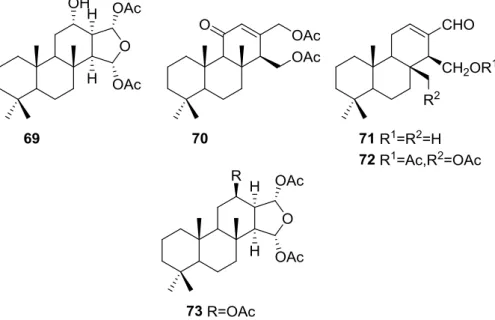
Zubía et al. [31] reported the isolation of four new metabolites 12-deacetyl-aplysillin
69, 15,16-diacetoxy-11-oxo-ent-isocopal-12ene 70, 15-hydroxy-ent- isocopal-12-en-16-al 71, 15,17-diacetoxy-ent-isocopal-12-en-16-al 72, and seven already known structures 40, 47, 49, 44,
73,46and45, from a Mediterranean sponge,S. zimoca, Schmidt 1862, collected in the channel of Sicily (Figure16).
The known compounds were identified by comparison with literature data while the new compounds were identified by comparison of the NMR spectra with those of50,72and47. Acetylation of69afforded a compound in all respects identical with50(including optical rotation). The absolute stereochemistry at C-12 was ascertained by applying a modified Mosher’s method. Attempts to acetylate71to obtain47failed, probably because of the existence of an intramolecular hydrogen bond between C-15-OH and the aldehyde group. Alternatively methanolysis of47with Na2CO3/MeOH
Mar. Drugs2016,14, 139 13 of 71
multiplicity of the olefinic proton allowed the correct localization of all the functionalities. The relative stereochemistry at C-14 was supported by comparison of the13C NMRδvalue for C-7, similar to that reported for47. The authors suggest that most probably all the metabolites have the same absolute stereochemistry as40. This suggestion has been proven for69(Mosher’s method) and is supported by the fact that45,47and72show CD curves opposite to the curves of known compounds (polydiglyal, scalaradial and 12-deacetoxy-scalaradial) supporting theent-isocopalane skeletons.
(Mosher’s method) a
δ α
β
Figure 16. Structures of 12-deacetyl-aplysillin 69, 15,16-diacetoxy-11-oxo-ent-isocopal-12ene 70, 15-hydroxy-ent-isocopal-12-en-16-al71, 15,17-diacetoxy-ent-isocopal-12-en-16-al72, and compound73.
Li et al. [32] reported the isolation of two new metabolites74and75(Figure17) together with the previously reported53,54,63and a furanoterpene, from the spongeS. matamatade Laubenfels, 1954, collected in Yap island, Micronesia. This specimen was later reclassified asS. zimocca sensude Laubenfels by the same authors in a subsequent study [33].
(Mosher’s method) a
δ α
β
Figure 17.Strutures of furanoterpenes74and75.
The known compounds were identified by comparison of the spectral data with the literature. The new compounds were identified by1H,13C, HMQC, HMBC and nOe experiments and comparison with54,53and63, that showed that they only differed in ring A. The A/B ringtransfusion of74
Mar. Drugs2016,14, 139 14 of 71
to the existence of a restricted conformation in which there is significant coupling between only one of the methylene protons and the hydroxyl proton. The authors suggest this is due to a hydrogen bond between the hydroxyl and one of the lactone oxygens. The brine shrimp lethality test was carried out for the purified compounds: 74was inactive, and the remaining compounds showed mild toxicity with LC50values of approximately 50–100µg/mL.
In the subsequent study of S. matamata de Laubenfels collected in Yap island, Micronesia, from the same authors [33] six new terpenoids, 16β-methoxy-15-oxospongi-13-en-19-oic-acid76, 16α-methoxy-15-oxospongi-13-en-19-oic-acid77, 15-oxospongi-13-en-19-oic acid78, 15α -methoxy-16-oxospongi-13-en-19-oic-acid79, 16-oxospongi-13-en-19-oic acid80, 13β,14α-dihydroxy-15α,16ξ -dimethoxyspongian-19-oic-acid81(Figure18) and the known spongia-13(16),14-dien-19-oic acid41
were isolated.
– μ
β α
α
β α α ξ
16β 16α
15α
13β,14α 15α,16ξ
α β γ
α
α β γ
α
α
β
α β γ
β
α β γ
– μ
– –
Figure 18.Strutctures of 16β-methoxy-15-oxospongi-13-en-19-oic-acid76, 16α -methoxy-15-oxospongi-13-en-19-oic-acid 77, 15-oxospongi-13-en-19-oic acid 78, 15α -methoxy-16-oxospongi-13-en-19-oic-acid79, 16-oxospongi-13-en-19-oic acid80, and 13β,14α-dihydroxy-15α,16ξ -dimethoxyspongian-19-oic-acid81.
The known metabolite41was identified by comparison with literature data. Comparison of1H and13C NMR data of the new compounds with 41showed that all compounds had identically
substituted rings A and B. Confirmation of these rings stereochemistry was obtained from pyridine-induced solvent shifts. For76the NMR data indicated the presence of a carbonyl group, a tetrasubstituted double bond, methoxyl and acetal functions. An α,β-unsaturated-γ-lactone was identified by UV and IR and confirmed by HMBC. The stereochemistry at C-16 of 76 was established on the basis of NOESY of H-16 with H-12α. Although a similar study could not be performed with77due to overlapping of the two allylic protons at C-12, the similarity of spectral data allowed its identification as an epimer of76. For79aα,β-unsaturated-γ-lactone with acetal function was confirmed by HMBC. Irradiation of Me-17 in NOESY studies caused an enhancement of H-15, confirming theα-orientation of the methoxyl group. The structure of81was established by HMBC data. NOESY experiments confirmed theα-orientation of the C-14-OH and C-15-OMe groups. The deshielding effect on Me-17 on running the NMR spectra in pyridine proved that C-13-OH isβ-oriented. The configuration at C-16 could not be resolved. NMR analysis of78and
80indicated the presence of anα,β-unsaturated-γ-lactone (confirmed by UV and IR data) in ring D. The downfield shift of C-14 in80when compared to78confirmed that this was theβ-carbon of the an α,β-unsaturated-γ-lactone and made possible the distinction of both compounds. The brine shrimp lethality test was carried out for all the purified compounds but76. Only41showed mild toxicity, with an LC50value 10–100µg/mL.
Mitchell et al. [34] reported the isolation of four new diterpenes, spongiabutenolides A–D,82–85
(Figure19), together with the known spongia-13(16),14-dien-19-oic acid41, from a sample ofSpongiasp. collected in the Philippines.
Mar. Drugs2016,14, 139 15 of 71
identified. The natural products were eventually separated and their spectral data was obtained. The structures were identified by1H,13C, HMQC, HMBC and 1D-TOCSY NMR spectra. The relative stereochemistry of the82(and83) was established by ROESY correlations of82and its methyl ester. Correlations were seen between Me-20/Me-17/COOMe and Me-18/H-5. The relative stereochemistry of84was established by ROESY spectra, while that of85was assumed.82and83were synthesized by singlet oxygen oxidation starting from41. All the compounds were tested for anti-cancer activity in a 25 cell-line panel but none showed significant cytotoxicity.
– –
β β
α α
3β,19 , 3β
3α
2α
Figure 19.Structures of spongiabutenolides A–D,82–85.
Zeng et al. [35] and Su et al. [36] reported the isolation of the new zimoclactone A86, zimoclactone B87and zimoclactone C88fromS. zimoccasubspeciesirregularia(Figure20).
– –
β β
α α
3β,19 , 3β
3α
2α
Figure 20.Structures of zimoclactone A86, zimoclactone B87, and zimoclactone C88.
The structures were determined by 1D and 2D NMR and X-ray diffraction analysis. Zimoclactone A86was isolated with 7-dehydrocholesterol and showed moderate cytotoxic activity against P388 cells.
Ponomarenko et al. [37], isolated five new diterpenes, 19-acetoxyspongia-13(16),14-dien-3-one89, 3β,19-diacetoxyspongia-13(16),14-diene90, 3β-acetoxyspongia-13(16),14-diene91, 3α -acetoxyspongia-13(16),14-diene92and 2(R),3(S),4(S)-3,18-methylene-2α-acetoxyspongia-13(16),14-diene93, together with the known 19-acetoxyspongia-13(16),14-diene94, fromS. Heterofibriacollected in Northern Cook Islands (Figure21).
Mar. Drugs2016,14, 139 16 of 71
nature and orientation of the substituents was established by NMR data analysis (including1H–1H COSY, HSQC, HMBC, and NOESY spectra, and irradiation experiments). For90the configuration at C-3 came from theJcoupling value of H-3 and its NOESY with H-5 and Me-18. NOESY of CH2-19
with Me-20 established theβ-orientation of the former. For91and92the orientation of the acetoxy group was inferred from theJcoupling values of H-3, and NOESY with H-5 and Me-18 in the case of91. The unusual cyclopropane ring in93was identified by the high field1H NMR signals and its location in C-3, C-4 and C-18 was established on the basis of the HMBC spectra. Itsα-orientation was established by nOe of one H-18 with H-5. Theα-orientation of the acetoxy group at C-2 was established by theJcoupling observed for H-2 and its nOe with Me-20. Compounds90and94were tested for immunomodulatory properties by the methods reported in the literature and demonstrated a slight lysosomal activation (about 130% of control) of mice spleenocytes at concentrations of 100µg/mL.
– –
β β
α α
3β,19 , 3β
3α
2α
Figure 21. Structures of 19-acetoxyspongia-13(16),14-dien-3-one 89, 3β ,19-diacetoxyspongia-13(16),14-diene 90, 3β-acetoxyspongia-13(16),14-diene 91, 3α-acetoxyspongia-13(16),14-diene 92, 2(R),3(S),4(S)- 3,18-methylene-2α-acetoxyspongia-13(16),14-diene93, and 19-acetoxyspongia-13(16),14-diene94.
Carroll et al. [38] reported the isolation of four new spongian diterpenes 20-acetoxy-19-hydroxyspongia-13(16),14-diene 95, 19-acetoxy-20-hydroxyspongia-13(16),14-diene 96, 19,20-diacetoxyspongia-13(16),14-diene 97 and 19,20-dihydroxyspongia-13(16),14-diene 98 (Figure 22) together with the known spongia-13(16),14-diene43, from an extract ofSpongia sp. collected in Wreck Reef, Coral Sea, that showed TRH-R2 binding affinity.
—
–
β
α α
μ
Figure 22.Structures of 20-acetoxy-19-hydroxyspongia-13(16),14-diene95, 19-acetoxy-20-hydroxyspongia-13(16),14-diene96, 19,20-diacetoxyspongia-13(16),14-diene97, and 19,20-dihydroxyspongia-13(16), 14-diene98.
Mar. Drugs2016,14, 139 17 of 71
spectra allowed their full characterization. All four compounds showed positive Cotton effects in their CD spectra, confirming the 4S,5R,8R,9R,10Sconfigurations. For95Me-18 was established as equatorial on basis of the chemical shift. ROESY correlations between CH2-19 and CH2-20 confirmed a 1,3-diaxial
relationship. For96,comparison with95revealed an isomeric relationship. HMBC confirmed the structure. For97the comparison with95and96led to the proposed structure. For98the lack of the acetate band in IR, of the corresponding methyl signal in1H NMR and the upfield shifts of CH2-20
when compared to95led to the proposed structure. TRH is a tripeptide that has been proposed to play an important role in neurotransmitter signaling. Two subtypes of the TRH receptor, TRH-R1 and TRH-R2 are found in rat brain tissues. Agonists and antagonist of TRH binding show potential therapeutic value in regulating endocrine function, in controlling pain, and in the treatment of spinal cord injury. Compound95was the most active of the five compounds in the TRH-R2 receptor binding assay, exhibiting an IC50of 23µM. Compounds96,97,98and43were only weakly active, displaying
IC50’s of 70µM, 400µM, 600µM and 1 mM, respectively. The reference compound TRH had an IC50of
23 nM.
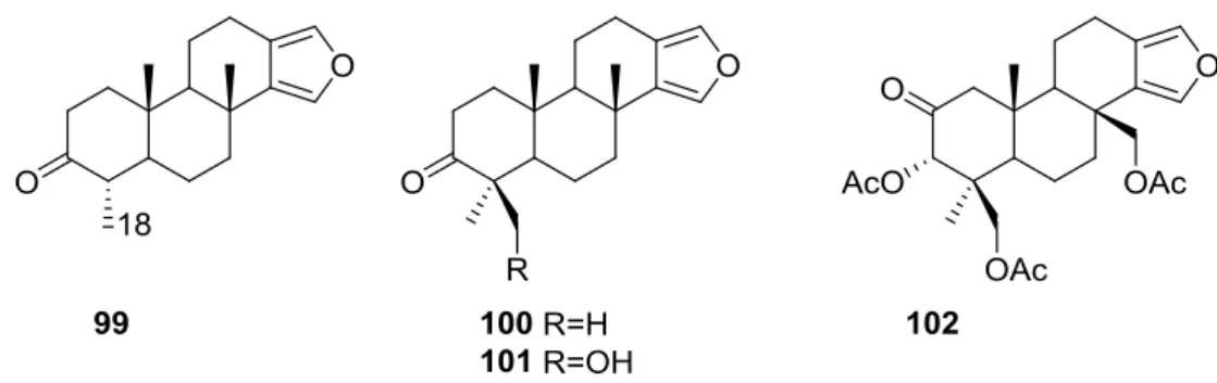
Ponomarenko et al. [39] reported the isolation of the new 19-norspongia-13(16),14-dien-3-one
99, together with the known93,102,100,91,92,94,89,90and101from aSpongiassp. (subgenus Heterofibria) collected in Northern Cook Islands. From aSpongiassp. (subgenusHeterofibria) collected in Vietnam the known43and91 were isolated. Compound99had previously been synthesized (Figure23).
μ
’ μ μ μ
–
β
μ μ
Figure 23.Structures of 19-norspongia-13(16),14-dien-3-one99, and compounds100–102.
MS and13C NMR data for99suggested a norditerpenoid structure. In the NMR spectra, signals
corresponding to the furan ring, two methyls at quaternary carbons, one methyl group at a tertiary carbon and a carbonyl group were observed. HMBC confirmed that only one methyl was attached to C-4. nOe enhancements of H-4 and Me-17 upon irradiation of Me-20 proved that H-4 isβ-oriented. The effects of89, 90,91,99, 100,101 and102on the biosynthesis of nucleic acids and embryonic development of the sea urchinStrongylocentrotus intermediuswere studied. All the compounds inhibited sea urchin embryo development at concentrations of 20µg/mL and above and DNA biosynthesis at the dose of 10µg/mL. The inhibitory effect of these diterpenoids may partly be explained by the inhibition of thymidine kinase activity. The same compounds stimulated RNA synthesis in the developing sea urchin embryos.
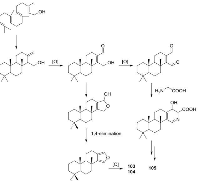
Parrish et al. [40] reported the isolation of three new diterpenes 18-nor-3,17-dihydroxyspongia-3,13(16),14-trien-2-one 103, 18-nor-3,5,17-trihydroxyspongia-3,13(16),14-trien-2-one 104 and spongiapyridine105(Figure24) together with the known62, from an unidentifiedSpongiasp. collected in Sulawesi, Indonesia.
Structure of compound103was established on the basis of1H,13C, HMBC and COSY NMR spectra. The relative configuration was ascertained by ROESY spectra where correlations between Me-20 and CH2-17 indicated they weresyndiaxial. Correlation between H-9 and H-5 (axial, on the
basis ofJcoupling values with H-6) identified H-9 as axial. The presence of the 5-OH substituent in
Mar. Drugs2016,14, 139 18 of 71
Of the four stereocenters of104 only two could be determined by NOESY: Me-20 and CH2-17 in
asyndiaxial relationship. The coupling constant of H-9 indicated it was axial as well. C-5 could not be determined due to rapid exchange of the alcoholic proton in aprotic solvents, and at lower temperatures. For compound105comparison with103showed identical rings A and B.1H and13C NMR data analysis, including1JC–Hvalues for H-16, were consistent with the presence of a pyridine
ring, which was confirmed by1H–15N HMBC. Additional structural features were deduced based on HMBC correlations that connected the pyridine ring to ring B, and indicated that the carbonyl was at C-12. The relative configuration were established by NOESY: again a correlation between Me-20 and CH2-18 confirmed both substituents assyndiaxial;1HJvalues for H-5 and H-9 suggested both to be
axial oriented. For62, NMR data analysis was in agreement with the known structure, including the configuration of C-4. Although this is not a new structure full1H and13C NMR data are presented since the former characterization was for the 17-acetyl methyl ester derivative [29]. Since all the spongian diterpenes for which absolute configurations were determined belong to the same enantiomeric series, the authors suggest that all the compounds in this study have the 5R,8R,9R,10Rconfiguration. The authors also propose a biosynthetic route to compounds103,104and105(Figure25).
μ
’ μ μ μ
–
β
μ μ
Figure 24. Structures of 18-nor-3,17-dihydroxyspongia-3,13(16),14-trien-2-one 103, 18-nor-3,5,17-trihydroxyspongia-3,13(16),14-trien-2-one104, and spongiapyridine105.
Several bioactivity tests were performed in search of the chemopreventive capacity of the isolated compounds. Modest inhibition of TNF-α-activated NF-κB activity was observed for62,103,104and
105 with ED50 values around 50µM. No significant activity was observed for inhibition of iNOS
activity in LPS-induced RAW 264.7 murine macrophage cells, and no significant induction occurred in a retinoic X receptor response element luciferase reporter gene assay. Compound104inhibited aromatase in a dose-dependent manner with an IC50value of 34.4µM. The other compounds did not
achieve 50% inhibition at a concentration of 50µM.104was also tested as an QR1 (NAD(P)H: quinone reductase 1) inducer. With cultured Hepa 1c1c7 cells104showed a CD (concentration required to double the specific activity) value of 11.2µM, which is similar to the CD value of resveratrol (21 mM), a weak QR1 inducer. None of the compounds showed any significant activity towards the aspartic protease BACE1 (<100µM).
Pham et al. [41] reported the isolation of an unusual nitrogenous spongian metabolite, haumanamide106, together with the known spongia-13(16),14-dien-19-oic acid41from aSpongiasp. collected in Pohnpei, Micronesia (Figure26).
The structure of 106 was established by comparison of the 13C NMR data with that of 41. Theα,β-unsaturated-γ-lactam in D ring was confirmed by the chemical shift of C-15, an IR band at 1665 cm´1, and HMBC spectrum analysis. Difference nOe measurements confirmed that the relative
Mar. Drugs2016,14, 139 19 of 71
– –
α κ
μ
Figure 25.Proposed biosynthesis route for compounds103,104and105. μ μ
μ
μ
α β γ
−
μ μ
– –
– –
Figure 26.Structure of haumanamide106.
De Marino et al. [42] reported the isolation of the new spongidines A–D107–110from aSpongiasp. collected in Vanuatu Islands, Australia (Figure27).
For107, mass spectrum, IR and13C NMR data indicated the presence of a carboxyl group.
Mar. Drugs2016,14, 139 20 of 71
with comparison with107 and108allowed the determination of the proposed structure. For110
the comparison with107and the differences observed for the pyridine salt moiety, together with COSY and IR data allowed the determination of the taurine residue. HMBC established its location. Inhibition of specific PLA2 enzymes constitutes a potentially useful approach for treating a great
variety of inflammatory disorders. Compounds 107,108,109and110were tested as inhibitors of sPLA2(secretory phospholipase A2) enzymes belonging to the groups I (Naja najavenom and porcine
pancreatic enzymes), II (human synovial recombinant and rat air pouch secretory enzymes) and III (bee venom enzymes). All compounds inhibited human synovial PLA2at 10µM, compound110,
containing a sulfonic acid group, being the most interesting inhibitor. In this regard these compounds can offer new structural requirements for further studies about mechanistic interactions between PLA2enzymes and inhibitors. All compounds were inactive to cPLA2. The results are summarized in
Table1.
μ μ
μ
μ
α β γ
−
μ μ
– –
– –
Figure 27.Structures of spongidines A–D107–110.
Table 1.Effect of compounds107–110on different sPLA2activitiesa.
Compound N. najaVenom %I (10µM)
Pancreas %I (10µM)
Human Synovial %I (10µM) IC50 (µM)
RAPb+ Zymosan %I (10µM)
Bee Venom %I (10µM) IC50 (µM)
107 0.5˘0.5 18.0˘8.1 40.1˘7.7d 17.1˘4.6 33.1˘6.0c
108 0.4˘0.4 14.2˘5.1 34.6˘5.8d 17.9˘4.2 32.2˘6.0c 109 3.1˘2.2 9.1˘3.5 40.4˘5.7d 30.9˘5.3c 36.2˘5.4d 110 0.0˘0.0 7.6˘4.0 48.2˘3.8d 19.6˘5.4 37.6˘6.5c
manoalide 17.0˘1.7c 32.3˘2.7d 93.2˘0.2d3.9 38.4˘0.5d 62.5˘3.8d7.5
aResults show percentages of inhibition at 10µM and IC
50(µM) values determined only for those compounds that reach 50% of inhibition. Mean˘S.E.M. (n= 6);bRAP: Rat air pouch PLA2;cp< 0.05;dp< 0.01.
Mori et al. [43] reported the isolation of spongolactams A–C,111–113(Figure28), together with the known spongia-13(16),14-dien-19-oic acid41, from aSpongiasp. collected in Okinawa, Japan, whose extract showed a 70% inhibition of FTase (Farnesyl transferase) at 20µg/mL, in a new assay described by the authors.
Mar. Drugs2016,14, 139 21 of 71
from NOESY spectra where correlation between H-12/H-16 specified the direction of the lactam group. For112the reversal of the chemical shifts of C-15 and C-16, together with NOESY between H-7/H-15 confirmed the structure. Compound113was identified by comparison with111: significant differences in the spectra were the absence of the imidazole moiety and the replacement of the C-22 methylene by a carbonyl group. The structure and absolute stereochemistry of111and112were confirmed by synthesis from41. The structure of113was also confirmed by synthesis from the same precursor. The synthesis of other spongolactam related compounds are also presented. FTase inhibitors are believed to be candidates for novel chemotherapeutic drugs. In the FTase inhibition assays the synthetic sample of111showed an IC50 23 mM (natural sample 22 mM). The activity of spongolactams B and C was determined only with synthetic samples due to inadequate amounts of natural material (130µM and >260µM, respectively). Cytotoxicity of these compounds against a human vulval-derived epidermoid carcinoma cell line, A431, was also evaluated and apparently some correlation exists between the two assays. The authors suggest that FTase could be a molecular target in the expression of spongolactam cytotoxicity.
μ
–
μ μ μ μ μ μ μ
μ μ
– –
μ
– –
δ
Figure 28.Structures of spongolactams A–C,111–113.
Other Studies
Kazlauskas et al. [44] reported the isolation of 3α,19-dihydroxyspongia-13(16),14-dien-2-one55, 3β,19-dihydroxyspongia-13(16),14-dien-2-one54, 3α,17,19-trihydroxyspongia-13(16),14-dien-2-one
58and, 3β,17,19-trihydroxyspongia-13(16),14-dien-2-one59, together with their acetyl derivatives 3α,19-diacetoxyspongia-13(16),14-dien-2-one 114, 3β,19-diacetoxyspongia-13(16),14- dien-2-one
115, 3α,17,19-triacetoxyspongia-13(16),14-dien-2-one 102 and 3β,17,19-triacetoxyspongia-13(16), 14-dien-2-one 116 from several Spongia sp. collected in the Great Barrier Reef (Figure 29). These specimens were subsequently reclassified asRhopaloeides odorabile[45].
For114fragment ions in mass spectra indicated successive losses of CH3, AcOH and 2xAcOH.
The1H NMR spectrum indicated three quaternary methyls, two acetoxy methyls, an oxygenated methylene and methine and two furan protons. For102mass spectra showed the successive losses of CH2OAc and AcOH, which suggested that a quaternary methyl group had been replaced by an
acetoxymethyl. This was confirmed by1H NMR, where the remaining signals were very similar in both compounds. IR and13C NMR showed the presence of a ketone group for both compounds. Analysis of the13C NMR spectra of both compounds allowed the identification of the furan ring and establishment of the functionality at C-17.1H NMR analysis together with biogenetic considerations established ring A. The position of an acetoxymethyl group at C-4 was assigned for114by an1H NMR study in the presence of Eu(fod)3. Definite proof of stereochemistry came from a single crystal
X-ray diffraction study of102where ring A was shown to be present as a boat conformation with atoms C-1, C-2, C-4 and C-5 coplanar, ring B formed a chair, ring C a distorted half-chair and ring D was practically flat. CD and ORD established the absolute configuration of102. Acetylation of59
Mar. Drugs2016,14, 139 22 of 71
μ μ
α
β α
β
α β
α β
3α,19 , 3β,19
and 3β,17,19
Figure 29. Structures of 3α,19-diacetoxyspongia-13(16),14-dien-2-one114, 3β ,19-diacetoxyspongia-13(16),14-dien-2-one115, and 3β,17,19-triacetoxyspongia-13(16),14-dien-2-one116.
Puliti and Matia [46] determined the relative configuration ofent-isocopal-12-en-15,16-dial45as 5S*,8R*,9R*,10S*,14S* by X-ray analysis. Atransfused tricyclic system with four methyl substituents, three of which are axiallyβ-oriented (at C-4, C-8, and C-10) was determined. Theβ-orientation of the aldehyde substituent at C-14 was confirmed.
An independent study by Yong et al. [47] determined the absolute configurations and conformations of100,54and55by X-ray analysis. For100 a twisted-boat ring A, chair B and C rings and a planar furan ring was determined, with an absolute stereochemistry of 5R,8R,9R,10R. In54ring A adopts a chair conformation and the hydroxymethylene group donates an intramolecular hydrogen bond to the C-3-OH. An absolute stereochemistry of 3R,4S,5R,8R,9R,10Rwas determined. For55a disordered ring A with a dominant chair conformer with the C-3-OH in an axial position was observed. The minor contribution was a distorted-boat conformer where the hydroxyl group adopts an equatorial position. An absolute stereochemistry of 3S,4S,5R,8R,9R,10Rwas determined. The authors point out that all the literature spongian diterpenes for which absolute stereochemistry had been reported belonged to the same enantiomeric series, even though some configurations had been assigned by Mosher esters analysis or CD data (as is the case of64,89,95and102).
An independent study by Betancur-Galvis et al. [48] tested several spongian diterpenes for their activity against herpes simplex virus type 2 and cytotoxic effect on tumor cells. Compound40
showed low cytotoxicity and43was poorly active against HSV-2. Compound100showed no anti viral activity but was cytotoxic to HeLa (human cervix epithelioid carcinoma-CC10030µg/mL), Hep-2
(human larynx epidermoid carcinoma-CC10040µg/mL), CHO (Cricetulus griseusChinese hamster
ovary cells ATCC CCL-61-CC10030µg/mL) and Bon-Fib (primary culture of bovine ear subcutaneous
fibroblasts-CC10040µg/mL) cells.
4. C21 and Other Linear Furanoterpenes
Work of Fattorusso et al. [8] and Cimino et al. [49–51] on the chemistry ofS. nitensandS. officinalis, both from the Mediterranean, allowed the isolation and characterization of the furanoterpenes, nitenin
Mar. Drugs2016,14, 139 23 of 71
α β
∆
α
γ α β
β γ –
Figure 30. Structures of nitenin117, dihydronitenin118, furospongin-1119, anhydrofurospongin-1 120, furospongin-2121, isofurospongin-2122, dihydrofurospongin-2123, tetrahydrofurospongin-2124, furospongin-3125, and furospongin-4126.
The structures were identified on the basis of UV, IR,1H NMR data with double irradiation experiments, mass spectra, and chemical transformation and degradation. Typical of the furan moiety seem to be a positive Ehrlich test, aλmaxca. 220 nm in UV (cyclohexane), characteristic IR bands at 3140, 1570, 1500, 875 and 780 cm´1,1H NMR (CCl
4) signals atδ7.26–7.15 ppm andδ7.14–7.05 ppm for
theα-protons of both rings (usually equivalent), one proton signal atδ6.16–6.14 ppm for theβ-protons of both rings (usually equivalent), and mass fragments atm/z67, 81 and 95. The isoprene unit is usually recognized by a methyl singlet atδ1.58 ppm, broadened by long range coupling to thetrans olefinic proton. For nitenin117and dihydronitenin118the configuration of C-11 was assigned as R, by applying Horeau’s method to the C-7 unsaturated and saturated diols, respectively, obtained after LAH reduction. This assignment was confirmed by1H NMR analysis of the Mosher’s esters in a subsequent study by Fontana et al. [52] that isolated both compounds fromS. agaricina from NE Spain. These authors also determine aRabsolute stereochemistry for C-8 of118 on the basis of nOe spectra. For furospongin-1119the absolute configuration at C-11 was established asSby applying Horeau’s method. Subsequent studies by Kobayashi et al. [53] corrected this assignment toRby applying Mosher’s method, further supported by nOe studies and pyridine induced shift. Although the authors [49] assigned aRconfiguration to C-13 on the basis of chemical degradation of the dehydrated derivative, this was later corrected toSin a subsequent paper [50].
The same correction is applied to the configuration of C-13 of123. For121UV and IR indicated anα,β-unsaturated ketone, confirmed by1H NMR with irradiation experiments of the signals of the corresponding H-12 and vinylic methyl. The low field resonance of the vinylic methyl at C-13 suggested it wascisto the carbonyl group. Further1H NMR analysis led to the proposal of the structure.
Mar. Drugs2016,14, 139 24 of 71
corresponding to the cleavage of the C-10/C-11 bond, together with1H NMR analysis, led to the proposal of the structure. For124the ketone group identified by IR,1H NMR data analysis, the fact that it presented no optical rotation, and that in the mass spectrum only one fragment forα-cleavage of the carbonyl group was observed, led the authors to propose themesocompound depicted.125and
126were isolated as a mixture resistant to separation. IR spectra indicated the presence of conjugated ester and carboxylic acid substituents that justify the intensity of the UV absorption observed, further confirmed by1H NMR shifts of the corresponding olefinic protons. The location of the carboxylic
acid was inferred from mass spectra and thetransorientation of the carboxyl substituents to the corresponding olefinic proton was established by spin decoupling experiments.
Further work by the same authors [54] on the sameS. officinalisled to the isolation of eight new structures (isolated as mixtures) related to furospongin-1119, withγ-hydroxy-α,β-butenolide and β,γ-epoxybutenolide rings,127–134(Figure31).
γ α,β
β,γ –
– –
β γ
Horeau’s method). In the already mentioned study of Fontana et al. −
MR analysis of the Mosher’s esters. Comparison of
Figure 31.Structures of furospongin-1119related compounds withγ-hydroxy-α,β-butenolide and β,γ-epoxybutenolide rings,127–134.
The structures were identified as mixtures on the basis of UV, IR,1H NMR data, mass spectra and
comparison with furospongin-1119. The mixtures of127and128, and129and130were also identified by chemical correlation with119. Since the structure of the latter was reviewed after the publication of this study, the structures here presented are also corrected. The mixture of131–134readily underwent decomposition to127–130. The fact that the authors were unable to identify any of the metabolites after exposure of a methanolic solution of furospongin-1119to the light, reinforces the nature of the β,γ-epoxybutenolides as natural products.
Kazlauskas et al. [55] reported the isolation of tetradehydrofurospongin-1135from an Australian Spongiasp (Figure32). The proposed structure was subsequentially corrected.
γ α,β
β,γ –
– –
β γ
Horeau’s method). In the already mentioned study of Fontana et al. −
Mar. Drugs2016,14, 139 25 of 71
The structure was identified on the basis of IR,1H NMR data with double irradiation experiments, mass spectra and chemical transformation. Capon et al. [56] in a subsequent study in 1982 assigned
13C data, established the Econfiguration of the double bonds based on the13C NMR resonances
of the vinylic methyls and J coupling values of H-6 and H-7, and an R configuration at C-11 (Horeau’s method). In the already mentioned study of Fontana et al. [52] of 1996 the enantiomer of (´)-untenospongine B is isolated fromS. virgultosafrom NE Spain. The NMR data of this new compound136(Figure33) was assigned by 1D and 2D experiments and anRconfiguration at C-11 was confirmed by1H NMR analysis of the Mosher’s esters. Comparison of the obtained NMR data with that of the reported for135led the authors to reassign the structure of136to tetradehydrofurospongin-1.
γ α,β
β,γ –
– –
β γ
Horeau’s method). In the already mentioned study of Fontana et al. −
MR analysis of the Mosher’s esters. Comparison of
Figure 33.Structure of tetradehydrofurospongin-1136.
Another study of AustralianSpongiasp. by Kazlauskas et al. [57] led to the isolation of two new compounds, furospongenol137and furospongenone138(Figure34).
∆
Figure 34.Structures of furospongenol137and furospongenone138.
The structures were identified on the basis of IR, UV,1H NMR data with irradiation experiments, mass spectra and chemical transformation. No absolute configuration was assigned to137.
Walker et al. [58] reported the isolation of the new idiadione139and the known furospinulosin-1
140fromS. idiade Laubenfels collected in San Diego, California (Figure35).
∆
Figure 35.Structures of idiadione139and furospinulosin-1140.
Mar. Drugs2016,14, 139 26 of 71
mass fragmentation of the diketone-tetrahydrofuran obtained after full hydrogenation; NMR analysis of the products synthesized by reduction and acetylation, followed by ozonolysis and hydrogenation of the ozonides, confirmed the position of the double bonds; their geometry was determined by the chemical shifts of the vinylic methyls.139was toxic to the sea starPisaster giganteusat a concentration of 5 mg/L, immobilized the larvae of the red abaloneHaliotis rufescensat 1 mg/L in sea water, and was toxic to the ectoproctMembranipora membranaceaat 10 mg/L. Both compounds were toxic to brine shrimpArtemiasp. at 10 mg/L.
From anWestern AustralianSpongiasp., Capon et al. [56] isolated a new C-21 furanoterpene,141
(Figure36).
∆
Figure 36.Structure of C-21 furanoterpene141.
The structure was elucidated on the basis of UV, IR, 1H and 13C NMR, and mass spectra.
The presence of a tertiary carbinol system came from IR,1H and13C NMR data. Significant downfield shifts were observed for the∆6,7vinylic protons and the methyl singlet upon recording the1H NMR spectrum in the presence of tris[3-(trifluoromethylhydroxymethylene-d-camphorato]europium(III). Confirmation of the proposed structure came from ozonolysis. TheEconfiguration of the double bond at C-11 came fromJH,Hcoupling analysis of H-11 and H-12; the same configuration was assigned to the
double bond at C-13 on the basis of the high field resonance observed for Me-14 (shielded bycis-allylic methylene group). Analysis of theJH,H coupling of H-6 and H-7 obtained by spectral simulation
allowed the assignment of theEconfiguration to the∆6,7double bond.
Subsequent work of Capon et al. [59] on Spirastrella papilosa led to the reisolation of the compound, and revision of the proposed structure to 142 (Figure 37), for which the name of (´)-isotetradehydrofurospongin-1 is proposed.
∆
(−
−
∆
∆
α
α β γ
by applying Horeau’s partial resolution method to the
μ
–
μ
Figure 37.Structure of (´)-isotetradehydrofurospongin-1142.
The revision was based on 2D NMR data. TheEconfiguration of∆5,6was confirmed byJcoupling analysis and the chemical shift of Me-14 in13C NMR; theEconfiguration of∆10,11is confirmed by NOESY. Ozonolysis was repeated and (R)-dimethyl citramalate was recovered: its assignment was confirmed by1H NMR, [α]Dand chiral HPLC comparison with authentic samples of both theRand Senantiomers.
Tanaka and Higa [60] reported the isolation of the new kurospongin 143 from aSpongiasp. collected in Miyako Island, Japan (Figure38).
∆
(−
−
∆
∆
α
α β γ
by applying Horeau’s partial resolution method to the
μ
–
μ
Mar. Drugs2016,14, 139 27 of 71
The structure was identified by mass spectrometry, IR, 1H and 13C NMR and irradiation experiments. These allowed the identification of the furan rings, the α,β-unsaturated γ-lactone, a vinyl methyl, atransdi-substituted double bond and a tri-substituted double bond. The geometry of the latter was assigned asEby the value of the13C chemical shift of the vinylic methyl. The absolute stereochemistry at C-11 was assigned asSby applying Horeau’s partial resolution method to the diol obtained after treatment with ethylmagnesiumbromide. Compound143was ichthyotoxic, killing goldfish at the concentration of 5µg/mL within 4 h. In feeding experiments using the omnivorous fish Tilapia mosambica143impregnated in feed completely deterred its consumption at the concentration level of 0.3%.
De Giulio et al. [61] reported the isolation of furospongin-2 121, together with its three new isomers144–146(Figure39), from aS. officinalisL. collected in northern Adriatic, whose extract showed cytotoxic activity (LD50 45µg/mL) in the brine shrimp assay.
–
α β
– μ
β Figure 39.Structures of furospongin-2121isomers144–146.
For all compounds, the presence of anα,β-unsaturated ketone was established by UV and IR and confirmed by1H and13C NMR spectra. The use of COSY and HETCOR spectra allowed the assignment of all resonances for144. For this compound the fact that the13C NMR spectra only
showed 11 signals led to the conclusion that it was symmetrical. Comparison of its data with the remaining compounds led to identification of the latter. The stereochemistry of the double bonds of all compounds were assigned on the basis of the chemical shifts in1H and13C spectra for the vinylic methyls and allylic methylenes. 13C NMR data for121is assigned based on COSY and HETCOR.
All compounds showed high activity (LD50 0.09–1.6µg/mL) in theArtemia salinashrimp bioassay, an in-house substitute for 9 KB and 9 PS cytotoxicities.
Mar. Drugs2016,14, 139 28 of 71 –
α β
– μ
β Figure 40.Structure of tetronic acid147.
Compound 147 was identified by NMR, where resonances for a β-substituted furan, three substituted double bonds with vinylic methyls and a tetronic acid moiety were observed. The presence of this latter feature and confirmation of the structure came by comparison with palominin, its known geometrical isomer. The observed chemical shifts for the olefinic methyl resonances of147confirmed theEgeometry of all double bonds. CD data supported a 21Rstereochemistry. The antibiotic activity of the extract was attributed entirely to147. Preliminary testing suggested that this compound was also responsible for the inhibitory activity detected in the crude ethanol extract. This compound reversibly blocked contractions, evoked by acetylcholine, 5-hydroxytryptamine and histamine, of isolated guinea-pig ileum, and electrical stimulation of intrinsic nerves. Purification of the extract appeared to remove the contracting substance detected, due to its loss or to a synergistic activity between the isolated compounds.
Urban and Capon [63] reported the isolation of the new cometins A–C 148–150 (Figure41), together with the known furospinosulin-1 140, from a Spongia sp. collected in the Great Australian Bight.
– –
– –
∆
δ
γ
∆
– μ μ
Figure 41.Structures of cometins A–C148–150.
Compound148was identified by mass spectrometry and1H and13C NMR where resonances for the difuran and tetronic acid moieties were identified. Confirmation came from comparison with literature compounds. The 1,1,4-trisubstituted 1,3 diene functionality was further identified by NMR. The geometry of the∆16,17double bond was determined asEby theJcoupling value observed for the olefinic protons. Comparison of theδvalue in13C NMR for the olefinic methyl with reference compounds allowed the determination of theEconfiguration for the trisubstituted double bond. Although stereochemistry at C-18 was not determined, aRconfiguration is proposed on the basis of CD data. Comparison of the NMR data of149with that of148allowed the replacement of the tetronic acid moiety by a conjugatedγ-butenolide. Confirmation of the structure came from COSY spectra and nOe. The geometry of the double bond was determined asEby the13C NMR shift of the olefinic methyl. Stereochemistry at C-18 was not determined. Comparison of the data of150with that of