RADIATION SCIENCES
07-03B (2019) 01-13
ISSN: 2319-0612 Accepted: 2019-10-21
BJRS
Dosimetric evaluation of extremities in
18F-FDG PET/CT
procedure using Monte Carlo Geant4 code
Pessanha
aP.R., Queiroz-Filho
aP.P., Souza-Santos
aD.
a Instituto de Radioproteção e Dosimetria – IRD/CNEN, 22783-116 , Rio de Janeiro-RJ, Brasil. [email protected]
ABSTRACT
A stylized phantom of forearm and hand was developed to assess extremities doses of workers in Positron Emission Tomography practices. This hand phantom was then coupled to a whole body ADAM phantom in order to evaluate eye lens doses as well. A case study, using 18F-FDG as the radiopharmaceutical, was simulated with the Geant4 Monte Carlo toolkit. Equivalent Dose values were obtained in 45 different points - in dominant and non-dominant hands and eye lens - for radiopharmaceutical administration procedure, with the use of injection syringe shielding. According to the data obtained, it can be observed that the most exposed points tend to be the index finger tips and the middle fingers for the dominant hand and the index finger for the non-dominant hand, the highest dose reaching 23 times higher than the pulse dose. Both the dose estimates for extremities and eye lenses remain below the limit established by the CNEN standard NN-3.01 of 2014. From the obtained data, we can conclude that a good alternative for monitoring of eye lens is done positioning the dosimeter near the temples. For extremities it is recommended to use dosimetric rings at the base of the most exposed finger.
1. INTRODUCTION
Positron Emission Tomography (PET) - increasingly used also for the purpose of evaluating the therapeutic response and staging of pathological manifestations - uses radionuclides, introduced into the body generally by intravenous route, of extremely short half-life, to provide biochemical body images. The introduction of this technology in Brazil was only possible with the beginning of the production of fluordesoxyglucose (FDG) radiopharmaceutical by the year 1999, as a tracer for this modality of tomography, in parallel with the use of conventional radiopharmaceuticals. Interest in this practice increased further after the introduction of the PET technique combined with the computed tomography (PET/CT) system, which allowed obtaining anatomical and metabolic or functional information in vivo in a single image scanning session. [1,2,3].
The energy of the annihilation photons (511 keV) generated by positrons of the 18F is 3.65 times
higher than the energy of the photons generated by 99mTc (140 keV) and 1.4 times higher than those
produced by 131I. Nuclear Medicine professionals who are involved in activities related to high
energy emitters are considered to be the group with the highest radiation exposure [4].
According to the CNEN NN 3.05 Brazilian norm, of December, 2013 ("Requirements of Security and Radiological Protection for Services of Nuclear Medicine") it is recommended the use of dosimeters located in the thorax, and dosimeters of extremities for external individual monitoring, for workers in controlled areas.
Workers executing PET procedures are routinely monitored using dosimetric rings and wristbands and / or dosimetric films and pocket dosimeters. However, dosimetric analysis of these workers is required, since the exposure of extremities during administration of 18F-FDG is
considered relatively high and non-uniform, and the distribution of doses in the hands of a worker varies greatly during a procedure, as well as from person to person [5]. The main objective of this work is to obtain information about the dose distribution in extremities and eye lens - from Monte Carlo simulations with the Geant4 toolkit - of occupationally exposed individuals in Positron Emission Tomography (PET/CT), resulting from administration of the 18F-FDG
Considering that extremities dosimeters are only recommended in activities where doses in the hands may be significantly higher than in the chest and that the annual dose limit for the eye lens has been reduced, in 2011, from 150 mSv to 20 mSv/year, a better grasp on exposure conditions in nuclear medicine is necessary. This work aims to obtain more information about the dose distribution in extremities and eye lens in the procedure of administering the 18F-FDG
radiopharmaceutical with the use of injection syringe, with and without shielding.
2. MATERIALS AND METHODS
For this work, the Geant4 Monte Carlo toolkit was used to implement a geometric phantom representing the hand of the professional involved in this clinical procedure, with hand and forearm dimensions of the standard man, to evaluate the extremity exposure.
Considered a software package, Geant4 provides the possibility of physical modeling in a fully flexible structure. It encompasses particle tracking - including leptons, photons, hadrons, and ions; geometry description; interface for physical processes that encompass interactions over a wide range of energy, - external graphical interface, and user interface systems [6]. The trend of modern experimental science is the growing reliance on tools that allow the simulation and interpretation of data in fields such as high energy physics [7].
For the construction of the phantom in Monte Carlo Geant4 code, simple geometric solids were used to model the bones, soft tissue and skin of the upper limbs (forearm and hand). It was considered only cylinders for the representation of the bones, as well as the fingers; for the projection of the region comprising the palm of the hand, a rectangular prism was used, and an elliptical base prism was used for the forearm segment (Figure 1).
The specifications for anatomical parts representing the fingers and phalanges were composed of three groups of cylindrical solid segments (proximal, middle and distal phalanges). From the specificity of these vertices the geometric phantom becomes capable of promoting movements simulation, by defining angles in the joints between the phalanges.
The geometric phantom, defined from a simple configuration of angles for joints in its phalanges, allows investigations in any positioning that requires only the malleability of the fingers, such as procedures that use injection syringe.
The program was validated from the irradiation of a REMAB® physical phantom, air kerma measurements with thermoluminescent crystals and comparison with values obtained in the simulation. The criteria for validation of the program obeyed the anthropometric parameters of width and length, as well as materials, which make up the REMAB® phantom described by ICRU 48 [8,9].
Figure 1: Geometrical hand and forearm geometry implemented in code Monte Carlo
Geant4.
Next to the hand and forearm phantom the ADAM phantom was implemented for evaluation of dose in eye lens and extremities concomitantly. The ADAM phantom has its body and organs described by mathematical expressions that represent cylinders, ellipses, spheres, cones, among others, besides combinations between these solids [10]. The masses of organs, mass and body height of the ADAM phantom correspond to the data recommended by ICRP 23 for standard man [11] (Figure 2). A representation of both the stylized whole body phantom and forearm and hand phantom in the Geant4 code can be seen in (Figure 3).
For each point of analysis, sensitive volumes with the same dimensions of a real thermoluminescent dosemeter (TLD), i.e., 0,9 x 0,9 x 0,3 mm3 were simulated. Each of these
volumes was numerically identified according to phantom placement. Positions used for dosimetric evaluation can be observed in Figures 4 and 5.
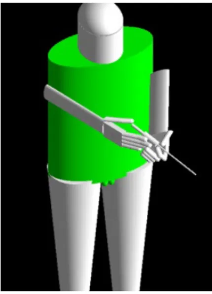
For data collection, the phantom had the angles of the finger joints positioned to hold a 3 ml injection syringe (polyethylene material) used in PET procedures during administration of 18F-FDG
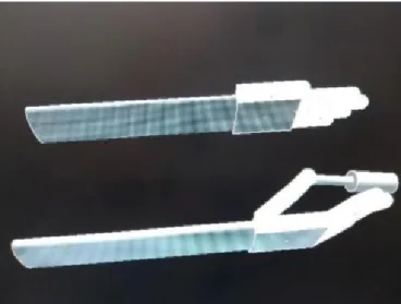
to the patient. For a PET radiopharmaceutical procedure, the syringe is protected with a 6 mm tungsten shield. Figure 6 shows an actual injection procedure and the simulation of this procedure in the Geant4 code.
Figure 3: ADAM phantom implemented alongside mathematical phantom of hand and
forearm
Figure 4: Positions where the TLDs were simulated. The points of analysis for the dominant
hand were numbered from 01 to 20, receiving numbers from 21 to 40 the points of analysis that comprise the non-dominant hand, respecting the same positioning and numerical order.
Figure 5: Analysis points for eye lens. Points 4 and 5 are positioned on the phantom's temples on
Figure 6: 18F-FDG administration geometry. Left: administration geometry of 18F in nuclear
medicine service [12]. Right: simulation in Monte Carlo Geant4 code.
During the simulation, in each segment that corresponded to the syringe plunger, random emissions of medium energy positrons were generated, according to the decay spectrum of the 18F
radionuclide [13]. The values of absorbed doseobtained in simulation - for all the TLD positioning points considering homogeneous media - allowed the use of shielded syringe (of 6 mm thick tungsten material) according to the actual shielding used in nuclear medicine in PET scanners.
3. RESULTS AND DISCUSSION
Estimates of daily and annual equivalent doses in the skin for dominant and non-dominant hand were made from the absorbed dose values obtained by Monte Carlo Geant4 code simulation. To obtain the daily dose values, it was considered that a professional in nuclear medicine service manipulate the radiopharmaceutical using a syringe shield, in a 40 seconds average time (exposure time), performing five procedures per day (on average). For these calculations, the recommended administration of an activity of 370 MBq (10 mCi) of 18F-FDG in injection syringe was also
considered for a standard adult of 70 kg mass [12,14].
For an annual equivalent dose estimate, it should be considered this same professional performing a total of six PET procedures per work day, twice a week, which leads to about 530 procedures per year.
Theobtained values for absorbed dose and the estimated equivalent dose values can be found in Tables 1 and 2, where each value is related to the corresponding analysis segments (Figure 4).
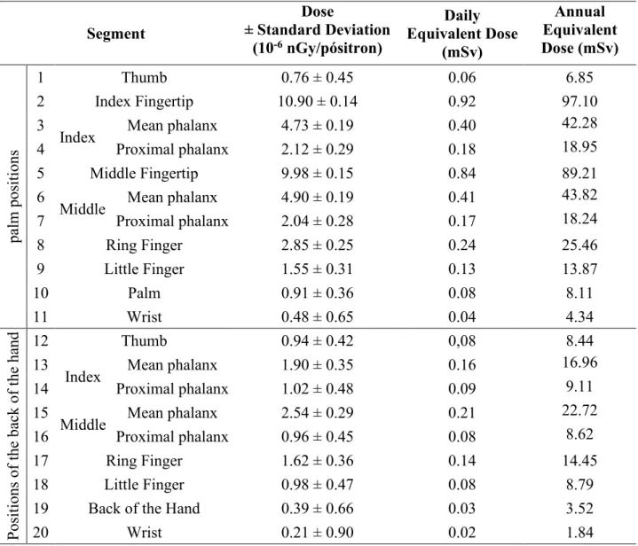
Table 1: Dose values obtained in dominant hand, for administration of 18F-FDG.
Segment ± Standard Deviation Dose (10-6 nGy/pósitron) Daily Equivalent Dose (mSv) Annual Equivalent Dose (mSv) pa lm pos iti ons 1 Thumb 0.76 ± 0.45 0.06 6.85 2 Index Fingertip 10.90 ± 0.14 0.92 97.10 3
Index Mean phalanx 4.73 ± 0.19 0.40 42.28
4 Proximal phalanx 2.12 ± 0.29 0.18 18.95
5 Middle Fingertip 9.98 ± 0.15 0.84 89.21
6
Middle Mean phalanx 4.90 ± 0.19 0.41 43.82
7 Proximal phalanx 2.04 ± 0.28 0.17 18.24 8 Ring Finger 2.85 ± 0.25 0.24 25.46 9 Little Finger 1.55 ± 0.31 0.13 13.87 10 Palm 0.91 ± 0.36 0.08 8.11 11 Wrist 0.48 ± 0.65 0.04 4.34 Pos iti ons of the ba ck of the ha nd 12 Thumb 0.94 ± 0.42 0,08 8.44 13
Index Mean phalanx 1.90 ± 0.35 0.16 16.96
14 Proximal phalanx 1.02 ± 0.48 0.09 9.11
15
Middle Mean phalanx 2.54 ± 0.29 0.21 22.72
16 Proximal phalanx 0.96 ± 0.45 0.08 8.62
17 Ring Finger 1.62 ± 0.36 0.14 14.45
18 Little Finger 0.98 ± 0.47 0.08 8.79
19 Back of the Hand 0.39 ± 0.66 0.03 3.52
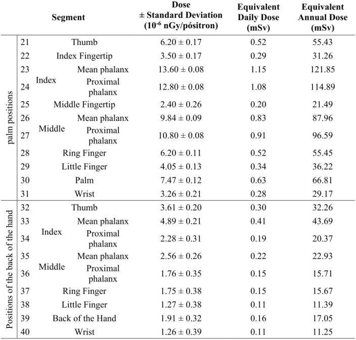
Table 2: Dose values obtained in non-dominant hand, for administration of 18F-FDG.
Segment ± Standard Deviation Dose (10-6 nGy/pósitron) Equivalent Daily Dose (mSv) Equivalent Annual Dose (mSv) pa lm pos iti ons 21 Thumb 6.20 ± 0.17 0.52 55.43 22 Index Fingertip 3.50 ± 0.17 0.29 31.26 23
Index Mean phalanx 13.60 ± 0.08 1.15 121.85
24 Proximal phalanx 12.80 ± 0.08 1.08 114.89
25 Middle Fingertip 2.40 ± 0.26 0.20 21.49
26
Middle Mean phalanx 9.84 ± 0.09 0.83 87.96
27 Proximal phalanx 10.80 ± 0.08 0.91 96.59 28 Ring Finger 6.20 ± 0.11 0.52 55.45 29 Little Finger 4.05 ± 0.13 0.34 36.22 30 Palm 7.47 ± 0.12 0.63 66.81 31 Wrist 3.26 ± 0.21 0.28 29.17 Pos iti ons of the ba ck of the ha nd 3233 Thumb 3.61 ± 0.20 0.30 32.26
Index Mean phalanx 4.89 ± 0.21 0.41 43.69
34 Proximal phalanx 2.28 ± 0.31 0.19 20.37
35
Middle Mean phalanx 2.56 ± 0.26 0.22 22.93
36 Proximal phalanx 1.76 ± 0.35 0.15 15.71
37 Ring Finger 1.75 ± 0.38 0.15 15.67
38 Little Finger 1.27 ± 0.38 0.11 11.39
39 Back of the Hand 1.91 ± 0.32 0.16 17.05
40 Wrist 1.26 ± 0.39 0.11 11.25
According to the data obtained and related in Tables 1 and 2, it can be observed that the most exposed points - for the studied geometry (Figure 6) - tend to be the index finger tips and the middle fingers for the dominant hand and index finger for non-dominant hand.
It is important to highlight that the dose estimates found for the wrist are 23 times lower than the most exposed point for the dominant hand, for example. Thus, measurements with a dosimetric ring, being placed at the base of the most exposed finger would only underestimate at five times the
estimated equivalent dose in the hand. Therefore, measurements taken with dosimetric rings would be better estimating the equivalent dose at the ends than wristbands.
As in Kubo & Maurício [12], the data obtained indicate that the use of dosimetric wristbands does not prove to be the best alternative for dose estimation in extremities.
Merce et al. [15] estimated the dose received by workers during the injection and fractionation practices of the radiopharmaceutical in 11 points between the thumb, index, middle and ring fingers, as well as pulse - for dominant and non-dominant hands. As results, we observed that placements in the index finger and thumb presented higher doses. Thus, the use of dosimetric rings at the base of the index finger is a better alternative for the monitoring of workers, since the pulse is the least exposed point. Non-dominant hand positions were also identified as more exposed.
For the studied geometry, according to the data presented in Tables 1 and 2, none of the analyzed points registered a dose greater than 150 mSv. However, points at the base of the index and middle fingers of the non - dominant hand are very close to the level for investigation at annual equivalent dose for extremities (150 mSv) defined by CNEN - NN - 3.01 brazilian norm, of March 13, 2014 [16].
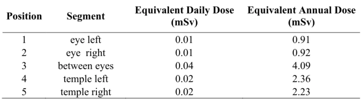
Table 3 shows the data on the dose estimates for eye lens.
Table 3: Dose estimates for eye lens test points
Position Segment Equivalent Daily Dose (mSv) Equivalent Annual Dose (mSv)
1 eye left 0.01 0.91
2 eye right 0.01 0.92
3 between eyes 0.04 4.09
4 temple left 0.02 2.36
5 temple right 0.02 2.23
Regarding the data presented in Table 3, the estimated dose values for eye lens remain below the limit established by CNEN brazilian norm (20 mSv annually). Positions for monitoring the dosimeter near the temples are a good alternative since the values obtained in these points are close to the values obtained directly in the lens of the ADAM phantom.
4. CONCLUSION
The simulations shows that the most exposed points tend to be the index finger tips and the middle fingers for the dominant hand and the index finger for the non-dominant hand, with the highest dose reaching 23 times higher than the pulse dose. Moreover, the base of the most exposed finger presents values five times smaller than the tips of the fingers. Thus, measurements with a dosimetric ring, being placed at the base of the finger more exposed, would be underestimating in five times the equivalent dose estimate in the hand.
In case of option for the use of dosimetric ring by the Nuclear Medicine workers in an 18F-FDG
injection procedure, the tendency would be to place it in the dominant hand. However, the bases of the fingers of the non-dominant hand are the points which would be most exposed.
As in Kubo & Maurício [12], the data obtained show that the use of dosimetric wristbands does not prove to be the best alternative for estimation of dose at the extremities. According to the data in Table 5, it is also clear the importance of the use of injection syringe shielding.
The estimated dose values for eye lens remain below the limit established by CNEN brazilian norm (20 mSv annually). Positions for monitoring the dosimeter near the temples are a good alternative since the values obtained in these points are close to the values obtained directly in the lens of the ADAM phantom.
REFERENCES
[1] IEN. Exames PET com FDG são utilizados em pesquisas médicas no Rio de Janeiro. 2006. Available at: <http://www.ien.gov.br>, Last accessed: 20 Apr. 2011.
[2] ROBILOTTA, C. C. A tomografia por emissão de pósitrons: uma nova modalidade na medicina nuclear brasileira. Rev. Panam. Salud Publica. Vol.20, No. 2-3, pp.134–142, 2006
[3] BOELLAARD, R. Standards for PET Image Acquisition and Quantitative Data Analysis. The Journal of Nuclear Medicine. Vol. 50, No. 5 (Suppl), 2009.
[4] BIXLER, A.; SPRINGER, G.; LOVAS, R. Practical Aspects of Radiation Safety for Using Fluorine-18. Journal of Nuclear Medicine Technology. Vol. 27. No. 1. pp 14-16, 1999.
[5] COVENS, P.; BERUS, D.; VANHAVERE, F.; CAVELIERS, V. The Introduction of automated dispensing and injection during PET procedures: A step in the optimization of extremity doses and whole-body doses of nuclear medicine staff. Radiation Protection Dosimetry. Vol. 140, No.3, pp. 250–258. 2010.
[6] ALLISON, J. et al., 2006. “Geant4 developments and applications”. IEEE Transactions on Nuclear Science. Vol. 53, 1, part 2. pp 270-278.
[7] ALLISON, J. et al., 2016. “Recent developments in GEANT4”. Nuclear Instruments and Methods in Physics Research A 835 (2016). pp 186–225.
[8] INTERNATIONAL COMISSION ON RADIATION UNITS AND MEASUREMENTS, 1992. “Phantoms and Computational Models in Therapy, Diagnosis and Protection”, ICRU-48, Bethesda, MD
[9] PESSANHA, P. R.; QUEIROZ-FILHO, P. P.; SOUZA-SANTOS, D. Implementação de fantoma geométrico de mão e antebraço no Código de Monte Carlo Geant4. Brazilian Journal of Radiation Sciences, 03-1A, pp. 01-15, 2015.
[10] KRAMER, R.; VIEIRA, J. W.; KHOURY, H. J.; ANDRADE-LIMA, F. MAX meets ADAM: a dosimetric comparison between a voxel-based and a mathematical model for external exposure to photons. Physics in Medicine and Biology, No. 49, pp. 887–910, 2004.
[11] INTERNATIONAL COMISSION ON RADIOLOGICAL PROTECTION. Report on the Task Group on Reference Man. ICRP Publication 23, Oxford: Pergamon Press; Ann. ICRP. 1975
[12] KUBO, A. L.S.L.; MAURICIO, C. L. P., 2014. TLD occupational dose distribution study in
nuclear medicine. Radiation Measurements. Available at:
http://dx.doi.org/10.1016/j.radmeas.2014.04.021
[13] JAN, S.; COLLOT, J.; GALLIN-MARTEL, M.; MARTIN, P.; MAYET, F.; TOURNEFIER, E. “GePEToS: A Geant4 Monte Carlo Simulation Package for Positron Emission Tomography”. IEEE Transaction on Nuclear Science, Vol. 52, No. 1. pp.102–106. 2005.
[14] VILLAS BOAS. [18F]FDG - Bula para o professional de saúde. Villas Boas Radiofármacos Brasil S/A. Available at: <http://www.vbrf.com.br/download/bulafdg.pdf>, Last accessed: 18 Jan 2012.
[15] MERCE, M. S. et al. (2011). “Extremity Exposure in Nuclear Medicine: Preliminary Results of a European Study”. Radiation Protection Dosimetry (2011), Vol. 144, No. 1–4, pp. 515–520 [16] COMISSÃO NACIONAL DE ENERGIA NUCLEAR, 2014, “CNEN – Diretrizes Básicas de
![Figure 2: Side and front view of the ADAM phantom [10].](https://thumb-eu.123doks.com/thumbv2/123dok_br/18277986.881311/5.892.295.616.389.792/figure-view-adam-phantom.webp)
![Figure 6: 18 F-FDG administration geometry. Left: administration geometry of 18 F in nuclear medicine service [12]](https://thumb-eu.123doks.com/thumbv2/123dok_br/18277986.881311/7.892.168.745.262.500/figure-administration-geometry-administration-geometry-nuclear-medicine-service.webp)