Ana Catarina da Silva Pereira
BS Biochemistry
Structural investigation of the
Bacillus subtilis
morphogenic factor
RodZ
A thesis to obtain a Master degree in Structural and Functional Biochemistry
Supervisor: Doctor Manolis Matzapetakis
Principal Investigator, Biomolecular NMR Group, ITQB, Oeiras, Portugal
Board Members
Committee Chair: Doctor José Ricardo Ramos Franco Tavares
Assistant Professor, Faculdade de Ciências e Tecnologia, Universidade Nova de Lisboa
Examiner: Doctor Jorge da Silva Dias
Assistant Investigator, Faculdade de Ciências e Tecnologia, Universidade Nova de Lisboa
Ana Catarina da Silva Pereira
BS Biochemistry
Structural investigation of the
Bacillus subtilis
morphogenic factor
RodZ
A thesis to obtain a Master degree in Structural and Functional Biochemistry
Supervisor: Doctor Manolis Matzapetakis
Principal Investigator, Biomolecular NMR Group, ITQB, Oeiras, Portugal
Board Members
Committee Chair: Doctor José Ricardo Ramos Franco Tavares
Assistant Professor, Faculdade de Ciências e Tecnologia, Universidade Nova de Lisboa
Examiner: Doctor Jorge da Silva Dias
COPYRIGHTS Ana Catarina da Silva Pereira FCT/UNL UNL
Acknowledgments
I would have never been able to finish my dissertation without the guidance of my supervisor, help from friends, and support from my family.
After this time I do come to a conclusion that I overcame major doubts about myself and my work. This would have never been possible without the help of my supervisor, Manolis Matzapetakis, who invited me to work again with him knowing beforehand about my limitations but also recognizing and encouraging my major strengths. I would like to express my sincere gratitude for his continuous support, for his patience, motivation, enthusiasm, and immense knowledge. His guidance helped me in all the time of research and writing of this thesis. Also, knowing that it could delay the progress of this project, he met my request to get my hands “dirty” on the wet lab and by that allowing me to gain new molecular biology skills, which I thank for.
I must mention and acknowledge Adriano Henriques and the Microbial Development Laboratory members Teresa Costa and Ana Paiva for providing all the required RodZ samples and detailed information of undergoing developments, without which I couldn’t have written this report.
I thank my fellow colleagues from the Biomolecular NMR laboratory: Meire Almeida, Mariana Palma and Ivo Saraiva, for the stimulating discussions during the many group meetings we had for the past year. Meire Almeida was also a key person during my training in the wet lab, being always available to help me solve any question.
My sincere thanks also goes for Claudia Almeida for helping me with equipment training and Isabel Pacheco and all members from NMR and Inorganic Chemistry laboratory for always being available to help me in any way possible.
I would like to thank my family for standing by me through good and bad times: my parents Leonor and Aires Pereira for giving me emotional and financial support, allowing me to complete another cycle of studies; my sister Márcia Pereira whose organizational skills were crucial to help me finish writing this thesis.
I would also like to thank Vicente Canhoto for always being there to cheer me up and for always supporting me and encouraging me with his best wishes.
Abstract
RodZ is a protein widely conserved in bacteria and a core component of the morphogenic apparatus of the cell. It is known to be required for assembly of the bacterial actin homologue, MreB, that controls cell wall synthesis and cell shape. The domain organization of RodZ consists of a well-conserved N-terminal (RodZn) with helix-turn-helix motif (HTH), a well-conserved transmembrane domain, and a conserved C-terminal domain (RodZc). RodZn, located in the cytoplasm, has been shown to interact with MreB actin-homologue by x-ray studies in T. maritima. However, the structure of RodZn from gram-positive B. subtilis showed low homology with the published one from gram-negative T. maritima. Here we present the solution structure of RodZn from B. subtilis determined for the first time, by NMR spectroscopy. Compared to previous structural data obtained from the crystallized RodZn from T. maritima and more recently from S. aureus, several differences could be observed, namely the length of the alpha-helices and the presence of an extended coil. Interaction studies were preformed between RodZn domain and MreB from which no significant results could be extrapolated. Since HTH motif is frequently associated with DNA interaction, the involvement of RodZn in DNA organization is being investigated. At the same time, RodZc domain, which structure has never been reported, was subject of study. Bioinformatic, biophysical and biochemical methodologies were employed to study this domain. A model based in a pseudo-ab initio methodology was built, revealing an Ig-like fold. The Ig superfamily is a large group of cell surface and soluble proteins that are involved in the recognition, binding, or adhesion processes of cells. Therefore, RodZ is thought to be a protein that establishes a link between the inner side of the cell membrane and the outer side, promoting spatiotemporal coordination between peptidoglycan synthesis and cell division.
Resumo
RodZ é uma proteína amplamente conservada em organismos bacterianos, fazendo parte do complexo aparelho morfogénico celular. Em estudos anteriormente publicados, RodZ foi considerada necessária à organização celular da proteina MreB, um homólogo da actina igualmente conservado em bactérias, responsavél por controlar a síntese da parede celular e a morfologia adoptada pela célula . A proteina RodZ é composta por três domínios distintos: um domínio N-terminal (RodZn) detentor de um motivo estrutural de hélice-volta-hélice (HTH) localizado no citoplasma, um domínio transmembranar (TM ), e um domínio C-terminal (RodZc) localizado na região periplasmática da célula. Em T. maritima (gram-negativo), a interacção entre o domínio RodZn e a proteina MreB foi demonstrado através de estudos de cristalografia de raios-x. No entanto , a estrutura do domínio RodZn presente em B. subtilis (gram-positivo) revelou uma baixa homologia com a estrutura publicada. No presente trabalho, foi determinada pela primeira vez através de Espectroscopia de RMN, a estrutura em solução do domínio RodZn de B. subtilis,. Comparando-a com os dados estruturais anterioremente publicados a partir do domínio RodZn presente em T. maritima e, mais recentemente, em S. aureus, várias diferenças foram observadas , nomeadamente, o comprimento das hélices alfa e a presença de uma zona alongada não estruturada. Estudos de interacção entre o domínio RodZn e a proteina MreB não levaram a resultados significativos. Sendo que o motivo estrutural HTH é frequentemente associado ao estabelecimento de interacções com a molécula de DNA, o envolvimento de RodZn em organização do DNA plasmídico encontra-se a ser investigado. Ao mesmo tempo , o domínio RodZc , cuja estrutura nunca antes fora revelada, foi objecto de estudo. Ferramentas bioinformáticas conjugadas com técnicas biofísicas e bioquímicas foram utilizadas para estudar este domínio. Foi construido um modelo tridimensional para o domínio RodZc. A metodologia usada baseou-se na utilização de ferramentas de modelação com um princípio de pseudo-ab initio, revelando um motivo estrutural rico em folhas beta, característico da super-família de imunoglobulinas. Esta super-família é composta por um variado grupo de proteinas que embora possuam um motivo estrutural semelhante, variam bastante em termos de função celular (processos de reconhecimento, ligação e adesão celular, entre outras funções). Sendo assim, uma hipótese foi levantada em que a proteina RodZ possa ser o elo de ligação entre o lado interno e o lado externo da parede da célula promovendo, desta forma, a coordenação espaço-temporal entre a síntese do peptidoglicano e a divisão celular.
INDEX
Chapter 1: Biological significance ... 1
1.1 Cell Wall ... 3
1.2 Cell elongation and division ... 4
1.3 Protein RodZ ... 7
1.4 Bacillus subtilis ... 11
1.5 Aims ... 13
Chapter 2: Methodology ... 17
2.1 Homology Modelling ... 19
2.2 Pattern-based Homology Modelling ... 21
2.3 Ab-initio ... 23
2.4 Data-driven structure prediction ... 25
2.5 Fundaments of NMR spectroscopy ... 27
2.5.1 Protein sample preparation for NMR ... 28
2.5.2 NMR assignment methodology ... 29
2.5.3 Structure calculation... 31
2.5.4 Structure refinement ... 32
2.5.5 Structure validation ... 33
Chapter 3: High-resolution NMR solution structure of RodZ cytoplasmic domain ... 36
3.1 Introduction ... 36
3.2 Material and Methods ... 38
3.2.1 NMR sample preparation ... 38
3.2.2 NMR data acquisition and structure calculation ... 38
3.2.3 NMR titration for protein-protein interaction studies ... 39
3.3 Results and Discussion ... 40
3.3.1 RodZn Structure analysis ... 40
3.4 Conclusion ... 51
Chapter 4: RodZ periplasmic domain: Structural and functional insight ... 54
4.1 Introduction ... 54
4.2 Material and methods ... 55
4.2.2 Bacterial Growth and Protein Expression ... 55
4.2.3 Protein Purification ... 56
4.2.4 NMR sample preparation ... 57
4.2.5 NMR data acquisition and 3D-model prediction ... 59
4.3 Results and Discussion ... 62
4.3.1 Bioinformatic analysis ... 62
4.3.2 Loss of structure after elimination of the predicted linker ... 68
4.3.3 RodZc protein present in solution in a 3-state... 71
4.3.4 Structure analysis ... 76
4.3.5 Protein dynamics studies ... 80
4.4 Conclusions ... 85
Chapter 5: Concluding remarks and future perspectives ... 88
Chapter 6: Appendix ... 90
LIST OF FIGURES
Figure 1 – Gram-positive and Gram-negative bacteria are differentiated by their cell wall structure; Picture comparing Gram-positive and -negative cell wall. ... 2 Figure 2 - Peptidoglycan synthesis in B. subtilis. The peptidoglycan layer is formed by polymerized
represented what is seen using decovolution fluorescence microscopy, where a stack of images taken through the cell body described MreB to form an helical structure along the bacterial cell wall . In B) through TIRFM microscopy, a high resolution technique able to capture the surface of one side of a bacterium found both MreB and a selection of several PG elongation proteins that move in short patches as opposed to long helical filaments with bidirectional motility. Schematic representations are not drawn to scale. Adapted from Courtney et al., 2012. ... 6 Figure 5 – Phylogenetic tree of bacterial species showing the conserved presence of RodZ (inred). Subtrees indicate phyla, except for the proteobacterial phylum, which is further subdivided by class (Alyahya et al., 2009). ... 8 Figure 6 – A) Cartoon of the proposed PG elongation machinery in Gram-positive rod-shaped bacteria like B. subtilis. Peptidoglycan layer is not shown. The elongation machinery is represented by proteins shown in various colours. Not all protein from the complex are portrayed. Adapted from Ana Paiva, 7th International Conference on Gram-positive Microorganisms Poster (June, 2013); Scheme credits: Patrícia Amaral); B) Xray structure of MreB complexed with the cytoplasmic domain of RodZ (RodZn) from rod-shaped Gram-negative T. maritima; PDB code: 2WUS.. ... 9 Figure 7 – Electron micrograph picture showing rod-shaped Bacillus subtilis cells. Image credit from
highlighted from the ribbon cartoon as sticks; B) X-ray structure of RodZ HTH moiety from T. maritima (PDB code: 2WUS). Residues K36, Y53 and Y57 are highlighted from the ribbon cartoon as sticks. (Van den Ent et al., 2010)... 50 Figure 13 - Schematic drawing of the apparatus for stretching the gel and inserting it in the open-ended NMR tube. The funnel-like device used for radial compression of the gel consists of four pieces: the funnel, the gel cylinder, and the piston, all made of Teflon, and a brass piston driver. Loading apparatus developed by Chou et al., 2001. ... 59 Figure 14 – Scheme of the IPAP approach for determining 15N-1H residual dipolar couplings. ... 61 Figure 15 – Full aminoacid sequence of RodZ from B. subtilis.The periplasmic domain of RodZ (RodZc) is highlighted in blue. The remainin residues (in black) belong to the transmembrane and cytoplasmatic domain. ... 63 Figure 16 - PSIPRED results for the secundary structure prediction of the full RodZ protein. (www.psipred.org). Predicted β-strands are shown as yellow arrows, and unstructured regions as black lines. ... 64 Figure 17 – Order/disorder profile of the full RodZ protein plotted with DISOPRED from the PSIPRED server. The disorder prediction is built against each protein residue. Region squared in grey corresponds to residues from the periplasmic domain of RodZ. ... 64 Figure 18 – RodZc model predicted with I-TASSER server. C-score: -3.18; RMSD: 12.4±4.3; TM-Score: 0.36±0.12. ... 66 Figure 19 - Topology diagrams of observed hydrogen bonding patterns. The 7—9 strands (a, b, c, c’,
Figure 22 – Proton NMR spectra of RodZn and RodZc. 1H NMR spectra of RodZC 201-304 (A, in blue) and RodZC 131-304 (B, in red). The resonances from 6 to 10.5 ppm in (B) are consistent with a predominantly folded protein. The resonances around 5 ppm in (B) are consistent with proton signals in the beta sheet conformation; the amide signals are very well resolved spanning more than a 2 ppm window. Also for B, the signals of methyl resonances close or below 0 ppm are indicative of a folded protein. Both groups of resonances are absent from RodZC 201-304 (A). The group of peaks around 8.2 ppm in panel B are attributed to an unfolded section in RodZC 131-304, presumably corresponding to the linker region. ... 70 Figure 23 - Size exclusion chromatography chart of protein sample collected from IMAC chromatography, with the corresponding 15%Tris-Gly SDS-PAGE of the two peaks eluted at 58.10 mL and 63.43 mL after being loaded into the separating column. The molecular weight values are standard calibration curved values for Superdex 75HiLoad column. ... 73 Figure 24 – Analytical size exclusion chromatography chart of protein sample collected from SEC chromatography after being loaded into the separating column. The molecular weight values are standard calibration curved values for Superdex 75 small column. ... 74 Figure 25 – 1D 1H NMR spectra collected, with a range of temperatures between 283.15 K and 318.5
K, and back to 298.15 K (b). Investigation of the downfield (10.25 ppm) and upfield (9.97 ppm) amidic proton peaks from the side chain of two forms of the only tryptophan present in RodZc domain. ... 76 Figure 26 - Experimental restraints for RodZn protein, including sequential, short- and medium-range
LIST OF TABLES
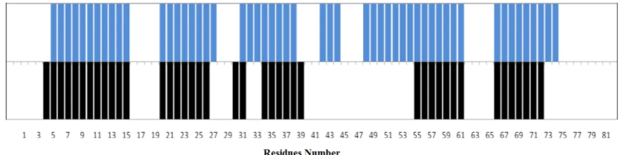
Table 1 - Report of the completeness of the Assignments of RodZn resonances from residue 1 to78, and for the full construct, separately. ... 42 Table 2 - Comparison of alpha helical secondary structure of the Homology model obtained from SWISS
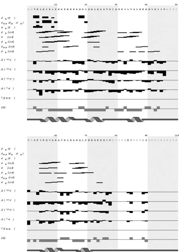
MODEL (shown in blue colour) and the NMR based CSI derived of RodZn1-101 (shown in black colour). Each bar represents the tendency of each assigned residues for a helical secondary motif. Residues that show no bars, either no assignment was performed or CSI predicted it to be in a coiled-coil region of the protein. ... 42 Table 3 - Final statistics of RodZn structure calculation. Output from UNIO10 software (Serrano et al., 2012). ... 45 Table 4 - Experimental restraints for RodZn protein, including sequential, short- and medium-range NOEs and HA, CA, CB and CO secondary shifts along with the secondary structure deduced from the data. The amino acid sequence and numbering are shown at the top. Sequential N-N NOEs are indicated by black bars; the thickness of the bar represents the strength of the observed NOE. The presence of medium-range N-N NOEs is indicated by solid lines. The chemical shift indices calculated from Cα, Cβ, CO and Ha are also shown by black bars at the bottom. The locations of the secondary structure elements identified in the calculated family of structures are shown at the bottom. ... 45 Table 5 – Resume of the evaluation of the obtained structures with RECOORD and AMBER refinement
methodologies performed with different sets of restraints. Evaluation output from ICING server. RECOORD and AMBER1 structures were obtained with the full set of restraints (NOE distance restraints, hydrogen bonds restraints and dihedrals restraints in a total of 1777 restraints). AMBER2 structure was obtained with the full set of restraints except the dihedral angles restraints. ... 47 Table 6 - Report of the completeness of the Assignments of RodZc considering only residues from
ABREVIATIONS
1HA Also Hα; proton atom attached to CA 1HB Also Hβ; proton atom attached to CB 1NH Also NH or HH; proton atom attached to N
13CA Also CA or Cα; carbon alpha from protein backbone 13CB Also CB or Cβ; carbon beta from protein side-chain 13CO Also C’; carbonyl carbon from protein backbone 15N Also N; nitrogen atom
ASEC Analytical Size Exclusion Chromatography BSA Bovine serum albumin
CD Circular Dichroism Spectroscopy D, ASP Aspartic Acid
Da Dalton
ddH2O Bi-distilled Water DNA Deoxyribonucleic acid
EDTA Ethylenediaminetetraacetic acid
g g force
IMAC Immobilized metal affinity chromatography IPTG Isopropyl β-D-1-thiogalactopyranoside
LB Lisogeni - Broth
M Molar
mAU mili units of absorbance
MW Molecular Weight
MWCO molecular weight cut off NMR Nuclear Magnetic Resonance O.D. Optical Density
PMSF phenylmethylsulfonyl fluoride ppm Parts per million
rpm Rotations per minute
RT Room Temperature
Chapter 1
Biological significance
Almost all bacteria are surrounded by a giant cell wall. The bacterial cell wall is a complex three-dimensional structure mainly composed by a peptidoglycan layer (PG) that plays a central role in the maintenance of bacterial shape, prevention of cellular stress due to differences in osmotic pressure, and formation of daughter cells during cell division. This biopolymer also known as murein is an important target of several classes of antibiotics. The clinical value of many of these antibiotics diminishes in the face of an increasing prevalence of various resistance mechanisms. As more is learned about it, new roads for research are being opened for novel drug targets and design of antimicrobials. In this light, there has been a huge effort in the science community to understand the highly complex enzymatic machinery that synthesizes the cell wall and how its activity is coordinated with cell growth and division. Spherical-shaped bacteria only synthesize PG at the plane of division (the septum), which bulges out after cell division leaving a round cell. Cells with a more complex shape, such as rod-shaped cells, exhibit an additional growth mode responsible for cell elongation. The rod-shaped bacteria such as Bacillus subtilis has been extensively used as a model in cell wall synthesis studies (reviewed by Scheffers and Pinho, 2005, and Young, 2010).
Chapter 1 – Biological significance
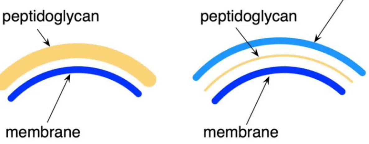
In this opening chapter I will attempt to summarize the wide range of information that is now available about the bacterial cytoskeleton, focusing on the Gram positive B. subtilis organism whose bacterial cell wall machinery is addressed in this report. Other Gram negative rod-shaped bacteria, such as E. coli and C. crescentus, will be mentioned for comparison purposes. The structure of the peptidoglycan of E. coli and C. crescentus and B. subtilis is very similar except for a few minor modifications. For instance, the major cell wall constituents, such as teichoic acids, are not attached to the peptidoglycan in E. coli and in C. crescentus, contrary to B. subtilis. The thickness of the cell walls in B. subtilis and the presence of an additional cell membrane in E. coli and in C. crescentus are other important differences in the cell wall (Figure 1). E. coli and C. crescentus possess an inner and outer membrane, with the space between the membranes (called the periplasmic space) containing one to two layers of peptidoglycan. In contrast, B. subtilis do not possess an outer membrane, but contains a thick peptidoglycan with 10-20 layers. The structure of the peptidoglycan though is very similar in both E. coli and B. subtilis (Foster et al., 2002; Vollmer et al., 2008a; Vollmer et al., 2008b; Archibald et al., 2002).
Figure 1 – Gram-positive and Gram-negative bacteria are differentiated by their cell wall structure; Picture comparing Gram-positive and -negative cell wall.
Chapter 1 – Biological significance
1.1
Cell Wall
The biosynthesis of the main component of the cell wall, the peptidoglycan, involves the coordination of the activity of proteins present in the cytoplasm, the membrane, and the periplasm. The high complexity of the cell wall elongation and division process has created a significant challenge for the study of the macromolecular interactions that regulate peptidoglycan biosynthesis. The availability of new structural and biochemical data on a number of components of peptidoglycan assembly machineries now provide novel insight into the basis of a complex molecular machinery.
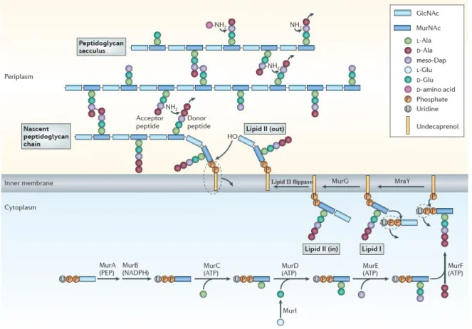
The peptidoglycan layer is formed by glycan strands of repeating disaccharide residues, cross-linked via peptide side chains (Archibald et al., 1993). It has a dynamic structure, continuously being synthesized, modified, and hydrolyzed to allow for cell growth and division, among many other roles (Foster et al., 2002).In gram-positive bacteria such as B. subtilis, PG is presented as a thick layer to which teichoic acids and cell wall specific proteins are covalently bound. Peptidoglycan synthesis in B. subtilis is summarized in Figure 2. Precursors are synthesized in the cytoplasm, linked to the transport lipid and flipped across the inner membrane followed by attachment of the newly synthesized chain (Bhavsar et al., 2006).
Figure 2 - Peptidoglycan synthesis in B. subtilis. The peptidoglycan layer is formed by polymerized chains
Chapter 1 – Biological significance
in several stages. Initially, the UDP-MurNAc-pentapeptide precursor is synthesized in six cytoplasmic
reactions catalysed by the MurA to MurF synthetases. MraY transferase then catalyses the reaction of this
precursor with the membrane acceptor, undecaprenyl phosphate, to yield lipid I. Lipid II, which comprises
the complete disaccharide-pentapeptide unit, is formed by the addition of N-acetylglucosamine to lipid I in
a reaction catalysed by MurG. Lipid II is then transferred to the outside of the membrane by lipid II
flippase. The next stage of peptidoglycan synthesis involves polymerization reactions on the outside surface
of the cytoplasmic membrane catalysed by penicillin binding proteins (PBPs) and the incorporation of the
newly formed material into the existing peptidoglycan by lytic transglycosylases (MltA). Not all
intervenients are characterized in this scheme. Notes: meso-Dap, meso-diaminopimelic acid; MraY,
MurNAc-pentapeptide phosphotransferase; MurA, GlcNAc enolpyruvyl transferase; MurB,
UDP-MurNAc dehydrogenase; MurC, UDP-UDP-MurNAc–l-Ala ligase; MurD, UDP-MurNAc-l-Ala–d-Glu ligase; MurE, UDPMurNAc- l-Ala-d-Glu–meso-Dap ligase; MurF, UDP-MurNAc-tripeptide–d-alanyl-d-Ala ligase; MurG, UDP-GlcNAcundecaprenoyl- pyrophosphoryl-MurNAc-pentapeptide transferase; MurI,
Glu racemase; PEP, phosphoenolpyruvate. Adapted from Typas et al., 2011.
The rod shape of the B. subtilis cell is maintained during its whole life cycle, being clear that all factors that control cell shape must be present in all phases of its growth. The coordinated action of two mechanisms of cell wall synthesis, one specific for cell elongation and the other for cell division is thought to be responsible for maintaining the rod shape. During cell division, the tubulin homologue FtsZ is the main player, whereas elongation is driven by the actin-homologue MreB and its paralogues MreBH and Mbl (Reviewed by Young, 2010).
1.2
Cell elongation and division
Chapter 1 – Biological significance
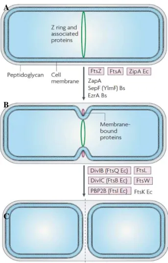
Figure 3 - Cell division in Gram-positive rod-shaped bacteria. A) Z ring formation occurs at midcell,
and recruits various FtsZ-binding proteins. B) The membrane-bound cell-division proteins are then
recruited, resulting in invagination of the cell wall and membrane to form a division septum. C) Septum
formation is complete and peptidoglycan hydrolases hydrolyse the completed cross wall, producing two
newborn cells. Proteins listed are the major ones known for B. subtilis and/or E. coli, and those in purple
are potential targets. Adapted from Rowena et al., 2008.
While cocci rely exclusively on the division machinery driven by FtsZ to grow as spheres, with each division producing two new hemispheres, rod-shaped bacteria like B. subtilis add an elongation phase before each division. The actin homolog MreB typically plays an essential role in this elongation stage by maintaining a constant cell width (Jones et al., 2001; Figge et al., 2004; Gitai et al., 2005; Kruse et al., 2003;Cabeen et al., 2011; den Blaauwen., 2008; White et al., 2011).
During the elongation stage, MreB form helical structures that are thought to guide the insertion of new peptidoglycan (PG) cell wall along the cell circumference. When MreB function is lost, cells become progressively larger as they grow and adopt spheroid morphology over time (Jones et al., 2001; Kawai et al., 2009).
How this occurs is not fully understood, but other proteins of the core morphogenic apparatus, A
B
Chapter 1 – Biological significance
with both cytoplasmatic and periplasmatic domains thought to be part of this multienzyme complex, mediating peptidoglycan synthesis on the lateral walls of B. subtilis cells. (Henriques et al., 1998; White et al., 2011; Dominguez-Escobar et al., 2011;Garner et al., 2011). This hypothesis was supported by the observation of MreC forming helical structures that alternate with the MreB helices (Dye et al., 2005). And experiments showed that MreC interacts with the penicillin-binding proteins that synthesize the cell wall. These results raised the possibility that the MreB filaments interact with MreCD complexes located in the inner cell membrane and thereby control the activity of the external cell wall-synthesizing protein complexes (Divakaruni et al., 2007; Van den Ent et al., 2006). This link between peptidoglycan synthesis and the cytoskeletal system was also confirmed by the observation of interactions between MurG and MreB in E. coli (Mohammadi et al., 2007). It was also shown that MurG localization is dependent on MreB in C. crescentus (Divakaruni et al., 2007). The interaction of MraY with MreD and the dependence of its localization on MreB in C. crescentus indicate that the morphogenic proteins MreD and MreB play a role in the organization of cell wall synthesis complexes (White et al., 2010).
While the exact function of these membrane-bound proteins remains somewhat unclear, evidence suggests that they regulate PG growth by linking MreB to cell wall enzymes or by working in concert with MreB to spatially restrict cell wall activities (Leaver et al., 2005; Levin et al., 1992; Varley et al., 1992; Wagner et al., 2005; Divakaruni et al., 2007; Kruse et al., 2005; Dye et al., 2005). The first observations of MreB were performed through fluorescence microscopy, which showed MreB filaments forming bundles moving continuously through growing B. subtilis cells (Defeu et al., 2004). However, recent reports came out suggesting that all three MreB paralogs would rather form patches moving independently (Courtney et al., 2012).
Chapter 1 – Biological significance
represents cell wall synthetic enzymes and cell shape determining proteins. In A) is represented what is seen
using decovolution fluorescence microscopy, where a stack of images taken through the cell body described
MreB to form an helical structure along the bacterial cell wall. In B) through TIRFM microscopy, a high
resolution technique capable of capturing the surface of one side of a bacterium found both MreB and a
selection of several PG elongation proteins that move in short patches as opposed to long helical filaments
with bidirectional motility. Schematic representations are not drawn to scale. Adapted from Courtney et
al., 2012.
These observations along with the dynamic directional MreB movement led to a model where MreB serves as a spiral track spanning the cell length, acting as a scaffold to organize cell wall synthesis.
1.3
Protein RodZ
Any missing players of the core morphogenic apparatus would represent a significant limitation to our understanding of cell morphogenesis. However, in recent years a new common player in bacterial cell morphogenesis has been discovered (Shiomi et al., 2008; Bendezu et al., 2008; Alyahya et al., 2009).
Chapter 1 – Biological significance
Figure 5 –Phylogenetic tree of bacterial species showing the conserved presence of RodZ (inred). Subtrees
indicate phyla, except for the proteobacterial phylum, which is further subdivided by class (Alyahya et al., 2009).
Protein RodZ from B. subtilis is encoded in ymfM gene and is composed by 304 residues. The domain organization of RodZ consists of a well-conserved N-terminal helix-turn-helix domain (HTH; residues 19-90), a conserved transmembrane domain (TM; residues 102-132) rich in hydrophobic residues, and a conserved C-terminal domain (residues 204-304) whose structure has not been determined. The conserved domains are capped by N and C terminal extensions and are separated by cytoplasmic and periplasmic linkers of variable length (29 and 117 residues, respectively). The linker separating the conserved C-terminal domain from the TM is enriched in prolines and small residues, such as glycine and alanine (Gerdes et al., 2009).The domain organization of RodZ by itself has led to the proposal that this protein could provide a direct link between the cytoplasmatic and periplasmatic peptidoglycan elongation machinery (Alyahya et al., 2009; Bendezu et al., 2009; Shiomi et al., 2008; Mitobe et al., 2011).
RodZ exhibits a localization pattern during the cell cycle corresponding to sites of active peptidoglycan synthesis. The temporal transition of RodZ from uniformly distributed patched-pattern to mid-cell localization depends on the actin-like MreB cytoskeleton. More recently the cytoplasmic domain of RodZ has been, in fact, reported to be required for assembly of MreB. And the cytoplasmic domain of RodZ (RodZn) has been shown to interact with MreB by functional and crystallographic studies in Thermotoga maritima as shown in Figure 6 (Van den Ent et al., 2010).
The interaction of the cytoplasmic part of RodZ with MreB enforces the hypothesis that this protein may be an additional transmembrane stabilizing factor of the bacterial cell wall elongation complex.
However, the cytoplasmic domain of RodZ form of Bacillus subtilis has a low homology with the published one from Thermotoga maritima (around 22%) (Gerdes et al., 2009; Mattei et al., 2010). And there is no structural evidence of direct interaction between RodZ and MreB in B. subtilis (Alyahya et al., 2009).
Chapter 1 – Biological significance
that maintenance of cell shape depended critically on a proper MreB/RodZ ratio. (Shiomi et al., 2008, Bendezu et al., 2009).
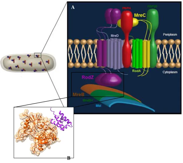
Figure 6 – A) Cartoon of the proposed PG elongation machinery in Gram-positive rod-shaped bacteria like B. subtilis. Peptidoglycan layer is not shown. The elongation machinery is represented by
proteins shown in various colours. Not all protein from the complex are portrayed. Adapted from Ana
Paiva, 7th International Conference on Gram-positive Microorganisms Poster (June, 2013); Scheme
credits: Patrícia Amaral); B) Xray structure of MreB complexed with the cytoplasmic domain of RodZ
(RodZn) from rod-shaped Gram-negative T. maritima; PDB code: 2WUS.
The cytoplasmic domain of RodZ (RodZn) alone is required for proper localization of the protein and mutations that affect the interaction between RodZn and MreB also result in mislocalization of the protein and cell shape defects (Van den Ent., 2010). In B. subtilis cells expressing RodZ without the cytoplasmic domain, the remain protein lost its characteristic localization pattern (patches), becoming uniformly distributed along the cell membrane, from cell division septa to the poles of the cell, a location
B
A
Chapter 1 – Biological significance
rarely seen for the wild type (unpublished data from The Microbial development Laboratory from ITQB). The function of the transmembrane domain of RodZ (TM ) has also been investigated. Through mutation of TM domain, this resulted in the production of short and wide rod cells, with asymmetric division. Besides, the mutated RodZ protein was shown to be distributed along the cell membrane, with only a few cells displaying the patch-like pattern. These observations led to the conclusion that the TM domain may be essential for the function and localization of B. subtilis RodZ.
As for the periplasmic domain of RodZ (RodZc), this domain is also conserved and is considered to have an important role in cell shape control, possibly through interactions with extracytoplamic components of the cell wall elongation machinery, such as MreC and the PBPs (Alyahya et al., 2009; Shiomi et al., 2008; Bendezu et al., 2009).
Unpublished results from the Microbial development laboratory from ITQB also showed that in B. subtilis, rodZ mutants lacking the periplasmic domain led to enlarged cells, roundish or with irregular shapes. And cells that still remained rod-shaped were shorter and wider than those of the wild type, similar to those of the rodZ deletion mutants. Proper localization of RodZ requires its cytoplasmic domain via interaction with MreB. But these new studies indicate that the periplasmic domain may also contribute to the sub-cellular localization of RodZ, thus inferring that interactions on either side of the cell membrane are required for the correct sub-cellular positioning of RodZ.
So, both RodZn and TM domains are essential to maintain the normal pattern of midcell division, and the right positioning of the nucleoid. Nevertheless, the overall rod-shape of the cell is maintained in mutants lacking these two domains. In contrast, deletion of RodZc also causes a severe change in the cell shape phenotype. So, the presence of the periplasmic domain in most RodZ orthologues suggests an important function but no meaningful hint about its role has been published so far (Alyahya et al., 2009).
Studies in E. coli have revealed direct interactions of RodZ with MreC and possibly with MreD and the cell wall elongation PBP2 (Bendezu et al., 2009). Therefore, RodZ seems to interact with proteins of the cell elongation machinery on both sides of the cytoplasmic membrane. MreC and PBPs are likely to interact with the periplasmic portion of RodZ (RodZc).
Chapter 1 – Biological significance
1.4
Bacillus subtilis
B. subtilis has approximately 4,100 genes. Of these, only 192 were shown to be indispensable and 79 were predicted to be essential as well. A vast majority of essential genes were categorized in relatively few domains of cell metabolism, with about half involved in information processing, one-tenth related to cell energetics and one-fifth involved in the synthesis of cell envelope and the determination of cell shape and division (Kunst et al., 1997; Kobayashi et al., 2003).
Originally named Vibrio subtilis in 1835 by Christian Gottfried Ehrenberg, this organism was renamed by Ferdinand Cohn Bacillus subtilis in 1872 (Ehrenberg, 1835; Cohn, 1872).Bacillus subtilis is a bacterial microorganism commonly found in the environment, mainly in soil, being categorized as a saprophyte organism (Brock et al., 2005). Nevertheless, this microorganism is well known by modern science to be very friendly to the human system, being able to promote dramatic healing benefits, even though it isn't one of the native microbes that normally inhabit the human body (Hong et al., 2009). Even though B. subtilis has been historically classified as a strictly aerobe microorganism, recent research shows that this species can actually leave under anaerobic conditions (Nakano et al., 1998).
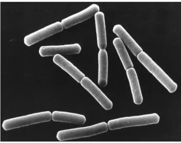
Similar to Gram-negative Escherichia coli and Caulobacter crescentus bacteria, Gram-positive B. subtilis has a rod-shape, as shown in Figure 7 , being 3-5 µm long, of about 1 µm width (Sargent, 1975) and with hemispherical cell poles (Burdett et al., 1978).
Figure 7 – Electron micrograph picture showing rod-shaped Bacillus subtilis cells. Image credit from NASA.
Chapter 1 – Biological significance
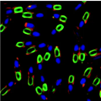
But B. subtilis has also the ability to multiply in an asymmetrical fashion, producing a single endospore that can remain viable for decades, being resistant to unfavourable environmental conditions such as drought, salinity, extreme pH, radiation and solvents (Errington, 2003) (Figure 8).
Figure 8 - Fluorescence micrograph of sporulating Bacillus subtilis cells, showing nucleoids (blue),
membranes (red) and YwcE protein (green; protein required for spore morphogenesis and germination).
Cover photograph, American Society for Microbiology. Copyright © 2005. All Rights Reserved.
Prior to the process of sporulation, cells might become motile by producing flagella, take up DNA from the environment, or produce antibiotics. These responses are viewed as attempts to seek out nutrients by searching a more favourable environment, enabling the cell to make use of new beneficial genetic material or simply by killing of competition. (Bandow et al., 2002).
Chapter 1 – Biological significance
1.5
Aims
As reviewed, recent work suggests that the widely conserved protein RodZ affects the processes of cell division site selection and chromosome positioning, in addition to its central role as a cell shape determinant. RodZ is a multidomain transmembrane protein, responsible for synchronizing inner and outer cell processes of the bacterial cell wall synthesis.
In the present thesis, bioinformatic, biophysical and biochemical tools/techniques are used to study the cytoplasmatic (RodZn) and periplasmatic (RodZc) domains of RodZ individually from B. subtilis.
Our first goal is to determine for the first time through NMR spectroscopy the solution structure of RodZn. NMR data acquisition, processing and analysis of RodZn has already been reported in 2011, in my bachelors’ graduation thesis. There I also reported the chemical shifts assignment routine. At the end of that work period we were able to complete the assignment of RodZ and from the chemical shifts index (CSI) obtained we were able to determine the secondary structure of RodZn1-98 construct. In the current work we concluded the studies of that system with the NMR structure elucidation of that N-terminal domain. From our analysis we concluded that our construct has similarities with previously determined structures but also some meaningful differences (Pereira, 2011). The functional role of RodZn in B. subtilis hasn’t been fully identified yet, but recent data indicate that it may interact with MreB. So, after determining the tertiary structure of RodZn, we intend to use the identified chemical shift resonances of the protein to perform protein-protein interaction studies with MreB.
Next, we’ll focus our studies in the uncharacterized periplasmic domain of RodZ (RodZc) from B. subtilis. Very little has been published regarding the function/structure of this domain. Since no structure has ever been determined for this domain, our main goal will be to calculate a 3-dimensional structure of RodZc through solution state NMR experiments. We will also study the dynamics of this domain, by collecting 15N relaxation data of the protein backbone and understand if this domain may possibly promote protein-protein interaction with other key players from the peptidoglycan synthesis machinery.
Levinthal’s Paradox
“The length of time in which a protein chain finds its folded state is many orders of magnitude shorter than it would be if it freely searched all possible configurations.”
Levinthal, C. 1969. Mossbauer Spectroscopy in Biological Systems. Proceedings of a meeting held at Allerton House. P. Debrunner, J. C. M. Tsibris, and E. Munck, editors. University of Illinois
The Central Dogma
“The three-dimensional structure of a protein is determined by its sequence and its environment without the obligatory role of extrinsic factors”.Chapter 2
Methodology
Proteins are linear chains of amino acids that adopt a three-dimensional structure in their native environment. The biological role of a protein is determined by its function, which is in turn largely determined by its structure (Dickerson et al., 1969; Petsko, 2000). Thus there are enormous benefits in knowing the three dimensional structure of all the proteins.
Levinthal’s paradox raised the question why and how a sequence of amino acids can fold into its functional native structure given the abundance of geometrically possible structures (Levinthal, 1969). The pioneering experiments of Anfinsen shed light on this problem. According to Anfinsen’s thermodynamic hypothesis, proteins are not assembled into their native structures by a biological process, but folding is a purely physical process that depends only on the specific amino acid sequence of the protein and the surrounding fluid. Anfinsen’s hypothesis implies that a protein structure can be predicted if a model of the free energy is available, and if the global minimum of this function can be identified.
The first three-dimensional protein structure in crystalline state, namely Myoglobin, was experimentally determined in 1959 by John Kendrew using x-ray crystallography (Kendrew, 1959).
Chapter 2 – Methodology
of proteins inside the cells. Therefore, x-ray crystallography and NMR are in many aspects complementary and are still, nowadays, the two major techniques used for protein structure determination at high resolution.
The last RCSB protein data bank (PDB; http://www.rcsb.org) annual report ((http://www.rcsb.org/pdb/statistics/holdings.do accessed in 07/10/2013) listed 94,336 atomic coordinate entries available in the PDB repository, from which 88% were determined by x-ray crystallography and 11% determined by NMR (Berman et al., 2000). However, the gap between known
protein sequences and structures is increasing rapidly.
Statistics released in October 2013 show that UniProtKB/Swiss-Prot contains 540,958 sequence entries (http://web.expasy.org/docs/relnotes/relstat.html). This gives an idea how experimental methods alone will not be able to fill in this gap. Therefore it is necessary to use computational methods to predict protein structures (Dill et al., 2007).
Template based homology modelling methods could be used for sequences that have detectable relationship with sequences of experimentally determined protein structures. On the other hand, for predicting the structure of proteins that do not share a detectable sequence relationship with experimental structures, Ab initio protein structure prediction techniques must be used. The methods under Ab initio protein structure prediction category aim to predict the structure of a protein from the sequence information alone, without any explicit use of previously known structures. These methods use thermodynamic principles and try to identify the native structure of a protein as the global minimum of a potential energy landscape.
Since such methods are computationally complex and are extraordinarily challenging, over the past few years there has been a huge effort in developing ab inito protein structure prediction methods. A major milestone in computer-based native structure prediction is the creation of CASP (Critical Assessment of Techniques for Structure Prediction) by John Moult (Moult, 2006). In the CASP experiments, research groups apply their prediction methods to amino acid sequences for which the native structure has not been publicly disclosed but has been determined and are to be published soon. These competitions provide a good measure to benchmark methods and progress in the field in an arguably unbiased manner (http//:www.forcasp.org).
These Ab initio prediction methods are based on physical and energetic principles that perform the search through the conformational space. Models used are usually simplified and search methods that are often used are Monte Carlo Algorithms.
Chapter 2 – Methodology
In the following chapter will be described the protein structure prediction methods available so far. They will be divided into four categories: 1) Homology Modelling, 2) Pattern-based Homology Modelling, 3) Ab-initio, and 4) Data-driven structure prediction.
Essentially, the classification reflects the degree to which different methods utilize the information content available from the known structure database and experimental data. I will focus on the specific methods that were used on the present work. I will go over the accuracy, applicability and shortcomings of each kind of tool. Protein structure calculation, refinement and evaluation methods are also discussed. In addition, for simplification purposes, I will be referring to the available not pure-ab initio tools (pseudo-ab initio) just as ab initio.
2.1
Homology Modelling
Homology Modelling relies on the principle that similar sequences exhibit similar three-dimensional structures (Floudas, 2007). Strong sequence similarity often indicates strong structure similarity, although the opposite is not necessarily true.
There are usually four steps in homology based protein structure prediction methods: (1) identify one or more suitable structural templates from the known protein structure databases; (2) align the target sequence to the structural template; (3) build the backbone from the alignment, including the loop region and any region that is significantly different from the template; and (4) place the side-chains.
In homology modelling, local sequence comparison methods are usually used since the sequence similarity is most likely over segments of the two sequences. The local sequence comparison can either be pair wise or profile based. Pair wise comparisons, such as the BLAST can detect sequence similarities better than 30% (Altschul et al., 1990). To increase the chance of detecting weak homologues, PSI-BLAST (Position Specific Iterated BLAST) was build to search the database iteratively until no new hits are found (Altschul et al., 1997). Methods such as PSI-BLAST encode the information about a whole protein family for the target sequence in a model to increase the chance of detecting remote homologies. To further increase the detection sensitivity, the sequences in the structure database can also be encoded in profiles. This forms the basis of the pattern-based homology modelling methods that will be explained in detail in the next chapter (Koehl and Levitt, 2002).
Chapter 2 – Methodology
On the other hand, structurally similar proteins can have low sequence identities (8-10%) and still be identified with sensitive profile-profile based comparison, but the RMSD can be as large as 3-6. The error largely comes from the misalignment from sequence comparison. At such low sequence identity, comparison method that can detect the remote homology as well as align the sequences close to the optimal from structure-structure alignment would be desirable.
SWISS-MODEL
SWISS-MODEL (http://swissmodel.expasy.org) is one of the most widely used web-based servers for automated homology modelling-based structure prediction (Peitsch et al., 2003). In addition to a fully automated mode requiring minimum user input, i. e. protein sequence only, SWISS-MODEL offers two more advanced user modes in which users can submit their own multiple sequence alignment or manually adjust the modelling parameters (Guex and Peitsch, 1997; Schwede et al., 2003).
To generate a 3D structure from a provided sequence, SWISS-MODEL utilizes rigid body assembly, in which sections from aligned regions of the template are connected together by separately constructed non-conserved regions to form the model backbone. Suitable template structures, those with similar sequences to the query, are first identified by a gapped BLAST search of the SWISS-MODEL template library ExPDB, a subset of PDB. The selected templates are then superimposed using an iterative algorithm, the backbone atom positions averaged, and the query sequence fitted to the template to optimize placement of insertion and deletion regions. Fragments that cannot be modeled by homology to the template are computed based on energy considerations or, if the region cannot be solved, searched against a library of loop structures to find an appropriate match. Finally, side chain conformations and intermolecular interactions are adjusted to minimize conformational energy and correct any irregularities in overall 3D structure that resulted from the assembly process.
Assessments of the SWISS-MODEL prediction algorithm reveal variable accuracy dependent on the degree of query-template sequence similarity. If the predicted protein models show 40% sequence identity between query and template, the RMSD will be less than 3 Å from their experimental structures. However, proteins with lower sequence identities failed to exhibit such modelling accuracy.
Chapter 2 – Methodology
numbers of 3D models that failed to converge (i.e. >3 Å RMSD) with the backbone structure, reducing the fidelity of the final model to its original template (Wallner and Elofsson, 2005).
2.2
Pattern-based Homology Modelling
For evolutionary related proteins, even if the sequence similarity is difficult to detect with sequence comparison methods, there could still be identifiable structural similarity. Structure alignment has been shown to be able to identify homologous protein pairs with sequence similarities less than 10% (Rost, 1997; Brenner et al., 1998; Gerstein et al., 1998). Pattern recognition-based methods involve similar steps as in homology modelling. The difference is in the pattern identification step. First of all, a structure library needs to be defined (Sitbon and Pietrokovski, 2007). Natural choices for building the library of protein clusters are protein families (Henikoff et al., 1997), sequence motifs that separate proteins into either conserved or random regions (Henikoff et al., 2000), and the four basic secondary structure elements namely alpha helices, beta strands, turns, and loops (Henikoff et al., 1995). Once the library is defined, the target sequence will be fitted to each library entry and an energy function is used to evaluate the fit between the target sequence and the library entries to determine the best possible templates.
Protein sequence comparison has become one of the most powerful tools for characterizing protein sequences because of the enormous amount of information that is preserved throughout the evolutionary process. A general approach for functional characterization of unknown proteins is to infer protein functions based on sequence similarity. One of the approaches is to define signatures of known families of biologically related proteins. These signatures usually identify conserved regions among the family of proteins, revealing the importance for the function of their structural properties. A representative example of this approach is the well-known PROSITE database, which gathers protein sequence patterns and profiles for a large number of families (Hulo et al., 2004). Another web tool based in pattern homology modelling is PSIPRED.
PSIPRED
Chapter 2 – Methodology
PSIPRED carries the secondary structure prediction on a protein and gives its name to the prediction server itself. α-helical (H), β-stranded (E) and Coiled-coil (C) protein motifs are predicted based on the analyses of the output obtained from PSI-BLAST combined with the DSSP algorithm (Define Secondary Structure of Proteins). To these predictions are assigned individual confidences (Altschul et al., 1997). Using a rigorous cross validation procedure to evaluate performance, PSIPRED has been shown to be capable of achieving an average Q3 score (% of 3-state symbols that are correct) of 81.6% (PSIPRED v3.2). This is one of the highest levels of accuracy published for any method to date (Jones, 1999).
PSIPRED server allows users to select a variety of prediction methods they may find useful. Here will be reviewed two recently developed tools: MENSAR-SVM which predicts the topology of a protein and DISOPRED that predicts disordered regions from the amino acid sequence.
Transmembrane protein prediction: Transmembrane proteins (TM) are involved in a wide range of important biological processes such as cell signalling, transport of membrane-impermeable molecules, cell-cell communication, cell recognition and cell adhesion. However, due to experimental difficulties, this class of proteins is severely under-represented in structural databases. In the absence of structural data, sequence-based prediction methods allow TM protein topology to be investigated. PSIPRED server also supports the vector machine-based TM protein topology predictor MENSAT-SVM.
This web-tool integrates both signal peptide and re-entrant helix prediction, cross validated on a novel data set of 131 sequences with known crystal structures. The method achieves topology prediction accuracy of 89%, while signal peptides and re-entrant helices are predicted with 93% and 44% accuracy respectively. An additional SVM trained to discriminate between globular and TM proteins detected zero false positives, with a low false negative rate of 0.4%. The high accuracy of TM topology prediction, which is able to discriminate signal peptides and identify the cytosolic and extra-cellular loops, makes this method ideally suited to whole genome annotation of alpha-helical transmembrane proteins (Nugent and Jones, 2009).
Most efforts in structural bioinformatics have been directed to the prediction of globular protein structures but there is an increasing interest in disordered regions for studying the function of proteins.
Order/Disorder prediction: Interest in intrinsically disordered proteins (IDPs) grew as a result of the realization that such proteins are unexpectedly common in human and other genomes (Ward et al., 2004; Oldfield et al., 2005; Tompa et al.,, 2006).
Chapter 2 – Methodology
reasons why IDPs are frequently associated with cellular control mechanisms and signalling, and have been identified at the “heart” of protein interaction networks (Dunker et al., 2005).
In recent years there has been a growing interest in intrinsically unstructured proteins and their role in biology. However, it’s yet poorly understood the relationship between the primary sequence of a protein and its susceptibility to a disordered conformation. A signature of probable intrinsic disorder is the presence of low sequence complexity and amino-acid compositional bias, with a low content of bulky hydrophobic amino acids (Val, Leu, Ile, Met, Phe, Trp and Tyr), and a high proportion of polar and charged amino acids (Gln, Ser, Pro, Glu, Lys, Gly and Ala) (Romero et al., 2001; Vucetic et al., 2003).
A number of computer programs are now available for the prediction of unstructured regions from amino acid sequences. Disordered regions are dynamically flexible and are distinct from irregular loop secondary structures, which are static in solution. But many disordered proteins do adopt more highly ordered conformations upon interactions with other cellular components (Dyson and Wright, 2002; Iakoucheva et al., 2002; Dunker and Obradovic, 2001).
The DISOPRED server present in the PSIPRED platform uses a knowledge-based method to predict dynamically disordered regions from the amino acid sequence. This server allows users to submit a protein sequence, and returns a probability of each residue in the sequence being disordered. The DISOPRED method is developed from the original DISOPRED predictor (Jones and Ward, 2003).
2.3
Ab-initio
Only when no suitable structure templates can be found, Ab initio methods can be used to predict the protein structure from the sequence information only. In addition, they can also predict protein mechanisms, motions, folding processes, conformational transitions and other situations in which protein behaviour requires more than just knowledge of the static native structure (Dill et al., 2007). In recognition of the developments and the importance of this field, the Nobel Prize of Chemistry of 2013 was focused on such approaches.
Chapter 2 – Methodology
successful prediction of a protein structure are generation of a vast number of conformations and accurate scoring functions.
The Ab initio methods are rigorous in calculations but are limited by the compute power and time involved which emphasizes the need for faster structure prediction methods. Also, the accuracy of these methods is dependent upon the potential energy functions used during simulations (Karplus and McCammon, 2002).
For practical reasons, most Ab initio prediction methods use reduced representations of the protein to limit the conformational space to convenient size and use empirical energy functions that capture the most important interactions that drive the folding of the protein sequence toward the native structures.
TASSER
TASSER is a successful free modelling approach that constructs 3D models based on a hybrid between ab initio and homology modelling approach (Zhang and Skolnick, 2004). The target sequence is first threaded through a set of representative protein structures to search for possible folds. Contiguous fragments (>5 residues) are then excised from the threaded aligned regions and used to reassemble full-length models, while unaligned regions are built by ab initio modelling (Zhang et al., 2003).
The protein conformation in TASSER is represented by a trace of Cα atoms and side chain centres of mass, and the reassembly process is conducted by Monte Carlo simulations (Zhang et al., 2002). The energy terms of TASSER include information about predicted secondary structure propensities, backbone hydrogen bonds, a variety of short- and long-range correlations and hydrophobic energy based on the structural statistics from the PDB library. Weights of knowledge-based energy terms are optimized using a large-scale structure decoy set (computer-generated set of protein structures) which coordinates the correlations between various interaction terms (Zhang et al., 2003).
Chapter 2 – Methodology
ROSETTA
ROSETTA has been extensively reviewed as being the best Ab initio method so far (Bonneau and Baker, 2001; Bonneau et al., 2001; Simons et al., 2001).
The ROSETTA method also illustrates many features and techniques that are common to the majority of the Ab initio based-methods. The ROSETTA method, like many others, uses a reduced representation of the protein as short segments, since local segments of the protein sequence have statistically important preferences for specific local structures and that the tertiary structure has to be consistent with this preference (Go, 1983; Simons et al., 1997).
The energy function is defined as probabilistic model of protein sequence/structure matches in terms of structural segments to formulate secondary structure prediction and this forms the basis of the Monte Carlo sampling of the reduced protein conformational space (Simons et al., 1999). The non-local potential, which drives the protein toward compact folded structure, includes terms that favour paired strands and buried hydrophobic residuals. The solvation effect can also be incorporated into the energy function.
A problem intrinsic to the reduced representation of the protein and the simplified empirical potential is that the energy function is not sensitive enough to differentiate the correct native structures from conformations that are structurally close to the native state. The energy landscape calculated from such energy functions will not be properly funnelled but flattened around the native structure. In fact, as the native state is approached, the correlation between the calculated energy and the measure of similarity between predicted and native structures are no longer valid. The usual practice is then to produce a large number of decoy structures and then use various filtering and clustering techniques to pick up the more native like structures. Filters can be used to eliminate structures with poorly formed secondary structures and low contact orders compared with that for sequences with compatible length (Bonneau et al., 2001).
2.4
Data-driven structure prediction
Even small amounts of experimental data can dramatically improve the quality and reliability of ab initio structure prediction with the application of spatial constraints. For instance, the Rosetta method can produce moderate to high-resolution structures when combined with limited NMR constraints (Standley et al., 1999; Bowers et al., 2000; Rohl and Baker, 2002).
Chapter 2 – Methodology
CS-ROSETTA (www.csrosetta.org) is a framework for structure calculation of biological macromolecules on the basis of structural information derived from NMR data, which is build on top of the biomolecular modelling and design software called ROSETTA. The name arise from its origin in combining NMR chemical shift data (CS) with ROSETTA prediction protocols (Shen et al., 2008). Further advantages of using chemical shifts are that these are among the most reliable parameters that can be obtained from NMR spectroscopy and that they can potentially be obtained for larger macromolecules for which NOEs become impractical.
Structure determination using CS-ROSETTA requires as only input the amino acid sequence and a list of chemical shifts and a number of parameters to control the process that can be changed from the default values. Backbone chemical shifts for 13CA, 13CB, 13CO, 1HA, 1HN, and 15N that are provided by the user, are validated and stored as the target shifts. These chemical shifts are first used to select a fragment library that contains many possible conformations (fragments) for a given set of degrees of freedom specific for the protein backbone. This selection is made from a structure database, e.g. the RCSB Protein Data Bank (PDB), based on the list of chemical shifts as predicted with SPARTA. SPARTA is a database system for empirical prediction of backbone chemical shifts (N, HN, HA, CA, CB, CO) using a combination of backbone phi, psi torsion angles and side chain chi1 angles from a given protein with known PDB coordinates (Yang and Bax, 2007).
Then the regular ROSETTA protocol for Monte Carlo assembly and relaxation is used to reassemble the protein from the fragments. For the resulting models the chemical shifts are back predicted using SPARTA and the deviations between the predicted and target values are used as a pseudo-energy term in the scoring of the models, yielding a ranking based on both overall structural quality as well as on the match with the experimental data.
The computationally most expensive step in the process is the construction of a model using Monte Carlo assembly and relaxation. To obtain a reliable prediction, a set of 10,000 to 50,000 models has to be built, each starting from the same fragment library. Using different seeds for generation of random numbers ensures independence of the results from different runs. The computational cost involved in chemical shift based structure determination makes CS-ROSETTA a typical example of a program that is beyond the capacity of most local sites.
More recently, a new protocol was added to CS-ROSETTA. RASREC protocol was designed to improve sampling close to the native structure by recombination of frequently occurring structural features such as α-helix or β-strand topologies.
Chapter 2 – Methodology
The RASREC protocol is characterized by first using chemical shift data to pick and choose a fragment library, as described previously. Then, individual structure calculations employ the abrelax protocol that combines ab initio fragment assembly in centroid mode with the all-atom refinement using the Rosetta full-atom force field (relax), by interleaving packing of side chains with gradient based minimization of torsional degrees of freedom. The advantage of this protocol is that it is relatively time efficient since Ab initio folding is faster and the relax step is more time-consuming (Das et al., 2007).
Around 200-1000 of independent structure calculations run in parallel and a pool of the best output decoy set is continuously updated from the results of the independent structure calculation. Specific features (like α-helix or β-strand topologies) in the pooled structures are then used to focus further sampling (Raman et al., 2010; Lange and Baker, 2012).
The CS-ROSETTA software package has recently been extended to include additional NMR conformational parameters, such as Residual Dipolar Couplings (RDC) and NOE distance restraints (Raman et al., 2010; Lange et al., 2012).
2.5
Fundaments of NMR spectroscopy
Nuclear magnetic resonance (NMR) spectroscopy is a very versatile tool that can detect the frequency at which certain nuclei are resonating under the influence of a magnetic field. Slight variations in this resonant frequency give us detailed information about the molecular structure in which the atom resides. It has a very wide variety of applications and it can also be used to study the structure and properties of biomolecular compounds.
The available nuclei suitable for nuclear magnetic resonance are those with non-zero spin quantum number. For technical reasons, mostly unpaired nuclear spins with I=1/2 are used for NMR spectroscopy. The most widely used nuclei in biomolecular NMR are 1H, 15N and 13C, being the main components of proteins.