Biomecánica, 17 (1), 2009, pp. 7-14
Abstract
Biodegradable polymers have been used in implantable medical devices, such as suture fibers, fixation screws and soft tissue engineering devices. Apart from biological compatibility, these devices shall also be functional compatible and perform adequate mechanical temporary support during the healing process. In regenerative medicine, the scaffold that will provide this temporary support should simultaneously enhance cellular adhesion, proliferation and remodeling of new tissue. In soft tissue applications, biodegradable polymers are the materials of election. These materials undergo through a process of degradation, mainly controlled by hydrolysis, leading to a reduction of molecular weight, followed by reduction of strength and finally a reduction of mass until it is totally absorbed and assimilated by the host. Fatigue/creep damage also contribute to the progressive decrease of mechanical properties. Meanwhile, cells cultured over the scaffold will produce the new tissue that will gradually replace the material biomechanical functions. Keywords: biomechanics, biomaterials, soft tissue, regenerative medicine.
Resumo
Os polímeros biodegradáveis têm sido utilizados em dispositivos médicos implantáveis, como fios de sutura, parafusos e dispositivos para engenharia de tecidos moles. Além da compatibilidade biológica, tais dispositivos devem apresentar compatibilidade funcional e desempenhar funções temporárias de suporte mecânico durante o processo de cura. Em medicina regenerativa, este suporte temporário deve favorecer a adesão celular, a sua proliferação e a remodelação de novo tecido. Em engenharia de tecidos moles os polímeros biodegradáveis são os materiais de eleição. Estes materiais sofrem um processo de degradação, controlado sobretudo hidrólise, e que resulta numa redução do peso molecular, seguida de uma redução da resistência mecânica e finalmente uma redução da massa até a completa absorção e assimilação pelo organismo. O dano por fadiga/ fluência também contribui para a progressiva diminuição das propriedades mecânicas. Entretanto, as células cultivadas sobre o suporte vão produzir o novo tecido que vai gradualmente substituir as funções biomecânicas do material.
Palavras-chave: biomecânica, biomateriais, tecidos moles, medicina regenerativa.
A.F.C. VIEIRA1, J.F.C. VIEIRA1, A.T. MARQUES2, R.M. GUEDES2. 1INEGI – Instituto de Engenharia Mecânica e Gestão Industrial. 2Dpto. Engenharia Mecânica e Gestão Industrial (DEMEGI),
Faculdade de Engenharia, Universidade do Porto (FEUP).
Correspondencia: [email protected] [email protected] [email protected] [email protected]
Biomechanics of biomaterials used in soft tissue
regenerative
(1r Premio Congreso SIBB 2008)
Introduction
In reconstructive medicine, traditional materials have been used to make implant devices able to replace the biological functions of a tissue. Polyurethane (PU), poly(tetrafluoroethylene) (PTFE), Polyetheretherketone (PEEK), polyethylene (PE) have been used in soft tissue devices due to there biocompatibility. Examples of these are vascular or valvular devices, ligament tissues augmentation devices, etc. These materials are considered bioinert and they don’t evoke any deleterious biologic response. However, at later time points, biomedical devices are prone to fatigue rupture or laxity due to creep, resulting in long term inefficiency. This usually implies revision surgeries, onerous for the patient and for the health care system.
New concepts of regenerative medicine are based on cells seeded over scaffolds, that can temporary replace the biomechanical functions, and will progressively be replaced by the new tissue formed. Scaffolds, cells and the environment (physical and chemical) constitute the three major components in regenerative medicine, and successful engineering of any tissue requires that the various components be optimized.
Such scaffolds should possess an optimized architecture that provides favourable porosity and surface area to allow the seeded cells to proliferate and regenerate the tissue. A minimum pore diameter of 200-250ìm is suggested for soft tissue ingrowth [1]. The presence of pore interconnectivity extending through an implant increases the overall surface area for cell attachment, which in turn can enhance the regenerative properties of the implant by allowing tissue ingrowth into the interior of the scaffold. [2] Overall scaffold porosity can modulate gross cellular response to the implant but also their biomechanical behaviour.
A successful tissue engineered scaffold must display similar mechanical behavior (shape of the stress–strain and stress relaxation response), have endurance strength (fatigue resistance at the same frequency of the application) have mechanical properties that are similar to or greater than the tissue it is regenerating, promote tissue ingrowth, and degrade at a rate that does not cause stress shielding or rupture of the new tissue. In the design and optimization process, damage simulation due to hydrolysis and fatigue/creep is a helpful tool in predicting outcomes and to analyze functional compatibility. To be biocompatible and ensure final clinical use, neither the scaffold nor its degradation
products should be harmful to the surrounding tissue and they should not result in unresolved inflammation.
Materials
The most popular and important biodegradable polymers are aliphatic polyesters, such as polylactic acid (PLA), polyglycolic acid (PGA), polycaprolactone (PCL), polydioxone (PDO), polyhydoxyalkanoates (PHA’s), polyethylene oxide (PEO), poly(propylene succinate) (PPSu), poly (butylene succinate) (PBSu), poly(ethylene succinate) (PESu), poly(butylene succinate-co-butylene adipate) (PBSA), etc. Processing technologies of these materials are the same of traditional thermoplastic polymers. In table 1, the chemical structures of the common aliphatic polyesters are presented (Table 1).
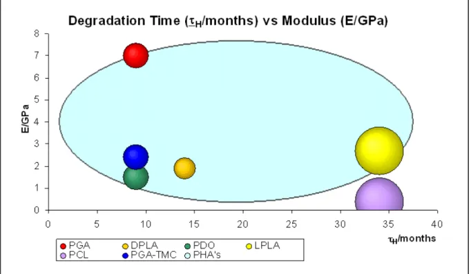
Aliphatic polyesters are a central class of biodegradable thermoplastic polymers, because hydrolytic and/or enzymatic chain cleavage of these materials leads to á-hydroxyacids, which in most cases are ultimately metabolized. For soft tissue applications, depending on material selection, a large range of mechanical properties are possible (Figure1).
Synthetic biodegradable PLA, PGA, and copolymers of these, have been manufactured for biomedical applications since the 1970s. Since then, FDA has approved its use for a variety of clinical applications, [3-7], e.g. for surgical sutures [8, 9], internal bone fixation [10] and drug delivery systems [11], because they present good biodegradability, biocompatibility, reasonably good mechanical properties and processibility. They have also been investigated for use in tissue engineering and regenerative medicine. Since they display different degradation rates, their combination enables to regulate the degradation time.
Polycaprolactone is also an important member of the aliphatic polyester family [12], suitable for long-term use in implants presenting a slower degradation rate than those of PLLA and PGA [13]. Due to its low stiffness, it is commonly combined with PLA to decrease its brittle behavior in slow degrading devices.
Hydrophilic materials are more prone to swell, corresponding to a greater decrease of the mechanical properties. By presenting higher saturation level they degrade faster, since degradation time is proportional to water concentration. That is the case of hydrophilic PGA compared to PLA. Poly(ethylene glycol) PEG is
Table 1. Chemical structures of the common aliphatic polyesters. Figure 2. Tensile strength vs time for different initial molecular weight [22].
another hydrophilic, water soluble and fully biodegradable polymer. PEG’s lack of toxicity allows its usage in many biomedical and pharmaceutical applications [14, 15].
PHA’s, particularly poly 3-hydroxybutyrate (PHB), copolymers of hydroxybutyrate and 3-hydroxyvalerate (PHBV), poly 4-hydroxybutyrate (P4HB), copolymers of hydroxybutyrate and hydroxyhexanoate (PHBHHx) and poly 3-hydroxyoctanoate (PHO) and its composites have been used to develop a great variety of biodegradable devices [16] with a large range of degradation times and mechanical properties.
However, each of these alone may have some shortcomings which restrict its applications, due to inappropriate stiffness, strength or degradation rate. It is possible to tune the hydrolytic rate constant of the material, and also its final mechanical properties, by block copolymerization or blending with other biodegradable polymers, having different hydrolytic rate constants. Copolymers of several lactides or lactones can be synthesized by ring opening polymerization resulting in high molecular weight polyesters [17]. The mixture of different polymers to produce blends, with controlled hydrolytic rate and mechanical properties can be preformed in two ways: mixing the melted polymers, or mixing polymers solutions using a common solvent. The miscibility is limited depending on polymers used and their volume fractions. A composite solution of several materials with different degradation rates also enables tuning the rate of degradation of the scaffold.
Hydrolytic Degradation
The degradation process of biopolymers is composed of a sequence of reactions, mainly diffusion of water and/or enzymes, and then hydrolysis. The overall rate is determined by the slowest reaction, named the rate-limiting step [18]. In the particular case of biodegradable polymers, the penetrating water rapidly creates a negative gradient of water concentrations from the surface to the centre. Since water diffusion is very fast compared to hydrolysis, one can consider that hydrolysis of ester bonds starts homogeneously.
The water concentration (W) is determined using Fick’s equation, presented for 1D [19]:
(1)
were the diffusion rated can be determined by measuring moisture absorption increased weight
during incubation. The amount of absorbed water is deduced from:
(2)
where Ws, and Wr, are the weight of the swollen specimen (after wiping the surface with paper) and the weight when dry, respectively. The swelling has a major significance on the visco-elastic properties of the material. When saturated, its ductile/fragile transition will occur at higher strain rates.
The macromolecular skeleton of many polymers is comprised of ester groups. These groups can go through hydrolysis leading to chain scissions. Hydrolysis has traditionally been modeled using a first order kinetics based on the kinetic mechanism of hydrolysis, according to the Michaelis–Menten scheme [20]. In the ideal case of hydrolytic degradation the following first-order equation describes the hydrolytic process to occur relative to the ester concentration (E) and water concentration (W):
(3)
umis the medium hydrolysis rate of the material, and W is constant as water is spread out uniformly in the sample volume (no diffusion control). In this ideal case, the hydrolysis rate constant, k, depends only on temperature and does not vary with the conversion (no autocatalysis).
Using the molecular weight, and since the concentrations of carboxyl end groups
t n
M
E
1
/
; the equation 3 turn:(4)
where
t n
M
andM
n0, are the number-average molecular weight, at a given time t and initially at t=0, respectively.Or using the scission numberntper mass unit, as in [20], at time t is given, and the initial ester concentration E0, the equation 3 becomes:
(5)
The experimental measurement of molecular weights by SEC (Size Exclusion Chromatography)
2 2 x W d dt dW ! ! r r s W W W W 100( " )/ W u kEW dt dE m " " kWt n n
M
e
M
t " 0 W n E k dt dn t t ( ) 0" "or GPC (Gel Permeation Chromatography) allows the determination of nt.
(6)
These equations lead to a relationship Mn =f(t). However, in the design phase of a biomedical device, it is important to predict the evolution of mechanical properties like stiffness and tensile strength, instead of molecular weight. It has been shown [21] that the fracture strength of a polymer can, in many cases, be related to Mn through the relationship:
(7)
where σ is the fracture strength, σ is the fracture strength at infinite molecular weight, and B is a constant.
As this is an empirical equation, the constant B
must be determined for each material. One can thus determine the limit strength for the device that weakens during the tissue recovering,
σ
d= f(t). The
variation of the tensile strength along with degradation time is presented in figure 3, changing with initial molecular weight. According to Farrar and Gilson [22] the rate constant depends on the structure of the polymer, and is independent of its molecular weight (Figure 2).
In order to perform computer simulation based on these models, the parameters diffusion coefficient (d) and degradation rate constant (k) must be previously determined by inverse parameterization, on specimens of two different thicknesses. For thin devices damage model can be simplified assuming homogeneous hydrolyses. On that ground, the mixture law may be used to determine the hydrolysis rate of composite ucof n degradable materials combined:
(8)
Mechanical properties of polymeric materials, mainly thermoplastics, and also natural tissues, highly depend on the strain rate applied to the specimen. As the strain rate increase, there is a transition from ductile to fragile behaviour. Material tensile properties must so be determined at the same strain rate. A general consequence of hydrolytic process is the lowering of the plastic flow ability of the polymer, thus causing the change from ductile behavior into a brittle one or, if the behavior was initially brittle, an increase in the brittleness. Moreover, after hydrolysis started, brittleness may occur at lower strain rates.
The degradation rate and the mechanical properties of biodegradable polymers are also strongly influenced by its morphology and 0 1 1 n n t M M n t ! kWt n n M e B M B t " " ! ! 0
#
#
#
$
! ! n i n n c u V u 1 *crystallinity. Generally, the biodegradation occurs first in the lower crystalline regions. There are numerous variables, such as the material’s chemical structure, crystallinity, molecular weight, processing conditions, shape and size, affecting the polymer degradation mechanisms.
Damage Modeling
The extent of distributed damage in a material can be characterized as Wang et al. [23] for polycarbonate, by a measurable macroscopic parameter, such as the residual fracture strain. Damage variable, including fatigue/creep and degradation, can be defined as:
(9)
where f is the fracture strain for a virgin sample, and ~f is the residual fracture strain after damage occurs by creep/fatigue and biodegradation. Damage is a time dependent variable that can be added to the stiffness, including fatigue and hydrolytic degradation along time. This simple analytical model allows the simulation of tissue stiffness during its degradation. The resulting stiffness must be compatible to natural tissue stiffness recovering. Having determined the molecular weight and the mechanical properties at the nth discrete time point, the damage equation due
to hydrolyzes and fatigue can be solved and it can be proceed to the next time step.
In homogeneous hydrolyses conditions we defined damage due to hydrolysis as:
(10)
where is the fracture strength for a virgin sample, and is the residual fracture strength after hydrolytic damage occurs.
Continuum damage mechanics (CDM) are widely used for the determination of material failure under creep, fatigue and particularly creep–fatigue-interacted processes [24-26]. As the temperature increases, and minimum stress become significant the interaction between the processes of creep and fatigue can lead to significant reductions in product life, due to early rupture or excessive laxity. Unified constitutive models in terms of internal variables
representing the damage caused by creep and fatigue, and the damage caused by their interaction, have thus been proposed [25, 27, 28]. After a polymeric structure is subjected to a certain number of loading cycles, damage such as shear bands, micro-crazes, micro-cracks, macroscopic crazes and cracks may be formed. This damage can also reduce the material integrity, leading to a residual fracture strain that is lower than that for a perfect virgin structure.
The fatigue damage, D
f, evolution per cycle can
be expressed as [23]: (11)
where s
0and S0are the material characteristic constants.
Assuming that the damage variable Dfis zero at
the beginning of the cyclic loading, that is, when N=0, Df=0. Then the damage value at any cycle, due exclusively to fatigue, can be determined by integrating Eq. (11),
(12)
Thus, the relation between the damage variable D
fand the number of cycles N is:
(13)
The fatigue life N
fcan be represented by:
(14)
Eq. (14) can be readily used to predict the fatigue life, and this equation can also be expressed in the following form [23]: 0 1 1 0 h n n M B M B D ! ! t " "
#
#
#
#
$
s%
f f s f f D D ES dN dD#
& 0 0 2 1 2 0 2 ) 1 ( 1 ) 1 ( 2 ' ( ( ) * + + , - . !$
%
$
%
/
0
dN ES s D D d s D N s f s f f f 0 0 0 0 0 0 2 0 2 1 2 1 2 2 1 ) 1 ( 1 ) 1 ( 11
1
22 3 4 55 6 7 . ' ! ' '#
&$
%
/
0
/
0
N ES s D s f s f f 0 0 0 2 0 1 2 1 2 2 1 1 ) 1 ( 1 22 3 4 55 6 7 . ' ! ! '#
&
&/
0
/
0
/
0
/
0
/
0
0 0 0 2 0 0 1 0 2 02
1
1
2
2
2
1
1
s f s s f fs
ES
ES
s
N
.
'
!
!
(
(
)
*
+
+
,
-22
3
4
55
6
7 .
'
!
#
&
#
&
f f f f f D ~ 1 ~ ! " ! " 0 !(15)
where (16)
Young’s modulus E and fracture strain
ε
f can
be determined from material tests at the same strain rate of the application. The stress amplitude
∆σ
versus fatigue life Nf curve can be obtained by
fatigue tests for polymers, at the same frequency of the application. Thus coefficients A and B can be determined through this curve. Finally s0and S0
can be calculated by Eq. (16). The fatigue damage evolution equation can be obtained as [23]:
(17)
Conclusions
In the conception of a tissue-engineered device, both functional and biological compatibility should be attended. In this review paper, a generic approach towards the biomechanical requirements is presented. This approach may obviate many of the shortcomings of current techniques.
The material selection and dimensioning of a biodegradable structure, able to deform with the convalescent tissue avoiding its stress shielding, able to support dynamic tensile loads, without excessive laxity associated to material creep, and able to degrade being gradually substituted by natural tissue during the process, has a major influence on optimal healing. The dimensioning method proposed here is an iterative process based on a damage model that includes fatigue and enzymatic hydrolyses. This model allows the simulation of the mechanical properties evolution during biodegradation. Material conditions like temperature, loading conditions and surrounding environment must be considered on the dimensioning and validation.
Acknowledgements
The authors would like to thank FCT (Science and Technology Foundation) for financial support under the grant PTDC/EME-PME/70155/2006
References
Von Recum, A., Handbook of biomaterials evaluation: scientific, technical and clinical testing of implant materials. 1986, New York: Macmillan.
Freeman, J. W., Woods, M. D., Laurencin, C. T., Tissue engineering of the anterior cruciate ligament using a braid–twist scaffold design. J Biomech, 2006. 9.
Wise, D. L., Trantolo, D. J., Altobelli, D. E., Yaszemski, M. J., Gresser, J. D., Schwartz, E. R., Encyclopedic handbook of biomaterials and bioengineering—Part A: materials. 1995, New York: Marcel Dekker.
Cooper, J. A., Lu, H. H.; Ko, F. K.; Laurencin, C. T., Fiber-based tissue engineered scaffold for ligament replacement: design considerations and in vitro evaluation, ed. Mt. Laurel., 2000, New Jersey, Society for Biomaterials.
Laurencin, C. T., Ko, F. K., Borden, M. D., Cooper, J. A., Li, W. J., Attawia, M., Fiber based tissue engineered scaffolds for musculoskeletal applications, in vitro cellular response. in Biomedical materials: drug delivery, implants and tissue engineering: Symposium; 1998, November, 30 to December, 1; Boston: Materials Research Society.
Morgan, J. R., Yarmush, M. L., Tissue engineering methods and protocols. 1999, New Jersey: Humana Press Inc.
Wise, D. L., Trantolo, D. J., Altobelli, D. E., Yaszemski, M. J., Gresser, J. D., Schwartz, E. R., Encyclopedic handbook of biomaterials and bioengineering — Part B: applications.; 1995, New York: Marcel Dekker.
Bendix, D., Polym Degrad Stab, 1998. 59: 129–35. Vert, M., Angew Makromol Chem, 1989. 166/167: 155–68.
Stähelin, A. C., Weiler, A., Ru¨fenacht, H., Hoffmann, R., Geissmann, A., Feinstein, R., Arthroscopy, 1997. 13: 238–44.
Edlund, U., Albertsson, A. C., Adv Polym Sci, 2002. 157: 67–112.
Hollinger, J. O., Battistone, G. O., Biodegradable bone repair materials. Synthetic polymers and ceramics. Clin Orthop, 1986. 207: 290–305. Chen, C. C., Chueh, J. Y.; Tseng, H.; Huang, H. M.; Lee, S. Y., Preparation and characterization of biodegradable PLA polymeric blends. Biomaterials, 2003. 24: 1167–1173.
!
!
!
"
"
#
$
%
&
&
$
'
%
'
&
log
log
2
2
1
1
2
log
log
0 0 0 0A
B
s
s
ES
N
f s f 0 2 1 1 1 1 1 1 s f f f N N D % ' ( ( ( ) * + + + , -. . / 0 1 1 2 3 ' ' & #!
!
0!
02
1
1
2
log
0s
ES
B
f s%
'
&
#
and A&'2s0 1. 2. 3. 4. 5. 6. 7. 8. 9. 10. 11. 12. 13.Bogdanov, B., Vidts, A., Van Den Bulcke, A., Verbeeck, R., Schacht, E., Polymer, 1998. 39: 1631. Zhu, K. J., Xiangzhou, L., Shilin, Y., J Appl Polym Sci, 1990. 39: 1.
Chen, G. Q., Wu, Q., Review:The application of polyhydroxyalkanoates as tissue engineering materials. Biomaterials, 2005. 26: 6565–6578. Endo, M., Aida, T., Onoue, S., Macromolecules, 1987. 20: 2982
Hill, C.R., An Introduction to Chemical Engineering Kinetics and Reactor Design. 1977, New York: John Wiley and Sons.
Crank, J., The mathematics of diffusion. 2nd ed. Polymer, Issue 11, ed. G.S. Park. Vol. Volume 16. 1975, Oxford: Clarendon Press. 855.
Bellenger, V., Ganemt, M., Mortaigne, B., Verdu, J., Lifetime prediction in the hydrolytic ageing of polyesters. Polymer Degradation and Stability, 1995. 49: 91-7.
Ward, I., Mechanical properties of solid polymers. 2nd ed. 1983, Chichester,UK: Wiley & Sons.
Farrar, D.F., Gillson, R. K., Hydrolytic degradation of polyglyconate B: the relationship between degradation time,strength and molecular weight. Biomaterials, 2002. 23: 3905– 3912.
Wang, B., Lu, H., Tan, G., Chen, W., Strength of damaged polycarbonate after fatigue. Theoretical and Applied Fracture Mechanics, 2003. 39: 163– 168.
Dunne, F. P. E., Hayhurst, D. R., Proc. R. Soc. Lond. A, 1992. 437: 545.
Dunne, F. P. E., Hayhurst, D. R., Proc. R. Soc. Lond. A, 1992. 437: 567.
Lemaitre, J., Chaboche, J. L., Mechanics of Solid Materials. 1990, Cambridge: Cambridge University Press.
Tong, J., Vermeulen, B., Int. J. Fatigue, 2003. 25: 413.
Yeom, J. T., Williams, S. J., Kim, I. S., Park, N. K., Met. Mater. Int., 2001. 7: 233.
14. 15. 16. 17. 18. 19. 20. 21. 22. 23. 24. 25. 26. 27.
![Figure 2. Tensile strength vs time for different initial molecular weight [22].](https://thumb-eu.123doks.com/thumbv2/123dok_br/19289542.991047/3.892.135.770.422.1126/figure-tensile-strength-time-different-initial-molecular-weight.webp)