Effect of chronic stress on the repair of cutaneous
wounds in rats
Efeito do estresse crônico no reparo de feridas em dorso de ratos
Alex Semenoff Segundo a
Tereza A. Delle Vedove Semenoff b
Eder Ricardo Biasoli c
Samyra Lopes Buzelle d
Suellen Lara Guirra Rosa d
a University of Cuiabá – Hospital Dentistry, and
Center for Dentistry Specialties for Special Patients (CEOPE/MT), Cuiabá, MT, Brazil
b Graduate Program in Dentistry, UNESP, Araçatuba,
SP, Brazil
c Departmento of Oral Pathology, UNESP,
Araçatuba, SP, Brazil
d Graduate Program in Biosciences, Federal
University of Mato Grosso State, Cuiabá, MT, Brazil
Correspondence:
Alex Semenoff Segundo
Rua Prof. Azélia Mamoré de Melo, 318/63 Cuiabá, MT – Brazil
78005-700
E-mail: [email protected]
Received: August 3, 2008 Accepted: February 2, 2009 Abstract
Purpose: The aim of this study was to determine the effect of different types of chronic stress on the contraction of cutaneous wounds on the back of rats.
Methods: Nineteen rats were randomly divided into two groups: chronic stress - GCS (n=9) and control – GC (n=10). The GCS were subjected to testing of chronic stress (exposure to light flashing, isolation, crowded environment, noise and smell of blood) throughout the experiment for a period of 10 days. The animals were anesthetized, and a surgical wound with area of 1 cm2 was made on the back of each animal, preserving the muscle tissue. The measurements
of the wounds were made by a blinded examiner at 3, 7 and 12 days after surgery, using a digital caliper. The contraction of the wound was measured using the following formula: (initial area – area on the day of measurement) / initial area × 100 = percentage of wound contraction. Data were analyzed by Student t test for independent samples.
Results: The results showed a lower rate of wound contraction in stressed animals at 7 and 12 days after surgery in comparison with the control group.
Conclusion: Based on the methods and the animal model used, it was possible to conclude that rats subjected to chronic stress have delayed wound contraction during repair.
Key words: Stress; wound healing; rats
Resumo
Objetivo: Avaliou-se o efeito de diferentes tipos de estresse crônico sobre a contração de feridas em dorso de ratos.
Metodologia: Dividiu-se aleatoriamente 19 ratos em dois grupos: estresse crônico – GEC (n=9) e controle – GC (n=10). O GEC foi submetido a ensaio de vários modelos de estresse (exposição à luz piscante, isolamento, ambiente congestionado, odor de sangue e barulho) durante todo o experimento. Decorridos 10 dias do início do estudo, os animais foram anestesiados e uma ferida com área de 1 cm2 foi realizada no dorso de cada animal,
preservando-se o músculo. As mensurações das feridas foram realizadas, por um examinador cego, nos períodos de 3, 7 e 12 dias, através de um paquímetro digital. A contração das feridas foi avaliada através da seguinte fórmula: (área inicial – área do dia da medida) / área inicial × 100 = percentual da contração da ferida. Os dados foram analisados por teste t
de Student para amostras independentes.
Resultados: Houve menor velocidade de contração nos animais submetidos a estresse nos tempos experimentais de 7 e 12 dias em comapração com o grupo controle.
Conclusão: De acordo com a metodologia empregada neste modelo animal, conclui-se que ratos submetidos a ensaio de estresse crônico apresentaram atraso na velocidade de contração de feridas durante o reparo.
Introduction
Stress is a major problem in modern society, and its effects can lead to the occurrence of various diseases or modulate the action of disease causal agents (1). Currently the epidemiologic relation between oral diseases and stress is controversial (2-5). This issue is less amenable to correlations because of the subjective aspects that humans demonstrate, besides the complexity of the different biological systems involved with the neuro-endocrino-immunological system (6). Studies measuring the rates of wound contraction in animals may offer a satisfactory model to evaluate tissue repair. This method is easy and of low cost, and can generate useful information for the understanding of this question (7-9).
The interference of stress in the process of tissue repair is reported in the literature (7,10). Upon chronic stressor stimulus, the hypothalamic-pituitary-adrenal (HPA) axis releases electrical and chemical stimuli capable of inducing the formation of substances such as cortisone, adrenalin, and noradrenaline; these hormones suppress or excite cells
involved in tissue repair, such as ibroblasts, neutrophils,
macrophages, and lymphocytes, which naturally induce a delay in tissue repair (7,9-11).
Tissue repair is important in the dental clinics because many odontologic interventions are invasive and promote tissue injury. Therefore, experimental and clinical studies are
needed to clarify the inluence of different patient variables
on the healing process.The aim of the present study was to determine the effect of chronic stress on wound contraction of cutaneous lesion in an animal model using rats.
Methods
The experiment was approved and registered by the Committee of Ethics in Animal Experimentation (CEEA) of the Universidade Estadual Paulista, Araçatuba, SP, Brazil, under the protocol no. 74/05. Nineteen Wistar rats
(Rattus Novergicus) were selected, with an initial weight
ranging between 220 and 240 g. The animals were allowed to acclimate to the environmental conditions where the study was carried out, since they were obtained from an animal breeding house with different conditions. The rats were kept in housing cages (polyethylene, 16×40×30 cm), four or
ive animals per cage, with standardized food and water
ad libitum, under a light/dark cycle of 12 h, temperature
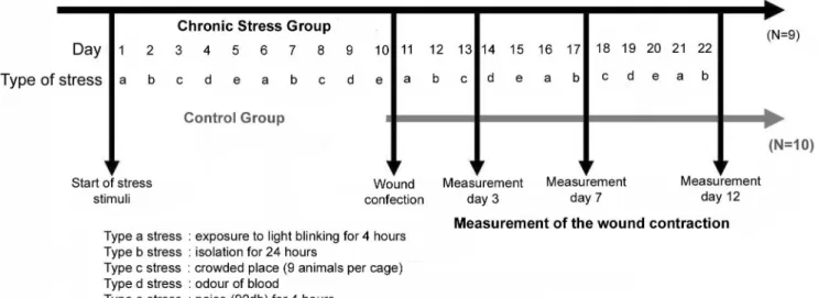
controlled at 23°C, and relative humidity of ±40%. Initially, the animals were randomly divided into two groups: chronic stress (GCS; n=9) and control (GC; n=10) groups. The GCS animals were submitted to different modes of stress for 10 days, while the GC animals were not, after which a surgical cutaneous wound was made in all animals. The stress conditions were maintained for the GCS group until the end of the experiment, which was 12 days after surgery (Fig. 1).
The models chosen for inducing chronic stress were: (a) exposure to blinking light for 4 h; (b) isolation for 24 h; (c) crowded environment (9 rats per cage); (d) odor of blood; and (e) noise (90 db) for 4 h.
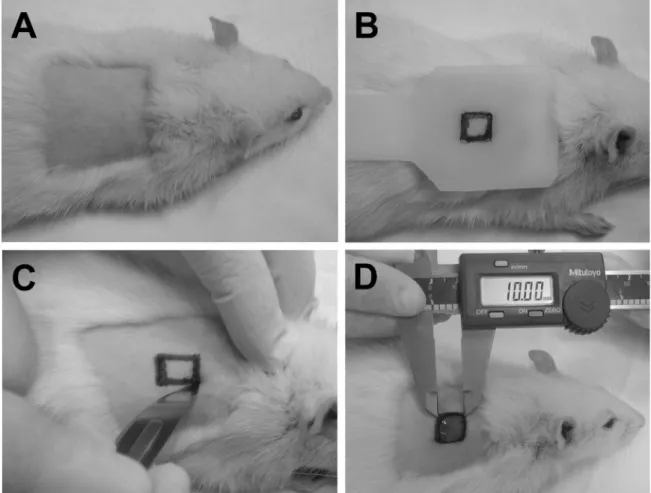
All surgical procedures were performed under general anesthesia, by intramuscular administration of 0.1 mL of ketamine hydrochloride (Dopalen, Agribrands. Saude Animal, Paulinia, SP, Brazil), combined with 0.05 mL of xylazine hydrochloride (Rompun, Bayer. Saude Animal, São Paulo, SP, Brazil), per 100 g weight of the animal. After anesthesia, the region on the right side of the midline of the dorsum was selected for the wound, which was shaved with scissors and disposable razor and antiseptically cleaned with 2% chlorhexidine (Fig. 2A). Before the incision, the borders of the wound were outlined, utilizing a pre-fabricated plastic template and ballpoint pen (Fig. 2B). The wounds were made with a scalpel and iris scissors (Fig. 2C), removing the skin, subcutaneous tissue and fat, leaving the muscle tissue intact, and standardizing all wounds to 1 cm2 (Fig. 2D), as
recommended by Semenoff-Segundo et al. (8). The same investigator performed all the surgical procedures.
In order to prevent the animals from coming in contact
with their feces and/or urine, a permeable metal loor was
installed in the cages, separating the rats from the lower part of the cage. The animals in a state of alert were counted manually, and the examiner, blinded to the groups, performed the measurements. The wounds were measured on day 0 (immediately after the surgery) and 3, 7, and 12 days postoperative, by using a digital pachymeter.
Based on the measurements of the wound edges, standardized on the right and lower edges, the area was obtained, and the contraction of the wound was determined by the following formula: (initial area – area on the day of measurement) / initial area × 100 = percentage of contraction on the day of measurement (13). The percentage of wound contraction was raised to the second power, and transformed into hyperbolic cosine, and the means of each experimental time were calculated for GCS and GC and compared for the experimental times of 3, 7 and 12 days. Data were analyzed by Student t test for independent samples. The alpha error established for this analysis was 5%.
Results
Table 1 shows the means and standard deviation values of wound contraction (%) for the control and experimental groups on days 3, 7, and 12 after surgery. There were no statistical differences between the two groups in relation to
initial and inal body weights of the animals.
Fig. 2. (A). Area of the dorsum of the animal after shaving and cleaning. (B) Template used for the initial outlining
of the shape and size of the wound with ballpoint pen. (C) Surgical procedure using a scalpel to make the wound.
(D) Demonstration of how the dimensions of the wound were determined with the concurrent initial measurement
using a digital pachymeter (baseline measurement).
Table 1. Comparison of the percentage of wound contraction in the control (GC) and chronic stress (GCS) groups at days 3, 7, and 12 after surgery.
Time GC (n=10) GCS (n=9)
Mean SD Mean SD
Day 3 6.21 A 2.20 7.57 A 0.67
Day 7 8.84 A 0.48 7.79 B 0.58
Day 12 9.83 A 0.07 9.76 B 0.06
In comparing the control and chronic stress groups at each
time after surgery, there was no signiicant difference of
wound contraction after 3 days. On the contrary, at 7 and 12 days, GCS had less wound contraction than the control group (P<0.05).
Discussion
The association between stress and tissue repair is of great interest for all health care professionals, particularly oral surgeons. The repair process starts soon after the tissue injury and consists of three periods: i) release of blood constituents resulting in platelet aggregation, blood coagulation and
the migration of inlammatory cells to the site of the
wound, ii) the proliferative phase, iii) and lastly, tissue maturation (9).
In the present study, the results demonstrated that stress slowed tissue repair at days 7 and 12, which demonstrates the effectiveness of the stress model utilized in slowing wound healing. In a clinical trial in humans, Marucha et al. (10) made wounds in the mouth of stressed students (close
to their inal exams) and non-stressed students (during
vacation) and their results of stress-related repair delay were similar to those obtained in this study. Another clinical study carried out in stressed individuals – caretakers of dependent persons – also showed a slower time of wound contraction compared with the control group (7).
We chose to study wound healing by second intention because it is a clinical condition that is frequently encountered by the oral surgeon. This condition could have been led to a bias due to infection, but there were no symptoms of acute or chronic infection from a qualitative point of view. We attribute the absence of symptoms to the housing techniques utilized,
i.e., the elevation of the cage loor, preventing animals from
coming in contact with their feces and urine (8).
In this stress model, the wounds were made 10 days after chronic stress was initiated. It is known that stress has an initial alarm phase which often conditions the animal to an adaptation. This fact led us to examine the next phase which involves the condition of resistance. In this phase, the animals try to establish homeostasis, which results in a series
of systemic changes. The results of this study demonstrated that chronic stress probably acts in the stage of tissue repair involving chemotaxis and tissue maturation (9,14,15,). A large number of substances such as cortisone, adrenaline and noradrenaline are able to interfere with re-epithelization and organization of connective tissue. These hormones diminish
the inlammatory response, acting on important cells such as macrophages, neutrophils and ibroblasts, (9). Another
factor linked to the stress model used is to search for a condition close to that of chronic stress in daily life (16). An experimental time period of 10 days was chosen because stress can be induced in rats within this period (12). One of the limitations of the present study is the lack of
histologic data to possibly correlate clinical indings with
those involved in the histopathologic process of tissue repair. Nevertheless, the method of wound contraction is a good
reference in the study of factors that inluence tissue repair,
and other authors have used very similar methods (7,8). One difference aimed at preventing error in wound evaluation, due to the variability of the dorsum, is the formula for the calculation of the measurements used in the present study (13). The authors calculated the contraction of the wound based on the initial area, which avoids a bias of the wound due to the mobility of the tissues in the area treated.
Studies in rats are of low cost and provide useful information
that could be dificult to obtain in humans. The proposed
stress model aimed to determine the response of wound contraction to stress in rats. In studies with humans, it is
dificult to eliminate biases in relation to their behavioral
variables, and standardize and maintain the same level of stress during the entire experiment. Thus, the stress animal model proposed in this work yielded simple information but still capable of triggering further investigations in this area of knowledge.
Conclusions
Based on the method employed, it was possible to conclude that rats subjected to chronic stress have delayed wound contraction.
References
Anisman H, Baines MG, Berczi I, Bernstein CN, Blennerhassett MG, 1.
Gorczynski RM, et al. Neuroimmune mechanisms in health and diseases: 2. Disease. CMAJ 1996;155:867-740.
Genco RJ, Ho AW, Grossi SG, Dunford RG, Todesco LA. Relationship 2.
of stress, distress, and inadequate coping behaviors to periodontal disease. J Periodontol 1999;70:711-23.
Moss ME, Beck JD, Kaplan BH, Offenbacher S, Gary W, Genco RJ 3.
et al. Exploratory case-control analysis of psychosocial factors and adult periodontitis. J Periodontol 1996;67:1060-9.
Solis AC, Lotufo RF, Pannuti CM, Brunheiro EC, Marques AH, Lotufo-4.
Neto F. Association of periodontal disease to anxiety and depression symptoms, and psychosocial stress factors. J Clin Periodontol 2004;31:633-8.
Castro GD, Oppermann RV, Haas AN, Winter R, Alchieri JC. 5.
Association between psychosocial factors and periodontitis: a case-control study. J Clin Periodontol 2006;33:109-14.
Maier FS. Bi-directional immune–brain communication: Implications 6.
for understanding stress, pain, and cognition. Brain Behav Immun 2003;17:69-85.
Kiecolt-Glasser JK, Marucha PT, Malarkey WB, Mercado AM, 7.
Glasser R. Slowing of wound healing by psychological stress. Lancet 1995; 346:1194-6.
Semenoff-Segundo A, Bosco AF, Maia D, Ribeiro RV, Aguiar EBH, 8.
Marucha PT, Sheridan JF, Padgett D. Stress and wound healing. 9.
Psychoneuroimmunology. 3. ed. San Diego: Academic Press; 2000. Marucha PT, Kiecolt-Glaser JK, Favagehi M. Mucosal wound 10.
healing is impaired by examination stress. Psychosom Med 1998;60:362-5.
Semenoff-Segundo A, Semenoff TA, Bosco AF, Biazoli ER, Ribeiro RV, 11.
Rocato GE et al. Efeito do estresse crônico na progressão de periodontite induzida por ligadura em ratos. Rev Periodontia 2007;17:5-9.
Semenoff-Segundo A. Efeito do estresse crônico na modulação da 12.
periodontite induzida por ligadura em ratos [tese]. Araçatuba (SP): Universidade Estadual Paulista; 2007.
Agren MS, Mertz PM, Franzén L. A comparative study of three 13.
occlusive dressing in the treatment of full-thickness wounds in pigs. J Am Acad Dermatol 1997;36:53-8.
Selye H. Thymus and adrenals in the response of the organism 14.
to injuries and intoxication. Br J Exp Pathol 1936;17:234-48. Selye H. The general adaptation syndrome and the diseases of 15.
adaptation. J Clin Endocrinol 1946;6:117-230.
Fazel S, Danesh J. Serious mental disorder in 23000 prisoners: 16.