45 45 45 45 45 Mem Inst Oswaldo Cruz, Rio de Janeiro, Vol. 93(1): 45-55, Jan./Feb. 1998
Morphological Features of Trypanosomes from Squirrel
Monkeys from the Brazilian Amazon
Mariangela Ziccardi
+, Ricardo Lourenço-de-Oliveira
Laboratório de Transmissores de Hematozoários, Instituto Oswaldo Cruz, Av. Brasil 4365, 21045-900 Rio de Janeiro, RJ, Brasil
A morphometric analysis of blood trypomastigotes identified as Trypanosoma minasense,T. saimirii,
and T. rangeli harbored by squirrel monkeys from the Brazilian Amazon was performed. Additionally,
morphological and biological comparative analyses were conducted of T. saimirii-like and T. rangeli development forms from haemoculture and xenodiagnosis. Illustrations are given of blood trypomastigotes as well as of developing flagellates in triatomine and axenic culture. Mean values of blood trypomastigotes
of T. saimirii differ statistically from those of T. rangeli in only two out of ten morphological characters
measured, and ranges overlapped. The developing forms of T. saimrii-like parasites were essentially identical in both xenodiagnosis and haemoculture to those of T. rangeli. Trypanosomes confirmed as T.
rangeli were transmitted to mice by the bites of the great majority of triatomines that fed on T.
saimirii-like infected monkeys. We conclude that, based on morphology and on the development in triatomine bugs and haemoculture, T. saimirii should not be considered a distinct species. We therefore propose T.
saimirii to be a junior synonym of T. rangeli.
Key words: Trypanosoma rangeli - Trypanosoma saimirii - Trypanosoma minasense - trypanosomes measurements - trypanosomatid flagellates - neotropical primates-culture - xenodiagnosis
Nearly all of the surveys on the prevalence of trypanosomes in non-human primates have been based on Giemsa-stained blood films. Although
some species of trypanosomes, like Trypanosoma
(Schizotrypanum) cruzi Chagas and T. ( Mega-trypanum) lambrechti Marinkelle, develop blood trypomastigotes that are morphologically very characteristic, identification of other species is often difficult due to the interspecific morphologi-cal similarities and intraspecific variability (Dunn et al. 1963, Marinkelle 1966, Dunn 1968). For
ex-ample, Trypanosoma (Herpetosoma) saimirii
Rodhain, because it ispoorly characterized, has
seldom been identified by most of the authors dur-ing surveys of trypanosomes in squirrel monkeys.
It is believed that T. saimirii resembles
Trypano-soma (Megatrypanum) minasense Chagas in blood trypomastigotes, but the former infects triatomine bugs and develops profusely in Novy, McNeal and
Nicolle (NNN) medium, while T. minasense does
not (T. minasense may be cultured under special
condictions, but not only in NNN, see Ziccardi et
al. 1996). When Rodhain (1941) described T.
saimirii he seemed to be much more influenced by
biological features than morphological ones in the
distinction between this parasite and T. minasense.
To learn more about the morphological character-istics of blood trypomastigotes as well as the mul-tiplying forms of these parasites we performed a morphometric analysis. In addition, we discuss
the taxonomic status of T. saimirii based on both
morphological and biological features. MATERIALS AND METHODS
The parasites analyzed were found in isolates as well in blood smears from 165 squirrel mon-keys during the trypanosome survey conducted by Ziccardi and Lourenço-de-Oliveira (1997). Details about the collection sites as well as procedures for haemoculture and xenodiagnosis are available in Ziccardi and Lourenço-de-Oliveira (1997).
A light microscope was used to observe trypomastigotes in Giemsa-stained thin blood
smears from two squirrel monkey species, Saimiri
sciureus and Saimiri ustus, from the Amazon (Ziccardi & Lourenço-de-Oliveira 1997), and from
marmosets, Callithrix penicillata (Geoffroy), from
Felixlândia, State of Minas Gerais. T. (Tejeraia)
rangeli Tejera trypomastigotes were analyzed mor-phometrically in thin blood smears of naturally infected squirrel monkeys as well as from a mar-moset and from mice experimentally infected with
isolates of this parasitefrom squirrel monkeys from
Balbina and Samuel. A standard strain of T. rangeli
(stock R1625-CDC) was used for comparisons
+Corresponding author. Fax: + 55-21-290.9339. E-mail:
47 47 47 47 47 Mem Inst Oswaldo Cruz, Rio de Janeiro, Vol. 93 (1), Jan./Feb. 1998 46
46 46 46
46 Trypanosomes in Squirrel Monkeys from Amazon M Ziccardi, R Lourenço-de-Oliveira
(Miles et al. 1983). A sample of these blood trypomastigotes, as well as multiplying forms from haemoculture and xenodiagnosis (triatomine bugs), was sketched using a Leitz Dialux 20 EB
micro-scope with a camera-lucida. Classification of T.
rangeli in the subgenus Tejeraia follows Añez
(1982).Morphometric analysis was performed
ac-cording to Hoare (1972). T. cruzi was not included
in the analysis. Statistical analysis was done using a t-Student test. In groups where values showed a great variability the non-parametric test of Mann-Whitney was used. The differences were
consid-ered to be significant when p £ 0.01. In Table III,
because we had a non-normal distribution, data were transformed by logarithm.
RESULTS AND DISCUSSION
One of the most widely distributed species of
trypanosome in Neotropical monkeys is T.
minasense. This trypanosome was originally de-scribed by Chagas (1908) from the blood of a
mar-moset, C. penicillata, from Lassance, State of
Minas Gerais, Brazil. Carini (1909) found
trypomastigotes similar to those of T. minasense
in the blood of a Callithrix jacchus (Linnaeus)
purchased in Rio de Janeiro. He redescribed the species in much greater detail and included its physical dimensions (Table Ic).
Rodhain (1937a) found trypanosomes similar to T. minasense in the blood of a squirrel monkey, S. sciureus, from Brazil. Although he noted that these flagellates were smaller than those reported by Carini (1909), he conditionally identified them as T. minasense. In his first trials with the
trypano-some from S. sciureus, he obtained a transitory
in-fection in a splenectomized rat inoculated with the infected blood, but he did not succeed in infecting
five other rats, three mice, a hamster, a Rhesus
monkey or a marmoset (C. penicillata). In spite of
the scanty parasitemia in the blood of squirrel monkeys, he obtained haemocultures in NNN me-dium. Later, Rodhain (1937b, 1941), suspicious of
the size differences in comparison with T.
minasense, decided to feed fleas, ticks and
he-matophagous Hemipteraon the infected
squir-rel monkey. Unlike T. minasense, the trypanosome
from S. sciureus successfully infected
Pans-trongylus megistus (Burmeister) and Cimex lectularius Linnaeus. Since the squirrel monkey trypanosome multiplied readily in NNN medium and in triatomine bugs, while that of the
marmo-set, T. minasense, besides being larger, appeared
to be incapable of infecting such insects or of be-ing cultured, Rodhain (1941) decided to describe the squirrel monkey parasite as a new species,
which he called T. saimirii.
Studying and measuring 15 blood try-pomastigotes (Table Ia) from two squirrel monkeys,
Rodhain (1941) concluded that T. saimirii had a
thinner posterior end than that of the T. minasense,
which is also gradually tapered, and that its kine-toplast was frequently elongated, in the form of a short rod. Otherwise, it was morphologically simi-lar to the marmoset trypanosome. According to Rodhain (1941) the nucleus is oval and situated at the junction between the middle and anterior thirds and there are frequently vacuoles near the nucleus. The undulating membrane is well developed, with several folds. The end of the flagellum sometimes presents a punctiform thickening, which is also
displayed by T. minasense (Carini 1909, Rodhain
1937a, Deane & Damasceno 1961). Deane and Damasceno (1961), studying trypanosomes found
in the blood of four S. sciureus and comparing them
to those found in a C. jacchus, corroborated the
observations by Rodhain (1941). They observed
that T. saimirii had smaller dimensions than T.
minasense (Table Ib).
In the present paper, T. minasense is
distin-guished from the other trypanosome species from blood smears by taking into account several mor-phological features which included mean measures used by previous workers (Table I). However, among the several analyzed parameters, the body length and width, the distance from posterior end of the body to kinetoplast and the shape of the pos-terior end of the body were given more weight for
species identification. In contrast to both T. rangeli
and T. saimirii (Table I) the posterior end of the
body (PK) in T. minasense is usually large but not
gradually tapered to a point and the cytoplasm is stained deep blue. But, even in the blood of its
origi-nal host (marmosets) T. minasense sometimes
dis-plays a posterior end as short as 5 µm (Fig. 2, Tables I, II). Notwithstanding, these trypomastigotes with
a short PK displayed all other features of typical T.
minasense. Our morphometric measurements of T. minasense from marmosets (Table II) agree with those observed by Carini (1909), Rodhain (1941), Deane and Damasceno (1961) and Dunn et al. (1963) (Table I).
A priori, all large trypomastigotes resembling T. minasense, but not displaying the above men-tioned features and, with PK around 4.5 µm were
diagnosed as T. saimirii. The general features of
the trypomastigotes identified as T. saimirii were
body width around 3 µm, cytoplasm generally pale blue, posterior end gradually tapered and undulat-ing membrane with several folds. However, some of these blood trypomastigotes did not agree in all respects with the above mentioned morphological characters. For instance, some parasites had PK in
T
ABLE I
Measurements and comparisons between blood trypomastigotes of
T
rypanosoma minasense, T
. rangeli
and trypanosomes similar to
T
. saimirii
with previously published
data on natural or experimentally infected animals. Ranges given with means in parentheses in (µm) except for ratios
Species and/or strains (host)
No. measured L P K K K N N NA F B K I N I T
. saimirii (S. sciur
eus ) a 15 28.5-33.5 5.38 ___ 7-8 ___ 8.84 6.2-9.3 2.5-3 ___ ___ (31) (7.75) T . saimirii ( S. sciur eus ) b 22 19.2-26 3.8-7.6 0.4-1 4.6-8 2.2-3.2 6-9.8 6-10.4 2.4-3.4 0.6-1.1 0.8-1.8 (23.3) (5.3) (0.8) (6.6) (2.7) (8.3) (7.6) (2.8) (0.8) (1.4) T . minasense ( C. penicillata ) a 13 30.7 7.82 ___ 6.56 ___ ___ 4.87 2.48-3.88 ___ ___ T . minasense ( C. jacchus ) b 39 23-39 6.8-14 0.4-1.2 5.6-13.6 2-3.6 5.6-1 1.2 5.4-9 2.6-4.4 0.8-1.8 1.6-3 (30.6) (10.7) (0.8) (8.7) (2.6) (8.4) (6.6) (3.5) (1.2) (2.1) T . minasense ( C. jacchus ) c ___ 38-45 10-15 ___ 4-5 2.5 ___ 8-10 4-6 ___ ___ T . minasense ( S. nigricollis ) d 43 30-46 8.5-17 ___ 5-5.9 ___ 12-16.5 4.5-6.5 1.5-2.5 ___ ___ (38) (1 1.5) (6.5) (15) (5.5) (2 ) T . minasense (several authors) e ___ 28.4-48 6.8-15 ___ 4-13.6 ___ ___ 4-10 2-6 2-2.7 1.2-2.1 T . rangeli
(mice, our data)
32 25-40 2-8.3 0.6-1 7-10 1-2 7-12 5-1 1 1-2.3 0.2-0.8 0.9-1.7 (31) (3.2) (0.8) (8.1) (1.7) (8.9) (8.8) (2 ) (0.4) (1.3) T . rangeli (man, various) e ___ 25-37 1.8-7 0.7 8.2-10 ___ ___ 7.9-9.5 ___ 1.2-1.7 1.6-2 (27-32.2) T . rangeli R1625 (man) f 23 28-36 2.7-4 ___ 9.3-12.7 ___ 6.3-1 1.7 7-10.7 ___ 1.2-1.4 1.1-2.2 (32.1) (3.3) (10.7) (8.8) (9.4) (1.3) (1.6) T . rangeli
(BUG 1798 GL)
f 25 27-37 1.7-5.7 ___ 7-12.7 ___ 6.6-1 1.7 5-10.3 ___ ___ ___ (29.1) (3.3) (9.2) (9.1) (7.5) T . rangeli
(BUG 1801 GL)
f 10 25.7-33.3 2.7-4 ___ 7-12 ___ 5-10.6 7.3-12.7 ___ 1.2-1.5 1-2.7 (30.8) (3.2) (9.9) (7.9) (9.7) (1.3) (1.8) T . rangeli SC58 ( E. dasythrix ) g 25 25.5-34 3.5-5 ___ 5.5-1 1 ___ 7-10 7-1 1 __ ___ ___ (30.7) (4) (9.2) (9) (9.5) T . rangeli
(man and mice, various) h___ 26.4- 33.8 3.4-4.4 0.7 9.5-9.7 ___ 6.9-8.9 8.1-9.5 ___ ___ 1.6-2 (25-37) (1.8-7) (8.2-10) (5-12) (5-1 1) (1.1-2.8)
L: total length (flagellum included); PK: distance from posterior end of the body to kinetoplast; K: kinetoplast; KN: distance
from kinetoplast to anterior mar
gin of nucleus; N:
nucleus length; NA: distance from nucleus to anterior end of body; F: free flagellum; B: body width (at nucleus level); KI: kin
etoplast index (PK/KN, according to Deane &
Damasceno 1961); NI: nuclear index (PN/NA);
a
- Rodhain 1941;
b
- Deane & Damasceno 1961;
c
- Carini 1909;
d
- Dunn et al. 1963;
e
- Hoare 1972;
f
- Miles et al. 1983;
g
- Steindel et al. 1991 and
h
49 49 49 49 49 Mem Inst Oswaldo Cruz, Rio de Janeiro, Vol. 93 (1), Jan./Feb. 1998 48
48 48 48
48 Trypanosomes in Squirrel Monkeys from Amazon M Ziccardi, R Lourenço-de-Oliveira
the range for T. saimirii (Table II), but the body
length, width and coloration as well as the shape
of the posterior end were like T. minasense. In those
cases the parasites were arbitrarily identified as T.
minasense,because PK of this parasite may have a large range (Table II).
Based on Table II and Figs 1, 2 and 3 we can make a comparative analysis of the measurements
and feature of the blood trypomastigotes of T.
minasense, T. rangeli and trypanosomes similar to T. saimirii found in the thin blood smears from a dozen primates. The differences were statistically
significant between blood parasites of T. saimirii
and T. minasense in the total length, in PK, in the nucleus length, in body width and kinetoplast in-dex (Table II).
Both T. minasense and T. saimirii display a
de-gree of morphological variation (Table I, II) that makes their identification difficult, particularly in thick blood smears. For example, there are trypomastigotes in the blood of marmosets and squir-rel monkeys, respectively, that are smaller than those
described for T. minasense and larger than those
considered to be T. saimirii (Deane & Damasceno
1961, Dunn et al. 1963 and see Figs 1, 2). Although the dimensions are larger in the marmoset trypano-some than in the trypomastigotes found in squirrel
monkeys (identifiable as T. saimirii), the minimum
and maximum measurements for the former gener-ally overlap those of the latter, a fact that was also noted by Dunn et al. (1963).
The trypomastigotes we identified as T. rangeli
(Table II and Fig. 3a) were typical forms, similar in dimensions and feature to those described by other authors in human and non-human primates, rodents, and marsupials, both naturally and artifi-cially infected (Table I and Fig. 3b,c; Groot et al. 1951, Herbig-Sandreuter 1957, Deane 1958, D’Alessandro 1976, Miles et al. 1983, Steindel et al. 1991, Urdaneta-Morales & Tejero 1992). The T. rangeli trypomastigotes we detected in the blood of squirrel monekys and marmosets had a mean length of 30.9 µm, were narrower (width of 1.9 µm) and, had a cytoplasm paler than those
belong-ing to both T. minasense and T. saimirii-like. The
kinetoplast was generally closer to the posterior end (around 3.8 µm from the posterior end) and 7.8 µm from the nucleus (Table II and Fig. 3a).
Blood trypomastigotes of T. saimirii and T. rangeli
were statistically different only in nucleus length and in body width. These results show that indeed there is a higher morphological similarity between T. saimirii and T. rangeli than between T. saimirii and T. minasense (Table II).
We were faced with a range of forms, some
typical of T. minasense and T. rangeli, others
simi-lar to T. saimirii, and still others intermediate,
of-T
ABLE II
Measurements of blood trypomastigotes of trypanosomes similar to
T rypanosoma saimirii , T . minasense and T . rangeli
found in thin blood smears from naturally infected
squirrel monkeys and natural or experimentally infected marmosets. Ranges given with means
±
SE in (µm) except for ratios
S pecies (host) No. measured L PK K K N N N A F B K I N I T . saimirii b (squirrel monkeys) 36 24-40 2-8 0.5-1 5-9 2-3 5-1 1 5-10 1.5-4 0.4-1.3 0.7-2.2
31.4 ± 0.73
d
4.5 ± 0.27
d
0.9 ± 0.02
7.3 ± 0.23
2.3 ± 0.07
d
8.2 ± 0.26
8.2 ± 0.36
3 ± 0.1
1
d
0.6 ± 0.03
d
1.5 ± 0.06
T . minasense (squirrel monkeys) 33 25-49 5-10 0.6-1.1 5-1 1 1.4-3 5-12 5-10 3.5-6.3 0.5-2 1-2.7
36 ± 0.84
e
7.1 ± 0.27
e0.9 ± 0.02
d
7 ± 0.24
2 ± 0.04
e
8.8 ± 0.36
d
7.6 ± 0.33
4.9 ± 0.1
1
e1.1 ± 0.06 e1.7 ± 0.08
d T . minasense (marmosets) c 31 25-53 5-13 0.8-1.2 4-9 2-5 7-17 4-1 1 4-9 0.8-2.3 0.7-2.4
36.2 ± 1.37
8.8 ± 0.40
f
1 ± 0.02
e
6.3 ± 0.20
2.6 ± 0.14
f11.5 ± 0.54
e
8 ± 0.36
5.6 ± 0.29
f
1.4 ± 0.07
f1.4 ± 0.07
e
T
. rangeli
(marmoset
a
10 and 7
29-36 2-6 0.5-1.1 7-9 1.5-2 6-13 5-1 1 1.2-3 0.3-0.9 0.8-2
and squirrel monkeys)
30.9 ± 0.49
3.8 ± 0.30
0.9 ± 0.03
7.8 ± 0.18
2 ± 0.03
e
8.6 ± 0.49
9.2 ± 0.41
1.9 ± 0.12
e
0.5 ± 0.04
1.4 ± 0.09
‘
L: total length (flagellum included); PK: distance from posterior end of the body to kinetoplast; K: kinetoplast; KN: distance
from kinetoplast to anterior mar
gin of nucleus; N:
nucleus length; NA: distance from nucleus to anterior end of body; F: free flagellum; B: body width (at nucleus level); KI: kin
etoplast index (PK/KN, according to Deane &
Damasceno 1961); NI: nuclear index (PN/NA);
a
: experimentally infected marmosets;
b
: morphologically indistinguishable from those described by Rodhain (1941) and Deane
& Damasceno (1961),
c
:
statistically compared with
T
. minasense
from squirrel monkeys.V
alues in a column followed by a dif
ferent letter are statistically distinct at
p
£
0.01.
Fig. 1: trypomastigotes found in thin blood smears from naturally infected squirrel monkeys from Balbina and Samuel: a: Trypa-nosoma minasense; b: T. saimirii-like; and c: a red blood cell from Saimiri sciureus for comparison. Fig. 2: T. minasense found in thin blood smears from naturally infected marmosets, Callithrix penicillata, from Felixlândia, Minas Gerais. A marmoset red blood cell is show for comparison.
Fig. 3: Trypanosoma rangeli. Parasites found in thin blood smears from a: naturally infected squirrel monkey, Saimiri ustus; b: a marmoset, Callithrix jacchus, experimentally infected with haemoculture from S. sciureus; c: mice, experimentally infected with isolates from squirrel monkeys from Balbina and Samuel (Ziccardi & Lourenço-de-Oliveira 1997).
ten in the same animal (Table II). Squirrel mon-keys harbored trypanosomes that lacked diagnos-tic parameters to be distinguished. This wide range of forms was also reported by Dunn et al. (1963).
Sometimes, the parameters to distinguish T. saimirii
from the above mentioned species can not be es-tablished except arbitrarily. The morphometry and the general features did not assure the identifica-tion of this parasite.
The pleomorphism previously described in T.
rangeli (Herbig-Sandreuter 1957, Hoare 1972,
D’Alessandro 1976, Miles et al. 1983, Steindel et al. 1991, Urdaneta-Morales & Tejero 1992) also includes blood trypomastigotes that are wider (0.9-5.1 µm), longer (9.3-39 µm), and sometimes with a greater distance between the posterior end and the kinetoplast (0.5-8 µm), with a well-developed undulating membrane and a vacuoled, densely stained cytoplasm. Such robust or “mature” forms of T. rangeli, with a large PK, have dimensions and feature that are similar to those of
trypano-somes identified as T. saimirii in the blood of
squir-m
51 51 51 51 51 Mem Inst Oswaldo Cruz, Rio de Janeiro, Vol. 93 (1), Jan./Feb. 1998 50
50 50 50
50 Trypanosomes in Squirrel Monkeys from Amazon M Ziccardi, R Lourenço-de-Oliveira
rel monkeys (Tables I, II). It is likely that the trypomastigotes from the blood of squirrel
mon-keys that have been identified by others as T.
saimirii correspond to either (1) T. minasense, con-sidering its pleomorphism (resulting from the length of the infection or interaction with a differ-ent host (Ziccardi 1995) or, (2) the “mature” forms of T. rangeli. Indeed, the morphometry of T. saimirii-like blood trypomastigotes differs
statis-tically from those of T. rangeli in only two out of
ten characters analyzed.
This conclusion strengthens the hypothesis made by Deane and Damasceno (1961). They got, as did Rodhain (1941), positive haemocultures (in NNN) as well as xenodiagnosis (triatomine bugs) from squirrel monkeys infected with what they
called T. saimirii (trypomastigotes resembling T.
minasense, but with smaller dimensions). They did not find metacyclical forms of the parasite in these invertebrates nor did they determine the form of transmission (i.e., inoculative or contaminative). However, Deane and Damasceno (1961) did not rule out the possibility that the forms observed in culture and in the triatomine might be of another trypanosome, not detected by direct examination (Giemsa-stained blood smears), but nevertheless circulating in the blood of the squirrel monkeys they examined. But what are the multiplying forms
supposely belonging to T. saimirii? The
develop-ing forms found in haemoculture of squirrel mon-keys in whose blood we had found only parasites
identifiable as T. saimirii were essentially
identi-cal to those of T. rangeli (strain R1625), both in
the general features and morphometry (Fig. 4, Table III). The haemoculture of primates supposedly
in-fected with T. saimirii were always positive and
profuse, with the predominance of long and slen-der epimastigotes.
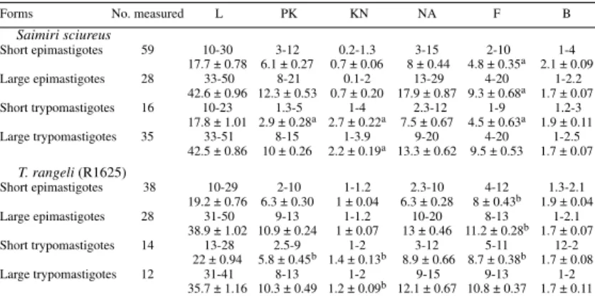
The statistical analysis of measures of cultured
forms isolated from S. sciureus supposedly infected
with T. saimirii,and of those of T. rangeli (strain
R1625), showed that among short and large epimastigotes the only significant difference is the length of free flagellum. In short trypomastigotes the differences are PK, KN and in the length of the free flagellum, and ranges overlapped. This shows a morphometric similarity between these parasites (Table III).
The flagellates found in the gut of triatomine bugs fed on squirrel monkeys, in whose blood only T. saimirii-like parasites had been detected, were also
essentially identical to those of T. rangeli (Table IV,
Fig. 5a). When haemocultures of T. saimirii-like
infected primates were injected in the hemocoel of R. prolixus, both the hemocoel and the salivary glands became infected (Ziccardi & Lourenço-de-Oliveira 1997) with flagellates also
indistinguish-able from those belonging to T. rangeli (Tables V,
TABLE III
Measurements of flagellates found in axenic culture, isolated from a Saimiri sciureus infected with trypanosomes similar to Trypanosoma saimirii, and comparisons with T. rangeli R1625, isolated from man. Ranges given with
means ± SE in (µm)
Forms No. measured L PK KN NA F B
Saimiri sciureus
Short epimastigotes 59 10-30 3-12 0.2-1.3 3-15 2-10 1-4
17.7 ± 0.78 6.1 ± 0.27 0.7 ± 0.06 8 ± 0.44 4.8 ± 0.35a 2.1 ± 0.09
Large epimastigotes 28 33-50 8-21 0.1-2 13-29 4-20 1-2.2
42.6 ± 0.96 12.3 ± 0.53 0.7 ± 0.20 17.9 ± 0.87 9.3 ± 0.68a 1.7 ± 0.07
Short trypomastigotes 16 10-23 1.3-5 1-4 2.3-12 1-9 1.2-3
17.8 ± 1.01 2.9 ± 0.28a 2.7 ± 0.22a 7.5 ± 0.67 4.5 ± 0.63a 1.9 ± 0.11
Large trypomastigotes 35 33-51 8-15 1-3.9 9-20 4-20 1-2.5
42.5 ± 0.86 10 ± 0.26 2.2 ± 0.19a 13.3 ± 0.62 9.5 ± 0.53 1.7 ± 0.07
T. rangeli (R1625)
Short epimastigotes 38 10-29 2-10 1-1.2 2.3-10 4-12 1.3-2.1
19.2 ± 0.76 6.3 ± 0.30 1 ± 0.04 6.3 ± 0.28 8 ± 0.43b 1.9 ± 0.04
Large epimastigotes 28 31-50 9-13 1-1.2 10-20 8-13 1-2.1
38.9 ± 1.02 10.9 ± 0.24 1 ± 0.07 13 ± 0.46 11.2 ± 0.28b 1.7 ± 0.07
Short trypomastigotes 14 13-28 2.5-9 1-2 3-12 5-11 12-2
22 ± 0.94 5.8 ± 0.45b 1.4 ± 0.13b 8.9 ± 0.66 8.7 ± 0.38b 1.7 ± 0.08
Large trypomastigotes 12 31-41 8-13 1-2 9-15 9-13 1-2
35.7 ± 1.16 10.3 ± 0.49 1.2 ± 0.09b 12.1 ± 0.67 10.8 ± 0.37 1.7 ± 0.11
L: total length (flagellum included); PK: distance from posterior end of the body to kinetoplast; KN: distance from kinetoplast to anterior margin of nucleus; NA: distance from nucleus to anterior end of body; F: free flagellum; B: body width (at nucleus level). Values in a column followed by a different letter are statistically distinct at p £ 0.01.
Fig. 4: forms found in haemoculture. a: Trypanosoma rangeli R1625; b: parasites isolated from a naturally infected squirrel mon-key, Saimiri sciureus from Balbina, with parasitemia by trypanosomes similar to T. saimirii.
VI and Fig. 5b,c; Zeledon 1956, Hoare 1972, Cuba-Cuba 1973, Steindel et al. 1991). Trypanosomes
confirmed as T. rangeli were transmitted to mice by
the bites of the great majority of triatomines that we
fed on a T. saimirii-like infected monkey (Ziccardi
& Lourenço-de-Oliveira 1997).Indeed, the
multi-plying forms of T. saimirii in both the triatomine
53 53 53 53 53 Mem Inst Oswaldo Cruz, Rio de Janeiro, Vol. 93 (1), Jan./Feb. 1998 52
52 52 52
52 Trypanosomes in Squirrel Monkeys from Amazon M Ziccardi, R Lourenço-de-Oliveira
P. megistus was the only triatomine species used by Rodhain (1937b) in his experimental infections
with T. saimirii. However, it is belived that in P.
megistus, as well as in some Triatoma species, that T. rangeli infects only the gut, with invasion of the hemocoel rare, and the parasite has never been found in their salivary glands (Coutinho & Nussenzweig 1952, Zeledon & Blanco 1965, Hoare 1972, Cuba-Cuba 1973).
The invasion of the triatomine hemocoel by T.
rangeli may not occur until 30 days after an infec-tive blood meal (Hoare 1972, Cuba-Cuba 1973). This period may not have been taken into account by the authors (Rodhain 1941, Deane & Damasceno 1961) in their examination of xendodiagnosis of squirrel monkeys infected with T. saimirii-like parasites. Both T. saimirii and T. rangeli experimentally infect the bed bug C. lectularius, as well as may display non-metacyclic
trypomastigotes in the gut of triatomine (Rodhain 1937b, 1941, Herbig-Sandreuter 1957, Deane 1958, Zeledon & Blanco 1965, Hoare 1972, Cuba-Cuba 1973).
In conclusion, there are neither reliable
mor-phological nor biological differences between T.
rangeli and T. saimirii in the developmental cycle in the invertebrate hosts nor in axenic culture. The flagellates found in haemocultures and in xenodi-agnosis of squirrel monkeys displaying parasitemia by T. saimirii are actually developing forms of T. rangeli.
Besides, most of xenodiagnosis and
haemo-culture of squirrel monkeys infected with T.
minasense was positive, and the respective
devel-oping forms were actually found to belong to T.
rangeli (Ziccardi & Lourenço-de-Oliveira 1997).
However, T. minasense does not develop in
triatomine bugs and its developing forms in cul-TABLE V
Measurements of flagellates found in hemolymph of Rhodnius prolixus experimentally infected with a trypanosome culture isolated from Saimiri sciureus, with parasitemia by trypanosomes similar to T. saimirii.
Ranges given with means (µm) in parentheses
Forms No. measured L PK KN NA F B
Short epimastigotes 3 11-2 1.2-4 __ 3-3.2 10-12 1.5-3
(16) (2.7) (3.1) (11) (2.2)
Intermediate epimastigotes 9 30-40 12-25 1 5-10 6-12 1.3-3
(37.1) (20.1) (7.3) (9.6) (2)
Large epimastigotes 42 45-100 21-65 1-1.5 5-20 4-35 1.3-3.5
(65.4) (44.1) (1.2) (11.2) (10.5) (2.2)
Short trypomastigotes 9 11-20 2 1-2 3-9 5-11 1.9-4
(16.4) (1.4) (5.6) (9) (2.8)
L: total length (flagellum included); PK: distance from posterior end of the body to kinetoplast; KN: distance from kinetoplast to anterior margin of nucleus; NA: distance from nucleus to anterior end of body; F: free flagellum; B: body width (at nucleus level).
Fig. 5: forms found in triatomine bugs (Rhodnius prolixus) that fed on squirrel monkeys Saimiri sciureus, and in adult of R. prolixus injected in the hemocoel with samples of haemoculture from S. sciureus and S. ustus. The squirrel monkeys are naturally infected, with parasitemia by trypanosomes similar to T. saimirii: a: gut contents; b: hemolymph and c: salivary glands.
TABLE VI
Measurements of flagellates (metacyclic) found in salivary glands of Rhodnius prolixus experimentally infected with a trypanosome culture isolated from Saimiri ustus which had parasitemia by trypanosomes similar to Trypanosoma saimirii, and compared to previously published data on T. rangeli. Ranges given with means (µm)
in parentheses
Species (host) No. measured L PK KN NA F B
T. rangeli (Saimiri ustus, our data) 35 9-16 1-3 1-4 2-6 2-5 1-2
(11.6) (1.5) (2.4) (3.7) (3.2) (1.6)
T. rangeli SC58 (Echimys dasythrix)a 50 8.4-10.2 0.6-1.2 1.9-2.7 2.6-3.4 2.7-3.5 ___
(9.3) (0.9) (2.3) (3) (3.1)
T. rangeli b ___ 10-13 ___ ___ ___ 3 ___
T. rangeli (R. prolixus - El Salvador)c ___ 8.3-13.3 ___ ___ ___ ___ ___
L: total length (flagellum included); PK: distance from posterior end of the body to kinetoplast; KN: distance from kinetoplast to anterior margin of nucleus; NA: distance from nucleus to anterior end of body; F: free flagellum; B: body width (at nucleus level); a: Steindel et al. 1991; b: Hoare 1972 and c: Zeledon 1956.
TABLE IV
Measurements of flagellates found in gut contents of Rhodnius prolixus that fed on Saimiri sciureus, infected with trypanosomes similar to Trypanosoma saimirii and comparisons with T. rangeli strain R1625 isolated from
man. Ranges given with means ± SE in (µm)
Species/forms No. measured L PK KN NA F B
Saimiri sciureus
Short epimastigotes 16 12-29 4-11 1 3.5-15 1.5-9 1-4
20 ± 1.26 7.8 ± 0.48 9.4 ± 0.84 a 4 ± 0.45 2.1 ± 0.17
Large epimastigotes 14 39-51 10-19 1.0-1.5 15-30 4-15 1-2
47 ± 1.28 a 13.8 ± 0.87 1.0 ± 0.08 23.9 ± 1.54 8.3 ± 0.88 1.7 ± 0.10
Short trypomastigotes 1 19 4 1 8 5 2
Large trypomastigotes 1 50 15 3 21 10 1.5
T. rangeli (R1625)
Short epimastigotes 14 20-25 3-10 1 10-15 2-9 1.2-3
22.6 ± 0.61 6.9 ± 0.53 12.4 ± 0.56 b 4.4 ± 0.60 2 ± 0.12
Large epimastigotes 9 31-51 5-10 1-2 15-21 1-11 1-2
35.8 ± 2.07 b 9.9 ± 1.26 1.2 ± 0.20 19.2 ± 0.62 5.7 ± 1.04 1.7 ± 0.12
Short trypomastigotes 1 20 4 ___ 6 6 2
Large trypomastigotes 1 50 15 3 20 5 2
L: total length (flagellum included); PK: distance from posterior end of the body to kinetoplast; KN: distance from kinetoplast to anterior margin of nucleus; NA: distance from nucleus to anterior end of body; F: free flagellum; B: body width (at nucleus level). Values in a column followed by a different letter are statistically distinct at p £ 0.01.
of T. rangeli (see Herbig-Sandreuter 1957, Zeledon
1966 apud Hoare 1972, Cuba-Cuba 1973).
Even though T. saimirii has developing forms
essentially identical to those of T. rangeli in
triatomine bugs, it is believed that the infection by T. saimirii is restricted to the gut while in T. rangeli the infection reaches the hemocoel and the flagel-late invades the salivary glands. However, several
strains of T. rangeli, mainly those from long-term
culture in axenic media, may fail to invade the
hemocoel of triatomine bugs and subsequently are not transmitted by the bite to susceptible hosts (Coutinho & Nussenzweig 1952, Tobie 1961, Zeledon 1965, Hoare 1972, Cuba-Cuba 1973, D’ Alessandro 1976). If the invasion and infection of
the hemocoel by T. rangeli is eventual, depending
on several circunstances, such as the parasite strain and the triatomine bug species, this biological event
does not assure the distinction between T. rangeli
55 55 55 55 55 Mem Inst Oswaldo Cruz, Rio de Janeiro, Vol. 93 (1), Jan./Feb. 1998 54
54 54 54
54 Trypanosomes in Squirrel Monkeys from Amazon M Ziccardi, R Lourenço-de-Oliveira
ture media are rather distinct from those of T.
rangeli (Dias & Campos-Seabra 1943, Deane & Damasceno 1961, Ziccardi et al. 1996). Those primates were therefore considered to have mixed
infections of T. minasense and T. rangeli.
Wild animals may be simultaneously infected with more than one trypanosome species, although only one may be detected in blood smears. There-fore, the parasite developing in xenodiagnosis or/ and haemoculture may not belong to the same spe-cies found in blood smears. This possibility was not often taken into account, and mixed infections have already led some authors to describe new
spe-cies, e.g., T. sanmartini Garnham and
Gonzales-Mugaburu (Deane 1969, Hoare 1972, Marinkelle
1976). This was probably the case in T. saimirii.
That is, in view of the morphological and
biologi-cal features of T. saimirii in both vertebrate and
invertebrate hosts discussed above and its
resem-blance to T. rangeli (although also to T. minasense
in some blood trypomastigotes), we conclude that
in his description of T. saimirii, Rodhain (1941)
actually worked with squirrel monkeys infected
with T. rangeli or with both T. rangeli and T.
minasense. Indeed, T. rangeli has been the most frequent trypanosome detected in squirrel monkeys from Brazil and other countries in the Americas
and mixed infections of T. minasense and T. rangeli
have often been reported in these primates (Dunn el al. 1963, Ayala 1964, Marinkelle 1966, Baker 1972, Deane et al. 1972, Hoare 1972, D’ Alessandro et al. 1986, Sullivan et al. 1993, Ziccardi & Lourenço-de-Oliveira 1997).
Results of on-going biochemical analysis us-ing SDS-PAGE show a great similarity with the
parasite growing in the haemoculture of T.
saimirii-like infected squirrel monkeys to T. rangeli and
that T. minasense has a particular peptideme quite
distinct from other assayed trypanosome species (unpublished data).
We concluded that based on morphology and on the development in the triatomine bugs and
haemoculture, T. saimirii cannot be considered a
distinct species. We therefore propose T. saimirii
as junior synonym of T. rangeli.
ACKNOWLEGMENTS
To Dr Pedro Cabello for the guidance on statistical procedures, Dr R Wilkerson and Dr LP Lounibos for critical review of the manuscript and Teresa F Silva for the aid with the illustrations.
REFERENCES
Añez N 1982. Studies on Trypansoma rangeli Tejera, 1920. IV. A reconsideration of its systematic posi-tion. Mem Inst Oswaldo Cruz 77: 405-415. Ayala FM 1964. Presencia de un hemoflagelado
semejante al Trypanosoma rangeli Tejera, 1920 en
el mono Saimiri boliviensis, en la region amazonica, Peru. Rev Inst Med Trop São Paulo 6: 47-50. Baker JR 1972. Protozoa of tissue and blood (Other than
the Haemosporina), p. 29-56. In RNTW Fiennes, Pathology of Simian Primates, Karger, Basel. Carini A 1909. Über Trypanosoma minasense. Arch f
Schiffs Tropenhyg 13: 447-448.
Chagas C 1908. Trypanosoma minasense. Brazil Médico 22: 471.
Coutinho JO, Nussenzweig V 1952. Infecção experimen-tal de Triatomíneos pelo T. rangeli Tejera, 1920. Fol Clin Biol 18: 181.
Cuba-Cuba CA 1973. Evolução de uma Cepa Peruana de T. rangeli em Rhodnius ecuadoriensis e Panstrongylus herreri, MSc. Thesis, UFMG, Belo Horizonte, 84 pp.
D’Alessandro A 1976. Biology of Trypanosoma (Herpetosoma) rangeli Tejera, 1920, p. 327-403. In WHR Lumsden & DA Evans (eds), The Biology of Kinetoplastida. Academic Press, London. D’Alessandro A, Eberhard M, Hincapie O, Halstead S
1986. Trypanosoma cruzi and Trypanosoma rangeli in Saimiri sciureus from Bolivia and Saguinus mistax from Brazil. Am J Trop Med Hyg 35: 285-289. Deane LM 1958. Encontro de tripanosomas do tipo
rangeli em gambás da espécie Didelphis marsupialis no Estado do Pará. Rev Brasil Malariol D Trop 10: 451-458.
Deane LM, Almeida FB, Ferreira Neto JA, Evangelista da Siva J 1972. Trypanosoma cruzi e outros tripanosomas em primatas brasileiros. Rev Soc Bras Med Trop 4: 361.
Deane LM, Damasceno RG 1961. Tripanosomídeos de mamíferos da região amazônica II. Tripanosomas de macacos da Zona do Salgado, Estado do Pará. Rev Inst Med Trop São Paulo 3: 61-70.
Deane MP 1969. A discussion on Trypanosoma sanmartini Garnham and Gonzalez-Mugaburu, 1962. Rev Lat Amer Microbiol Parasitol 11: 91-96. Dias E, Campos-Seabra CA 1943. Sôbre o Trypanosoma
conorrhini, hemoparasito do rato transmitido pelo Triatoma rubrofasciata. Presença do vector infectado na cidade do Rio de Janeiro. Mem Inst Oswaldo Cruz 39: 301-333.
Dunn FL 1968. The parasite of Saimiri: in the context of platyrrhine parasitism, p. 31-68. In LA Rosenblum & RW Cooper (eds), The Squirrel Monkey, Academic Press, New York and London.
Dunn FL, Lambrecht FL, Du Plessis R 1963. Trypano-somes of South American monkeys and marmosets. Am J Trop Med Hyg 12: 524-534.
Groot H, Renjifo S, Uribe C 1951. Trypanosoma ariarii, n. sp., from man, found in Colombia. Am J Trop Med 31: 673-691
Herbig-Sandreuter A 1957. Further studies on Trypano-soma rangeli Tejera, 1920. Acta Tropica 4: 193-207. Hoare CA 1972. The Trypanosomoses of Mammals, Blackwell Scientific Publication, Oxford, 749 pp. Lourenço-de-Oliveira R 1988. Hemoparasitos
encon-trados em alguns mamíferos de Balbina, Estado do Amazonas. Mem Inst Oswaldo Cruz 83: 233. Marinkelle CJ 1966. Observations on human, monkey
and bat trypanosomes and their vectors in Colom-bia. Trans R Soc Trop Med Hyg 60: 109-116
Marinkelle CJ 1976. The biology of the trypanosomes of non-human primates, p. 217-256. In WHR Lumsden & DA Evans (eds), The Biology of Kinetoplastida, Academic Press, New York. Miles MA, Arias JR,Valente SAS, Naiff RD, De Souza
AA, Povoa MM, Lima JAN, Cedillos RA 1983. Vertebrate hosts and vectors of Trypanosoma rangeli in the amazon basin of Brazil. Am J Trop Med Hyg 32: 1251-1259.
Rodhain J 1937a. Notes sur Trypanosoma minasense Chagas. C R Soc Bio 125: 1034-1036. Rodhain J 1937b. Notes sur Trypanosoma minasense
Chagas. Evolution du trypanosome du Saimiri chez divers arthropodes. C R Soc Bio 126: 69-72. Rodhain J 1941. Notes sur Trypanosoma minasense
Chagas. Identité spécifique du trypanosome du Saimiri: Chrysothrix sciureus. Acta Biol Belg 1: 187-192.
Steindel M, Carvalho Pinto JC, Toma HK, Mangia RHR, Ribeiro-Rodrigues R, Romanha AJ 1991. Trypano-soma rangeli (Tejera, 1920) isolated from a sylvatic rodent (Echimys dasythrix) in Santa Catarina island, Santa Catarina state: first report of this trypanosome in Southern Brazil. Mem Inst Oswaldo Cruz 86: 73-79.
Sullivan JJ, Steurer F, Benavides G, Tarleton RL, Eberhard ML, Landry S 1993. Trypanosomes and microfilariae in feral owl and squirrel monkeys main-tained in research colonies. Am J Trop Med Hyg 49:
254-259.
Tobie EJ 1961. Experimental transmission and biologi-cal comparison of strains of Trypanosoma rangeli. Exp Parasitol 11: 1-9.
Urdaneta-Morales MS, Tejero F 1992. Trypanosoma rangeli (Tejera, 1920): observations upon pleomor-phism. Mem Inst Oswaldo Cruz 87: 511-516. Zeledon R 1956. Hallazgo de formas evolutivas de
Try-panosoma rangeli Tejera, 1919, en glándulas salivales de Rhodnius prolixus Stal, 1859, salvadoreños. Rev Biol Trop 4: 1-8.
Zeledon R 1965. Trypanosoma rangeli en glándulas salivales de Rhodnius pallescens de Panamá. Rev Biol Trop 13: 157.
Zeledon R, Blanco E 1965. Relaciones huésped-parasito en Trypanosomiasis rangeli. I. Infeccion intestinal y hemolinfático comparativa de Rhodnius prolixus y Triatoma infestans. Rev Biol Trop 13: 143. Ziccardi M 1995. Tripanosomas de Macacos-de-cheiro:
O que é o Trypanosoma saimirii Rodhain, 1941? MSc. Thesis, Instituto Oswaldo Cruz, Rio de Janeiro, 100 pp.
56 56 56 56