https://doi.org/10.1590/0004-282X20170193
ARTICLE
Chronic treatment with carvacrol improves
passive avoidance memory in a rat model of
Parkinson’s disease
O tratamento com carvacrol melhora a memória de esquiva passiva em um modelo da
doença de Parkinson em ratos
Hossein Haddadi1, Ziba Rajaei1, Hojjatallah Alaei1, Somayeh Shahidani1
Parkinson’s disease (PD) is a chronic neurodegenera-tive disorder, which is caused mainly by the degeneration of dopaminergic neurons in the substantia nigra, leading to motor dysfunctions such as resting tremor, muscle rigidity
and bradykinesia1. In addition to motor dysfunctions,
cogni-tive deficits such as learning and memory impairments and
dementia are seen in a high percentage of PD patients2. The
proportion of PD patients with dementia is 25–30%, up to six
times higher than in healthy people3. Moreover, PD patients
suffer from painful sensations that have been described as five different types: musculoskeletal pain (due to parkinso -nian rigidity, rheumatological disease or skeletal deformity), radicular-neuropathic pain (due to a root lesion, focal or
peripheral neuropathy), dystonic pain (related to antipar-kinsonian medication), central neuropathic pain (related
to antiparkinsonian medication) and akathisia (during
off-periods or drug induced)4.
Pharmacotherapy with L-DOPA and DOPA-decarboxylase
inhibitors is still the most effective treatment for motor symp
-toms, but this type of therapy has no effect on cognitive defi
-cits in PD5,6. Considering the clear impact of cognitive deficits
on the quality of the PD patient’s life, it is valuable to
investi-gate the treatments affecting non-motor symptoms, such as cognitive deficits and pain, in animal models of PD.
Carvacrol (CAR, 2-methyl-5-isopropylphenol) is a pheno-lic monoterpene abundantly present in the essential oil of the
1Isfahan University of Medical Sciences, School of Medicine, Department of Physiology, Isfahan, Iran.
Correspondence: Ziba Rajaei; Department of Physiology, School of Medicine, Isfahan University of Medical Sciences, Isfahan, Iran; E-mail: [email protected] Conflict of interest: There is no conflict of interest to declare.
Received 09 May 2017; Received in final form 29 October 2017; Accepted 08 November 2017. Support: This study was supported by Isfahan University of Medical Sciences.
ABSCRACT
The present study investigated the effects of carvacrol on motor and memory deficits as well as hyperalgesia in the 6-OHDA-lesioned rat model of Parkinson’s disease. The animals were subjected to unilateral microinjection of 6-OHDA into the medial forebrain bundle and treated with carvacrol (25, 50 and 100 mg/kg, ip) for six weeks after surgery. The 6-OHDA-lesioned rats showed contralateral rotations towards the lesion side, which was accompanied by learning and memory deficits in a passive avoidance test and a decrease in tail withdrawal latency in a tail flick test at the end of week 6. The results also showed that treatment with carvacrol at a dose of 25 mg/kg ameliorated memory deficits, with no effect on rotations and hyperalgesia in lesioned rats. In conclusion, carvacrol improves memory impairments in rats with Parkinson’s disease; therefore, it may serve as an adjunct therapy for the alleviation of memory deficits in Parkinson’s disease patients.
Keywords: Carvacrol; 6-hydroxydopamine; memory; motor activity; hyperalgesia; Parkinson’s disease.
RESUMO
O presente estudo investigou os efeitos do carvacrol nos déficits motores e de memória, bem como na hiperalgesia, em um modelo da doença de Parkinson (DP) em ratos com lesões 6-OHDA. Os animais foram submetidos a microinjeção unilateral de 6-OHDA no feixe mediano do prosencéfalo e tratados com carvacrol (25, 50 e 100 mg / kg, ip) durante 6 semanas após a cirurgia. Os ratos com lesões 6-OHDA mostraram rotações contralaterais para o lado da lesão, que foram acompanhadas de déficits de aprendizagem e de memória em um teste de evitação passiva, e de uma diminuição da latência de retirada da cauda em um teste de cauda no final da semana 6. Os resultados também mostraram que o tratamento crônico com carvacrol a uma dose de 25 mg / kg aliviou os déficits de memória, sem efeito sobre rotações e hiperalgesia em ratos lesados. Em conclusão, o carvacrol melhora a deficiência de memória em ratos com DP e, portanto, pode servir como uma terapia complementar para aliviar os déficits de memória em pacientes com DP.
family lamiaceae. Carvacrol has been reported to have many
pharmacological benefits, including antibacterial, antifungal, antioxidant, antinociceptive, anti-inflammatory, anti-apop
-tosis and anti-cancer activities7. Carvacrol also exerts several
actions on the neuronal system including
acetylcholinester-ase inhibition8,9, as well as having anxiolytic10 and
antide-pressant11 properties. It also modulates central
neurotrans-mitter pathways, such as dopaminergic, serotonergic and
GABAergic systems7.
Based on the antioxidant activity of carvacrol and its
effects on cholinergic and dopaminergic systems, we decided
to examine whether carvacrol could improve the motor
and memory deficits and pain in the 6-OHDA model of
Parkinson’s disease.
METHODS
Experimental animals
The experimental animals used in the present study were adult male Wistar rats weighing 250–350 g. They were main
-tained at a controlled temperature (22 ± 2°C) with a 12-hour dark/light cycle and had free access to water and food. The
Ethics Committee for Animal Experiments at Isfahan University of Medical Sciences approved the study (Approval No. 194036) and all experiments were conducted in accordance with the
National Institute of Health Guide for the Care and Use of Laboratory Animals (NIH Publication, 8th edition, 2011).
Experimental design
The animals were randomly assigned to five groups, as follows:
Group 1: sham-operated group (injection of 0.2% ascor-bate-saline into the left medial forebrain bundle (MFB), 1%
Tween 80 ip, n = 7);
Group 2: lesioned group (16 μg 6-OHDA into the MFB, 1%
Tween 80 ip, n = 7);
Group 3: Carvacrol-treated lesioned group (16 μg 6-OHDA
into the MFB, 25 mg/kg carvacrol ip, n = 10);
Group 4: Carvacrol-treated lesioned group (16 μg 6-OHDA into the MFB, 50 mg/kg carvacrol ip, n = 10);
Group 5: Carvacrol-treated lesioned group (16 μg 6-OHDA into the MFB, 100 mg/kg carvacrol ip, n = 7).
Carvacrol was emulsified with 1% Tween 80 (Sigma, USA) and dissolved in normal saline. The animals were treated with
carvacrol at doses of 25, 50 and 100 mg/kg, intraperitoneally,
one week before the surgery until six weeks after surgery. The
sham-operated group received 1% Tween 80 dissolved in nor-mal saline at the same volume as the treated groups.
Surgery and 6-OHDA lesion
Under chloral hydrate (450 mg/kg, ip) anesthesia, rats were
positioned in the stereotaxic apparatus (Stoelting, USA). The
scalp was cleaned with an iodine solution and lidocaine was injected (2% solution, Sc). A midline skin incision was made
with subsequent drilling of the skull. The 6-OHDA compound
(16μg/4μl 0.2% ascorbate-saline) was injected into the left
MFB by a Hamilton microsyringe according to the coordi
-nates: AP: -3.6 mm; ML: -1.8 mm; DV: -8.2 mm12. The rats in the
sham-operated group also received an identical volume of the
ascorbate-saline as the vehicle. The injection rate was 1 µl/min and the needle was kept in place for a further five minutes after
injection for complete absorption of the toxin. After surgery, the rats were placed singly into a clean page and kept warm until recovery from anesthesia was complete.
Apomorphine-induced rotations
The hemiparkinsonian rats were diagnosed by observ -ing the rotational behavior after an injection of apomor-phine hydrochloride (Sigma-Aldrich, USA) at the end of the 2nd and 6th week after surgery. Apomorphine hydrochloride was dissolved in normal saline and injected intraperitone-ally at a dose of 2 mg/kg. On the test day, the animals were allowed to habituate to a transparent plexiglass container (28×28×50 cm) for 10 minutes. One minute after the injection of apomorphine, full rotations were counted at 10 minute
intervals for 30 minutes in a dimly-lit, quiet room. The num -ber of ipsilateral rotations was counted as positive scores and
those of contralateral rotations as negative scores. The net number of rotations were defined as the difference between
the rotations in both directions13.
Passive avoidance memory
Passive avoidance learning was assessed by a shuttle box
at the end of week 6. The apparatus consisted of a light and a
dark compartment, connected by a guillotine door. In the train-ing session, animals were placed individually in the light com-partment for one minute. After the opening of the door and the movement of the rat into the dark chamber, the door was closed and a 0.5 mA foot electric shock was delivered through the grid
floor for three seconds. In the test session, each rat was again placed into the light compartment. The step-through latency
to entering the dark compartment was measured as a positive
index of memory performance, with a 300 second cut-off time14.
Tail-flick test
The animal’s response to phasic pain was tested by mea
-suring the latency of tail flick to a high intensity light beam. The test was performed with a Tail Flick instrument that gives an automatic recording of tail-flick latency to radiant heat. The animal was placed on the recording platform of the appa -ratus where it was kept under painless restraint, with its tail
placed on the radiant heat window. When the animal flicked
its tail, a photocell was activated and the time between
acti-vation of the heat source and tail flick latency was recorded.
For each animal, the thermal stimulus was applied on three
different parts of the tail and the latency was considered as
the mean of three measurements15. A cut-off exposure time of
Dissection and homogenization
After completion of the behavioral testing, the animals were euthanized and the brains were removed from the skulls
on day 42. The striatum was dissected out and weighed. A
10% (w/v) tissue homogenate was prepared in NaCl solution.
Lipid peroxidation levels
The lipid peroxidation level of the striatum was measured
as malondialdehyde, which reacts with thiobarbituric acid as a thiobarbituric acid reactive substance (TBARS) to pro-duce a red-colored complex that has a peak absorbance (A) at 535 nm. A mixture of trichloroacetic acid, thiobarbituric
acid, and HCl were added to 1 ml of homogenate, and the
mixture was heated for 45 minutes in a boiling water bath. After cooling and centrifugation at 1000 g for 10 minutes, the
absorbance was measured at 535 nm16. The level of TBARS
was calculated by: C (M) = absorbance/1.65 × 105.
Total thiol concentration
Total sulfhydryl groups were measured using
2.2´-Dinitro-5.5´-dithiodibenzoic acid (DTNB) as the reagent. This
reagent reacts with the sulfhydryl groups to produce a yel-low-colored complex that has a peak absorbance at 412 nm.
Briefly, 1 ml tris-EDTA buffer was added to 50 µl homogenate
and the sample absorbance was read at 412 nm against the
tris-EDTA buffer alone (A1). Then, 20 µl of the DTNB reagent
(10 mM in methanol) was added to the mixture and after 15 min (minutes), the sample absorbance was read again
(A2). The absorbance of the DTNB reagent was also read as a blank (B). The total thiol concentration (mM) was calculated by: (A2-A1-B) × 1.07/0.05 × 13.616.
Histology
The brains were removed and stored in 10% formalin for 72 hours. The brains were sectioned coronally at 40 μm by a freezing microtome (Leica, Germany). Sections were mounted on gelatin-coated slides and studied using a light
microscope. The track of the needle and the injection site of 6-OHDA (Figure 1) was determined by reference to a rat
brain atlas12.
Statistical analysis
The results are presented as mean ± SEM. Data were ana
-lyzed by one-way ANOVA followed by Tukey’s post hoc test.
Results were considered significant at P < 0.05.
RESULTS
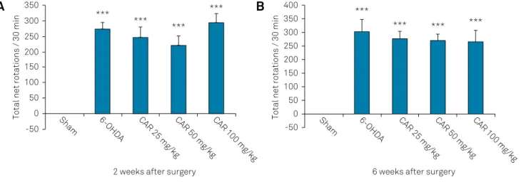
Effects of carvacrol on rotational behavior
The results showed that apomorphine hydrochloride
administration (2 mg/kg, ip) produced contralateral
rota-tions towards the lesion side in 6-OHDA-lesioned rats at
the end of the 2nd (Figure 2A) and 6th week (Figure 2B),
indicating unilateral damage to the left MFB. No such rota-tions were observed in the sham group rats.
Analyzing data with one-way ANOVA revealed a signifi
-cant difference in rotations between groups (p < 0.001) at
weeks 2 and 6. Further analysis with Tukey’s post hoc test showed that the number of contralateral rotations in the
6-OHDA-lesioned group and the carvacrol-treated lesioned groups were significantly increased compared to the sham group (p < 0.001) at weeks 2 and 6, but there was no differ -ence between the treated and not-treated groups (Figures 2A and 2B).
Effects of carvacrol on passive avoidance memory
As shown in Figure 3, the step-through latency of 6-OHDA-lesioned rats (128.8 ± 47.38) was shorter than the sham group rats (293 ± 7) at the end of week 6 (p < 0.05, Figure 3).
Moreover, treatment of lesioned rats with carvacrol at a dose
of 25 mg/kg significantly increased the latency (281 ± 19) compared with lesioned rats (128.8 ± 47.38) (p < 0.05).
Effects of carvacrol on tail flick latency
According to the results, there was a significant decrease
in tail flick latency in 6-OHDA-lesioned rats compared to sham group rats at week 6 (p < 0.05, Figure 4). No signif -icant difference in latency to tail flick was observed
fol-lowing carvacrol administration in the 6-OHDA-lesioned
groups (Figure 4).
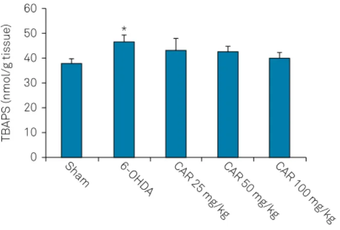
Effects of carvacrol on lipid peroxidation levels
Injection of 6-OHDA into the MFB resulted in signifi
-cant elevation of TBARS level in the striatum (p < 0.05).
In addition, the results showed that treatment of lesioned rats with carvacrol at doses of 25, 50 and 100 mg/kg for six weeks did not change the increased TBARS level in the stri-atum (Figure 5).
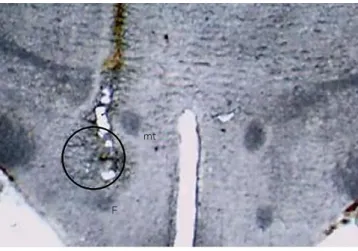
mt: mammilothalamic tract, F: fornix.
Figure 1. Photograph of the coronal section of a rat brain representing the injection site of 6-OHDA in the medial forebrain bundle (open circle).
Effects of carvacrol on total thiol concentration
Total thiol concentrations in the striatum were signifi
-cantly decreased in the 6-OHDA-lesioned animals compared to the sham group (p < 0.05). Treatment of lesioned rats with
carvacrol at doses of 25, 50 and 100 mg/kg for six weeks did not change the decreased total thiol concentrations in the striatum (Figure 6).
DISCUSSION
It has been demonstrated that 6-OHDA causes the
degeneration of nigrostriatal dopaminergic neurons and produces motor and non-motor impairments such as
cog-nitive deficits similar to those observed in patients with
PD14,17,18. Therefore, it was used as a valid model of PD. Our
results also confirmed these changes. The administration of
6-OHDA into the MFB produced motor and memory defi -cits and reduced the pain threshold.
The 6-OHDA compound is a dopaminergic neurotoxin
that undergoes auto-oxidation and produces cytotoxic hydrogen peroxide, reactive oxygen species and catechol-amine quinones, which attack intracellular nucleophilic
groups19. The increase in reactive oxygen species
lev-els causes abnormalities in cell structure and
metabo-lism and eventually leads to neuronal degeneration20.
In line with this, our results showed that microinjection
of 6-OHDA into the MFB caused oxidative damage to the
membrane, as evidenced by increased levels of TBARS and decreased total thiol concentration in the striatum at the end of week 6.
In the present study, the microinjection of 6-OHDA into the left MFB resulted in motor deficits that were observed by increased rotations. The unilateral lesion of the nigrostriatal
***p < 0.001 vs sham group.
Figure 2. Apomorphine-induced rotations in the sham, 6-OHDA-lesioned group and carvacrol-treated lesioned groups at the end of the 2nd (2A) and 6th (2B) week after surgery. Carvacrol was administered ip daily at doses of 25, 50 and 100 mg/kg.
Total net rotations / 30 min Total net rotations / 30 min
2 weeks after surgery 350
300
250
200
100 150
50
-50
0 Sham 6-OHDA CAR 25 mg/kgCAR 50 mg/kgCAR 100 mg/kg Sham 6-OHDA CAR 25 mg/kgCAR 50 mg/kgCAR 100 mg/kg ***
***
***
***
6 weeks after surgery 400
350 300 250 200
100 150
50
-50 0
***
*** *** ***
A
B
*p < 0.05 vs sham group, +p < 0.05 vs 6-OHDA-lesioned group.
Figure 3. Step-through latencies in the passive avoidance test in the sham, 6-OHDA-lesioned rats and lesioned rats treated with carvacrol at doses of 25, 50 and 100 mg/kg at the end of week 6. Data are mean ± SEM.
Step through latency (s)
350
300
250
200
100 150
50
0 Sham 6-OHDA CAR 25 mg/kgCAR 50 mg/kgCAR 100 mg/kg *
+
*p < 0.05 vs sham group.
Figure 4. Tail flick latencies in the sham, 6-OHDA-lesioned rats and lesioned rats treated with carvacrol at doses of 25, 50 and 100 mg/kg at the end of week 6. Data are mean ± SEM.
Latency (s)
10 9 8 7
5 4 6
1 2 3
dopaminergic system by 6-OHDA decreases dopamine lev -els in the striatum and upregulates dopamine postsynaptic
receptors on the same side. These changes produce a motor
asymmetry that can be evaluated by dopamine agonists
such as apomorphine21. Apomorphine-induced rotations in
6-OHDA-lesioned rats is a reliable marker for the nigrostria
-tal dopamine depletion. The symptoms of PD appear when
about 60–80% of the dopamine levels and 50–60% of
dopami-nergic neurons in the substantia nigra are lost22.
The present study also examined the potential therapeu
-tic effect of carvacrol, as an antioxidant agent, in a 6-OHDA
model of PD. Our results showed that treatment with carva-crol at doses of 25, 50 and 100 mg/kg did not decrease the
apomorphine-induced rotations in rats. This is in contrast to prior studies, which report the protective effects of carvacrol
on oxidative insults such as cerebral ischemia-reperfusion23.
The reasons for this discrepancy could be related to the
dosage and duration of treatment. The initial oxidative stress produced by 6-OHDA might be attenuated by the antioxi
-dant activity of carvacrol; however, when there is substantial
ongoing oxidative stress and neurodegeneration24 – as in the
6-OHDA model of PD – the antioxidant response wanes or is
overwhelmed over time and, at that point, carvacrol cannot act as an antioxidant.
Furthermore, our results also showed that unilateral
lesion to the left MFB significantly reduced the pain sensa
-tion threshold in the tail flick test. This is consistent with pre
-vious findings, which have reported that bilateral 6-OHDA
lesions decreased the latency of the hind paw lick in the hot
plate test25 and enhanced sensitivity to a wide range of
ther-mal and mechanical painful stimuli26. The precise mecha
-nisms underlying alteration in the withdrawal response to
thermal and mechanical stimulation in the 6-OHDA rats is
unclear. Neurophysiological, clinical and behavioral experi-ments indicate that, other than motor control, the basal
ganglia are also involved in pain processing27. Experimental
studies have shown that striatal dopamine is important for
modulating nociceptive behavioral responses27, an effect that
appears mediated through dopamine D2 receptors25. The stri
-atum has direct and indirect efferent connections to various
brainstem structures involved in the descending pain
modu-lation system27. It has been suggested that the upregulation
of dopamine D2 receptors in the striatum in the 6-OHDA rats
may lead to a decrease in the activity of the descending pain modulation system.
In the present study, the unilateral 6-OHDA lesioned rats were treated with carvacrol to evaluate its possible effect on pain sensation in an animal model of PD. The results showed
that treatment with carvacrol did not change the pain
sensa-tion threshold in the tail flick test in PD animals. This could
also be attributed to the lack of antioxidant activity of carva-crol in this study.
Moreover, cognitive decline is a main non-motor symp-tom of PD that predisposes the majority of patients to
pro-gression into dementia28. Parkinson’s disease patients
com-plain of executive dysfunction and deficiencies of working
memory that resemble those created by frontal lobe injury29.
The 6-OHDA-induced neuronal loss in the substantia nigra
has also been shown to result in learning and memory
impairments14,30. In line with previous studies, our results also
showed that short-term memory in the step-down avoidance
task had deteriorated in the rats with 6-OHDA-induced PD,
and treatment with carvacrol at a dose of 25 mg/kg improved short-term memory.
Cognitive deficits in PD patients might result from
non-dopaminergic dysfunction, as degeneration of noradrenergic, serotonergic and, most importantly, cholinergic systems have
been reported in PD31. Significant loss of cholinergic forebrain
neurons has been reported in PD-affected brains32. It also has
been reported that the loss of cholinergic cells in the nucleus basalis of Meynert (where Lewy bodies are frequently found)
*p < 0.05 vs sham group.
Figure 5. Lipid peroxidation levels in the striatum of the sham, 6-OHDA-lesioned rats and lesioned rats treated with carvacrol at doses of 25, 50 and 100 mg/kg at the end of week 6. Data are mean ± SEM.
TBAPS (nmol/g tissue)
60
50
40
30
10 20
0 Sham 6-OHDA CAR 25 mg/kgCAR 50 mg/kgCAR 100 mg/kg *
*p < 0.05 vs sham group.
Figure 6. Total thiol concentrations in the striatum of the sham, 6-OHDA-lesioned rats and lesioned rats treated with carvacrol at doses of 25, 50 and 100 mg/kg at the end of week 6. Data are mean ± SEM.
Total thiol concentration (mM)
100
80
60
40
20
is greater than that seen in Alzheimer’s disease33. The nucleus
basalis of Meynert is the main source of cholinergic projec-tions to the cerebral cortex and degenerates in PD. In addition,
choline acetyltransferase activity has been found to be signifi
-cantly decreased in the cortex of parkinsonian subjects34.
Our findings indicated that carvacrol ameliorated
short-term memory impairment in rats with PD. It has also been shown that carvacrol improved spatial memory impair-ments induced by scopolamine, a muscarinic receptor
antagonist, in rats35. More importantly, the
acetylcholines-terase inhibitory activity of carvacrol has been shown in
sev-eral studies8,9. Therefore, the effect of carvacrol on memory
improvement could be due to its anticholinesterase activity
References
1. Fahn S. Description of Parkinson’s disease as a clinical syndrome. Ann N Y Acad Sci. 2003 Jun;991(1):1-14. https://doi.org/10.1111/j.1749-6632.2003.tb07458.x
2. Brown RG, Marsden CD. How common is dementia in Parkinson’s disease? Lancet. 1984 Dec;2(8414):1262-5. https://doi.org/10.1016/S0140-6736(84)92807-1
3. Aarsland D, Andersen K, Larsen JP, Lolk A, Nielsen H, Kragh-Sørensen P. Risk of dementia in Parkinson’s disease: a community-based, prospective study. Neurology. 2001 Mar;56(6):730-6. https://doi.org/10.1212/WNL.56.6.730
4. Beiske AG, Loge JH, Rønningen A, Svensson E. Pain in Parkinson’s disease: prevalence and characteristics. Pain. 2009 Jan;141(1-2):173-7. https://doi.org/10.1016/j.pain.2008.12.004
5. Janvin CC. Cognitive impairment in patients with parkinson’s disease: profiles and implications for prognosis. Bora – Uib. 2007 [acess year Month day]. Available from: http://bora.uib.no/handle/1956/2242
6. Kulisevsky J, Avila A, Barbanoj M, Antonijoan R, Berthier ML, Gironell A. Acute effects of levodopa on neuropsychological performance in stable and fluctuating Parkinson’s disease patients at different levodopa plasma levels. Brain. 1996 Dec;119(Pt 6):2121-32. https://doi.org/10.1093/brain/119.6.2121
7. Baser KH. Biological and pharmacological activities of carvacrol and carvacrol bearing essential oils. Curr Pharm Des. 2008;14(29):3106-19. https://doi.org/10.2174/138161208786404227
8. Jukic M, Politeo O, Maksimovic M, Milos M, Milos M. In vitro acetylcholinesterase inhibitory properties of thymol, carvacrol and their derivatives thymoquinone and thymohydroquinone. Phytother Res. 2007 Mar;21(3):259-61. https://doi.org/10.1002/ptr.2063
9. Kaufmann D, Dogra AK, Wink M. Myrtenal inhibits acetylcholinesterase, a known Alzheimer target. J Pharm Pharmacol. 2011 Oct;63(10):1368-71. https://doi.org/10.1111/j.2042-7158.2011.01344.x
10. Melo FH, Venâncio ET, Sousa DP, Fonteles MMF, Vasconcelos SM, Viana GS et al. Anxiolytic-like effect of Carvacrol
(5-isopropyl-2-methylphenol) in mice: involvement with GABAergic transmission. Fundam Clin Pharmacol. 2010 Aug;24(4):437-43. https://doi.org/10.1111/j.1472-8206.2009.00788.x
11. Melo FH, Moura BA, Sousa DP, Vasconcelos SM, Macedo DS, Fonteles MM et al. Antidepressant-like effect of carvacrol (5-Isopropyl-2-methylphenol) in mice: involvement of dopaminergic system. Fundam Clin Pharmacol. 2011 Jun;25(3):362-7.
https://doi.org/10.1111/j.1472-8206.2010.00850.x
12. Paxinos G, Watson C. The rat brain in stereotaxic coordinates. 5th ed. Amsterdam, London: Elsevier Academic; 2005.
13. Fujita M, Nishino H, Kumazaki M, Shimada S, Tohyama M, Nishimura T. Expression of dopamine transporter mRNA and its binding site in fetal nigral cells transplanted into the striatum of 6-OHDA lesioned rat. Brain Res Mol Brain Res. 1996 Jul;39(1-2):127-36. https://doi.org/10.1016/0169-328X(96)00018-6
14. Rajaei Z, Hosseini M, Alaei H. Effects of crocin on brain oxidative damage and aversive memory in a 6-OHDA model of Parkinson’s disease. Arq Neuropsiquiatr. 2016 Sep;74(9):723-9. https://doi.org/10.1590/0004-282X20160131
15. Tassorelli C, Greco R, Wang D, Sandrini M, Sandrini G, Nappi G. Nitroglycerin induces hyperalgesia in rats: time-course study. Eur J Pharmacol. 2003 Mar;464(2-3):159-62. https://doi.org/10.1016/S0014-2999(03)01421-3
16. Ahmadi M, Rajaei Z, Hadjzadeh MA, Nemati H, Hosseini M. Crocin improves spatial learning and memory deficits in the Morris water maze via attenuating cortical oxidative damage in diabetic rats. Neurosci Lett. 2017 Mar;642:1-6. https://doi.org/10.1016/j.neulet.2017.01.049
17. Campos FL, Carvalho MM, Cristovão AC, Je G, Baltazar G, Salgado AJ et al. Rodent models of Parkinson’s disease: beyond the motor symptomatology. Front Behav Neurosci. 2013 Nov;7:175. https://doi.org/10.3389/fnbeh.2013.00175
18. Tadaiesky MT, Dombrowski PA, Figueiredo CP, Cargnin-Ferreira E, Da Cunha C, Takahashi RN. Emotional, cognitive and neurochemical alterations in a premotor stage model of Parkinson’s disease. Neuroscience. 2008 Oct;156(4):830-40. https://doi.org/10.1016/j.neuroscience.2008.08.035
19. Palumbo A, Napolitano A, Barone P, Ischia M. Nitrite- and peroxide-dependent oxidation pathways of dopamine: 6-nitrodopamine and 6-hydroxydopamine formation as potential contributory mechanisms of oxidative stress- and nitric oxide-induced neurotoxicity in neuronal degeneration. Chem Res Toxicol. 1999 Dec;12(12):1213-22. https://doi.org/10.1021/tx990121g
20. Blum D, Torch S, Lambeng N, Nissou M, Benabid AL, Sadoul R et al. Molecular pathways involved in the neurotoxicity of 6-OHDA, dopamine and MPTP: contribution to the apoptotic theory in Parkinson’s disease. Prog Neurobiol. 2001 Oct;65(2):135-72. https://doi.org/10.1016/S0301-0082(01)00003-X
21. Schwarting RK, Huston JP. Behavioral and neurochemical dynamics of neurotoxic meso-striatal dopamine lesions. Neurotoxicology. 1997;18(3):689-708.
22. Dauer W, Przedborski S. Parkinson’s disease: mechanisms and models. Neuron. 2003 Sep;39(6):889-909.
https://doi.org/10.1016/S0896-6273(03)00568-3
and modulation of the cholinergic system. In agreement with this, it has been reported that acetylcholinesterase inhibitors
have a beneficial effect on cognitive performance in PD36.
In conclusion, the present study demonstrated that carva-crol improves short-term memory impairment in rats with PD. Considering that L-DOPA therapy for PD just relieves motor symptoms, we suggest that carvacrol may serve as an adjunct
therapy for the alleviation of memory deficits in patients with PD.
Acknowledgements
The results presented in this work have been taken from a student’s thesis. This study was supported by Isfahan
23. Yu H, Zhang ZL, Chen J, Pei A, Hua F, Qian X et al. Carvacrol, a food-additive, provides neuroprotection on focal cerebral ischemia/reperfusion injury in mice. PLoS One. 2012;7(3):e33584. https://doi.org/10.1371/journal.pone.0033584
24. Sarre S, Yuan H, Jonkers N, Van Hemelrijck A, Ebinger G, Michotte Y. In vivo characterization of somatodendritic dopamine release in the substantia nigra of 6-hydroxydopamine-lesioned rats. J Neurochem. 2004 Jul;90(1):29-39.
https://doi.org/10.1111/j.1471-4159.2004.02471.x
25. Lin MT, Wu JJ, Chandra A, Tsay BL. Activation of striatal dopamine receptors induces pain inhibition in rats. J Neural Transm (Vienna). 1981;51(3-4):213-22. https://doi.org/10.1007/BF01248953
26. Takeda R, Ikeda T, Tsuda F, Abe H, Hashiguchi H, Ishida Y et al. Unilateral lesions of mesostriatal dopaminergic pathway alters the withdrawal response of the rat hindpaw to mechanical stimulation. Neurosci Res. 2005 May;52(1):31-6. https://doi.org/10.1016/j.neures.2005.01.005
27. Chudler EH, Dong WK. The role of the basal ganglia in nociception and pain. Pain. 1995 Jan;60(1):3-38. https://doi.org/10.1016/0304-3959(94)00172-B
28. Janvin CC, Larsen JP, Aarsland D, Hugdahl K. Subtypes of mild cognitive impairment in Parkinson’s disease: progression to dementia. Mov Disord. 2006 Sep;21(9):1343-9. https://doi.org/10.1002/mds.20974
29. Lewis SJ, Cools R, Robbins TW, Dove A, Barker RA, Owen AM. Using executive heterogeneity to explore the nature of working memory deficits in Parkinson’s disease. Neuropsychologia. 2003;41(6):645-54. https://doi.org/10.1016/S0028-3932(02)00257-9
30. Beppe GJ, Dongmo AB, Foyet HS, Tsabang N, Olteanu Z, Cioanca O et al. Memory-enhancing activities of the aqueous extract of Albizia adianthifolia leaves in the 6-hydroxydopamine-lesion rodent model of Parkinson’s disease. BMC Complement Altern Med. 2014 Apr;14(1):142. https://doi.org/10.1186/1472-6882-14-142
31. Jokinen P, Brück A, Aalto S, Forsback S, Parkkola R, Rinne JO. Impaired cognitive performance in Parkinson’s disease is related to caudate dopaminergic hypofunction and hippocampal atrophy. Parkinsonism Relat Disord. 2009 Feb;15(2):88-93. https://doi.org/10.1016/j.parkreldis.2008.03.005
32. Whitehouse PJ, Hedreen JC, White CL 3rd, Price DL. Basal forebrain neurons in the dementia of Parkinson disease. Ann Neurol. 1983 Mar;13(3):243-8. https://doi.org/10.1002/ana.410130304
33. Liu AK, Chang RC, Pearce RK, Gentleman SM. Nucleus basalis of Meynert revisited: anatomy, history and differential involvement in Alzheimer’s and Parkinson’s disease. Acta Neuropathol. 2015 Apr;129(4):527-40. https://doi.org/10.1007/s00401-015-1392-5
34. Dubois B, Danzé F, Pillon B, Cusimano G, Lhermitte F, Agid Y. Cholinergic-dependent cognitive deficits in Parkinson’s disease. Ann Neurol. 1987 Jul;22(1):26-30. https://doi.org/10.1002/ana.410220108
35. Azizi Z, Ebrahimi S, Saadatfar E, Kamalinejad M, Majlessi N. Cognitive-enhancing activity of thymol and carvacrol in two rat models of dementia. Behav Pharmacol. 2012 Jun;23(3):241-9. https://doi.org/10.1097/FBP.0b013e3283534301