87 Radiol Bras. 2018 Mar/Abr;51(2):87–94
Suspicious amorphous microcalcifications detected on
full-field digital mammography: correlation with histopathology
Microcalcificações amorfas suspeitas detectadas na mamografia digital: correlação histológica
Vera Christina Camargo de Siqueira Ferreira1, Elba Cristina Sá de Camargo Etchebehere2, José Luiz Barbosa
Bevilacqua3, Nestor de Barros4
Ferreira VCCS, Etchebehere ECSC, Bevilacqua JLB, Barros N. Suspicious amorphous microcalcifications detected on full-field digital mammography: correlation with histopathology. Radiol Bras. 2018 Mar/Abr;51(2):87–94.
Abstract
Resumo
Objective: To evaluate suspicious amorphous calcifications diagnosed on full-field digital mammography (FFDM) and establish cor-relations with histopathology findings.
Materials and Methods: This was a retrospective study of 78 suspicious amorphous calcifications (all classified as BI-RADS® 4)
de-tected on FFDM. Vacuum-assisted breast biopsy (VABB) was performed. The histopathological classification of VABB core samples was as follows: pB2 (benign); pB3 (uncertain malignant potential); pB4 (suspicion of malignancy); and pB5 (malignant). Treatment was recommended for pB5 lesions. To rule out malignancy, surgical excision was recommended for pB3 and pB4 lesions. Patients not submitted to surgery were followed for at least 6 months.
Results: Among the 78 amorphous calcifications evaluated, the histopathological analysis indicated that 8 (10.3%) were malignant/ suspicious (6 classified as pB5 and 2 classified as pB4) and 36 (46.2%) were benign (classified as pB2). The remaining 34 lesions (43.6%) were classified as pB3: 33.3% were precursor lesions (atypical ductal hyperplasia, lobular neoplasia, or flat epithelial atypia) and 10.3% were high-risk lesions. For the pB3 lesions, the underestimation rate was zero.
Conclusion: The diagnosis of precursor lesions (excluding atypical ductal hyperplasia, which can be pB4 depending on the severity and extent of the lesion) should not necessarily be considered indicative of underestimation of malignancy. Suspicious amorphous calcifications correlated more often with precursor lesions than with malignant lesions, at a ratio of 3:1.
Keywords: Breast stereotaxis biopsy; Calcification; Diagnosis; Amorphous morphology; Digital mammography; Breast cancer.
Objetivo: Correlacionar o achado mamográfico de calcificações amorfas suspeitas diagnosticadas na mamografia digital com seus diagnósticos anatomopatológicos.
Materiais e Métodos: Setenta e oito casos de calcificações amorfas suspeitas (todas classificadas como BI-RADS® 4) detectadas
na mamografia digital e submetidas a biópsia percutânea assistida à vácuo foram retrospectivamente avaliados. A classificação anatomopatológica utilizada na biópsia foi: pB2 para lesão benigna, pB3 para lesão com potencial incerto de malignidade, pB4 para lesão suspeita, e pB5 para lesão considerada maligna. O tratamento foi recomendado para as lesões pB5, a exérese cirúrgica foi indicada para lesões pB3 e pB4, para descartar malignidade, e o seguimento evolutivo foi adotado para as demais pacientes.
Resultados: A histologia demonstrou 8 (10,3%) casos malignos (6 lesões pB5 e 2 lesões pB4) e 36 (46,2%) casos benignos (pB2). As demais 34 (43,6%) lesões foram classificadas como pB3 (33,3% foram lesões precursoras – hiperplasia ductal atípica, neopla-sia lobular ou atipia epitelial plana – e 10,3% foram lesões de alto risco). A taxa de subestimação das lesões pB3 foi zero.
Conclusão: O diagnóstico de lesões precursoras (excluindo hiperplasia ductal atípica, que pode corresponder a lesão pB4 de-pendendo da severidade e extensão dos achados) na biópsia percutânea assistida à vácuo por calcificações amorfas suspeitas não necessariamente representa lesão subestimada. Calcificações amorfas suspeitas se associaram a lesões precursoras numa proporção de 3:1 em relação às lesões malignas.
Unitermos: Neoplasias mamárias/diagnóstico; Mamografia digital; Biópsia por agulha; Microcalcificações mamárias; Calcificações amorfas; Câncer de mama.
Study conducted at the Unidade Radiológica Paulista (URP)/Instituto Fleury, São Paulo, SP, Brazil.
1. MD, PhD, Hospital Sírio Libanês and Instituto do Câncer do Estado de São Paulo (Icesp), São Paulo, SP, Brazil.
2. MD, PhD, Faculdade de Ciências Médicas da Universidade Estadual de Cam-pinas (FCM-Unicamp), CamCam-pinas, SP, Brazil.
3. MD, PhD, Hospital Sírio Libanês, São Paulo, SP, Brazil.
4. MD, PhD, Tenured Professor, Faculdade de Medicina da Universidade de São Paulo (FMUSP), São Paulo, SP, Brazil.
Mailing address: Dra. Vera Christina Camargo de Siqueira Ferreira. Rua Profes-sora Carolina Ribeiro, 221, ap. 71, Jardim Vila Mariana. São Paulo, SP, Brazil,
04116-020. E-mail: [email protected]. Received February 17, 2017. Accepted after revision May 8, 2017. INTRODUCTION
In the last decade, the introduction of full-field digi-tal mammography (FFDM) for screening has yielded enhanced diagnostic benefits. Comparative studies of conventional mammography and FFDM have shown the latter to be superior in terms of the identification of mi-crocalcifications, thus increasing detection rates for
duc-tal carcinoma in situ (DCIS) and invasive carcinoma(4–6).
In the last year, the subject of mammographic screen-ing and percutaneous biopsies has gained visibility in edi-torials(7,8) and articles(9) published in the radiology litera-ture of Brazil. In fact, knowledge of clinical practice is fundamental to improving patient care.
Grouped amorphous microcalcifications constitute the most discrete morphology related to suspicious calci-fications detected by mammography. There is a need to understand how these findings detected by FFDM are re-lated to the presence of DCIS and invasive carcinomas. The aim of the present study was to correlate suspicious amorphous calcifications (identified on FFDM) with his-topathological findings.
MATERIALS AND METHODS
This was a retrospective study conducted at two health care clinics operated by a private institution. The study was approved by the research ethics committee of the institution. Because of the retrospective nature of the study, informed consent was deemed unnecessary.
Patient selection
In the first year after the introduction of the digital technique (2006 at one clinic and 2007 at the other), we reviewed all consecutive FFDM reports for female
pa-tients in whom the findings were classified as BI-RADS®
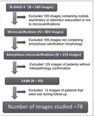
category 4(10). The FFDM examination was performed for either screening or diagnostic purposes (in the latter case to verify findings obtained at our breast care center, which is a referral center). All biopsy samples were obtained via the vacuum-assisted breast biopsy (VABB) technique. We included patients regardless if the following clinical con-ditions was also present: a family history of cancer; a his-tory of breast or ovarian cancer; previous biopsy-proven diagnosis of a precursor lesion; and bilateral suspicious lesions. We initially reviewed all FFDM images classified as BI-RADS category 4 (n = 589). Of those 589 exami-nations, 511 were excluded on the basis of the following criteria: the images showing a nodule, asymmetry, or dis-tortion, with or without microcalcifications (n = 185); the images not showing an amorphous calcification morphol-ogy (n = 185); patients without histopathological analy-sis confirmation by VABB (n = 129); and patients having been lost to follow-up (n = 12). Therefore, the final sample comprised 78 FFDM images of suspicious amorphous cal-cifications, in 77 patients (Figure 1). These patients were followed-up for at least six months or underwent surgical excision of suspicious or malignant lesions.
FFDM
Mammograms were obtained with a digital mam-mography system (Lorad Selenia; Hologic, Danbury, CT, USA). At least two projections, necessarily including cra-niocaudal and mediolateral oblique views, were obtained for analysis. True geometric (air-gap) magnification views were also obtained.
Images were displayed on a dedicated 5-megapixel display. The images were analyzed by two breast imaging radiologists, each with over 10 years of experience. In the event of disagreement, a final decision was made by a third experienced breast imaging radiologist.
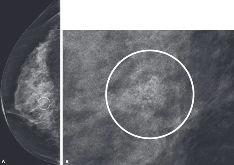
The suspicious amorphous microcalcifications (all classified as BI-RADS category 4 findings) showed the following morphologic characteristics: solely amorphous (Figure 2); or punctate and amorphous (Figure 3). In ad-dition, the distribution of the microcalcifications was clas-sified as grouped, linear (Figure 4), or segmental. There were no cases of microcalcifications with a regional dis-tribution.
VABB
were obtained. Two separate core vials—one containing microcalcifications and one containing other fragments— were sent to the pathology laboratory, together with their corresponding radiographs. At the end of the procedure, the biopsy site was marked with an identifying clip.
In all patients, craniocaudal and lateral radiographs were acquired after the procedure. Those radiographs were obtained for the following reasons: to confirm the removal of the lesion and the placement of the identifying clip; to guide the preoperative localization when surgical excision was required; and to establish a baseline for use in patient follow-up.
Histopathological findings
All histopathological analyses were performed in a pa-thology laboratory, by a pathologist with over 11 years of experience in evaluating breast lesions. The classification of the histopathological diagnoses of lesions submitted to diagnostic VABB were based on the guidelines for non-op-erative diagnostic procedures and reporting in breast can-cer screening established by the United Kingdom National
Coordinating Committee for Breast Pathology(11): B1,
nor-mal; B2, benign; B3, uncertain malignant potential; B4, suspicion of malignancy; and B5, malignant. Hereafter,
each of those pathological categories will be preceded by the letter “p” to avoid confusion with the BI-RADS catego-ries. Therefore, a diagnosis of atypical intraductal epithe-lial proliferation could be classified as pB3 or pB4 depend-ing on the severity and extent of the lesion(11).
In the presence of concurrent diagnoses within the same lesion, the primary diagnosis was designated as fol-lows, in descending order by severity: invasive carcinoma; DCIS; atypical ductal hyperplasia (ADH); lobular neopla-sia, comprising both lobular carcinoma in situ (LCIS) and atypical lobular hyperplasia (ALH); flat epithelial atypia (FEA); other high-risk lesions, including radial scar (RS) and papillary lesion; and benign lesions.
For statistical purposes, pB2 lesions were classified as benign, whereas pB4 and pB5 lesions were grouped to-gether and classified as malignant. Category pB3 included ADH, LCIS, ALH, FEA, RS, and papillary lesions. Among those, ADH, LCIS, ALH, and FEA were considered pre-cursor lesions.
Surgical excision and patient follow-up
All pB4 and pB5 lesions were submitted to surgical excision. All benign lesions (pB2) were followed through-up with clinical evaluation and FFDM (after a minimum Figure 2. Grouped amorphous microcalcifications in a DCIS, shown in craniocaudal and magnified lateral views (A and B, respectively).
Figure 4. Punctate microcalcifications together with amorphous microcalcifications, with a linear distribution, in a benign lesion—mediolateral and magnified lateral views (A and B, respectively).
A B
Figure 3. Grouped punctate and amorphous microcalcifications in a benign lesion—craniocaudal implant-displaced (Eklund) and magnified lateral views (A and B, respectively).
of 6 months). To avoid underestimation and to rule out ad-jacent malignancy, we recommended surgical excision for pB3 lesions. Patients who refused surgery were followed.
Statistical analysis
For patient ages, the median and range were de-scribed. To evaluate the equivalency between the group of patients studied and the population of patients that were excluded, the nonparametric Wilcoxon rank sum test was applied to test age and the Pearson’s chi-square test was applied to test the other demographic variables. Be-cause multiple comparisons were tested (for age and
de-mographic variables), the level of significance (p-value) of
each separate test was divided by the number of tests
per-formed (n = 5). The p-value required to indicate statistical
significance, with Bonferroni correction for multiple com-parisons (based on an α of 0.05), was calculated as 0.01.
RESULTS Patient data
Our sample included 78 examinations from 77 pa-tients (one with bilateral lesions). Papa-tients ages ranged from 40 to 81 years, with a median of 53 years.
The patients enrolled underwent FFDM for screening (in 72%); because of a family history of cancer (in 21%); because of a patient history of breast or ovarian cancer (in 5%); or because of a patient history of mammary atypia (in 3%). The group of patients studied was equivalent to the population of patients that were excluded regarding age (p
= 0.838); reason for performing FFDM (p = 0.015); family
history of cancer (p = 0.033); history of cancer (p = 0.047);
and bilateral BI-RADS category 4 findings (p = 0.815).
FFDM findings
The morphologic characteristics of the suspicious amorphous microcalcifications were as follows: solely amorphous, in 53 (68.0%) of the 78 cases, and punctate microcalcifications accompanied by amorphous microcal-cifications, in 25 (32.1%). Among the 78 cases, the dis-tribution of the suspicious amorphous microcalcifications was classified as grouped in 66 (84.6%), as linear in 8 (10.2%), and as segmental in 4 (5.1%). Of the 12 microcal-cifications with linear or segmental distribution, 6 (50.0%) showed a punctate/amorphous morphology.
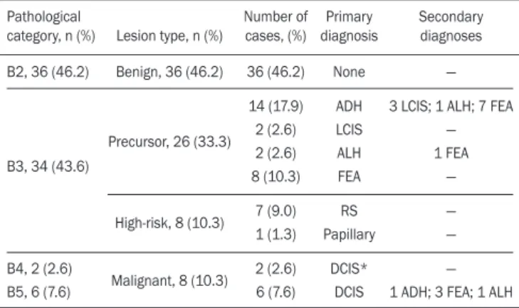
Histopathology findings and correlation to FFDM As can be seen in Table 1, 8 (10.3%) of the 78 sus-picious amorphous microcalcifications were classified as malignant—6 were DCIS (pB5) and 2 were on the borderline between DCIS and ADH (pB4)—malignancy being confirmed at surgery, and 36 (46.2%) were classi-fied as benign (pB2). In addition, 34 (43.6%) of the 77 patients were diagnosed with pB3 lesions. Of those 34 lesions, 26 (76.4%) were precursor lesions—ADH (n = 14), LCIS (n = 2), ALH (n = 2), and FEA (n = 8)—and
8 (23.5%) were other high-risk lesions—RS (n = 7) and papillary lesion (n = 1).
Surgical excision and patient follow-up
Surgical excision was performed in 18 (52.9%) of the 34 pB3 lesions, including17 precursor lesions— ADH (n = 10), LCIS (n = 1), ALH (n = 2), FEA (n = 4)—and 1 high-risk lesion—an RS. For the pB3 lesions, the underestima-tion rate was zero. Of the 34 patients with pB3 lesions, 16 (47.1%) were not submitted to surgical excision. All of those patients underwent mammographic follow-up. The mammographic follow-up of the 36 benign lesions and the 16 pB3 lesions not submitted to surgical excision showed no significant changes at the biopsy site. Follow-up was conducted for a minimum of 6 months and a maximum of 55 months (mean, 22 months).
Although the statistical analysis included only the pri-mary diagnoses, a high number of secondary precursor le-sions were identified in the VABB core samples. Of the 42 lesions classified as pB3, pB4, or pB5, 19 were precursor/ high-risk lesions. Among the cases of DCIS, there were 5 secondary lesions: 1 ADH, 1 ALH, and 3 FEAs. Among the cases of ADH, secondary lesions were found in 11: ALH (n = 1); LCIS (n = 3); and FEA (n = 7). Among the cases of ALH, 3 secondary lesions (all FEA) were observed.
DISCUSSION
Studies of screening programs have shown that can-cer detection rates are higher for digital mammography
than for conventional screen-film mammography(12–14),
especially among patients with microcalcifications(4–6,13).
However, such studies have evaluated microcalcifications of all morphologies together, despite the fact that different morphologies are known to be associated with different malignancy rates(15,16).
Digital mammography has a higher detection rate for microcalcifications than does conventional
mammogra-phy(17,18). All of the images evaluated in the present study
Table 1—Distribution of suspicious amorphous microcalcifications, by patho-logical category and type of lesion, together with the primary and secondary (associated) diagnoses.
Pathological category, n (%)
B2, 36 (46.2)
B3, 34 (43.6)
B4, 2 (2.6) B5, 6 (7.6)
Lesion type, n (%)
Benign, 36 (46.2)
Precursor, 26 (33.3)
High-risk, 8 (10.3)
Malignant, 8 (10.3)
Number of cases, (%) 36 (46.2) 14 (17.9) 2 (2.6) 2 (2.6) 8 (10.3) 7 (9.0) 1 (1.3) 2 (2.6) 6 (7.6) Primary diagnosis None ADH LCIS ALH FEA RS Papillary DCIS* DCIS Secondary diagnoses —
3 LCIS; 1 ALH; 7 FEA — 1 FEA — — — — 1 ADH; 3 FEA; 1 ALH
were obtained with FFDM, and we were therefore able to detect a large number of amorphous microcalcifications, because these are so small and/or hazy in appearance. The detection rate in our study was high in comparison with those reported in previous studies, which is probably due to the fact that our analysis was based solely on FFDM, whereas those of other studies have been either based solely on conventional mammography or based on a mix-ture of breast imaging techniques(15,16,19–21), as well as be-cause our study was conducted during the first year after the introduction of the digital technique into the breast cancer screening protocol of the clinics in question(22).
We detected malignancy in 10.3% of the suspi-cious amorphous microcalcifications. The malignancy rates reported in the literature range from 13% to 31%, and, again, most of the studies evaluating such micro-calcifications have employed conventional
mammogra-phy(16,19–21), as detailed in Table 2. The slight discrepancy
between the malignancy detection rates observed in our study and those reported in the literature could be at-tributable to several factors: we evaluated solely FFDM images; we included microcalcifications with amorphous or punctate/amorphous morphology; and the time span for the evaluation of lesions differed. For instance, one study, based exclusively on conventional mammography, analyzed clustered amorphous calcifications that were not clearly stable for at least 5 years(20).
suspicious breast lesions(24). The higher rate observed in the present study might be attributable to the fact that we evaluated only amorphous calcifications detected on FFDM. Also, microcalcifications distributed in a cluster with amorphous morphology is the most frequent
mam-mographic finding of FEA(25).
The high rate of detection of precursor lesions in comparison with that of detection of malignancy (33% vs. 10%), together with the fact that all cancers were DCIS and there was no underestimation of pB3 lesions, con-firmed the correlation between suspicious amorphous cal-cifications on FFDM and early diagnosis. That was most notable for precursor lesions. Therefore, the diagnosis of a precursor lesion (excluding ADH, which can be classified as pB4 depending on the severity and extent of the lesion) in VABB core samples should not necessarily be consid-ered indicative of underestimation of malignancy, because it could represent an appropriate diagnosis when the le-sion is fully excised and correlated with cores containing calcifications. In addition to the fact that amorphous cal-cifications correlated more strongly with precursor lesions than with malignant lesions, we believe that the lack of un-derestimation of the malignancy of pB3 lesions might be due to the experience of the diagnostic team with VABB, which therefore yielded considerably fewer false-negative results(26), as well as to the fact that a dedicated breast pa-thologist analyzed separate core vials, one containing
mi-crocalcifications and one containing other fragments(27).
The diagnosis and management of high-risk breast le-sions currently constitute a dilemma, especially because of recent improvements in detection(26). The use of VABB to diagnose precursor lesions within amorphous calcifica-tions seen on digital mammography allows us to identify patients at high risk for developing breast cancer, who could benefit from individualized preventive measures. In addition to special mammography screening, such pa-tients could benefit from the use of annual magnetic reso-nance imaging scans, as per the recommendations of the
American Cancer Society(28). Other promising approaches
include chemoprophylaxis(29), as well as the more radi-cal approach (prophylactic mastectomy) requested by some patients following the diagnosis of high-risk lesions. Therefore, mammography might serve not only as a form of secondary prevention of breast cancer but also as a pri-mary preventive measure. By diagnosing precursor lesions, we can intervene in the disease process prior to the emer-gence of breast cancer.
In a study involving the use of VABB with an 11-gauge needle, Liberman et al.(19) reported that the rate of non-retrieval of all calcifications was significantly higher for grouped amorphous calcifications than for all calcification morphologies, as it was for lesions smaller than 0.5 cm. However, it is important to attempt the retrieval of all cal-cifications during VABB. In our study, the underestima-tion rate was zero. That could be due to the great number Table 2—Comparison between the present study and others in the literature, in
terms of the distribution of malignant lesions and precursor lesions.
Reference
Present study Burnside et al.(16)
Liberman et al.(19)
Berg et al.(20)
Shin et al.(21)
Mammography technique
D D and C
C C C Number of cases 78 30 35 150 100 Malignant lesions n 8 4 9 30 31 (%) (10.3) (13.3) (25.7) (20.0) (31.0) Precursor lesions n 26* 4† — 30† 8 (%) (33.3) (13.3) — (20.0) (8.0)
D, digital; C, conventional.
*Including ADH, LCIS, ALH, and FEA; † Including ADH, LCIS, and ALH.
Amorphous calcifications diagnosed on FFDM can represent calcifications in the initial stages of formation and might be related to slight changes, on the spectrum of modifications associated with the formation of cancer, and FFDM thus allows the detection of precursor lesions(23). In the present FFDM study, suspicious amorphous micro-calcifications correlated more often with precursor lesions than with malignant lesions (in 33.3% and 10.3% of cases, respectively).
Precursor lesions (including ADH, LCIS, and ALH) are detected in 8–20% of patients presenting with grouped
amorphous calcifications(16,20,21). In the present study,
of fragments we removed and to the presence of
micro-calcifications within those fragments. Jackman et al.(30)
reported underestimation rates of 8% when the entire le-sion was removed, 13% when maximum lele-sion diameter was < 1.0 cm, 17% when calcifications were present, and 35% in the presence of mass lesions. In other words, the underestimation of calcifications was less than was that of mass lesions.
Our study has certain limitations. Our patient sam-ple was small, and not all patients in whom surgical ex-cision was recommended underwent the procedure. In addition, the follow-up period was, on average, relatively short. However, the bias was minimized by the fact that the group of patients studied was equivalent to the popu-lation that was excluded. Furthermore, interobserver vari-ability is inherent in the practice of radiology. Moreover, there is no consensus on the use of the term amorphous, which could lead to differences among treatment centers in terms of the rates of detection and underestimation of malignancy(31).
CONCLUSION
Suspicious amorphous calcifications diagnosed on FFDM and submitted to VABB correlate strongly with precursor lesions. That knowledge should be taken into consideration in the management of the patients affected.
Studies seek to find parameters that facilitate the management of patients diagnosed with precursor lesions on percutaneous biopsy, informing decisions regarding the choice between surgery and follow-up alone. A multidis-ciplinary team can offer individualized treatment options for patients with concordant findings in the imaging and histological analyses(32–34). Precursor lesions are of low grade, with a low risk for disease progression(35). There-fore, with appropriate screening for high-risk patients, we believe that, in the event of disease progression, the diag-noses can still be made without affecting the prognosis.
Further studies involving larger samples and longer follow-up are needed. Approaches can be tailored on the basis of risk factors, patient age, the type/size of the lesion on imaging, histopathology, the extent of lesion excised, and the correlation with microcalcifications.
REFERENCES
1. World Health Organization. Breast cancer: prevention and control. [cited 2016 Jun 9]. Available from: http://www.who.int/cancer/ detection/breastcancer/en/.
2. Tabár L, Fagerberg CJ, Gad A, et al. Reduction in mortality from breast cancer after mass screening with mammography. Ran
-domised trial from the Breast Cancer Screening Working Group of the Swedish National Board of Health and Welfare. Lancet. 1985;1:829–32.
3. Tabár L, Vitak B, Chen TH, et al. Swedish two-county trial: impact
of mammographic screening on breast cancer mortality during 3
decades. Radiology. 2011;260:658–63.
4. Del Turco MR, Mantellini P, Ciatto S, et al. Full-field digital versus screen-film mammography: comparative accuracy in concurrent screening cohorts. AJR Am J Roentgenol. 2007;189:860–6.
5. Vigeland E, Klaasen H, Klingen TA, et al. Full-field digital mam
-mography compared to screen film mam-mography in the prevalent
round of a population-based screening programme: the Vestfold
County Study. Eur Radiol. 2008;18:183–91.
6. Karssemeijer N, Bluekens AM, Beijerinck D, et al. Breast can
-cer screening results 5 years after introduction of digital mam
-mography in a population-based screening program. Radiology. 2009;253:353–8.
7. Koch H. Mammography as a method for diagnosing breast cancer. Radiol Bras. 2016;49(6):vii.
8. Canella EO. Percutaneous biopsy and radiological imaging of the breast. Radiol Bras. 2016;49(2):ix.
9. Badan GM, Roveda Júnior D, Piato S, et al. Diagnostic underesti -mation of atypical ductal hyperplasia and ductal carcinoma in situ at percutaneous core needle and vacuum-assisted biopsies of the
breast in a Brazilian reference institution. Radiol Bras. 2016;49:6– 11.
10. American College of Radiology. ACR BI-RADS Atlas 5th edition. Breast Imaging Reporting and Data System. Reston, VA: American College of Radiology; 2013.
11. The Royal College of Pathologists. Guidelines for non-operative
diagnostic procedures and reporting in breast cancer screening.
Sheffield: NHSBSP Pub N.50, 2016. [cited 2017 Apr 20]. Avail -able from: https://www.rcpath.org/resourceLibrary/g150-non-op-reporting-breast-cancer-screening-feb17-pdf.html.
12. Heddson B, Rönnow K, Olsson M, et al. Digital versus screen-film
mammography: a retrospective comparison in a population-based
screening program. Eur J Radiol. 2007;64:419–25.
13. Skaane P, Hofvind S, Skjennald A. Randomized trial of screen-film versus full-field digital mammography with soft-copy reading in population-based screening program: follow-up and final results of Oslo II study. Radiology. 2007;244:708–17.
14. Hambly NM, McNicholas MM, Phelan N, et al. Comparison of
digital mammography and screen-film mammography in breast can
-cer screening: a review in the Irish breast screening program. AJR Am J Roentgenol. 2009;193:1010–8.
15. Bent CK, Bassett LW, D’Orsi CJ, et al. The positive predictive value of BI-RADS microcalcification descriptors and final assessment categories. AJR Am J Roentgenol. 2010;194:1378–83.
16. Burnside ES, Ochsner JE, Fowler KJ, et al. Use of microcalcifica
-tion descriptors in BI-RADS 4th edi-tion to stratify risk of malig
-nancy. Radiology. 2007;242:388–95.
17. Kim HS, Han BK, Choo KS, et al. Screen-film mammography and soft-copy full-field digital mammography: comparison in the pa
-tients with microcalcifications. Korean J Radiol. 2005;6:214–20.
18. Fischer U, Baum F, Obenauer S, et al. Comparative study in pa
-tients with microcalcifications: full-field digital mammography vs screen-film mammography. Eur Radiol. 2002;12:2679–83. 19. Liberman L, Smolkin JH, Dershaw DD, et al. Calcification retrieval
at stereotactic, 11-gauge, directional, vacuum-assisted breast bi
-opsy. Radiology. 1998;208:251–60.
20. Berg WA, Arnoldus CL, Teferra E, et al. Biopsy of amorphous breast calcifications: pathologic outcome and yield at stereotactic biopsy. Radiology. 2001;221:495–503.
21. Shin HJ, Kim HH, Ko MS, et al. BI-RADS descriptors for mam
-mographically detected microcalcifications verified by histopathol
-ogy after needle-localized open breast biopsy. AJR Am J Roentgenol. 2010;195:1466–71.
22. Glynn CG, Farria DM, Monsees BS, et al. Effect of transition to digital mammography on clinical outcomes. Radiology. 2011;260: 664–70.
23. Evans AJ, Wilson AR, Burrell HC, et al. Mammographic features of
ductal carcinoma in situ (DCIS) present on previous mammogra
-phy. Clin Radiol. 1999;54:644–6.
24. Eby PR, Ochsner JE, DeMartini WB, et al. Frequency and upgrade
assisted breast biopsy: 9-versus 11-gauge. AJR Am J Roentgenol. 2009;192:229–34.
25. Solorzano S, Mesurolle B, Omeroglu A, et al. Flat epithelial atypia of the breast: pathological-radiological correlation. AJR Am J Roent
-genol. 2011;197:740–6.
26. Neal CH, Coletti MC, Joe A, et al. Does digital mammography in
-crease detection of high-risk breast lesions presenting as calcifica
-tions? AJR Am J Roentgenol. 2013;201:1148–54.
27. Margolin FR, Kaufman L, Jacobs RP, et al. Stereotactic core breast biopsy of malignant calcifications: diagnostic yield of cores with and cores without calcifications on specimen radiographs. Radiology. 2004;233:251–4.
28. Saslow D, Boetes C, Burke W, et al. American Cancer Society guidelines for breast screening with MRI as an adjunct to mam
-mography. CA Cancer J Clin. 2007;57:75–89.
29. Fisher B, Costantino JP, Wickerham DL, et al. Tamoxifen for prevention of breast cancer: report of the National Surgical Ad
-juvant Breast and Bowel Project P-1 Study. J Natl Cancer Inst. 1998;90:1371–88.
30. Jackman RJ, Birdwell RL, Ikeda DM. Atypical ductal hyperplasia:
can some lesions be defined as probably benign after stereotactic 11-gauge vacuum-assisted biopsy, eliminating the recommendation for surgical excision? Radiology. 2002;224:548–54.
31. Berg WA, Campassi C, Langenberg P, et al. Breast Imaging Re
-porting and Data System: inter- and intraobserver variability in feature analysis and final assessment. AJR Am J Roentgenol. 2000; 174:1769–77.
32. Atkins KA, Cohen MA, Nicholson B, et al. Atypical lobular hyper -plasia and lobular carcinoma in situ at core breast biopsy: use of careful radiologic-pathologic correlation to recommend excision or
observation. Radiology. 2013;269:340–7.
33. Neal L, Sandhu NP, Hieken TJ, et al. Diagnosis and management of
benign, atypical, and indeterminate breast lesions detected on core
needle biopsy. Mayo Clin Proc. 2014;89:536–47.
34. Meroni S, Bozzini AC, Pruneri G, et al. Underestimation rate of
lobular intraepithelial neoplasia in vacuum-assisted breast biopsy.
Eur Radiol. 2014;24:1651–8.
35. Ellis IO. Intraductal proliferative lesions of the breast: morphology, associated risk and molecular biology. Mod Pathol. 2010;23 Suppl 2:S1–7.