Cop
yright
© ABE&M t
odos os dir
eit
os r
eser
vados
.
Evaluation of cytopathological
indings in thyroid nodules with
macrocalciication: macrocalciication
is not innocent as it seems
Avaliação dos achados citopatológicos em nódulos tiroidianos com macrocalciicações: elas não são tão inocentes como parecem
Dilek Arpaci1, Didem Ozdemir2, Neslihan Cuhaci2, Ahmet Dirikoc2,
Aylin Kilicyazgan3, Gulnur Guler3, Reyhan Ersoy2, Bekir Cakir2
ABSTRACT
Objective: Microcalciication is strongly correlated with papillary thyroid cancer. It is not clear whether macrocalciication is associated with malignancy. In this study, we aimed to assess the result of ine needle aspiration biopsies (FNAB) of thyroid nodules with macrocalciications.
Subjects and methods: We retrospectively evaluated 269 patients (907 nodules). Macrocalci-ications were classiied as eggshell and parenchymal macrocalciication. FNAB results were divided into four groups: benign, malignant, suspicious for malignancy, and non-diagnostic.
Results: There were 79.9% female and 20.1% male and mean age was 56.9 years. Macrocalci-ication was detected in 46.3% nodules and 53.7% nodules had no macrocalciMacrocalci-ication. Paren-chymal and eggshell macrocalciication were observed in 40.5% and 5.8% nodules, respective-ly. Cytologically, malignant and suspicious for malignancy rates were higher in nodules with macrocalciication compared to nodules without macrocalciication (p = 0.004 and p = 0.003, respectively). Benign and non-diagnostic cytology results were similar in two groups (p > 0.05). Nodules with eggshell calciication had higher rate of suspicious for malignancy and nodules with parenchymal macrocalciication had higher rates of malignant and suspicious for malig-nancy compared to those without macrocalciication (p = 0.01, p = 0.003 and p = 0.007, respec-tively). Conclusions: Our indings suggest that macrocalciications are not always benign and are not associated with increased nondiagnostic FNAB results. Macrocalciication, particularly the parenchymal type should be taken into consideration. Arq Bras Endocrinol Metab. 2014;58(9):939-45
Keywords
Thyroid nodule; macrocalciication; malignancy; suspicious of malignancy
RESUMO
Objetivo: A microcalciicação está fortemente correlacionada com o câncer papilar de tiroide. Não está claro se a macrocalciicação também está associada com malignidade. Neste estudo, nosso objetivo foi avaliar o resultado da biópsia de aspiração por agulha ina (FNAB) de nó-dulos tiroidianos com macrocalciicações. Sujeitos e métodos: Avaliamos retrospectivamente 269 pacientes (907 nódulos). As macrocalciicações foram classiicadas como periféricas (cas-ca de ovo) ou parenquimatosas (interna). Os resultados da FNAB foram divididos em quatro grupos citológicos: benignos, com malignidade, suspeita de malignidade e não diagnósticos.
Resultados: Das amostras, 79,9% foram coletadas de mulheres e 20,1% de homens, e a idade média foi de 56,9 anos. A macrocalciicação foi detectada em 46,3% dos nódulos, e em 53,7% dos nódulos não havia macrocalciicação. A macrocalciicação parenquimatosa e periférica foi observada em 40,5% e 5,8% dos nódulos, respectivamente. Em termos citológicos, a maligni-dade e suspeita de malignimaligni-dade foram mais comuns em nódulos com macrocalciicação em comparação com nódulos sem macrocalciicação (p = 0,004 e p = 0,003, respectivamente). Re-sultados benignos e não diagnósticos da citologia foram similares em ambos os grupos (p > 0,05). Os nódulos com calciicações periféricas apresentaram uma taxa maior de suspeita de malignidade e os nódulos com macrocalciicação parenquimatosa apresentaram taxas maiores de malignidade e suspeita de malignidade em comparação com nódulos sem macrocalciica-ção (p = 0,01, p = 0,003 e p = 0,007, respectivamente). Conclusões: Nossos achados sugerem que as macrocalciicações não são sempre benignas e esses nódulos não estão associados com maiores resultados não diagnósticos da FNAB. A macrocalciicação, particularmente do tipo parenquimatoso, deve ser levada em consideração. Arq Bras Endocrinol Metab. 2014;58(9):939-45
Descritores
Nódulo tiroidiano; macrocalciicação; malignidade; suspeita de malignidade
1 Sakarya University, Sakarya Training and Research Hospital, Department of Endocrinology and Metabolism, Adapazari, Sakarya, Turkey
2 Yildirim Beyazit University, Ankara Ataturk Training and Research Hospital; Department of Endocrinology and Metabolism, Ankara, Turkey 3 Yıldırım Beyazit University, Ankara Ataturk Training and Research Hospital, Department of Pathology, Ankara, Turkey
Correspondence to:
Dilek Arpaci Korucuk, 1683 A/10 Adapazari, Sakarya, Turkey [email protected]
Received on July/20/2014 Accepted on Aug/10/2014
Cop
yright
© ABE&M t
odos os dir
eit
os r
eser
vados
.
INTRODUCTION
T
hyroid nodules are commonly observed in the adult population and the incidence is increasing largely related with widespread use of Doppler ultraso-nography (US) and other imaging techniques. Around 4-8% of thyroid nodules are found incidentally in as-ymptomatic adults, whereas 10-41% are detected by US (1). The majority of thyroid nodules are benign, with malignancy rates ranging from 9 to 13% in diffe-rent studies (2,3). Thyroid US has an important role in the diagnosis of thyroid nodules because it is a simple, non-invasive, effective, and useful method. Nodules with a diameter of 2-3 milimeter (mm) can be detec-ted by high resolution images. Also, vascularity can be determined by colour Doppler or power Doppler US. The use of US and US guided ine needle aspiration biopsy (FNAB) to assess thyroid nodules has reduced the number of unnecessary surgeries and increased the rate of diagnosis of thyroid cancer (4,5). The rate of ac-curacy of FNAB was reported up to 96%. Morphologi-cal features of nodules such as echogenicity, texture, margin regularity, presence of halo, presence and type of calciication can be assessed by high resolution US. Margin irregularity, hypoechogenicity and microcalcii-cation were considered to be important risk factors for malignancy, however, size of thyroid nodule alone was not considered as a risk factor (6).Thyroid nodular calciications can be classiied ac-cording to their diameter and location; calciications < 2 mm and without acoustic shadow at posterior are mi-crocalciications, calciications ≥ 2 mm and with posterior acoustic shadow are macrocalciications, and calciica-tions surrounding the nodule are peripheral (eggshell) calciications. Pathologically, microcalciication is a psam-moma body that contains 10-200 µm, rough, smooth, bright, calciic aggregations (7). Large and irregular bor-dered macrocalciication can exist secondary to tumor necrosis and it can be seen in both benign and malignant nodules (2,8). Peripheral calciications are believed to oc-cur secondary to chronic degenerative changes.
Although, microcalciications are known to be strongly associated with malignant nodules, the asso-ciation of macrocalciications with malignancy is con-troversial (8-15). Recent studies have revealed a rela-tionship between macrocalciication and malignancy, particularly in papillary thyroid carcinomas (9,10,16-18). In addition, despite the general belief that peri-pheral macrocalciication indicates benign situations, it
was shown that if it is irregular it can also be related with malignancy (8,19). Macrocalciication together with microcalciication in the same nodule or located in the middle of a hypoechoic nodule have a higher prob-ability of malignancy (20).
The role of FNAB in thyroid nodules with macro-calciications is unclear with 11 to 25% of the biopsies yielding false negative and 5 to 30% yielding non-di-agnostic cytologies (19-22). Calciied lesions detected by USG have been reported to be the most common cause of insuficient FNAB sampling (23). In this study, we aimed to evaluate FNAB results of thyroid nodules with parenchymal and peripheral macrocalciications. We also tried to ind out the impact of macrocalciica-tions on nondiagnostic cytology results.
SUBJECTS AND METHODS
We retrospectively evaluated 907 nodules from 269 pa-tients seen in our out-patient clinic. Papa-tients > 15 years of age with nodular or multinodular goiter and macro-calciication in at least one nodule were included. Pa-tients with a previous history of thyroid surgery, percu-taneous invasive procedures for nodules, radiotherapy to head and neck region or radioactive iodine therapy were excluded from the study. Preoperative thyroid functions, thyroid autoantibodies, thyroid US indings and FNAB results were obtained from medical records. The study was approved by the local ethical committee in accor-dance with the ethical standarts of Helsinki declaration. Blood samples were obtained between 08:00 to 10:00 in the morning from all patients. Serum sensitive thyrotrophin (TSH), free triiodothyronine (fT3), free thyroxine (fT4) and thyroid autoantibodies [antithy-roid peroxidase antibody (anti-TPO) and anti-thyro-globulin antibody (anti-TGAb)] levels were measured with chemiluminescent immunoassay (Immulite 2000, Diagnostic Products Corporation, Los Angeles, CA, USA, and the UniCel DxI 800, Beckman Coulter, CA, USA). Normal levels were as follows; TSH: 0.4 - 4.0 uIU/mL, fT3: 1.57 - 4.71 pg/mL, fT4: 0.61 - 1.12 ng/dL, anti-TPO < 10 U/mL and anti-Tg < 30 U/mL.
Cop
yright
© ABE&M t
odos os dir
eit
os r
eser
vados
.
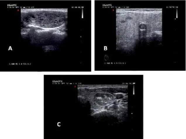
regularity (regular or irregular), presence of hypoechoic halo, presence and type of calciication (microcalci-ication, parenchymal macrocalci(microcalci-ication, peripheral macrocalciication) and vascularization pattern were recorded for all nodules evaluated with FNAB. We de-ined calciications < 2 mm as microcalciication and ≥ 2 mm in diameter and with an acoustic shadow as mac-rocalciication (Figure 1).
Thyroid FNAB was performed by an experienced cli-nician with 27-gauge needle and 20 mL syringe under US guidance. Each nodule was aspirated for 2 - 4 times and at least 4 - 6 preparations were obtained from each aspiration. Cytological assessment was conducted by an experienced cytopathologist. FNAB materials were air-dried and stained by May-Grunwald-Giemsa. The cy-tological diagnoses were classiied as benign, non-diag-nostic, suspicious for malignancy and malignant. FNAB results of nodules with parenchymal and peripheral mac-rocalciications were compared with nodules not includ-ing macrocalciication in the same patient group.
All the data were analyzed with SPSS (Statistical Package of Social Science for Windows) 15.0.
Descrip-tive statistics were expressed as mean ± standard devia-tion for continuous variables and as number of cases and percentage for nominal variables. Student’s t test was used to compare differences between independent groups for continuous variables and Chi-square test was used to compare nominal variables. A p value < 0.05 was considered statistically signiicant.
RESULTS
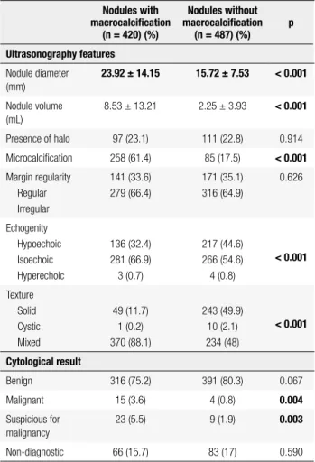
There were 215 female (79.9%) and 54 (20.1%) male patients and the mean age was 56.9 ± 13.1 years (21 - 87 years). One hundred and sixty-one (60%) pa-tients had multinodular goiter and 108 (40%) papa-tients had solitary thyroid nodule. Macrocalciications were observed in 420 (46.3%) nodules, and 487 (53.7%) nodules had no macrocalciication. Parenchymal and peripheral macrocalciications were present in 367 (40.5%) and 53 (5.8%) of 907 nodules, respectively. Mean diameters of nodules with macrocalciication and without macrocalciicaiton were 23.92 ± 14.15 mm and 15.72 ± 7.53 mm, respectively (p < 0.001) (Table 1).
Figure 1. Thyroid nodule calciications detected in ultrasonography. (A) Microcalciication, (B) peripheral (eggshell) macrocalciication, (C) parenchymal
Cop
yright
© ABE&M t
odos os dir
eit
os r
eser
vados
.
Nodules with macrocalciication had signiicantly higher volume compared to nodules without macro-calciication. Ultrasonographically, rates of presence of hypoechoic halo and margin regularity were similar in two groups. Microcalciications were observed more commonly in nodules with macrocalciication (p < 0.001). Thirty-two point four percent of nodules with macrocalciication and 44.6% of nodules without ma-crocalciication were hypoechoic (p < 0.001). In terms of texture, nodules with macrocalciication had a higher prevalence of solid-cystic mixed texture, while nodules without macrocalciication had a higher prevalence of solid texture (Table 1).
Cytological results of 420 nodules with macro-calciication were benign in 75.2%, non-diagnostic in 15.7%, suspicious for malignancy in 5.5% and malig-nant in 3.6%. Of the nodules without macrocalciica-tion, 80.3% were benign, 0.8% malignant, 1.9% suspi-cious for malignancy, and 17% non-diagnostic (Table 1). Accordingly, the rates of suspicious for malignancy and
malignant results were signiicantly higher in nodules with macrocalciication compared to nodules without macrocalciication (p = 0.004 and p = 0.003, respec-tively).
When we compared cytological results of nodules with peripheral macrocalciication and without mac-rocalciication, we found that the rate of suspicious for malignancy was higher in nodules with peripheral macrocalciication while rate of benign was higher in nodules without macrocalciication (p = 0.01and p = 0.036, respectively) (Table 2). Cytologically, 3.8% of nodules with parenchymal macrocalciication and 0.8% of nodules without macrocalciication were malignant (p = 0.003). Suspicious for malignancy rate was also higher in nodules with parenchymal macrocalciication compared to nodules without macrocalciication (p = 0.007) (Table 3). Although rate of nondiagnostic cy-tology was higher in nodules with peripheral macrocal-ciication, the difference was not statistically signiicant. In multiple logistic regression analysis, macrocalciica-tion was found to be related with malignant cytology results independent from presence of microcalciica-tion, irregular margins and absence of halo (p = 0.008).
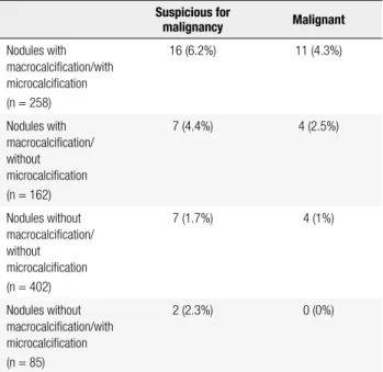
The numbers and rate of the thyroid nodules with or without micro/macro-calciications determined as suspicious for malignancy or malignant were shown in the table 4.
Table 1. Ultrasonography features and cytological results of thyroid
nodules with and without macrocalciication
Nodules with macrocalciication
(n = 420) (%)
Nodules without macrocalciication
(n = 487) (%) p
Ultrasonography features
Nodule diameter (mm)
23.92 ± 14.15 15.72 ± 7.53 < 0.001
Nodule volume (mL)
8.53 ± 13.21 2.25 ± 3.93 < 0.001
Presence of halo 97 (23.1) 111 (22.8) 0.914
Microcalciication 258 (61.4) 85 (17.5) < 0.001
Margin regularity Regular Irregular
141 (33.6) 279 (66.4)
171 (35.1) 316 (64.9)
0.626
Echogenity Hypoechoic Isoechoic Hyperechoic
136 (32.4) 281 (66.9) 3 (0.7)
217 (44.6) 266 (54.6) 4 (0.8)
< 0.001
Texture Solid Cystic Mixed
49 (11.7) 1 (0.2) 370 (88.1)
243 (49.9) 10 (2.1) 234 (48)
< 0.001
Cytological result
Benign 316 (75.2) 391 (80.3) 0.067
Malignant 15 (3.6) 4 (0.8) 0.004
Suspicious for malignancy
23 (5.5) 9 (1.9) 0.003
Non-diagnostic 66 (15.7) 83 (17) 0.590
Table 2. Cytological results of thyroid nodules with peripheral
macrocalciication and without macrocalciication
Cytological result
Nodules with peripheral macrocalciication
(n = 53) (%)
Nodules without macrocalciication
(n = 487) (%)
p
Benign 67.9 80.3 0.036
Malignant 1.9 0.8 0.442
Suspicious for malignancy
7.5 1.9 0.010
Non-diagnostic 22.7 17 0.309
Table 3. Cytological results of thyroid nodules with parenchymal
macrocalciication and without macrocalciication
Cytological result
Nodules with parenchymal macrocalciication
(n = 367) (%)
Nodules without macrocalciication
(n = 487) (%)
p
Benign 76.3 80.3 0.159
Malignant 3.8 0.8 0.003
Suspicious for malignancy
5.2 1.9 0.007
Cop
yright
© ABE&M t
odos os dir
eit
os r
eser
vados
.
Histopathological results were available in 43 pa-tients who underwent surgery for various reasons such as malignant or suspicous for malignancy cytol-ogy results, giant nodule, compression symptoms and suspicious US indings. There were 18 patients with malignant and 25 patients with benign histopathology. Ultrasonographically, micro and macrocalciication, particularly parenchymal macrocalciication were more prevalent in malignant nodules compared to benign nodules (Table 5).
in benign and malignant nodules (7,18). Previously, peripheral calciication was thought to occur secondary to chronic degenerative changes and therefore indicate a benign status. However, recent studies have found that macrocalciications including peripheral calciica-tion might also be an indicator of thyroid nodule ma-lignancy (9,10,16-18,24,25). In this study, we showed that cytologically malignant and suspicious for malig-nancy results are observed more frequently in nodules with parenchymal macrocalciication. Also, nodules with peripheral macrocalciication had a higher rate of suspicious for malignancy results.
Taki and cols. assessed preoperative US indings in 151 surgically resected thyroid nodules and found that 57 (38%) of nodules had calciication (14). Among 11 nodules with microcalciication, 9 (82%) were malig-nant and among 46 nodules with macrocalciication (intranodulary and peripheral) 22 (47.8%) were malig-nant. Additionally, malignancy was histologically iden-tiied in 6 (43%) of 14 nodules with peripheral calciica-tion. The authors concluded that all calciication types may be associated with malignancy and nodules with macrocalciication should be examined thoroughly.
In previous studies, histopathologically proven ma-lignancy rate of thyroid nodules with peripheral mac-rocalciication was reported to range between 18.5% to 70% with most of studies showing higher than 50% malignancy rate in these nodules (8-10,23). Majority of carcinomas were papillary type, with a few follicular car-cinoma histopathologically. Even, anaplastic carcar-cinoma was reported in nodules with peripheral macrocalcii-cation which was blamed for insuficient FNAB result (26). Although there are some US criteria known to be associated with malignancy, it is dificult to apply these criteria for nodules with peripheral macrocalciication due to posterior shadowing and inability to interpret marginal regularity. This has led to search for additional criteria to indicate malignancy in these nodules. In the study by Park and cols., thickening and interruption of peripheral calciications were suggested to be signii-cant indicators of malignancy (11).
Ugurlu and cols. (27) retrospectively assessed the FNAB results of 1,004 patients with thyroid nodules and found that the risk of malignancy was greater in nodules containing microcalciication than those without calciication. However, presence of macro-calciication was not associated with increased risk of malignancy in FNAB compared to nodules without macrocalciication. These results are contrary to our
Table 4. The numbers and rate of the thyroid nodules with or without
micro/macro-calciications determined as suspicious for malignancy or malignant
Suspicious for
malignancy Malignant
Nodules with macrocalciication/with microcalciication (n = 258)
16 (6.2%) 11 (4.3%)
Nodules with macrocalciication/ without microcalciication (n = 162)
7 (4.4%) 4 (2.5%)
Nodules without macrocalciication/ without microcalciication (n = 402)
7 (1.7%) 4 (1%)
Nodules without macrocalciication/with microcalciication (n = 85)
2 (2.3%) 0 (0%)
Table 5. Preoperative calciication types in ultrasonography in patients
with inal histopathological results
Calciication type Malignant (n = 18) (%)
Benign
(n = 25) (%) p
Microcalciication 14 (77.7) 12 (48) 0.049
Macrocalciication 13 (72.2) 10 (40) 0.037
Peripheral macrocalciication
2 (11.1) 2 (8) 0.473
Parenchymal macrocalciication
13 (72.2) 9 (36) 0.019
Without macrocalciication
2 (11.1) 11 (44) 0.021
DISCUSSION
Cop
yright
© ABE&M t
odos os dir
eit
os r
eser
vados
.
indings and those of some previous studies. We have observed cytologically higher malignant and suspi-cious for malignancy rates in nodules with macrocalci-ication compared to those without macrocalcimacrocalci-ication. Similarly, in a recent trial including 713 subcentimeter nodules, solid composition and macrocalciication in addition to hypoechogenicity, iniltrative margin, mi-crocalciication, and taller-than-wide shape were found to be signiicantly associated with malignant cytology (28). The authors showed that including solid compo-sition with or without macrocalciication improved the diagnostic performance in subcentimeter nodules for the identiication of malignant lesions. Park and cols., investigated sonographic indings of 854 macrocalci-ied nodules and reported that 171 (20.8%) were non-diagnostic cytologically, 470 (55.0%) were benign (18 were conirmed by histopathology) and 179 (20.9%) were malignant histopathologically (29). In that study, the rates of nondiagnostic and suspicious for malignan-cy malignan-cytologies were similar with our indings. However, rate of malignancy was higher and rate of benign result was lower compared to our study. As the authors have mentioned as a limitation of their study, patients with benign indings at US had not undergone biopsy or surgery which might have resulted in relatively fewer benign nodules.
In contrary to some previous reports suggesting that the presence of calciication is signiicantly asso-ciated with non-diagnostic FNAB cytology (30), we found no difference in terms of non-diagnostic cytol-ogy between nodules with or without macrocalciica-tion. This inding was also supported in a recent trial by Lee and cols. who retrospectively reviewed sono-graphic indings and histopathological results of 188 nodules with macrocalciication (23). They showed that 6.9% of nodules with macrocalciication was non-diagnostic cytologically and sensitivity, speciicity, pos-itive predictive value and negative predictive value of FNAB were all higher than 90% with a diagnostic ac-curacy of 96% in these nodules. The authors suggested that FNA of thyroid nodules with macrocalciication had a high diagnostic yield. In another study, ultraso-nographic features of 1,195 nodules with inadequate cytology were evaluated prospectively and neither mi-cro- nor macrocalciication was reported to be related with increased risk of inadequacy (31).
Our study has several limitations including the retrospective design and the fact that histopathologi-cal results were available only in a small percentage of
patients who underwent surgery. Thus, we could not determine the exact effect of macrocalciication on false positivity or negativity of FNAB in nodules with mac-rocalciication.
In conclusion, peripheral and parenchymal macro-calciications are associated with higher suspicious for malignancy and/or malignant results in FNAB. In ad-dition to hypoechogenicity, marginal irregularity, ab-sence of halo and vascularization pattern, the preab-sence of macrocalciication in a nodule might be accepted as one of the suspicious US features. However, further studies including histopathological conirmation of these cytological indings are required to support this suggestion. Also, presence of macrocalciication is not related with increased nondiagnostic cytology in FNAB and should not prevent clinicians from making further assessments in case of nondiagnostic results.
Disclosure: no potential conlict of interest relevant to this article was reported.
REFERENCES
1. Frates MC, Benson CB, Charboneau JW, Cibas ES, Clark OH, Coleman BG, et al. Management of thyroid nodules detected at US: Society of Radiologists in Ultrasound consensus conference statement. Radiology. 2005;237(3):794-800.
2. Papini E, Guglielmi R, Bianchini A, Crescenzi A, Taccogna S, Nardi F, et al. Risk of malignancy in nonpalpable thyroid nodules: pre-dictive value of ultrasound and color-Doppler features. J Clin En-docrinol Metab. 2002;87(5):1941-6.
3. Sherman SI, Angelos P, Ball DW, Beenken SW, Byrd D, Clark OH, et al.; National Comprehensive Cancer Network. Thyroid Carci-noma. J Natl Compr Canc Netw. 2005;3(3):404-57.
4. Campbell JP, Pillsbury HC 3rd. Management of the thyroid nod-ule. Head Neck. 1989;11(5):414-25.
5. Nguyen GK, Ginsberg J, Crockford PM. Fine-needle aspiration biopsy cytology of the thyroid. Its value and limitations in the diagnosis and management of solitary thyroid nodules. Pathol Annu. 1991;26 Pt 1: 63-91.
6. Gul K, Ersoy R, Dirikoc A, Korukluoglu B, Ersoy PE, Aydin R, et al. Ultrasonographic evaluation of thyroid nodules: comparison of ultrasonographic, cytological, and histopathological indings. Endocrine. 2009;36(3):464-72.
7. Moon WJ, Baek JH, Jung SL, Kim DW, Kim EK, Kim JY, et al. Ul-trasonography and the ultrasound-based management of thyroid nodules: consensus statement and recommendations. Korean J Radiol. 2011;12(1):1-14.
8. Sahin M, Gursoy A, Tutuncu NB, Guvener DN. Prevalence and prediction of malignancy in cytologically indeterminate thyroid nodules. Clin Endocrinol (Oxf). 2006;65(4):514-8.
9. Yoon DY, Lee JW, Chang SK, Choi CS, Yun EJ, Seo YL, et al. Peripheral calciication in thyroid nodules: ultrasonographic features and prediction of malignancy. J Ultrasound Med. 2007;26(10):1349-55.
Cop
yright
© ABE&M t
odos os dir
eit
os r
eser
vados
.
11. Park M, Shin JH, Han BK, Ko EY, Hwang HS, Kang SS, et al. So-nography of thyroid nodules with peripheral calciications. J Clin Utrasound. 2009;37(6):324-8.
12. Li QS, Chen SH, Xiong HH, Xu XH, Li ZZ, Guo GQ. Papillary thy-roid carcinoma on sonography. Clin Imaging. 2010;34(2):121-6. 13. Chammas MC, de Araujo Filho VJ, Moyses RA, Bresci MD, Mulatti
GC, Brandao LG, et al. Predictive value for malignancy in the ind-ing of microcalciications on ultrasonography of thyroid nodules. Head Neck. 2008;30(9):1206-10.
14. Taki S, Terahata S, Yamashita R, Kinuya K, Nobata K, Kakuda K, et al. Thyroid calciications: sonographic patterns and incidence of cancer. Clin Imaging. 2004;28(5):368-71.
15. Lu Z, Mu Y, Zhu H, Luo Y, Kong Q, Dou J et al. Clinical value of us-ing ultrasound to assess calciication patterns in thyroid nodules. World J Surg. 2011;35(1):122-7.
16. Cooper DS, Doherty GM, Haugen BR, Kloos RT, Lee SL, Mandel SJ, et al. Revised American Thyroid Association management guidelines for patients with thyroid nodules and differentiated thyroid cancer. Thyroid. 2009;19(11):1167-214.
17. Frates MC, Benson CB, Doubilet PM, Kunreuther E, Contreras M, Cibas ES, et al. Prevalence and distribution of carcinoma in pa-tients with solitary and multiple thyroid nodules on sonography. J Clin Endocrinol Metab. 2006;91(9):3411-7.
18. Moon WJ, Jung SL, Lee JH, Na DG, Baek JH, Lee YH, et al. Benign and malignant thyroid nodules: US differentiation--multicenter retrospective study: US differentiation – multicenter retrospec-tive study. Radiology. 2008;247(3):462-70.
19. Beliore A, La Rosa GL. Fine-needle aspiration biopsy of the thy-roid. Endocrinol Metab Clin North Am. 2001;30(2):361-400. 20. Khoo ML, Asa SL, Witterick IJ, Freeman JL. Thyroid calciication and
its association with thyroid carcinoma. Head Neck. 2002;24(7):651-5. 21. Amrikachi M, Ramzy I, Rubenfeld S, Wheeler TM. Accuracy of ine-needle aspiration of thyroid. Arch Pathol Lab Med. 2001;125(4):484-8.
22. Gharib H. Changing concepts in the diagnosis and manage-ment of thyroid nodules. Endocrinol Metab Clin North Am. 1997;26(4):777-800.
23. Lee J, Lee YS, Cha SH, Cho BS, Kang MH, Lee OJ. Fine-needle aspiration of thyroid nodules with macrocalciication. Thyroid. 2013;23(9):1106-12.
24. Seiberling KA, Dutra JC, Grant T, Bajramovic S. Role of intrathy-roidal calciications detected on ultrasound as a marker of malig-nancy. Laryngoscope. 2004;114(10):1753-7.
25. Kim BK, Choi YS, Kwon HJ, Lee JS, Heo JJ, Han YJ, et al. Rela-tionship between patterns of calciication in thyroid nodules and histopathologic indings. Endocr J. 2013;60(2):155-60.
26. Vescini F, Di Gaetano P, Vigna E, Pascoli A, Cacciari M. Anaplastic thyroid carcinoma in a 49 year-old woman with a long-standing goiter. A case report. Minerva Endocrinol. 2000;25(3-4):81-3. 27. Ugurlu S, Caglar E, Yesim TE, Tanrikulu E, Can G, Kadioglu P.
Evaluation of thyroid nodules in Turkish population. Intern Med. 2008;47(4):205-9.
28. Kim HG, Moon HJ, Kwak JY, Kim EK. Diagnostic accuracy of the ultrasonographic features for subcentimeter thyroid nodules suggested by the revised American Thyroid Association guide-lines. Thyroid. 2013;23(12):1583-9.
29. Park YJ, Kim JA, Son EJ, Youk JH, Kim EK, Kwak JY, et al. Thyroid nodules with macrocalciication: sonographic indings predictive of malignancy. Yonsei Med J. 2014;55(2):339-44.
30. Choi SH, Han KH, Yoon JH, Moon HJ, Son EJ, Youk JH, et al. Fac-tors affecting inadequate sampling of ultrasound-guided ine-needle aspiration biopsy of thyroid nodules. Clin Endocrinol (Oxf). 2011;74(6):776-82.