2,4-dichlorophenoxy
acetic acid-mediated
stress in tomato
plants: a biochemical
and molecular
approach.
Ana Águeda Ferreira Pinto
Mestrado Biologia Celular e Molecular
Departamento de Biologia2016
Orientador
Armando Jorge Gomes Teixeira, Professor Auxiliar, FCUP
Coorientador
O Presidente do Júri,
Agradecimentos
Quero expressar o meu mais sincero agradecimento a todas as pessoas que, através da sua colaboração, possibilitaram a realização desta tese.
Gostaria de dirigir as primeiras palavras de agradecimento ao meu orientador Professor Doutor Jorge Teixeira, pela disponibilidade na transmissão de conhecimento e exaustivo acompanhamento do meu trabalho, bem como pelo entusiasmo demostrado ao longo dos últimos anos e principalmente pelas críticas e sugestões construtivas que me fazem evoluir e progredir. Acima de tudo, obrigado pela confiança depositada em mim e pela liberdade de ação que me permitiu, pois foi decisiva para que este trabalho contribuísse para o meu desenvolvimento pessoal.
À Professora Doutora Fernanda, muito obrigada pelo profissionalismo, sinceridade e pela total disponibilidade que sempre revelou para comigo.
Aos meus colegas de laboratório obrigado por toda a ajuda e companheirismo. Em especial à Alexandra de Sousa, não só pela preciosa ajuda na realização da tese e partilha de conhecimentos, mas também pela amizade e companheirismo. Sem ela esta etapa teria sido muito mais difícil de completar.
Muito obrigada ao Mário Costa pela total disponibilidade em ajudar nos trabalhos de biologia molecular. Obrigada pelo tempo e paciência dispensados e pelas importantes sugestões que ajudaram a melhorar esta tese.
O meu agradecimento à Inês Valente e ao Professor Doutor José António Rodrigues, do Departamento de Química e Bioquímica, pela ajuda na técnica no HPLC. Às BioPrincesas - Rita, Íris, Maria João, Lia, Margarida e Ashley pelo apoio incondicional durante estes 5 anos. Obrigada pela vossa amizade, companhia e genuinidade, fatores que permitiram que cada dia fosse encarado com muita motivação. Obrigada por serem como são, por serem tão especiais.
Ao Tiago, o principal prejudicado com esta dissertação. Obrigada pelo modo como sempre me apoiou e acompanhou nestes anos. Sempre que necessário soube aconselhar, criticar e motivar. Agradeço a partilha de alegrias bem como a compreensão nas alturas de desânimo.
À Minha Família, em especial à Minha Mãe, Irmã, Tio António e Tia Amélia, um enorme obrigada por acreditarem sempre em mim e naquilo que faço e por todos os ensinamentos de vida. À Minha Mãe, espero que esta etapa, que agora termino, possa, de alguma forma, retribuir e compensar todo o amor, carinho, apoio e dedicação que, constantemente, me deste. À minha irmã pelo apoio constante e por estar sempre a torcer por mim. Aos meus tios, obrigada por todo o amor e carinho, por me motivarem a
fazer o melhor possível e a ter a confiança necessária para realizar os meus sonhos. A eles, dedico todo este trabalho.
Resumo
Nos últimos 60 anos, os herbicídas auxínicos como o ácido 2,4-diclorofenoxiacético (2,4-D) têm estado entre os herbicidas mais utilizados na agricultura. O 2,4-D é um herbicida seletivo que mata dicotiledóneas e que atua a nível molecular como a auxina nativa ácido indol-3-acético (IAA). Não obstante, são ainda necessários muitos estudos de forma a desvendar o preciso mecanismo de ação deste herbicida. É sabido que o etileno, o ácido abscísico (ABA) e espécies reativas de oxigénio (ROS) possuem um papel fundamental na toxicidade do 2,4-D, levando a alterações nefastas nos tecidos das plantas. Até ao momento, a forma como as células reagem aos ROS e como estes regulam a expressão de genes relacionados com a defesa e/ou o stress continua por se desvendada. Neste estudo, o tomateiro (Solanum
lycopersicum L.) foi utilizado para desvendar os efeitos dos ROS induzidos pelo 2,4-D
no sistema antioxidante, dando especial atenção à expressão dos genes da classe phi da glutationa S-transferase (GST). Quando as plantas S. lycopersicum foram expostas ao herbicida (2,26 mM) durante 48 h, os níveis de H2O2 e O2•− nas folhas aumentaram,
juntamente com uma redução do fecho dos estomas, da assimilação do CO2 e perda de
clorofilas. Contrariamente aos efeitos observados nas folhas, 2,26 mM de 2,4-D não foram suficientes para provocar sintomas claros de stress oxidativo nas raízes. Apesar das diferenças encontradas nos níveis de ROS em ambos os órgãos, a exposição do tomateiro ao 2,4-D levou a um aumento da atividade de enzimas chave do sistema antioxidante, excluindo a superóxido dismutase (SOD) que apenas aumentou nas raízes. As atividades da peroxidase do ascorbato (APX) e da catalase (CAT) aumentaram tanto nas folhas como nas raízes. Mais ainda, os tomateiros expostos a 2,26 mM de 2,4-D responderam ao herbicida aumentando os níveis de ascorbato (AsA) em ambos os órgãos enquanto que um aumento na acumulação de glutationa (GSH) foi apenas observado nas folhas. A exposição ao herbicida levou a um aumento tanto da síntese como da regeneração da GSH, assim como do seu uso para conjugar o 2,4-D, dado que as atividades das enzimas y-glutamil-cisteína-sintetase (γ-ECS), GST e glutationa redutase (GR) aumentaram. A atividade da enzima GST foi aumentada devido a um aumento da expressão dos genes SlGSTF4 e SlGSTF5. No entanto, não foi possível observar o aumento da expressão génica de nenhuma das GST estudadas ao nível das raízes. Este estudo mostra claramente que as folhas e as raízes do tomateiro foram diferencialmente afetadas pela exposição ao 2,4-D na concentração 2,26 mM. Mais ainda, os resultados obtidos sugerem que, no tomateiro, a destoxificação do 2,4-D ocorre principalmente das folhas, com a participação de GST específicas da classe
Abstract
In the last 60 years, auxinic herbicides like 2,4-dichlorophenoxyacetic acid (2,4-D) have been among the widest and successful herbicides used in agriculture. 2,4-D is a selective herbicide that kills dicots and mimics the natural plant phytohormone indol-3-acetic acid (IAA) at the molecular level. Nevertheless, concerted efforts are still being made to unravel the precise mechanism of action of this herbicide. It is known that ethylene, abscisic acid (ABA) and reactive oxygen species (ROS) play a central role in 2,4-D toxicity, leading to numerous unbeneficial changes in plant tissues. Yet, how ROS are perceived by the cell and how they regulate defense- and/or stress-related genes’ expressions remains to be elucidated. In this study, tomato plants (Solanum
lycopersicum L.) were used in order to unravel the effects of 2,4-D-related ROS in the
plant antioxidant system, a special attention being given to the expression of the GST phi class gene family members. When S. lycopersicum plants were root-treated with 2.26 mM 2,4-D for 48 h, H2O2 and O2•− levels increased in leaves and were accompanied by
a reduction in stomatal aperture, CO2 assimilation and chlorophyll loss. Contrary to their
effect on the leaves, in roots 2.26 mM 2,4-D did not provoke clear symptoms of oxidative stress, as lipid peroxidation, H2O2 and O2•− levels decreased. Despite the difference in
ROS levels observed in both organs, the exposure of tomato plants to 2,4-D lead to the activation of key antioxidant enzymes in both organs, apart from superoxide dismutase (SOD) whose activity increased only in roots. Ascorbate peroxidase (APX) and catalase (CAT) activities increased in leaves and in roots. Also, tomato plants responded to 2.26 mM 2,4-D by increasing Ascorbate (AsA) levels in both organs while an increase in Glutathione (GSH) was only observed in leaves. The herbicide increased both the synthesis and the regeneration of GSH, as well as its usage to conjugate 2,4-D, as leaf γ-glutamyl-cysteinyl synthetase (γ-ECS), glutathione reductase (GR) and glutathione S-transferase (GST) activities increased. Leaf GST increased activity was due to an increased expression of SlGSTF4 and SlGSTF5, and none of the SlGSTFs increased their expression in roots. This study clearly showed that leaves and roots of tomato plants were differentially affected by the exposure to 2.26 mM 2,4-D for 48 h. Moreover, the obtained results suggest that in tomato plants 2,4-D detoxification occurs mainly in leaves, with the participation of specific glutathione transferase phi class members
Key-words
2,4-dichlorophenoxyacetic acid (2,4-D); Solanum lycopersicum L.; reactive oxygen species (ROS); oxidative stress; antioxidant system; stress defense; plant hormones; ethylene; abscisic acid; glutathione S-transferase; plant xenome; detoxification.
Table of Contents
Agradecimentos ... i Resumo ... iii Abstract ... iv Key-words ... v Table of Contents ... vi Figure Index ... ix Table Index ... x Appendix Index ... xiAbbreviations and Acronyms ... xi
Introduction ... 1
1. Auxin: Effects and auxin herbicides ... 1
2. Auxin overdose and the deregulation of growth ... 3
2.1. Mode of action of 2,4-D: metabolic and physiological processes ... 3
2.1.1. 2,4-D action at subcellular level ... 4
2.2. Auxinic herbicides selectivity ... 5
2.3. Auxin Signaling and Gene Expression ... 6
2.4. Hormone interactions and growth response ... 7
3. Reactive Oxygen Species, Sites of Production and Their Effects ... 9
3.1. Types of Reactive Oxygen Species ... 10
3.2. Effects of Reactive Oxygen Species ... 12
4. Elimination of Reactive Oxygen Species ... 13
4.1. Non-enzymatic components of the antioxidant system ... 13
4.1.1. Ascorbate ... 13
4.1.2. Glutathione ... 14
4.1.3. Others ... 14
4.2. Enzymatic components of antioxidant system ... 15
4.2.1. Superoxide dismutase (SOD; EC 1.15.1.1) ... 15
4.2.2. Catalase (CAT; EC 1.11.1.6) ... 16
4.2.3. Enzymes of the Ascorbate-Glutathione cycle ... 16
5. 2,4-D detoxification by plants ... 19
5.1. Cytochrome P450 monooxygenases ... 20
5.2. Sugar and amino acid conjugation ... 20
5.3. Glutathione S-transferase ... 21
5.4. ABC transporters ... 21
6. Solanum lycopersicum L. as a fine tool for biochemical and molecular studies 22 Material and Methods ... 24
1. Plant material and growth conditions ... 24
2. Biochemical determinations ... 25
2.1. Photosynthetic pigments determination ... 25
2.2. Determination of lipid peroxidation ... 25
2.3. Determination of H2O2 ... 26
2.3.1. Colorimetrical measurement of H2O2 ... 26
2.3.2. Histochemical visualization of H2O2 ... 26
2.4. Determination of O2.- ... 26
2.5. Gas exchange (IRGA) ... 27
2.6. Quantification of non-enzymatic antioxidants ... 27
2.6.1. Free proline ... 27
2.6.2. Reduced, oxidized and total ascorbate ... 28
2.6.3. Reduced (GSH) and oxidized (GSSG) glutathione ... 28
2.7. Quantification of soluble proteins ... 29
2.8. Enzymes of the Antioxidant System ... 29
2.8.1. SOD (EC 1.15.1.1) ... 29 2.8.2. CAT (EC 1.11.1.6) ... 30 2.8.3. APX (EC 1.11.1.11) ... 30 2.8.4. GR (EC 1.8.1.7) ... 30 2.8.5. γECS (EC 6.3.2.2) ... 31 2.8.6. GST (EC 2.5.1.13) ... 31
3. Bioinformatics characterization of Solanum lycopersicum GSTFs ... 32
4. Evaluation of SlGSTF family expression by real-time RT-PCR ... 32
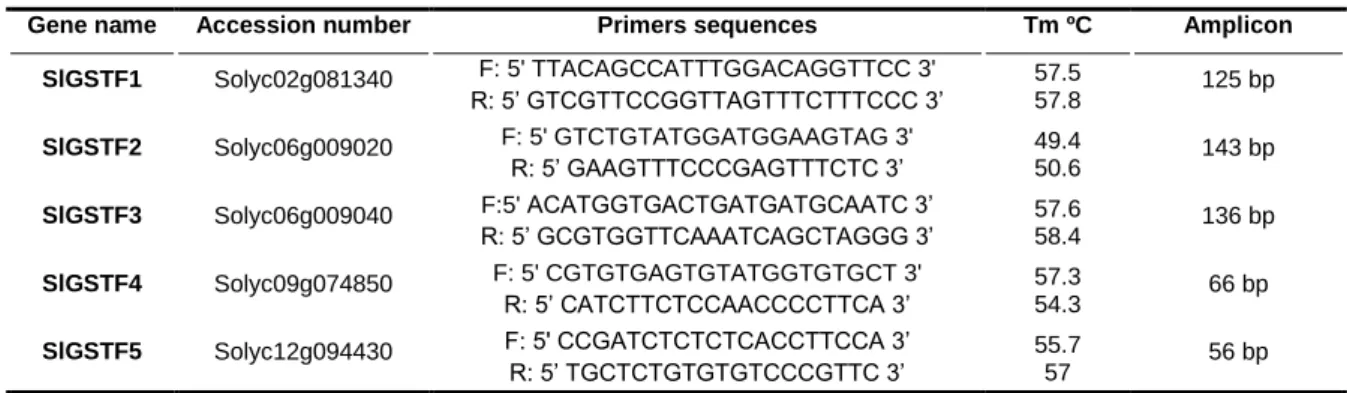
4.1. Primer design ... 32
4.2. Extraction, quantification and assessment of the state of purity of total RNA 33 4.3. Reverse Transcription (RT - cDNA Synthesis)... 34
4.4. Expression of SlGSTF genes by Real-Time PCR ... 34
5. Statistics ... 35
Results ... 36
1. Effects of increasing 2,4-D concentrations on several physiological parameters of S. lycopersicum ... 36
1.1. Effect of increasing 2,4-D concentrations on visible symptoms of toxicity .. 36
1.2. Effect of increasing 2,4-D concentrations in leaves and roots fresh weights 37 1.3. Effect of 2,4-D in total chlorophyll and carotenoid content ... 37
1.4. 2,4-D-induced oxidative stress in S. lycopersicum leaves ... 38
2. 2.26 mM 2,4-D treatment-induced responses of the antioxidant system ... 39
2.1. 2.26 mM 2,4-D-induced oxidative stress in S. lycopersicum roots ... 39
2.2. Effect of 2.26 mM 2,4-D on several physiological parameters of tomato leaves 40 2.3. Effect of 2.26 mM 2,4-D on soluble protein content ... 40
2.4. Effects of 2.26 mM 2,4-D in the enzymatic component of antioxidant system 41 2.5. Effects of 2.26 mM 2,4-D in the non-enzymatic components of the antioxidant system ... 44
3. Bioinformatics characterization of S. lycopersicum GSTFs ... 45
3.1. Phylogenetic analysis of S. lycopersicum GSTFs ... 45
3.2. Analysis of SlGSTFs relative expression using eFP browser... 46
3.3. Changes in transcript levels of selected SlGSTF genes in tomato plants under 2,4-D stress ... 48 Discussion ... 51 Conclusion ... 61 Future perspectives ... 62 References ... 63 Appendixes ... 76
Figure Index
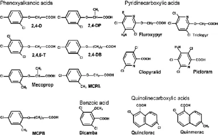
Figure 1. Chemical structures of the different groups of auxinic herbicides (Song, 2014).
... 2
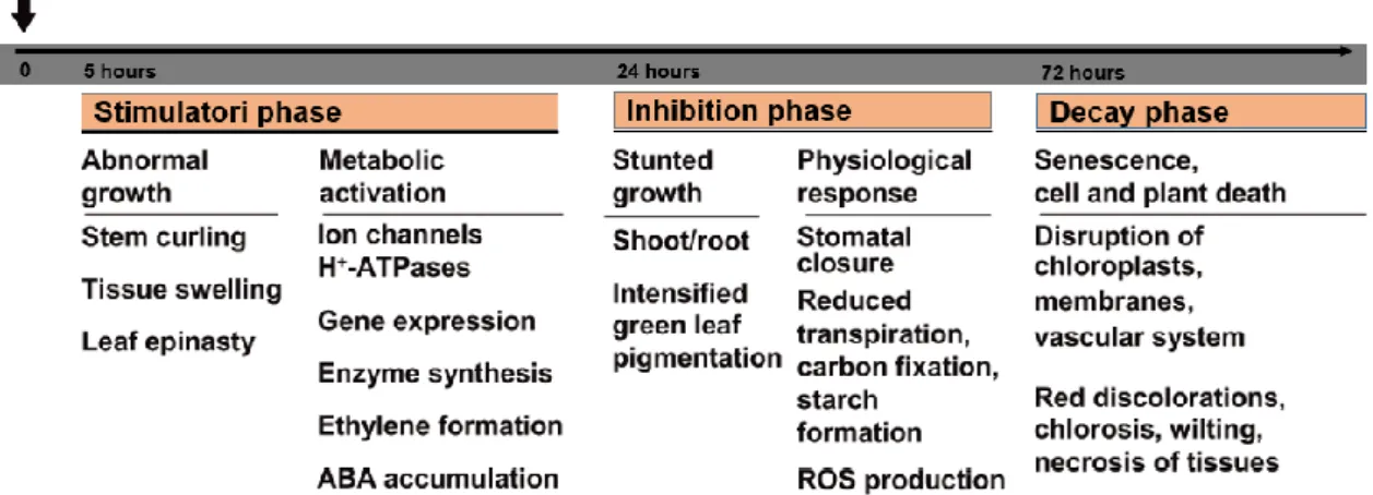
Figure 2. Three-phase response in auxin herbicide auxin action for dicot weed plants
(modified from (Grossmann, 2010)). ... 4
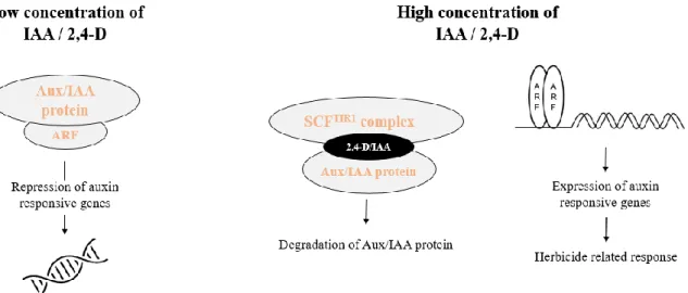
Figure 3. A simplified model of the molecular mechanism of IAA/2,4-D (modified from
(Song, 2014)). ... 7
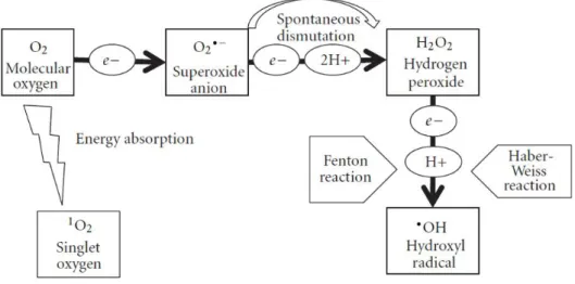
Figure 4. Schematic representation of generation of ROS in plants. Reduction of O2 can
occur by two different mechanisms. Sequential monovalent reduction of O2 leads to
formation of O2•−, H2O2 and •OH. On the other hand, energy transfer to O2 leads to
formation of 1O
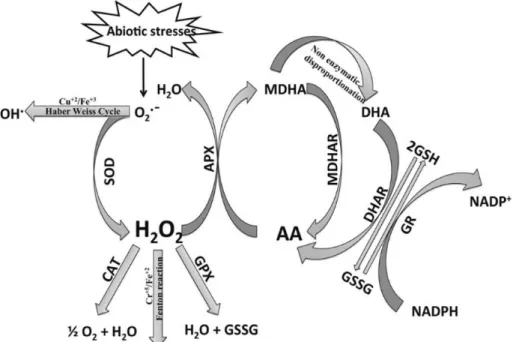
2. Adapted from (Sharma et al., 2012). ... 10 Figure 5. Enzymatic and non-enzymatic antioxidant defense pathways and ROS
homeostasis in plant cells (Gill and Tuteja, 2010). ... 15
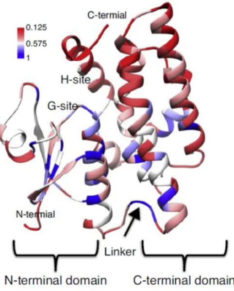
Figure 6. Sequence conservation of plant GSTs depicted in the crystal structure of
Glycine max GST (PDB id 2vo4). The color bar shows the level of conservation from low (red) to high (blue) (Labrou et al., 2015)... 18
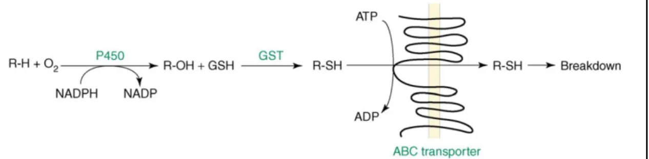
Figure 7. Schema of P450, GSTs and ABC transporter into a four-step detoxification
process. Adapted from (Yuan et al., 2007)………20
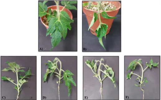
Figure 8. Effect of different concentrations of 2,4-D on 28-d tomato plants. Plants were
treated once with 0 (control), 2.26, 4.52 and 9.04 mM 2,4-D and then were grown for 2 d in a greenhouse. (A) Leaves of control plants (0 mM). (B) Leaves of plants treated with 2.26 mM 2,4-D. (C) Aerial organs of control plants. (D) Aerial organs of plants treated with 2.26 mM 2,4-D. (E) Aerial organs of plants treated with 4.52 mM 2,4-D. (F) Aerial organs of plants treated with 9.04 mM 2,4-D. The arrow indicates a constriction from the hypocotyl until above cotyledons.………36
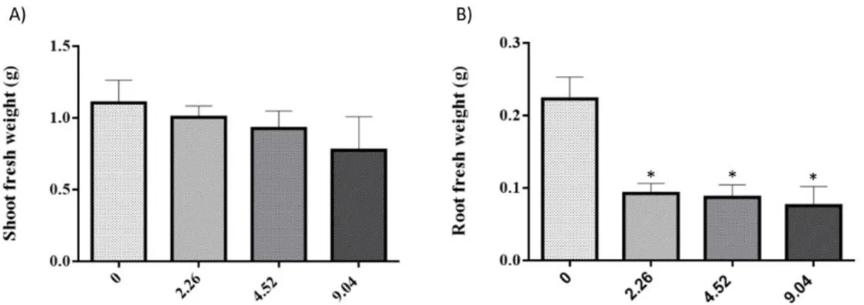
Figure 9. Shoot (A) and root (B) biomass of S. lycopersicum plants grown in nutrient
medium supplemented with different concentrations of 2,4-D. *above bar indicates
significant statistical differences from control at P < 0.05………37
Figure 10. Histochemical localization of H2O2 in terminal leaflets of plants treated with
different concentrations of 2,4-D and then grown for 48 h in a greenhouse. H2O2 labelling
was mainly detected as brown spots (arrows) in the central vein and in peripheral zones of 2,4-D treated plants………...38
Figure 11. Total soluble proteins content in leaves and roots of 4-weeks-old tomato
plants treated with 2.26 mM 2,4-D for 48 h. *above bar indicates significant statistical differences from control at P < 0.05………41
Figure 12. Response of ROS scavenging enzymes in 4-weeks-old tomato plants
exposed to 2.26 mM 2,4-D for 48 h. Panels represent SOD activity (A,B), CAT activity (C,D) and APX activity (E,F), in leaves and roots, respectively. *above bar indicates significant statistical differences from control at P < 0.05……….42
Figure 13. Response of GSH-related enzymes in 4-weeks-old tomato plants exposed to
2.26 mM 2,4-D for 48 h. Panels represent γ-ECS activity (A,B), GST activity (C,D) and GR activity (E,F), in leaves and roots, respectively. *above bar indicates significant statistical differences from control at P < 0.05……….43
Figure 14. Free proline content in leaves and roots of 4-weeks-old tomato plants
exposed to 2.26 mM 2,4-D for 48 h. *above bar indicates significant statistical differences from control at P < 0.05………45
Figure 15. Evolutionary relationships of SlGSTFs. The evolutionary history was inferred
using the Neighbor-Joining method and the percentage of replicate trees in which the associated taxa clustered together in the bootstrap test (1000 replicates) are shown next
to the branches.Evolutionary analyses were conducted in MEGA7………46
Figure 16. Total RNA extracted from control (A) and from plants exposed to 2.26 mM
2,4-D (B). For quality assessment of total RNA, it was separated on agarose gel at 0.8 % (w/v). 1, 28 S rRNA; 2, 18 S rRNA; 3, 5S + tRNA………48
Figure 17. Electrophoretic agarose gel (0.8 % (w/v)) evidencing the RT-qPCR products
of GSTF1, GSTF2, GSTF3, GSTF4 and GSTF5 of S. lycopersicum. The used Ladder
was NZYDNA Ladder VI (Nzytech®, Portugal)………..49
Figure 18. Comparison between the transcript levels of selected tomato GSTF genes in
leaves and roots of 4-week-old tomato plants without any treatment. Data were normalized using the tomato 18S gene as internal control and the relative transcript level of leaves control sample was arbitrarily considered as one for each gene. *above bar indicates significant statistical differences from control at P < 0.05………..50
Figure 19. Effect of 2.26 mM 2,4-D treatment on the transcript levels of selected tomato
GSTF genes in leaves (A) and roots (B) of 4-week-old tomato plants after 48h. Data were normalized using the tomato 18S gene as internal control and the relative transcript level in the control samples was arbitrarily considered as one for each gene………..50
Table Index
Table 2. Effect of different 2,4-D concentrations on several physiological parameters of
tomato plant leaves………39
Table 3. Effect of 2.26 mM 2,4-D on several physiological parameters of tomato plant
roots……….40
Table 4. Effect of 2.26 mM 2,4-D on physiological parameters of tomato plant……….40 Table 5. Ascorbate content in leaves (A) and roots (B) of 4-weeks-old tomato plants
exposed to 2.26 mM 2,4-D for 48 h. *above bar indicates significant statistical differences from control at P < 0.05……….44
Table 6. Glutathione content in leaves (A) and roots (B) of 4-weeks-old tomato plants
exposed to 2.26 mM 2,4-D for 48 h. *above bar indicates significant statistical differences from control at P < 0.05……….45
Table 7. Relative expression data of the 5 GST phy-encoding genes from tomato…...47
Appendix Index
Appendix 1. Genome loci of tomato GST coding sequences, the new and the former (if
any) names of the proteins, the fully deduced coding and protein sequences………….77
Appendix 2. Melting curves for all SlGSTF genes assayed. Red curve - SlGSTF1; Green
curve - SlGSTF2; Black curve - SlGSTF3; Pink curve - SlGSTF4; Blue curve - SlGSTF5. 80
Abbreviations and Acronyms
2,4-D - 2,4-dichlorophenoxyacetic acid;•OH - hydroxyl radical
1O
2 - singlet oxygen
ABA - abscisic acid
ACC - 1-aminocyclopropane-1-carboxylic acid ACX - acyl-CoA oxidase
APX - acorbate peroxidase ARF - auxin response factors AsA - ascorbate
AtGSTF - Arabidopsis thaliana glutathione S-transferase phi class
Aux - auxin
Carot - Carotenoids CAT - catalase
CER - CO2 exchange rate Chl - Chlorophyll
Chl a - Chlorophyll a Chl b - Chlorophyll b DHA - dehydroascorbate
DHAR - dehydroascorbate reductase ETC - electron transport chain
F.W. - fresh weight GR - glutathione reductase GS - glutathione synthetase GSH - reduced glutathione GSSG - oxidized glutathione GST - glutathione S-transferase H2O2 - hydrogen peroxide
HPLC–ESI/MS - high-performance liquid chromatography coupled with electrospray
ionization tandem mass spectrometry
IAA - Indole-3-acetic acid;
ICC - intercellular CO2 concentration MDHA - monodehydroascorbate
MDHAR - monodehydroascorbate reductase NAA - 1-Naphthaleneacetic
NCED - 9-cis-epoxycarotenoid dioxygenase O2•− - superoxide anion
P450 - cytochrome P450 monooxygenases PM - plasma membrane
PSI - photosystem I PSII - photosystem II
ROS - reactive oxygen species
RT-qPCR - real-time quantitative PCR SAM - S-adenosyl-L-methionine SC - stomatal conductance
SlGSTF - Solanum lycopersicum glutathione S-transferase phi class
SOD - Superoxide dismutase TR - transpiration rate
XOD - xanthine oxidoreductase
Introduction
1. Auxin: Effects and auxin herbicides
In higher plants, metabolic regulation and coordination, as well as, morphogenesis and responses to both biotic and abiotic factors are interceded by signaling molecules, called phytohormones. It is the balance between promoting and inhibiting agents in a network which ultimately governs the normal path of plant growth and development (Vanstraelen and Benkova, 2012). Natural auxins are an important class of phytohormones, consisting of indole-3-acetic acid (IAA), the principal natural auxin in higher plants, and related endogenous molecules such 4‐chloroindole‐3‐acetic acid, phenylacetic acid, and indole‐3‐butyric acid, which cause the same responses as IAA (Vanneste and Friml, 2009). Auxins play a critical role in plant growth and are involved in many developmental processes, such as cell division and elongation; in developmental processes including vascular tissue and floral meristem differentiation, leaf initiation, phyllotaxy, senescence, apical dominance and root formation. Auxins are also essential components in tropic responses (Taiz and Zeiger, 2010). As a critical plant hormone, auxin modulates plant growth and development from embryogenesis through all stages of development (Sieburth and Lee, 2010). Because IAA influences virtually every aspect of plant growth and development, the development of chemicals that mimic the behavior of natural auxins acquired great importance, not only for their use in in vitro systems, but also because of their effects on undesired plants.
It is well known that undesired plants compete with crops for water, carbon dioxide, light, nutrients and space. The discovery of synthetic herbicides in 1945 was a major technical achievement that quickly changed weed management practices. Herbicides are agrochemicals used to control the growth of undesired weeds, and aim to significantly increase crop productivity. Most herbicides are small molecules that inhibit specific molecular target sites within critical plant physiological and/or biochemical pathways, and consequently those inhibitions often have catastrophic and lethal consequences on the affected plants. Target sites of herbicides are usually enzymes involved in primary metabolic pathways (i.e., processes that are necessary for the growth and development of an organism) or proteins carrying out essential physiological functions. For this reason, target sites involved in secondary metabolism are less likely to cause deadly effects on plants (Dayan et al., 2010).
Herbicides are commonly classified as either nonselective or selective. A nonselective herbicide is used to kill or damage all growth and is generally reserved for agricultural use or for clearing large or heavily overgrown areas. In contrast, a selective herbicide controls specific weed species, while leaving the desired crop relatively unharmed, and usually works through some type of hormone disruption. Synthetic auxins opened a new era of weed control in crop production due to their systemic mobility in the plant and by exerting a selective action, primarily against dicot weeds in cereal crops (Grossmann, 2003).
Over the years, several chemical classes of auxin herbicides, with different weed spectra and types of selectivity, have been synthesized and commercially introduced. Auxinic herbicides have an aromatic ring and a carboxylic acid moiety, as does IAA, and contain four major chemical groups, including quinolinecarboxylic acids (e.g., quinmerac and quinclorac), pyridinecarboxylic acids (e.g., picloram, clopyralid, triclopyr), a benzoic acid (e.g., dicamba), and phenoxyalknoic acids (e.g., 2,4‐D and MCPA) (Grossmann et
al., 2001; Sterling, 1997) (Figure 1). This herbicide family is said to have initiated an
agricultural revolution and laid the corner stone of present‐day weed science.
As one of many so‐called phenoxy herbicides, 2,4-dichlorophenoxyacetic acid (2,4-D) was developed during the World War II by aiming to increase crop yields for a nation at war. The development of 2,4-D in the 1940s appears to have occurred through a series of multiple, independent experiments. Although it can be debated to whom should be given credit for the discovery of 2,4-D, its commercialization in 1946 revolutionized weed control. It was the first selective herbicide to be commercially released, which allowed greatly enhanced weed control in rice, maize, wheat, and other similar cereal crops because it specifically targets dicots. 2,4-D's low cost has led to
continued usage today and it remains one of the most commonly used herbicides in the world (Song, 2014).
2,4-D is the active ingredient of the most widely used herbicides in the world, existing over 600 2,4-D-related products currently on the market. However, the acid form of this herbicide is usually not formulated as the end-use product. In its pure form, 2,4-D acid is moderately nonvolatile and it is only slightly soluble in water (44.558 mg L-1). The
acid form is low in solubility and, for this reason, it has to be modified in other herbicide formulations that consist in more soluble forms. In general, there are two types of formulations with a big acceptance in the marketplace: amine salts and esters. Amine salts are formed when 2,4-D acid reacts with an amine. The amine salt formulations of 2,4-D include, triisopropanolamine salt, isopropylamine and dimethylamine. When in contact with water, these compounds dissociate into the acid part (negative charge) and the amine part (positive charge), being readily soluble in water and forming a true solution. On the other hand, the reaction of 2,4-D acid with an alcohol forms esters (butoxyethylester , ethylhexyl ester, etc.), which are readily dissolved in an organic solvent but insoluble in water. For this reason they are formulated as emulsifiable concentrates for applications in either water or soils (Charles et al., 2001; Peterson et
al., 2016).
2. Auxin overdose and the deregulation of growth
2.1.
Mode of action of 2,4-D: metabolic and physiological
processes
When applied as herbicides, synthetic auxins mimic the effects of the natural auxin IAA in plants. As shown in IAA-overproducing plants (Romano et al., 1993), high doses of auxin drives plants overgrowth, including stunting and twisting of stems, brittleness and general abnormal growth (Grossmann, 2007; Pazmiño et al., 2012). Although the concentration of natural auxins and its effects are tightly controlled, auxinic herbicides like 2,4-D escape to regulatory mechanisms of sensitive plants and cause an uncontrolled auxin response. Moreover, 2,4-D is long-lasting, particularly due to its higher stability in the plant, and, therefore, more effective than IAA (Song, 2014). This phenomenon has been described as an auxin overdose which leads to an imbalance in auxin homeostasis and interactions with other hormones at the tissue level.
According to several reports, when separating the time course of events, the deregulation of plant growth by 2,4-D (or IAA) at high concentrations can be divided into three phases. The first is the stimulation phase that leads to an induction of 1-aminocyclopropane-1-carboxylic acid synthase (ACCS) resulting in increased ethylene biosynthesis, followed by symptoms such as stem curling and tissue swelling. At this phase, abscisic acid (ABA) also begins to accumulate. In the second phase, which occurs within 24 h, starts abnormal growth and a series of physiological responses such as growth inhibition of root and shoots, decreased internode and leaf area elongation, and intensified green leaf pigmentation. Concomitantly, reductions in stomatal aperture, transpiration, carbon assimilation, starch formation and overproduction of reactive oxygen species (ROS) are observed. The third phase is the phase of tissue decay, which is marked by accelerated chloroplast damage and progressive chlorosis, and by the destruction of membrane and vascular system integrity, leading to cell death. As can be seen in Figure 2, these processes are exemplified for dicot weeds against the background of reported data in the literature (Grossmann, 2004; Grossmann, 2010).
2.1.1.
2,4-D action at subcellular level
2.1.1.1. Chloroplasts
In different species it has been demonstrated that 2,4-D affects the development, structure and function of chloroplasts (Pazmiño et al., 2012). While low concentrations of this chemical may increase carbon assimilation and photochemical reactions, higher doses have inhibitory effects on these reactions (Grossmann, 2000). When applied at high concentrations, 2,4-D reduces chlorophyll and carotenoid contents, as well as the Figure 2. Three-phase response in auxin herbicide auxin action for dicot weed plants (modified from (Grossmann, 2010)).
chlorophyll/benzoquinone relation (Saygideger and Okkay, 2008; Wong, 2000). Moreover, by inhibiting the Hill reaction, 2,4-D blocks the electron transport in photosystem II (Wong, 2000). Combined, all these effects result in chloroplast damage and consequent induction of senescence of the photosynthetic apparatus.
2.1.1.2. Mitochondria
One of the main effects of 2,4-D is the increment of the respiratory rate, culminating in an increased CO2 concentration (Kelly and Avery, 1949). Humphreys &
Dugger (1957) reported that 2,4-D increases respiration by causing more glucose to be catabolized via the pentose phosphate pathway. On the other hand, they also suggested that an increase in respiration may be due to the induction of cell division by 2,4-D and other auxins (Humphreys and Dugger, 1957).
2.1.1.3. Nucleic acids
The increase in nucleic acids is one of the most characteristic response of plant cells to 2,4-D treatment (Peterson et al., 2016). It has been established that in sensitive tissues (e.g. seedlings), treatment with 2,4-D results in a massive accumulation of DNA and RNA associated with induction of cell division (West et al., 1960). Experiments carried out by Chrispeels and Hanson (1962) showed that soybean seedlings’ RNA increased by 175 % after 48 h of exposure to 2,4-D, being that this RNA was mostly ribosomal (Chrispeels and Hanson, 1962).
2.2.
Auxinic herbicides selectivity
Auxinic herbicides have been widely used to control dicot weeds for several decades in agricultural and in nonagricultural settings. Major sites include pasture and rangeland, commercial golf courses, residential lawns, roadways, and cropland (http://npic.orst.edu/factsheets/archive/2,4-DTech.html). Yet, the underling mechanism of how auxinic herbicides selectively kill dicots and spare monocots is still not understood (McSteen, 2010). Initially, several studies tried to understand the correlation between uptake and tolerance; however, the results showed that there is little correlation between these two factors [See references in (Peterson et al., 2016)]. Early research has proposed that the resistance by weeds includes either altered vascular anatomy (Monaco et al., 2002), altered perception of auxin in monocots (Kelley and Riechers, 2007), limited translocation or rapid degradation of exogenous auxin (Gauvrit and
Gaillardon, 1991; Monaco et al., 2002). In 1982, a study conducted by Hall and collaborators, demonstrated that after exposure to radioactively labeled 2,4-D for 24 h, only 5 % of the total radioactivity moved from treated leafs in tolerant oat compared to 55 % in sensitive soybean (Hall et al., 1982). More recently, it was shown that 14
C-radiolabelled 2,4-D is not effectively transported throughout the resistant plant after uptake into the leaf, leading to localized retention of this herbicide (Goggin et al., 2016). These studies make a strong case for the hypothesis that variations in translocation between tolerant and sensitive species could explain differences in 2,4-D resistance.
2.3.
Auxin Signaling and Gene Expression
Although 2,4-D has been used in agriculture for several decades, it’s molecular mode of action is far from being completely characterized. Due to its similarity to the natural auxin IAA, it is thought that 2,4-D acts like IAA at the molecular level. For this reason, identification of receptors that mediate transcriptional and biochemical responses to auxin may provide basic clues about the molecular mode of action of 2,4-D.
The natural auxin IAA enters the cell through the plasma membrane
(PM)-resident auxin transporters like the amino acid permease-like AUXIN
RESISTANTS/LIKE AUX (AUX/LAX) proteins (Swarup et al., 2008; Yang et al., 2006). After IAA enters the cell through auxin-influx carriers, it rapidly controls auxin responsive gene expression by regulating the degradation of Aux/IAA repressor proteins, which are negative regulators of auxin-responsive genes (Mockaitis and Estelle, 2008). The main protein responsible for this response is the F-box protein TIR1. Aux/IAA proteins are recruited to TIR1 in an auxin-dependent manner and after binding, the Aux/IAA repressors are degraded by Skp1-cullin-F-box (SCF) E3 ubiquitin ligase (SCFTIR1)
(Dharmasiri et al., 2005; Hagen and Guilfoyle, 2002; Kepinski and Leyser, 2005). At low concentrations of auxin, Aux/IAA proteins bind to auxin response factors (ARF), repressing the expression of genes controlled by auxins; at high concentrations, IAA functions as a “molecular glue” to enhance TIR1-Aux/IAA protein interaction, mediating the degradation of Aux/IAA proteins. In that way, ARFs are alleviated from AUX/IAA repressors, allowing the homo-dimerization of ARFs and subsequently the expression of auxin-responsive genes (Tan et al., 2007) (Figure 3). Interestingly, crystal structures of TIR1 revealed that, in a slightly weaker manner, 2,4-D also binds at the base of TIR1 acting as a molecular glue to mediate the interaction between Aux/IAA proteins and TIR1 F-box protein (Calderon-Villalobos et al., 2010).
2.4.
Hormone interactions and growth response
Hormone interplay is important in the regulation of plant growth and development. IAA or auxinic herbicides at high concentrations are directly related to overexpression of auxin-responsive genes. The induction of ACCS, which is a key enzyme in ethylene biosynthesis, begins a cascade of physiological responses responsible for a sequential hormone interaction, which play a decisive role in the mode of action of 2,4-D on sensitive plants (Pazmiño et al., 2012). Ethylene is a gaseous phytohormone with a very simple structure that plays an important role in a wide range of physiological reactions including plant responses to stress and regulation of senescence and plant growth (Bleecker and Kende, 2000). Ethylene is biosynthesized from the amino acid methionine to S-adenosyl-L-methionine (SAM) by the enzyme SAM synthase. In a further reaction, that is considered to be the rate-limiting step, SAM is then converted to 1-aminocyclopropane-1-carboxylic acid (ACC) by the enzyme ACCS. The final step involves the action of the enzyme ACC-oxidase (ACCO), leading to the production of ethylene (Bleecker and Kende, 2000). Ethylene induces the reorientation of the microtubules of cells, promoting lateral cell expansion and consequent swelling of the stems. Additionally, an increase in the production of this phytohormone causes leaf abscission (Grossmann, 2003). A study conducted by Lin and collaborators showed that both enzymes required for the formation of ethylene can be regulated by several external factors, including 2,4-D (Lin et al., 2009). Moreover, tests with several auxins (IAA, 2,4-D and 1-Naphthaleneacetic acid (NAA)) revealed that 2,4-D and NAA produced more ethylene than IAA at all concentrations tested (Arteca and Arteca, 2008).
Unlike natural auxins that are rapidly eliminated by plants, 2,4-D lasts for a longer time resulting in high levels of ethylene (Song, 2014). Following the ethylene burst in response to induced ACCS activity, huge amounts of ABA were found in roots and even more in shoot tissues. As shown in Galium aparine, sensitive plants exposed to auxin treatment had increased levels of ACCS after 2h of treatment followed by increased levels of ABA (70 times more than in control plants) within 4 h (Hansen and Grossmann, 2000; Scheltrup and Grossmann, 1995). Nonetheless, while IAA and different auxin herbicides induce ACCS expression and de novo ABA synthesis in sensitive plants, the same was not observed for crop species (Grossmann, 2003; Hansen and Grossmann, 2000). In fact, IAA and different auxin herbicides induce de novo ABA synthesis in several sensitive plants while in crop species ACS and ABA levels did not present differences (Grossmann, 2003; Hansen and Grossmann, 2000). The key regulatory enzyme of ABA biosynthesis is the plastid enzyme 9-cis-epoxycarotenoid dioxygenase (NCED), which is encoded by a family of NCED genes (Taylor et al., 2005). Whereas increased levels of ethylene appear to stimulate de novo ABA biosynthesis, possible increasing synthesis, activity and/or its stability (Tan et al., 2007), IAA and auxin herbicides are also capable to directly trigger gene activation of NCED genes. In accordance, transcriptome analysis of 2,4-D-treated Arabidopsis thaliana showed increased expression of NCED1 (Raghavan et al., 2006). ABA is known as a critical phytohormone for plant growth and development and plays an important role in integrating various stress signals and controlling downstream stress responses (Taiz and Zeiger, 2010). While occurrence of leaf abscission and swelling of the steams may be associated to auxin-stimulated ethylene (Grossmann, 2003; Klee and Lanahan, 1995), phenomena like reduction in stomatal aperture with consequent inhibition of transpiration, carbon assimilation, plant growth and progressive foliar tissue damage are correlated with increased levels of ABA. In conclusion, these physiological responses support the hypothesis that ABA, together with ethylene, function as second hormones in auxin signaling (Grossmann et al., 2001).
It is known that a progressive foliar tissue damage is accompanied by an overproduction of ROS such as hydrogen peroxide (Grossmann et al., 2001). This effect appears to be triggered by the failure of photosynthetic activity due to ABA-mediated stomatal closure which leads to higher leakage of electrons from the photosystems to O2
in the chloroplasts (Grossmann, 2010). Moreover, the increase of ROS accumulation induced by 2,4-D is a direct consequence of the activation of specific enzymes such as xanthine oxidoreductase (XOD), involved in ureide metabolism; acyl-CoA oxidase (ACX), involved in fatty acid β-oxidation and jasmonic acid biosynthesis; and lipoxygenase (LOX) (Pazmiño et al., 2011). An accumulation of harmful ROS
concentrations leads to oxidative tissue damage through membrane lipid peroxidation and probable process signaling in senescence (Dat et al., 2000). In addition, 2,4-D can bind to certain phospholipids and alter interactions in membranes, which may increase the availability of lipids for peroxidation (Pogosyan et al., 1984).
Overall, auxin activity alone and auxin-stimulated ethylene and ABA appear to be the main responsible for the symptoms observed for 2,4-D at supraoptimal concentrations. In particular, overproduction of ABA and hydrogen peroxide link the auxin action to the main observed effects in sensitive plants: growth inhibition, senescence and tissue decay. Consequently, using synthetic auxins, new principles have been identified in auxin perception and hormone interactions of signaling.
3. Reactive Oxygen Species, Sites of Production and Their Effects
Earth's atmosphere contains 21 % of molecular oxygen (O2). The first trace of O2
appeared approximately 2.7 billion years ago due to photosynthetic organisms (Halliwell, 2006) and its introduction into the atmosphere enabled respiratory metabolism and a more efficient generation of energy, with O2 being the final electron acceptor. An
unavoidable consequence of aerobic metabolism is the production of ROS (Temple et
al., 2005).
Molecular oxygen itself is not a harmful molecule, which makes it unlikely to participate in reactions with biomolecules unless it is activated (Apel and Hirt, 2004). However, O2 can be converted in to reactive ROS forms either by electron-transfer
reactions, leading to production of free radicals such as superoxide anion (O2•−), hydroxyl
radical (•OH) and non-radical molecules like hydrogen peroxide (H2O2); or by high-energy
exposure leading to the production of the non-radical molecule singlet oxygen (1O 2)
(Figure 4) (Gill and Tuteja, 2010). In plants, it has been estimated that 1-2 % of O2
consumption is diverted to produce ROS in several subcellular organelles such as chloroplasts, mitochondria and peroxisomes (Asada, 2006). Under normal physiological conditions these molecules are scavenged by the antioxidant system components. However, when the equilibrium between ROS production and scavenging change, these unwelcome companions of aerobic life damage the biomolecules of plant’ cells (Gill and Tuteja, 2010). This equilibrium may be perturbed by different developmental signals or by various environmental factors such as drought, salinity, chilling, metal toxicity, and UV-B radiation as well as exposure to herbicides and pesticides (Mittler, 2002; Sharma
Despite their ability to damage plant cells, localized and temporal production of ROS is likely to be extremely important in the cellular and intracellular transduction of ROS signals. To date, it is known that ROS play key functions in the control and regulation of several biological processes such as growth, development, and responses to biotic and/or abiotic stresses in plants. Whether ROS will act as damaging or signaling molecules depends on the equilibrium between ROS production and scavenging (Mittler, 2002; Mittler et al., 2004).
3.1.
Types of Reactive Oxygen Species
The most common ROS are O2•−, H2O2, •OH and 1O2. As mentioned before, molecular
oxygen itself is not a harmless molecule. However, activation of O2 may occur by two
different mechanisms: absorption of sufficient energy to reverse the spin on one of the unpaired electrons and stepwise monovalent reduction (Figure 4).
The single electron reduction of O2 results in the generation of O2•−. This free radical
is mainly produced in the primary electron acceptor of photosystem I (PSI) in thylakoid membranes (Gill and Tuteja, 2010; Moller, 2001). However, superoxide is also produced in the apoplast via the function of respiratory burst oxidase homologues (RBOHs), named NADPH oxidases (Wi et al., 2012). O2•− is a moderately reactive, short-lived ROS
with approximately 2 - 4 µs of half-life. It is the primary ROS to be formed, which triggers a cascade of reactions that produce other ROS (Valko et al., 2005). O2•− has been shown
to reduce cytochrome C and oxidize enzymes that contain the [4Fe-4S] clusters (Imlay, 2003; Sharma et al., 2012). Furthermore, it can also give an electron to iron (Fe3+)
Figure 4. Schematic representation of generation of ROS in plants. Reduction of O2 can occur by two
different mechanisms. Sequential monovalent reduction of O2 leads to formation of O2•−, H2O2 and •OH. On
resulting in a reduced form of iron (Fe2+) which can then reduce H
2O2 to •OH (Gill and
Tuteja, 2010). At low pH, the dismutation of this free radical is inevitable and its added electron is given to other O2•−. Additionally, O2•− can also accept two more protons to
form H2O2. The formation of H2O2 from O2•− can easily occur either spontaneously (1) or
catalytically by the action of superoxide dismutase (SOD) (2). 2O2•− + 2H+ → H2O2 + O2, (1)
2O2•− + 2H+ → H2O2 + O2, (2)
H2O2 is formed in the cells under normal conditions as well as in a wide variety of
stressful conditions such as drought, exposure to intense light and UV radiation, chilling, as well as wounding and intrusion by pathogens (Sharma et al., 2012). Organelles with intense rate of electron flow such as electron transport chains (ETC) of chloroplasts, mitochondria and others are good sites of H2O2 production (Mittler, 2002). The production
of H2O2 during various stressful conditions results from pathways such as
photorespiration, in which the transformation of glycolate to glyoxylate by the enzyme glycolate oxidase leads to increased levels of this ROS (Fahnenstich et al., 2008). Furthermore, production of H2O2 can also occur in microbodies during β-oxidation of fatty
acids (Mittler, 2002; Pazmiño et al., 2011). H2O2 is moderately reactive, being able to
oxidize the cysteine (–SH) or methionine residues (–SCH3), and to inactivate enzymes
by oxidizing their thiol groups (Gill and Tuteja, 2010; Gutteridge and Halliwell, 1992). Moreover, it has a relatively long half-life (1 ms) (Bhattacharjee, 2005). Unlike other ROS, H2O2 has no unpaired electrons, a condition that allows it to cross biological membranes
and consequently cause oxidative stress far from the site of its formation (Gill and Tuteja, 2010). Because of its relatively long life and its ability to cross biological membranes, H2O2 is accepted as a cell-to-cell signaling molecule involved in the regulation of specific
biological processes (Desikan et al., 2007; Wi et al., 2012).
Both H2O2 and O2•− are only moderately reactive. The cellular damage observed in
plant cells appears to be due to their conversion into the more reactive specie •OH. The formation of •OH is dependent on both O2•− and H2O2. In the presence of metals such as
Fe, the reaction through O2•− and H2O2 generate •OH is called Fenton’s reaction (3)
(Desikan et al., 2007).
H2O2 + O2•− → OH- + O2 + •OH, (3)
•OH is the most reactive ROS. •OH interacts with all molecules, being the principal
responsible for oxygen toxicity in plants. Due to its high reactivity, •OH radicals will react with all molecules they encounter, whether they are proteins, lipid or nucleic acid, causing cellular damage such as lipid peroxidation and membrane destruction (Foyer et
SOD
al., 1997; Sharma et al., 2012). Since cells have no enzymatic pathways to eliminate •OH, its action can lead to cell dead (Pinto et al., 2003).
1O
2 is another form of ROS. However, contrarily to O2•−, H2O2 and •OH that suffer
addition of extra electrons to O2, 1O2 is formed when one electron of O2 is elevated to a
higher energy orbital (Asada, 2006). When energy from photosynthesis is not dissipated a chlorophyll (Chl) triplet state is formed (3Chl) (4) (Krieger-Liszkay, 2005), which can
transfer its electrons to molecular O2, resulting in the production of 1O2 (5) (Asada, 2006).
Chl → 3Chl, (4) 3Chl + 3O
2 → Chl + 1O2, (5)
Several environmental stresses can lead to closure of stomata resulting in limited CO2, a factor that favors the formation of 1O2 (Sharma et al., 2012). Moreover, formation
of 1O
2 during photosynthesis damages the photosynthetic machinery (Gill and Tuteja,
2010). This ROS has a limited live time in water, about 3 µs, and is capable to react with most of biological molecules such as proteins, unsaturated fatty acids, and DNA (Wagner
et al., 2004). 1O
2 can be efficiently quenched by β-carotene, plastoquinone and
tocopherol, as well as to activate genes involved in the photooxidative stress response (Asada, 2006; Gill and Tuteja, 2010).
3.2.
Effects of Reactive Oxygen Species
Lipid peroxidation is perhaps the primary cytotoxic effect of ROS. It triggers a series of changes in the cell, which makes it commonly used to assess the degree of oxidative stress (Gill and Tuteja, 2010). ROS react with the fatty acids of biological membranes leading to their gradual destruction and loss of integrity, which results in increased permeability and consequent loss of selectivity for the ion input and/or output, nutrients and toxic substances to the cell that may even lead to cell death (Sharma et al., 2012). As a consequence of excessive ROS production, site-specific amino acid modification, fragmentation of the peptide chains and increased susceptibility of proteins to proteolysis occur (Moller and Kristensen, 2004). Different amino acids have different susceptibilities to ROS, being the amino acids that contain thiol groups and sulphur the most susceptible (Sharma et al., 2012). ROS can also cause oxidative damage to nuclear, mitochondrial, and chloroplast DNA. It has been reported that •OH is the most reactive ROS towards DNA, damaging both pyrimidines and purines. DNA damage by ROS include: base deletions, pyrimidine dimers, cross-links, strand breaks and base modifications (Desikan et al., 2007; Halliwell, 2006).
4. Elimination of Reactive Oxygen Species
All ROS can be toxic when accumulated in excess. In plants, scavenging or detoxification of excess ROS is counteracted by antioxidant systems that include a variety of scavengers and non-enzymatic low molecular metabolites. It was the evolution of highly efficient scavenging mechanisms that enabled plant cells to overcome ROS toxicity and led to the use of several of these ephemeral reactive molecules as signal transducers (Gill and Tuteja, 2010).
4.1.
Non-enzymatic components of the antioxidant system
4.1.1.
Ascorbate
Ascorbate (AsA) is generally the most abundant and powerful antioxidant that acts in preventing or reducing the damage caused by ROS in plants (Gill and Tuteja, 2010). It is present in almost all plants tissues. However, it tends to be more concentrated in photosynthetic cells and meristems (Smirnoff, 2007). In plants, AsA is synthesized in the mitochondria and then transported to other cell components through a proton-electrochemical gradient or through facilitated diffusion (Shao et al., 2008). A study conducted by Wheeler and collaborators (1998) showed that vtc-1 mutants (deficient in the activity of a key enzyme of AsA biosynthetic pathway) were found to be more sensitive to UV-B treatment than wild type plants (Wheeler et al., 1998). Moreover, AsA levels have been reported to alter in response to several stresses (Hernández et al., 2001; Mishra et al., 2011; Sharma and Dubey, 2005). This metabolite protects biological membranes by directly reacting with O2•− and H2O2 and regenerating α-tocopherol due to
its ability to donate electrons in a large number of enzymatic and non-enzymatic reactions (Gill and Tuteja, 2010; Sharma et al., 2012). After removal of H2O2 by the
ascorbate-glutathione (AsA-GSH) cycle, AsA suffers two sequential oxidations, first producing monodehydroascorbate (MDHA) and subsequently dehydroascorbate (DHA) (Figure 5). For this reason, the regeneration of AsA is extremely important and counts with the activity of several enzymes involved in AsA-GSH cycle.
4.1.2.
Glutathione
The tripeptide glutathione (GSH; γglu-cys-gly) is a crucial metabolite in plants. Besides its importance in the defense against ROS oxidative stress, GSH is also important in several physiological processes, including signal transduction, regulation of sulfate transport, conjugation of metabolites, detoxification of xenobiotics (Xiang et al., 2001) and the expression of stress-responsive genes (Mullineaux and Rausch, 2005). GSH has been detected in all cellular compartments. However, its biosynthesis occurs in the cytosol and chloroplasts of plant cells by two specific enzymes: γ-glutamyl-cysteinyl synthetase (γ-ECS) and glutathione synthetase (GS). First, γ-ECS catalyzes formation of γ-glutamylcysteine from Cys and Glu, which is the rate limiting step of the pathway. In a second step, Gly is added by GS. Both steps are ATP-dependent (Mullineaux and Rausch, 2005). As synthesized, GSH can protect cells, either as proton donor in the presence of ROS (acting as a potential scavenger) or by the formation of adducts directly with reactive electrophiles (glutathiolation). Moreover, GSH also plays a key role in plant defense by regenerating AsA, by the ASH-GSH cycle (Gill and Tuteja, 2010; Xiang et al., 2001). The maintenance of reduced GSH by de novo synthesis or via recycling by Gluthatione Reductase (GR) is very important for the maintenance of cells’ redox homeostasis (Sharma et al., 2012) (Figure 5).
4.1.3.
Others
Proline was initially known for its action as an osmoprotectant and osmoregulator. However, recent studies showed that proline is an important protein stabilizer by acting as a scavenger of •OH and 1O
2, also protecting biological membranes from lipid
peroxidation (Ashraf and Foolad, 2007; Trovato et al., 2008).
In all photosynthetic organisms, carotenoids, like β-carotene and tocopherols, have protective roles in the photosynthetic apparatus. Besides acting like accessory pigments (Siefermann-Harms, 1987) carotenoids are also important in the photo-oxidative stress, either by dissipating the excess of energy or by scavenging ROS and suppressing lipid peroxidation (Gill and Tuteja, 2010).
4.2.
Enzymatic components of antioxidant system
4.2.1.
Superoxide dismutase (SOD; EC 1.15.1.1)
SOD is a metalloenzyme that acts as the first line of defense against ROS by catalyzing O2•− dismutation in H2O2 and O2 thereby decreasing the risk of •OH production
(Figure 5). O2•− dismutation by SOD is 10,000 fold faster than O2•− spontaneous
dismutation (Gill and Tuteja, 2010). It is ubiquitous in most of the subcellular compartments that generate O2•−. Depending on the metal present in the active center
of this enzyme, there are three types of SODs: Cu/ZnSOD, when they contain copper and zinc; MnSOD, if the metal is manganese; or FeSOD, if the metal is iron (Sharma et
al., 2012). Among the three metalloproteins, Cu/ZnSOD is the most abundant in plants,
being distributed throughout the cytosol, chloroplasts, peroxisomes and apoplast (Gómez et al., 2004). In general, total SOD activity is increased in response to unfavorable environmental conditions, confirming their importance in plant defense against oxidative stress caused by such situations (Arbona et al., 2008; Bhargava et al., 2007; Kochhar and Kochhar, 2005).
Figure 5. Enzymatic and non-enzymatic antioxidant defense pathways and ROS homeostasis in plant cells (Gill and Tuteja, 2010).
4.2.2.
Catalase (CAT; EC 1.11.1.6)
CAT enzyme as an important role in the protection against the harmful effects of H2O2, since it is one of the enzymes responsible for the control of the concentration of
this ROS inside plant cells (Figure 5). In the presence of high concentrations of H2O2,
CAT breaks down this molecule in water and molecular oxygen, without using reducing power (Scandalios, 2005). CAT has one of the highest turnover rates: CAT molecules can eliminate 6 million molecules of H2O2 per minute. Yet, this enzyme has a low affinity
for H2O2, being more relevant when high concentrations of this ROS are present in the
cell (Willekens et al., 1997). In a study conducted by Polidoros and Scandalios (1999), it was demonstrated that high levels of H2O2 induced the expression of CAT genes, while
low concentrations seem to inhibit it (Polidoros and Scandalios, 1999). CAT is found mainly in peroxisomes, which participates in the removal of the H2O2 generated during
photorespiration and β-oxidation of fatty acids (Gill and Tuteja, 2010). There are also reports that suggest the presence of this enzyme in the cytosol, chloroplast, and mitochondria, although at a much lower level that the ones found in peroxisomes (Sharma et al., 2012).
4.2.3.
Enzymes of the Ascorbate-Glutathione cycle
The change in the ratio of AsA to DHA and GSH to oxidized glutathione (GSSG) is crucial for the cell to sense oxidative stress and respond accordingly. The AsA-GSH cycle is the recycling pathway of AsA and GSH that involves consecutive oxidation and reduction of AsA, GSH and NADPH, with the participation of ascorbate peroxidase (APX;
EC 1.1.11.1), monodehydroascorbate reductase (MDHAR; EC 1.6.5.6),
dehydroascorbate reductase (DHAR; EC 1.8.5.1) and glutathione reductase (GR; EC 1.6.4.2) (Figure 5).
APX, one of the key enzymes involved in H2O2 scavenging, uses two molecules
of AsA as electron donors to reduce H2O2 to two molecules of MDHA and water. APX
enzyme has a great affinity to H2O2, even at low concentrations, and is present in all
organelles that produce this ROS (chloroplasts, mitochondria and peroxisomes) (Mittler, 2002). As observed for SOD and CAT, the literature reported enhanced activity of APX in response to abiotic stresses such as drought, salinity, chilling and herbicides, among others (Chool Boo and Jung, 1999; Hefny and Abdel-Kader, 2009; Sharma and Dubey, 2005).
The reduction of AsA is essential to cell redox homeostasis and the maintenance of its reduced levels can be assured by two different ways. First, MDHAR catalyzes the regeneration of AsA from MDHA using NADPH as the electron donor. Second, MDHA
produced by APX has a short lifetime and if not rapidly reduced suffers spontaneous dismutation in DHA. In this case, the DHA formed is then reduced to AsA by the action of DHAR, which uses GSH (Miller et al., 2010). The resulting GSSG is reduced to GSH by GR (Figure 5). This enzyme also needs NADPH as a reductant for this reaction (Sharma et al., 2012).
GR is mainly found in chloroplasts, whereas a small amount of this enzyme isozymes are also found in mitochondria, cytosol and peroxisomes. In higher plants, different GR isozymes are differentially stimulated depending of the environmental signals and plant stress (Yousuf et al., 2012). It was found that transgenic Nicotiana
tabacum with decreased GR activity (30-70 %) showed enhanced sensitivity to oxidative
stress, suggesting that GR has a key role in the response to oxidative stress. GR plays an important role in plant homeostasis both by regeneration of GSH and also by maintaining the ASH pool (Ding et al., 2008).
4.2.4.
Glutathione S-Transferase
GSTs play a key role in cell detoxification because they catalyze a wide range of reactions involving the conjugation of GSH to electrophilic compounds to form more soluble peptide derivatives. Usually, GSTs are responsible for the transference of GSH to a substrate (R-X) that contains a reactive electrophilic center, to form a polar S-glutathionylated reaction product (R-SG) (Edwards and Dixon, 2005).
To the ensemble of all GSTs expressed by an organism and their collective roles in it is called GSTome (Mannervik, 2012). The GSTome comprises three distinct families: cytosolic, mitochondrial and microsomal. The last two are membrane-associated proteins that are likely to be involved in eicosanoid and glutathione metabolism, not being described to have a role in detoxification reactions (Labrou et al., 2015). An extensive analysis by Liu and collaborators (2013) revealed that plant cytosolic GSTs are grouped into ten different classes: GSTU (tau), GSTF (phi), GSTL (lambda), GSTT (theta), GSTZ (zeta), DHAR, TCHQD, EF1Bg, hemerythrin and Iota. However, only phi, tau, DHAR, and lambda GSTs are specific to plants (Labrou et al., 2015). For example, it was found that A. thaliana contains 55 GST-encoding genes, distributed in 8 classes, with 28 belonging to tau, 13 belonging to phi and the rest distributed to the theta, zeta, lambda, DHAR, TCHQD and microsomal GSTs (Dixon et al., 2002; Wagner et al., 2002). In rice genome, a total of 79 GST-encoding genes were found, with the phi and tau classes been the largest ones comprising 17 and 52 genes, respectively (Jain et al., 2010). A similar picture arises in barley, in which tau and phi classes were the dominant of the 84
sequences divided in 9 classes were found. Among these, the tau class was the most heterologous comprising 56 members, while in the phi class only 5 gene sequences were represented (Csiszar et al., 2014). Despite the large number of GST gene sequences found in several species, especially for the major GSTs classes’ tau and phi, they share a relatively conserved gene structure, with some studies revealing that they usually are grouped in the same chromosome as tandem duplications (Lan et al., 2009; Rezaei et
al., 2013).
Plant GSTs have two domains: a N-terminal domain and a C-terminal domain (Figure 6). The N-terminal domain contains both α helices and β strands. The C-terminal domain is all α-helical and is connected to the N-terminal domain by a short linker sequence of ~ 10 residues. While the N-terminal domain is conserved, the C-terminal domain is variable, being responsible for the distinction of the several hydrophobic substrate specificities of the different plant GSTs. The active site is formed by two subsites: a glutathione-specific site (G-site) and a substrate binding site (H-site) (Cummins et al., 2011).
Whereas the functional genomics of the GST superfamily is still in the beginning, it is clear that tau and phi classes are primarily responsible for herbicide detoxification in plants, showing class specificity in substrate preference (Edwards and Dixon, 2005). Moreover, GSTs from tau, phi and theta classes also exhibit GSH-dependent peroxidase activity (GPx; EC1.11.1.9). In this case, GSH is not used to form stable conjugates with
Figure 6. Sequence conservation of plant GSTs depicted in the crystal structure of Glycine
max GST (PDB id 2vo4). The color bar shows the level of conservation from low (red) to high
substrates but instead undergoes oxidation to form the GSSG disulfide, which is subsequently reduced by GR (Cummins et al., 2011).
Recent studies reported that alterations in expression levels of specific GSTs appear to be associated with plant adaptations to non-optimal environmental conditions such as exposure to pathogens, high doses of chemicals, and UV-inducible signal transduction, as well as to hormone homeostasis through binding of auxins and cytokinin and transportation of endogenous substrates (Csiszar et al., 2014; Cummins et al., 2011; Dixon et al., 2010; Edwards and Dixon, 2005; Wagner et al., 2002). For these reasons, the elevated GST expression as a marker for plant response to stress is gaining attention. Additionally, the interest is being focused on the selection of GST that allows the genetically engineered plants to be resistant to abiotic and abiotic stresses. In particularly, the detoxification properties of tau and phi classes have been used to develop herbicide tolerance in some crops (Benekos et al., 2010; Yu and Powles, 2014). For instance, overexpression of a rice tau class in A. thaliana resulted in both reduced plant sensitivity and oxidative stress (Sharma et al., 2014). Although plants overexpressing GSTs have not been commercially used for increased tolerance to environmental stresses, much of the current understanding of GSTs resulted from these initiatives, in special of their genetic diversity and functional roles in endogenous metabolism and xenobiotic detoxification.
5. 2,4-D detoxification by plants
Herbicides are chemicals that are not the natural substrate for transporters or enzymes of plant’s cells. Even so, these compounds are capable to inhibit specific target sites leading to catastrophic consequences. In response to the constantly changing environment, plants evolved a sophisticate detoxification system against several xenobiotics (Shimabukuro et al., 1971). Plants usually metabolize herbicides via a four-phase schema that converts the parent molecule into a more polar and insoluble product (Hatzios et al., 2005). Phase I is activation, in which herbicide molecules are modified, via hydrolysis or oxidation, so they can be exposed to phase II enzymes. In phase II, detoxification, plants generally are able to conjugate a diverse range of hydrophilic molecules to the activated agrochemical, which enables the recognition by the phase III transporters. Phase III involves further transportation of the conjugates into the vacuole or deposited as bound residues into biopolymers found in the cell walls of plant cells (Coleman et al., 1997; Schroder and Collins, 2002). ABC transporters are the most common group involved in this phase (Yuan et al., 2007). In phase IV, after transportation
into the vacuole or extracellular spaces, conjugated molecules are further degraded (Cobb and Reade, 2010b) (Figure 7). Several plant detoxification pathways are involved in the metabolism of xenobiotics. However, to date, participation in 2,4-D detoxification has been established for: direct conjugation with glucose and amino acids, cytochrome P450 monooxygenases (P450s) activation, conjugation by GSTs and ABC transporters involvement.
5.1.
Cytochrome P450 monooxygenases
In recent years, genome sequencing revealed that the P450 gene family has a lot of diversity in both monocots and dicots, comprising 246 P450 genes and 26 pseudogenes in A. thaliana (Nelson et al., 2004). P450s are monooxygenases involved in the phase I that insert one atom of oxygen into inert hydrophobic molecules to make them more reactive and hydrosoluble (Figure 7) (Urlacher and Girhard, 2012). The function of P450s has been well stablished through the correlation of its activity with herbicide weed resistance (Kemp et al., 1990). In fact, ring hydroxylation of 2,4-D by P450 is more readily observed in tolerant monocots than in dicots, suggesting differences in 2,4-D metabolism between tolerant and sensitive species (Hatzios et al., 2005; Peterson et al., 2016).
5.2.
Sugar and amino acid conjugation
The conjugation of endogenous or xenobiotic toxicants with sugars is commonly described as glycosylation (Roberts, 2000). Herbicides like 2,4-D that have hydroxylated aromatic rings are mainly conjugated with glucose, forming glucosides. In fact, O-glycosylation of herbicides that were hydroxylated during the phase I has been shown to play an important role in the plant metabolism (Hatzios et al., 2005). On the hand, Figure 7. Schema of P450, GSTs and ABC transporter into a four-step detoxification process. Adapted from (Yuan et al., 2007).