UNIVERSIDADE DA BEIRA INTERIOR
Ciências da Saúde
Cyclooxygenase-2 Immunoexpression in Breast
Cancer: Progesterone Receptor Influence
Micaela Carina Pereira Almeida
Dissertação para obtenção do Grau de Mestre em
Ciências Biomédicas
(2º ciclo de estudos)
Orientador: Prof. Doutor José Fonseca Moutinho
Co-orientador: Prof. Doutor Javier Muñoz Moreno
ii
Agradecimentos
Agradeço ao meu orientador, Professor Doutor José Fonseca Moutinho por propor um trabalho inovador, por toda a disponibilidade, empenho, conhecimento e exigência ao longo de toda esta dissertação.
Agradeço ao meu co-orientador, Professor Doutor Javier Muñoz Moreno por toda a disponibilidade, empenho, conhecimento e exigência ao longo de toda a dissertação.
Agradeço à técnica de anatomia patológica Catarina Ferreira por toda a disponibilidade e ajuda indispensável ao trabalho prático.
Agradeço à Professora Sara Nunes por toda a disponibilidade e ajuda à análise dos resultados. Agradeço à Universidade da Beira Interior e ao Centro Hospitalar Cova da Beira, por contribuírem para que este trabalho fosse possível.
Uma vez que esta dissertação é o culminar de cinco anos de estudo, não poderia deixar de agradecer a todos os amigos que sempre me apoiaram, principalmente à Catarina, Margarida, Dina, Ivo, João Pedro, Marcelo, Amilcar, Liliana, Diana, André, Tiago, Gonçalo, Stephanie, Margarida G., Ana M. e Cátia.
Não poderia deixar de agradecer especialmente ao Bruno Gomes pelo apoio e paciência incansáveis ao longo de todos estes anos.
Por fim, agradeço e dedico esta dissertação aos meus pais, Dora e João, às minhas avós Angélica e Manuela, ao meu tio e padrinho Joaquim e à Maria Francisca. Não poderia deixar de referir os meus avôs Manuel e Alberto e o meu bisavô José. Porque vocês me ensinaram que na família não existe o “eu”, mas o “nós”, esta dissertação também é vossa.
iii
Resumo
No cancro da mama a expressão de ciclooxigenase-2 está relacionada com elevados níveis locais de receptor de estrogénio, e consequente pior prognóstico, mas a relevância clínica da ciclooxigenase-2 ainda não está bem esclarecida.
Foi analisada, por imunohistoquímica, a expressão de ciclooxigenase-2 e do receptor de progesterona em 31 casos de carcinoma ductal invasivo, de doentes do Departamento da Mulher e da Criança do Centro Hospitalar Cova da Beira.
A expressão de ciclooxigenase-2 e do receptor de progesterona foi verificada em 64.5% e 54.8% dos tumores, respectivamente. Verificou-se que os tumores com expressão do receptor de progesterona tinham menores dimensões, e a maioria das mulheres com estes tumores não apresentava metastização ganglionar, quando comparados aos tumores com receptor de progesterona negativo. Foram encontrados resultados semelhantes quando se correlacionou a expressão do receptor de progesterona e de ciclooxigenase-2 com os factores clinicopatológicos.
Estes resultados sugerem que o receptor de progesterona tem uma função protectora no cancro da mama, pela modulação da via inflamatória. Os carcinomas ductais invasivos da mama que exprimem COX-2+/PR+, têm melhor comportamento biológico, e sugerimos que a
determinação da ciclooxigenase-2 poderá vir a ter utilidade na prática clínica. Estudos posteriores poderão vir a clarificar o papel da determinação imunohistoquímica da ciclooxigenase-2 no cancro da mama.
Palavras-chave
iv
Resumo Alargado
O processo inflamatório crónico está associado com o cancro da mama, as citocinas libertadas por esta via aumentam a expressão de ciclooxigenase-2. Esta enzima induz a produção de prostaglandinas, com consequente aumento dos níveis de aromatase e de estrogénio. Vários estudos indicam que os tumores do tipo receptor de estrogénio positivo, receptor de progesterona negativo, são os tumores mais agressivos, quando comparados com tumores com receptores de progesterona positivos. No entanto, a função do receptor de progesterona, no cancro da mama, não é clara. Neste estudo tentou-se estabelecer uma possível relação entre o receptor de progesterona e a ciclooxigenase-2, correlacionando-os com factores clinicopatológicos.
Analisou-se, por imunohistoquímica, a expressão de ciclooxigenase-2 e de receptor de progesterona, em carcinomas invasivos ductais de 31 doentes do Departamento da Mulher e da Criança do Centro Hospitalar Cova da Beira, entre os anos de 2007 e 2009. A expressão de ciclooxigenase-2 e do receptor de progesterona foram correlacionadas com factores clinicopatológicos (idade, dimensão tumoral, grau de diferenciação tumoral e metastização ganglionar).
Verificou-se, que 64.5% (n = 20) dos tumores apresentavam expressão de ciclooxigenase-2 e 45.2% (n = 14) dos tumores apresentavam expressão do receptor de progesterona. Quando estes dados foram correlacionados com os factores clinicopatológicos, foi possível estabelecer uma relação entre o receptor de progesterona, a dimensão tumoral e a metastização ganglionar. Obtiveram-se resultados semelhantes, quando a expressão do receptor de progesterona e a expressão de ciclooxigenase-2 foram correlacionados com os mesmos parâmetros. Verificou-se que tumores com expressão de receptor de progesterona tinham menores dimensões e o número de casos com metastização ganglionar era reduzido. Estes dados sugerem que o receptor de progesterona é um modelador da ciclooxigenase-2, podendo desempenhar uma função anti-inflamatória, no entanto, são necessários mais estudos, com maior número de casos clínicos, de modo a que doentes com cancro da mama possam beneficiar de uma terapêutica mais eficaz.
Palavras-chave
v
Abstract
In breast cancer cyclooxygenase-2 expression is related with high local estrogen receptor levels and consequent poor outcomes, but the clinical relevance of cyclooxygenase-2 is still unclear.
We analyzed, by immunostaining, cyclooxygenase-2 and progesterone receptor expression in 31 cases of women with invasive ductal carcinoma, from Child and Women department of Cova da Beira Medical Center.
Cyclooxygenase-2 and progesterone receptor expression was observed in 64.5% and 54.8% of the tumors, respectively. We verified that tumors with progesterone receptor expression had lower size and the majority of women with these tumors had no axillary node metastasis, when compared to tumors with positive progesterone receptor. Similar results were found when a correlation between progesterone receptor, cyclooxygenase-2 and clinicopathological factors was performed.
These results suggest that progesterone receptor has a protective role in breast cancer by inflammatory pathway modulation. COX-2+/PR+ seems to be a marker of better behavior in
ductal invasive breast cancer.
We speculate if cyclooxygenase-2 determination may have be a clinical usefulness in clinical practice. It’s expected that further studies may clarify this issue.
Keywords
vi
Índice
Chapter 1: Introduction ... 1
Inflammation and Carcinogenesis ... 1
Carcinogenic Risk Factors ... 1
Inflammatory Pathway ... 2
Cyclooxygenase ... 6
Cyclooxygenase-2 Up-regulation Mechanisms ... 7
Prostaglandins Synthesis ... 8
Epidermal Growth Factor Receptor 2 Synthesis ... 9
Aromatase Synthesis ... 10
Steroid Hormone Receptors ... 11
Progesterone Receptor and Breast Cells ... 12
Chapter 2: Materials and Methods ... 14
Patients Selection ... 14
Immunostaining ... 14
Sample Pre-treatment ... 14
Cyclooxygenase-2 and Progesterone Receptor Immunostaining ... 14
Statistical Analysis ... 16
Chapter 3: Results Analysis ... 17
Descriptive Statistical Analysis ... 17
Immunohistochemestry Analysis ... 18
Cyclooxygenase-2 Immunoreactivity ... 18
Progesterone Receptor Immunoreactivity ... 18
Comparative Statistical analysis ... 19
Correlation between Clinicopathologic Factors and Cyclooxygenase-2 Expression ... 19
Correlation between Clinicopathologic Factors and Progesterone Receptor Expression . 23 Correlation between Progesterone Receptor Expression and Clinicopathologic Factors according to Cyclooxygenase-2 Expression ... 25
Chapter 4: Discussion ... 29 Chapter 5: Conclusion ... 33 Bibliography ... 34 Annexes ... 38 Annex 1 ... 38 Annex 2 ... 40 Annex 3 ... 42 Annex 4 ... 44
vii Annex 5 ... 46 Annex 6 ... 48 Annex 7 ... 50 Annex 8 ... 52 Annex 9 ... 54 Annex 10 ... 56 Annex 11 ... 58 Annex 12 ... 61 Annex 13 ... 64 Annex 14 ... 67 Annex 15 ... 70
viii
Lista de Figuras
Figure 1: Intrinsic pathway is activated by genetic events, whereas extrinsic pathway is activated by inflammatory or infectious events, both culminate on NF-κB activation, and other transcription factors, that recruit and active inflammatory cells through inflammation mediators as cytokines, more transcription factors are activated, resulting in an inflammatory micro-environment. ... 3 Figure 2: M1 class when exposed to IFN-γ induces IL-12, IL-23 and IFN-γ, which recruit more inflammatory mediators that participate in tumor suppression, by events such as phagocytosis and apoptosis. ... 4 Figure 3: M2 class when exposed to IL-4 induces IL-1, IL-6, IL-10 and TNF-α, which recruits more inflammatory mediators and transcription factors. COX-2 is expressed as a result of this mast cells class and together with other inflammatory mediators confers tumor proliferation, inhibiting apoptosis and stimulating angiogenesis. ... 5 Figure 4: COX-2 is up-regulated by PDGF that induces COX-2 via Ras/Raf-1/ERK and Ras/MEKK1/JNK, by IL-1β and TNF-α via PI3K/Akt and NF-κB and by LPS via MAPK and PKC-ζ, these signal transduction pathways bind to COX-2 human promoter, inducing its over-expression. ... 8 Figure 5: Phospholipase A2 synthesizes AA from phospholipids. AA is converted in PGH2 through COX-2, in turn PGH2 is converted in PG by specific isomerases. ... 8 Figure 6: HER-2 induces COX-2 up-regulation via Ras/MAPK and NF-κB, COX-2 is over-expressed and converts AA in PG. Synthesized PGE2 stimulates HER-2, suggesting a positive feedback between COX-2 and HER-2. Use of Celecoxib (COX-2 inhibitor) has proof this theory, because it inhibits PGE2 synthesis and as result HER-2 levels are diminished. ... 9 Figure 7: PGE2 induces estrogen synthesis. PGE2 via cAMP, PKA and CRE activates aromatase resulting in estrogen synthesis. ... 10 Figure 8: COX-2 synthesizes PGE2 which via cAMP induces CYP19 expression, resulting in estrogen synthesis by aromatase. Estrogen acts through its receptor and undergoes translocation to the nucleus where activates Scr, allowing EGF binding to its receptor. HER-2 acts through NF-κB activating COX-2 expression, and through ERK resulting in AA formation. More PGE2 is produced and as consequence more estrogen is synthesized by aromatase, suggesting a feedback between estrogen and HER-2. ... 11
ix Figure 9: COX-2 is expressed in chronic inflammation, converting AA into PGE, leading to aromatase increase with subsequent increase of ER and PR. PR through NF-κB inhibition results in COX-2 lower expression. ... 13 Figure 10: Immunohistochemical localization of COX-2 in ductal carcinoma (400 x magnifications). There is cytoplasmatic staining, with COX-2 light expression. ... 18 Figure 11: Immunohistochemical localization of PR in ductal carcinoma (400x magnification). There is nuclear strong staining, with PR high expression. ... 19 Figure 12: Correlation between COX-2 expression and age. For COX-2 negative expression the minimum value is 59 years and the maximum value is 91 years, with a mean of 75.45 years and a standard deviation of 8.251 years. For COX-2 positive expression the minimum value is 39 years and the maximum value is 87 years, with a mean of 65.65 years and a standard deviation of 16.027 years. ... 20 Figure 13: Correlation between COX-2 expression and tumor size. For COX-2 negative expression the minimum value is 1cm and the maximum value is 9 cm, with a mean of 3.95 cm and a standard deviation of 2.709 cm. For COX-2 positive expression the minimum value is 1cm and the maximum value is 15cm, with a mean of 3.36 cm and a standard deviation of 2.952 cm. ... 20 Figure 14: Correlation between COX-2 expression and age, considering 2 groups. For COX-2 negative expression there are 4 tumors with less than 2 cm and 7 tumors with 2 cm or more. For COX-2 positive expression there are 6 cases with less than 2 cm and 14 cases with 2cm or more. ... 21 Figure 15: Correlation between COX-2 expression and differentiation grade. For COX-2 negative expression there is 1 tumor with low differentiation grade and 10 tumors with G2 or G3 histologic grade. For COX-2 positive expression there are 6 tumors with low differentiation grade and 14 tumors with G2 or G3 histologic grade. ... 22 Figure 16: Correlation between COX-2 expression and axillary node metastasis. In 16 of the 20 women whose tumors overexpressed COX-2 there was no axillary node metastasis and in 4 of them there was axillary node metastasis. In tumors with negative COX-2 expression, 6 of 11 women had no axillary node metastasis and 5 of them had axillary node metastasis. ... 22 Figure 17: Correlation between PR expression and age. For PR negative expression the minimum value is 39 years and the maximum value is 81 years, with a mean of 67.59 years and a standard deviation of 11.609 years. For PR positive expression the minimum value is 39 years and the maximum value is 91 years, with a mean of 71 years and a standard deviation of 17.537 years. ... 23
x Figure 18: Correlation between PR expression and tumor size. For PR negative expression the minimum value is 1 cm and the maximum value is 15 cm, with a mean of 4.60 cm and a standard deviation of 3.471 cm. For PR positive expression the minimum value is 1 cm and the maximum value is 4 cm, with a mean of 2.32 cm and a standard deviation of 0.816 cm. ... 24 Figure 19: Correlation between PR expression and differentiation grade. For PR negative expression there are 2 tumors with low differentiation grade and 15 tumors with G2 or G3 histologic grade. For PR positive expression there are 5 tumors with low differentiation grade and 9 tumors with G2 or G3 histologic grade. ... 24 Figure 20: Correlation between PR expression and axillary node metastasis. In 13 of women whose tumors expressed PR there was no axillary node metastasis and in 1 of them there was axillary node metastasis. In tumors with negative PR expression, 9 women had no axillary node metastasis and 8 of them had axillary node metastasis. ... 25 Figure 21: Correlation between PR expression and age according to COX-2 expression. a) The COX-2-/PR- group is more heterogeneous than COX-2-/PR+ group, with a standard deviation of
6.989 years. COX-2-/PR+ has a higher mean and median than COX-2-/PR- group. b) COX-2+/PR+
group has a higher mean and standard deviation than COX-2+/PR- group ... 26
Figure 22: Correlation between PR expression and tumor size according to COX-2 expression. a) The COX-2-/PR- group is more heterogeneous and has higher mean than COX-2-/PR+ group,
with a standard deviation of 2.941 cm and a mean of 4.60 cm. b) COX-2+/PR+ group has a
lower mean and standard deviation than COX-2+/PR- group. ... 26
Figure 23: Correlation between PR expression and differentiation grade according to COX-2 expression. a) In COX-2-/PR- group all tumors have a high differentiation grade and in COX-2
-/PR+ group there is only one case with low differentiation grade. b) For COX-2 positive
expression group tumors with PR positive expression have higher number of cases with low differentiation grade (G1). ... 27 Figure 24: Correlation between PR expression and axillary node metastasis according to COX-2 expression. a) In COX-2-/PR- there is the same number of cases with and without axillary node
metastasis, for PR negative expression there are more cases without axillary node metastasis. b) COX-2+/PR- has a higher number of cases with axillary node metastasis than COX-2+/PR+
xi
Lista de Tabelas
Table 1: Patient characteristics: Year, Age, COX-2 expression, PR expression, Tumor Size, Differentiation Grade and Axillary Node Metastasis. ... 17 Table 2: Correlation between COX-2 and PR expression... 25
xii
Lista de Acrónimos
A
AA Arachidonic acid AF-1 Activation function 1 AF-2 Activation function 2 Akt Protein Kinase B
B BRCA1 Breast Cancer Type 1 BRCA2 Breast Cancer Type 2
C
cAMP Cyclic adenosine monophosphate COX Cyclooxygenase
COX-1 Cyclooxygenase-1 COX-2 Cyclooxygenase-2 CRE cAMP response element CTL Cytolytic T lymphocyte
CYP19 Cytochrome protein 450, subfamily XIX
D
DBD DNA binding domain DC Dendritic cells
DNA Deoxyribonucleic acid
E
EGF Epidermal growth factor ER Estrogen receptors ERα Estrogen receptors alpha ERβ Estrogen receptors beta
ERK Extracellular signal-regulated protein kinase
H
HER-2 Human epidermal growth factor receptor 2 HIF1-α Hypoxia inducible factor 1 alpha
HRP Horseradish peroxidase HSPG Prostaglandin H2 synthase
xiii
I
IL-1 Interleukine 1 IL-1β Interleukine 1beta IL-4 Interleukine 4 IL-6 Interleukine 6 IL-10 Interleukine 10 IL-12 Interleukine 12 IL-23 Interleukine 23 IκB Kappa b inhibitor IFN-γ Interferon-gamma
J
JNK c-Jun N-terminal kinase
L
LBD Ligand binding domain LPS Lipopolysaccharides
M
MAPK Mitogen-activated protein kinase MEKK1 MAPK kinase 1
N
NSAID's Non-steroidal anti-inflammatory drugs NF-IL6 Nuclear factor interleukin 6
NF-κB Nuclear factor kappa B NK Natural killer cells
P
PDGF Growth factor derived from platelets PG Prostaglandins PGD2 Prostaglandin D2 PGE2 Prostaglandin E2 PGF2 Prostaglandin F2 PGG2 Prostaglandin G2 PGH2 Prostaglandin H2 PGI2 Prostaglandin I2
xiv PKC-ζ Protein kinase C-zeta
PR Progesterone receptor PR-A Progesterone receptor-A PR-B Progesterone recepto-B PR-C Progesterone receptor-C
R
ROS Reactive oxygen species
Raf-1 Murine leukemia viral oncogene homolog 1 Ras Rat sarcoma viral oncogene
S
Scr Serum creatinine response
STAT3 Signal transducer and activator of transcription 3
T
Th1 T helper type 1
TNF–α Tumor necrosis factor-alpha TP53 Tumor Protein 53
TRAIL Apoptosis-inducing ligand related to the tumor necrosis factor TXA2 Thromboxane A2
V
1
Chapter 1: Introduction
Cancer is a worldwide disease, it was estimated that in Europe in 2008 there were 3.2 million new cancer cases and 1.7 million deaths from cancer, and breast cancer accounted for 420.850 new cases, leading to death 129.390 women (Ferlay et al. 2010). In the same year, in Portugal, it is estimated that there were 5.280 new cases and 1.520 deaths from breast cancer (Ferlay et al. 2010). This represents a matter of great concern because dead by breast cancer is the most important cause of cancer mortality in women between 40 and 50 years old (Danaei et al. 2005).
Despite being a disease more common in westernized countries, there is some variability in incidence and mortality in these countries, probably associated to different behavioral and environmental risk factors (Danaei et al. 2005; Ruddon 2007).
The etiology of breast cancer is unknown, but inflammation plays an important role in breast cancer development. The mechanisms involving the inflammatory process in breast cancer genesis are poorly understood (Lu et al. 2006).
Inflammation and Carcinogenesis
Carcinogenic Risk Factors
The most important factors associated with carcinogenic risk for breast cancer, are heredity, persistent hormonal stimulation and chronic inflammation (Henderson and Feigelson 2000; Ruddon 2007).
About 10% of the breast cancers are hereditary in origin, most of them associated with a mutation in the breast cancer type 1 (BRCA1), breast cancer type 2 (BRCA2), and tumor protein 53 (TP53) genes; therefore family history is a determinant risk factor (Polyac 2007; Schlehe and Schmutzler 2008).
Hormonal factors also stimulate breast tumor growth. Hormones are considered risk factors for breast cancer, due to the promoting effect of estrogen and progesterone in carcinogenesis (Pitot and Dragan 1991). Clinical factors related with hormonal stimulation, such as early menarche, late menopause, postmenopausal obesity, hormone replacement therapy and oral contraceptives, have been implicated as epidemiologic factors associated with breast cancer (Henderson and Feigelson 2000). In those women with history of early menarche and late menopause there are a lot of ovulatory cycles, with long and intensive exposure to estrogen
2 and progesterone (Henderson and Feigelson 2000). In adipose tissue androgens, by aromatase enzymatic action, are converted into estrogen (Henderson and Feigelson 2000; Ruddon 2007). Postmenopausal obese women have increase free estrogen levels in circulation (Henderson and Feigelson 2000; Ruddon 2007). Women in hormone replacement therapy, have increased mammographic breast density, consequence of estrogenic breast stimulation, which seems to be a risk factor for breast cancer (Ross et al. 2000). In the Ross et al. study 2.500 postmenopausal women in hormone replacement therapy for every 5 years of use, the risk of breast cancer increased 10% (Ross et al. 2000). The risk of oral contraceptives for breast cancer development is more controversial, however it should be considered when used before age of 20 (Henderson and Feigelson 2000; Ruddon 2007). Probably long and continued expositions to pill estrogen and progestin have a promoting effect on breast carcinogenesis (Henderson and Feigelson 2000). Hormonal factors have been considered as risk factors for breast cancer, due to the promoting effect of estrogen and progesterone in carcinogenesis (Pitot and Dragan 1991).
It is estimated that the pathogenesis of 15-20% human tumors is related to chronic inflammation (Karin and Greten 2005; Allavena et al. 2008). Chronic inflammation has been considered as a risk factor for breast cancer, probably, in consequence of continuous release of inflammatory mediators that impair the tissue regeneration and enhance tumor growth (Hu et al. 2008).
Inflammatory Pathway
Inflammation is a physiological process in response to foreign agents from self (Lu et al. 2006). The first line of defense of the inflammatory response is the innate immune response at this stage there is migration of neutrophils, macrophages, mast cells, dendritic cells (DC) and natural killer cells (NK) to inflammation site (Lu et al. 2006; DeNardo and Coussens 2007). These cells have the ability to release inflammatory mediators such as interleukin 10, 12 and 23 (IL-10, IL-12, IL-23) and tumor necrosis factor-alpha (TNF-α), which may enhance the capacity of phagocytosis, neutralization or elimination of the foreign agent (DeNardo and Coussens 2007; Lin and Karin 2007). Almost simultaneously to the innate immune response, adaptive immune response occurs, where the DC present antigens to T cells, leading them into inflammation site, followed by B cells, with subsequent clonal expansion and destruction or neutralization of the antigen (DeNardo and Coussens 2007). When foreign agents are completely neutralized or destroyed, the inflammatory process ceases, however, in the event of deregulation of acute inflammatory process, sets up a chronic inflammation (Lu et al. 2006; DeNardo and Coussens 2007).
Rudolf Virchow, in nineteenth century, was the first to suggest a link between chronic inflammation and cancer, noting that some irritants, when they were not eliminated, cause
3 chronic inflammation and cell proliferation, and increase cancer risk (DeNardo and Coussens 2007). Currently, epidemiological studies refer that inflammation associated with cancer accounts for 15-20% of worldwide deaths, and this relationship is due to two routes, intrinsic and extrinsic pathways (Mantovani et al. 2008).
The intrinsic pathway is activated by genetic events such as activation of oncogenes by mutation, chromosomal rearrangement or amplification and inactivation of tumor suppressor genes events that promote cellular alterations with subsequent release of inflammatory mediators (Mantovani et al. 2008). The extrinsic pathway is triggered by infectious or inflammatory events that increase the risk of developing cancer in certain anatomic sites (Mantovani et al. 2008).
The activation mechanisms of the two pathways are different, however, converge on activation of transcription factors like the nuclear factor kappa B (NF-κB), signal transducer and activator of transcription 3 (STAT3) and hypoxia inducible factor 1 alpha (HIF1-α) (Mantovani et al. 2008). These factors recruit and activate inflammatory cells and coordinate the production of inflammatory mediators such as cytokines, which activate transcription factors in inflammatory cells (Mantovani et al. 2008). Production of inflammatory mediators by transcription factors, results in an inflammatory micro-environment (see figure 1), characterized by tissue damage and malignant growth (Lu et al. 2006; Mantovani et al. 2008).
Figure 1: Intrinsic pathway is activated by genetic events, whereas extrinsic pathway is activated by
inflammatory or infectious events, both culminate on NF-κB activation, and other transcription factors, that recruit and active inflammatory cells through inflammation mediators as cytokines, more transcription factors are activated, resulting in an inflammatory micro-environment.
4 The microenvironment of chronic inflammation is dominated by macrophages, which are divided into two classes M1 and M2, having, respectively, anti-inflammatory or pro-tumorigenic properties and may be involved in carcinogenesis, tumor invasion and metastasis (Karin and Greten 2005; Lu et al. 2006; Mantovani et al. 2008).
M1 class when exposed to interferon-gamma (IFN-γ) results in IL-12, IL-23 and IFN-γ synthesis (see figure 2) (Karin and Greten 2005).
IL-12 confers host resistance to tumor, promoting T helper type 1 (Th1) adaptive immunity, cytolytic T lymphocyte (CTL) response and induction of IFN-γ (Lin and Karin 2007; Grivennikov et al. 2010).
IL-23 belongs to the IL-12 super-family, however, may have an anti or pro-tumorigenic effect (Lin and Karin 2007). The anti-tumorigenic effect increased proliferation of memory T cells, IFN-γ and IL-12 (Lin and Karin 2007).
IFN-γ has a toxic effect on cancer cells and angiogenic activity, induces apoptosis-inducing ligand related to the tumor necrosis factor (TRAIL) production by monocytes, NK and DC cells (Karin and Greten 2005; Lin and Karin 2007).
TRAIL induces apoptosis in various tumor cells, however, requires the inhibition of NF-κB or neutralization of the activator of NF-κB (TNF-α), if this inhibition does not occur, the tumors become resistant to the TRAIL cytotoxicity (Lin and Karin 2007).
Figure 2: M1 class when exposed to IFN-γ induces IL-12, IL-23 and IFN-γ, which recruit more
inflammatory mediators that participate in tumor suppression, by events such as phagocytosis and apoptosis.
Class M2 when exposed to interleukine 4 (IL-4) originates interleukine 1 (IL-1), interleukine 6 (IL-6), IL-10 and TNF-α formation (see figure 3) (Balkwill et al. 2005; Karin and Greten 2005).
5 Macrophages produce IL-1 that is abundant in tumorigenic sites (Lin and Karin 2007; Allavena et al. 2008). This cytokine expressed constitutively NF-κB, which up-regulates cyclooxygenase-2 (COX-2) expression and induces HIF1α expression, resulting in vascular endothelial growth factor (VEGF) production by stromal cells, increasing invasive and metastatic ability of tumor cells (Balkwill et al. 2005; Karin and Greten 2005; Lu et al. 2006). IL-6 is also an inflammatory mediator and an anti-apoptic factor and is considered a key factor in tumor growth (Lin and Karin 2007; Allavena et al. 2008). IL-6 levels increases with increasing age due to loss of inhibitory sex hormones, resulting in inhibition of DC maturation; cytokine signal transduction of this cytokine is performed by STAT3 (Balkwill et al. 2005; Lin and Karin 2007).
IL-10 is a cytokine with pro and tumorigenic effects (Lin and Karin 2007). Its tumorigenic effect is TNF-α and IL-6 inhibition, inhibiting NF-kB activation, having anti-angiogenic properties (Lin and Karin 2007). However, his pro-tumorigenic effect occurs by STAT3 activation, blocking DC maturation, repression of M1 polarization and suppression of cytotoxic activity of macrophages (DeNardo and Coussens 2007; Lin and Karin 2007).
Figure 3: M2 class when exposed to IL-4 induces IL-1, IL-6, IL-10 and TNF-α, which recruits more inflammatory mediators and transcription factors. COX-2 is expressed as a result of this mast cells class and together with other inflammatory mediators confers tumor proliferation, inhibiting apoptosis and stimulating angiogenesis.
TNF-α is a pro-inflammatory cytokine, eradicates anti-tumor immunity and accelerates progression, angiogenesis and metastasis of various cancers (Lin and Karin 2007; Schetter et al. 2010). TNF-α activates NF-κB, which by COX-2 induction activates HIF1α, which induces angiogenic factors, contributing for proliferation and cancer cells survival (Karin and Greten 2005; Lu et al. 2006; Allavena et al. 2008; Schetter et al. 2010), and produces reactive
6 oxygen species (ROS) which cause deoxyribonucleic acid (DNA) damage and inhibit DNA repair (Lu et al. 2006; Lin and Karin 2007).
Tumor development depends on the collective balance between cytokines, which can be pro or anti-inflammatory (Schetter et al. 2010). When anti-tumor activity overlaps immunosuppressive activity, there is activation of transcription factors that enhance invasion and metastasis of tumor cells (Lin and Karin 2007).
One of the transcription factors that potentiate tumor development is the HIF1-α, which promotes chronic inflammation, tumor growth and angiogenesis via the transcription of VEGF (Lu et al. 2006).
STAT3 promotes inflammatory microenvironment development, proliferation, apoptosis resistance and immune tolerance (Lu et al. 2006; Grivennikov et al. 2010). It is constitutively activated in inflammatory cells and tumor cells by pro-inflammatory cytokines, lying in different types of cancer (Lu et al. 2006).
NF-κB regulates several inflammatory molecules, phagocytosis and suppresses apoptosis leading to tumor growth and proliferation (Lu et al. 2006). NF-κB is considered the key factor between inflammation, promotion and tumor progression, because it becomes active after an inflammatory stimulus (Karin and Greten 2005; Schetter et al. 2010). This transcription factor acts binding to kappa B inhibitor (IκB) that is found in the cytoplasm, the IκB is phosphorylated and enters in proteasomal degradation after an inflammatory stimulus, NF-κB is released and enters in the nucleus where it activates COX-2 transcription, which is over-expressed in various types of cancer (Schetter et al. 2010).
Cyclooxygenase
The cyclooxygenase (COX) or prostaglandin H2 synthase (HSPG) is an enzyme that catalyzes the prostaglandins (PG) production from arachidonic acid (AA) (Singh-Ranger et al. 2008). Exists in two isoforms, cyclooxygenase-1 (COX-1) and COX-2, the first is constitutively expressed in different tissues including platelets, gastric mucosa, kidney, being responsible for maintaining homeostasis in these tissues (Isakson 2003; Schetter et al. 2010). The second isoform was discovered in 1992, is expressed in most tissues, is the induced form in response to growth factors, tumor promoters, hormones and cytokine (Isakson 2003; Singh-Ranger et al. 2008).
The COX-2 expression and subsequent increase of transcription factors such as HIF1-α and prostaglandins play a key role in mediating the inflammatory response, may affect cell
7 proliferation, DNA mutation rates, angiogenesis and metastasis, and is a potential therapeutic target in treating breast cancer (Isakson 2003; Singh-Ranger et al. 2008; Schetter et al. 2010). COX can be inhibited by non-steroidal anti-inflammatory drugs (NSAID's) or by selective inhibition of COX-2 such as celecoxib (Schetter et al. 2010). Continued use of these drugs has been associated with reduction of certain cancers such as breast cancer, confirming the relationship between COX and the inflammatory process; however, these drugs may cause adverse effects such as renal and cardiovascular toxicity (Schetter et al. 2010).
Cyclooxygenase-2 Up-regulation Mechanisms
There is evidence that several mechanisms contribute to the COX-2 overexpression in tumor tissues (Howe et al. 2001).
COX-2 human promoter contains multiple binding sites for transcription factors that respond to different stimuli such as cyclic adenosine monophosphate (cAMP), cAMP response element (CRE), nuclear factor interleukin 6 (NF-IL6) and NF-κB (Howe et al. 2001).
These factors induce COX-2 expression in response to stimuli like lipopolysaccharides (LPS), growth factor derived from platelets (PDGF), interleukin 1 beta (IL-1β) and TNF-α (see figure 4) (Howe et al. 2001; Davies et al. 2002; Dannenberg and Howe 2003; Bassères and Baldwin 2006).
PDGF induces COX-2 by activation of rat sarcoma viral oncogene/murine leukemia viral oncogene homolog 1/extracellular signal-regulated protein kinase (Ras/Raf-1/ERK) and rat sarcoma viral oncogene/mitogen-activated protein kinase kinase 1/c-Jun N-terminal kinase (Ras/MEKK1/JNK) signal transduction pathways and is predominantly mediated by the CRE (Howe et al. 2001).
The inflammatory mediators IL-1β and TNF-α degrade IκB via phosphoinositide 3-kinase/ protein kinase B (PI3K/Akt), NF-κB is released and induces COX-2 transcription (Bassères and Baldwin 2006).
COX-2 induction by LPS occurs via mitogen-activated protein kinase (MAPK) and via protein kinase C-zeta (PKC-ζ) (Davies et al. 2002). MAPK can also be activated by ceramide, which activates JNK; leading to COX-2 and AA increased expression, which results in PG increased production (Howe et al. 2001; Davies et al. 2002).
8
Figure 4: COX-2 is up-regulated by PDGF that induces COX-2 via Ras/Raf-1/ERK and Ras/MEKK1/JNK, by
IL-1β and TNF-α via PI3K/Akt and NF-κB and by LPS via MAPK and PKC-ζ, these signal transduction pathways bind to COX-2 human promoter, inducing its over-expression.
Prostaglandins Synthesis
AA released from phospholipids membrane by phospholipase A2 action is metabolized by COX action (Singh-Ranger et al. 2008). This process occurs in two steps, in first COX inserts molecular oxygen in AA, prostaglandin G2 is produced (PGG2) and is converted in prostaglandin H2 (PGH2) by COX peroxidase activity (Howe et al. 2001). PGH2 by specific prostanoid isomerases action is converted in prostaglandin E2, F2, D2, I2 (PGE2, PGF2, PGD2, PGI2) and thromboxane A2 (TXA2) (see figure 5) (Howe et al. 2001; Gately and Kerbel 2003).
Figure 5: Phospholipase A2 synthesizes AA from phospholipids. AA is converted in PGH2 through COX-2,
9 Prostaglandins presence and levels, especially PGE2, in human tumor tissues have been associated with the aromatase and human epidermal growth factor receptor 2 (HER-2) expression with consequent poor prognosis (Davies et al. 2002; Mendelson and Hardy 2006).
Epidermal Growth Factor Receptor 2 Synthesis
HER-2 is a transmembrane receptor protein that belongs to epidermal growth factor receptor family with over-expression occurring in approximately 30% of breast tumors (Benoit et al. 2004).
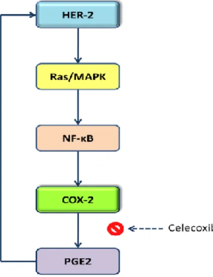
HER-2 induces NF-κB and activates COX-2 synthesis via Ras/MAPK, resulting in prostaglandins synthesis, primarily PGE2 (Benoit et al. 2004; Bassères and Baldwin 2006).
Benoit et al. found that COX-2 expression is related to HER-2 expression via PGE2, and PGE2 inhibition by celecoxib resulted in HER-2 reduced expression (see figure 6) in breast tumors (Benoit et al. 2004).
Figure 6: HER-2 induces COX-2 up-regulation via Ras/MAPK and NF-κB, COX-2 is over-expressed and
converts AA in PG. Synthesized PGE2 stimulates HER-2, suggesting a positive feedback between COX-2 and HER-2. Use of Celecoxib (COX-2 inhibitor) has proof this theory, because it inhibits PGE2 synthesis and as result HER-2 levels are diminished.
COX-2 increased expression by HER-2, establishes a relationship between HER-2 and aromatase, both present in breast tumors with high proliferation rate (Mendelson and Hardy 2006; Subbaramaiah et al. 2006).
10
Aromatase Synthesis
PGE2 stimulates cAMP signaling cascade, PKA and CRE that increase cytochrome protein 450, subfamily XIX (CYP19) gene transcription, with consequent aromatase activation (see figure 7) (Mendelson and Hardy 2006; Subbaramaiah et al. 2006). This enzyme is responsible for estrogen synthesis, Subbaramaiah et al. concluded that estrogen synthesis via the PGE2 is a consequence of COX-2 induction by the HER-2 (Mendelson and Hardy 2006; Subbaramaiah et al. 2006).
Figure 7: PGE2 induces estrogen synthesis. PGE2 via cAMP, PKA and CRE activates aromatase resulting
in estrogen synthesis.
Thomas et al. found that synthesized estrogen also had positive feedback on COX-2 expression (Thomas et al. 2008). Estrogen binds to its receptor and undergoes translocation to the nucleus; this interaction activates serum creatinine response (Scr), which stimulates metalloproteins cascade, allowing epidermal growth factor (EGF) release from heparin and its binding to HER-2, which activates ERK (Thomas et al. 2008). Phospholipase A2 is a ERK substrate and its active form releases phospholipids from cell membranes, AA is formed, with consequent synthesis of PGE2 (see figure 8) (Thomas et al. 2008).
11
Figure 8: COX-2 synthesizes PGE2 which via cAMP induces CYP19 expression, resulting in estrogen
synthesis by aromatase. Estrogen acts through its receptor and undergoes translocation to the nucleus where activates Scr, allowing EGF binding to its receptor. HER-2 acts through NF-κB activating COX-2 expression, and through ERK resulting in AA formation. More PGE2 is produced and as consequence more estrogen is synthesized by aromatase, suggesting a feedback between estrogen and HER-2.
Estrogen binding to its receptor increases inflammatory process and is involved in pathophysiology of various cancers, including breast cancer (Mendelson and Hardy 2006; Subbaramaiah et al. 2006).
Steroid Hormone Receptors
Approximately 75% of breast cancers express estrogen receptors (ER) (Cui et al. 2005). ER is a nuclear transcription regulator that can bind to transcription factors, proteins and estrogen (Cui et al. 2005; Mendelson and Hardy 2006).
There are two isoforms of this receptor that are expressed in mammary gland, estrogen receptors alpha and beta (ERα and ERβ), ERα over-expression is critical to breast cancer development (Cui et al. 2005).
It has long been thought that ER induced progesterone receptor (PR) and that serves as an indicator of ER functional capacity; new studies indicate that PR is an independent predictor risk factor and have protective actions which antagonize inflammatory response pathway (Mendelson and Hardy 2006).
12 PR exists in three isoforms, progesterone receptor-A (PR-A) (94kDa), progesterone receptor-B (PR-B) (110kDa) and progesterone receptor-C (PR-C) (60kDa) (Abdel-Hafiz et al. 2009). PR are proteins with several domains, a DNA binding domain (DBD), N-terminally that activates proximal activation function 1 (AF-1) common to PR-A and PR-B, and a C-terminally that upstream ligand binding domain (LBD) has a nuclear localization region with activation function 2 (AF-2) (Mendelson and Hardy 2006; Abdel-Hafiz et al. 2009).
PR-A and PR-B are equimolar in most tissues, but in breast cancer this balance does not exist, PR-A can block all three PR-B transcriptional activation domains, acting as PR repressor in breast cancer (Mendelson and Hardy 2006).
PR-C isoform is augmented in cancerous breast cells, binds to PR-B reducing its capacity to interact with progesterone response elements in progesterone-responsive genes (Mendelson and Hardy 2006).
Mendelson et al. concludes that NF-κB increased activation, results in PR-C over-expression that together with PR-A and PR-B ablation results in COX-2, HER-2 and aromatase over-expression, suggesting a protective role of RP in breast cells (Mendelson and Hardy 2006).
Progesterone Receptor and Breast Cells
In breast cells PR directly interacts with NF-κB to block its binding to DNA, or inhibits NF-κB activation and translocation to the nucleus via IκBα (Mendelson and Hardy 2006). NF-κB has a major role in COX-2 over-expression, which in turn up-regulate expression of the genes encoding aromatase and HER-2 via PGE2, resulting in estrogen up-regulation (Mendelson and Hardy 2006).
PR impairs NF-κB transactivation of COX-2 via induction of IκBα, these findings suggest that NF-κB inhibition by PR, results in estrogen lower levels and PR serves a crucial role in blocking breast tumor formation and progression (see figure 9) (Mendelson and Hardy 2006; Hardy et al. 2008).
13
Figure 9: COX-2 is expressed in chronic inflammation, converting AA into PGE, leading to aromatase
increase with subsequent increase of ER and PR. PR through NF-κB inhibition results in COX-2 lower expression.
Another suggestion of PR protective role is ER+/PR- receptor tumor subtype that can amplify
HER-2 expression, which results in a worse overall survival (Cui et al. 2005). There are four receptor tumor subtypes: ER+/PR+, ER+/PR-, ER-/PR+ and ER-/PR-, that differs with age,
hormone use and menopause (Cui et al. 2005). ER+/PR- is the most aggressive receptor tumor
subtype, it has high proliferation rates and has higher recurrence than ER+/PR+, this findings
also suggests that PR has a protective role in breast cancer (Arpino et al. 2005).
Based on these studies, which indicate that PR might have a protective role in breast cancer, we speculate that tumors with positive COX-2 expression and positive PRexpression had an overall better prognosis than tumors with positive COX-2 expression and negative PR expression. It was performed an immunohistochemical study to determine a possible relation between COX-2 and PR expression, correlating them with clinicopathologic factors (age, tumor size, histologic grade and axillary node metastasis), which are known to be classical prognostic factors in breast cancer.
14
Chapter 2: Materials and Methods
Patients Selection
Child and Women department of Cova da Beira Medical Center has initiated its Gynecological Oncology activity in 2005 and since then has given assistance to several women with breast cancer.
In 2007 the department had a restructuration and started to work with the current anatomic pathology laboratory, for these reason only clinical cases from 2007 to 2009 were selected. For immunohistochemical study, it was selected 31 clinical cases that meet the follow criteria:
1. Histological invasive ductal carcinoma confirmed by an independent pathologist 2. Tumor with 10 mm or more in longest diameter (in order to material preservation for
further studies)
3. Submitted to radical surgery with axilary assessment 4. No pregnant woman
5. No primary chemotherapy or hormonotherapy 6. No documented distant metastasis
Immunostaining
Sample Pre-treatment
It was selected representative pathologic material from paraffin blocks. From each selected paraffin block were obtained three 3µm sections: one slide was for hematoxylin-eosin staining and the remaining two slides were for COX-2 and PR immunostaining.
Cyclooxygenase-2 and Progesterone Receptor Immunostaining
In histological sections for immunostaining, a dewaxing was performed during 10 minutes with xylene, followed by rehydration in decreasing ethanol grades (absolute, 95% and 70%) and water. To proceed with the antigen retrieval, slides were put into pressure cooker and citrate buffer 0.01 M for 6 minutes at pH 6 and washed with water and buffer solution (K5006, ChemMat™, Dako).
The slides were then incubated with the primary antibody, for COX-2 staining was used clone SP21, rabbit monoclonal antibody (MC-16-240r, CellMarque™) at 1:10 dilution; for PR staining were used clone 16 and clone SAN27 (NCL-L-PGR-AB, Novocastra™) at 1:100 dilution, in both
15 cases the diluent used was S2022 (Dako Real) and incubation with the primary anti-body was made for 25 minutes at room temperature, then washed with buffer solution (K5006, ChemMat ™, Dako).
Incubation with biotinylated secondary antibody was made for 25 minutes, using the bottle A from K5001 kit (Dako Real™), followed by washing with buffer solution (K5006, ChemMat ™, Dako). The next step was to block endogenous peroxidase using bloking-Peroxidase solution (S2023, Dako Real™) for 7 minutes and 30 seconds, followed by washing with buffer solution (K5006, ChemMat ™, Dako).
After these procedures, it was added streptavidin conjugated horseradish peroxidase (HRP) (bottle B, K5001, Dako Real), for 25 minutes followed by washing with buffer solution (K5006, ChemMat ™, Dako), chromogen was added (Dako Real DAB + chromogen, bottle C + HRP substrate buffer bottle D, K5001, Dako Real) at a 20:1000 dilution for 15 minutes with washes at every 5 minutes, this was followed by washing with a buffer solution (K5006, ChemMat ™, Dako). The counterstaining was done with Mayer's hematoxylin for 1 minutes and washed with a buffer solution (K5006, ChemMat ™, Dako), dehydrated in increasing ethanol grades (75%, 90%, absolute) and xylene. Slides were preserved in synthetic mounting medium, and were analyzed with an optical microscope (x400), using 50 fields for slide.
For COX-2 was considered cytoplasmatic immunostaining and to PR was recorded nuclear immunostaining.
The intensity of immunostaining was recorded as: negative (-), moderate (+) or high (++). Positivity of immunostaining was recorded as percentage of the cells that stained. It was counted all cells in an optical high power field considered representative of all slide, in a minimum of 500 cells. For statically analysis we considered expression in less than 25% of the cell as a negative result; expression from 25 to 50% as a light expression; from 50 to 75% as moderate expression and 75 to 100% as a high expression. All cases with positive expression had a moderate or high intensity.
For comparative statistical analysis we considered as positive immunostaining cases with light, moderate or high expression.
16
Statistical Analysis
Was performed a comparative study between patients clinical factors (age, tumor size, differentiation grade and ganglion metastasis) and COX-2 and PR expression.
Statistical analysis was performed using SPSS, version 17. Mann-Whitney and Chi-Squared tests were used for the analysis, considering a statistical significance when P-value was < 0.05.
17
Chapter 3: Results Analysis
Descriptive Statistical Analysis
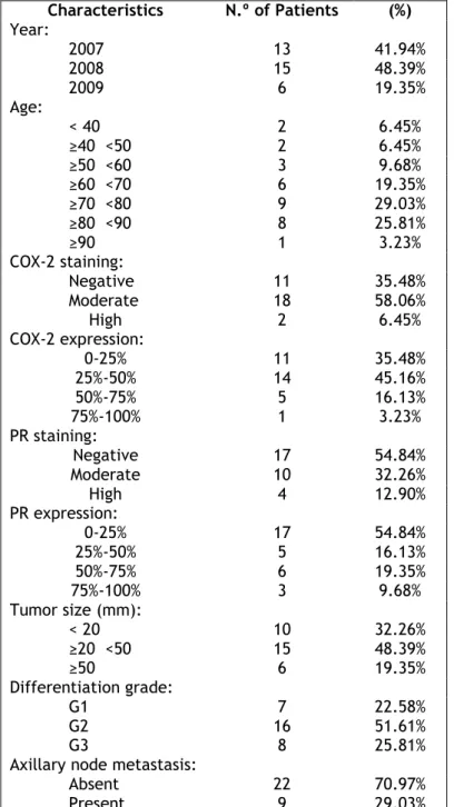
The analyzed clinical cases were between the year 2007 and 2009 and the age of patients varied from 39 to 91 years old. Most of the tumors expressed COX-2 (64.52%) and 45.16% of the tumors expressed PR. It was also taking into account the differentiation grade and axillary node metastasis, and we verified that twenty four of tumors had a high (G2 or G3) differentiation grade and only 29.03% of women had axillary node metastasis (see table 1).
Table 1: Patient characteristics: Year, Age, COX-2 expression, PR expression, Tumor Size,
Differentiation Grade and Axillary Node Metastasis.
Characteristics N.º of Patients (%) Year: 2007 2008 2009 13 41.94% 15 48.39% 6 19.35% Age: < 40 ≥40 <50 ≥50 <60 ≥60 <70 ≥70 <80 ≥80 <90 ≥90 2 6.45% 2 6.45% 3 9.68% 6 19.35% 9 29.03% 8 25.81% 1 3.23% COX-2 staining: Negative Moderate High 11 35.48% 18 58.06% 2 6.45% COX-2 expression: 0-25% 25%-50% 50%-75% 75%-100% 11 35.48% 14 45.16% 5 16.13% 1 3.23% PR staining: Negative Moderate High 17 54.84% 10 32.26% 4 12.90% PR expression: 0-25% 25%-50% 50%-75% 75%-100% 17 54.84% 5 16.13% 6 19.35% 3 9.68% Tumor size (mm): < 20 ≥20 <50 ≥50 10 32.26% 15 48.39% 6 19.35% Differentiation grade: G1 G2 G3 7 22.58% 16 51.61% 8 25.81% Axillary node metastasis:
Absent Present
22 70.97% 9 29.03%
18
Immunohistochemestry Analysis
Cyclooxygenase-2 Immunoreactivity
COX-2 expression was considered positive by semiquantitative scoring in 20 of 31 cases studied (64.52%), all 20 cases had at least 50% or more of COX-2 expression and all cases had a moderate or strong staining intensity. Immunostaining intensity was considered negative in 11 cases, moderate in 18 cases and strong in 2 cases (see annex 1). In figure 10 it can be seen a moderate immunostaining with moderate expression.
Figure 10: Immunohistochemical localization of COX-2 in ductal carcinoma (400 x magnifications).
There is cytoplasmatic staining, with COX-2 light expression.
Progesterone Receptor Immunoreactivity
PR expression was detected by semiquantitative scoring in 14 of 31 cases in study (45.16%), all 14 cases had at least 50% of PR expression and all cases had a moderate or strong staining intensity. Immunostaining intensity was negative in 17 cases, moderate in 10 cases and strong in 4 cases. In figure 11 it can be seen a strong immunostaing for PR with strong expression.
19
Figure 11: Immunohistochemical localization of PR in ductal carcinoma (400x magnification). There is
nuclear strong staining, with PR high expression.
Comparative Statistical analysis
Correlation between Clinicopathologic Factors and Cyclooxygenase-2
Expression
As described previously, 20 (64.52%) cases in study were positive for COX-2 expression and 35.48% were negative.
When COX-2 expression was correlated with age, it was verified that positive COX-2 expression group is more heterogeneous, have a higher standard deviation (16.027 years) than negative cases (8.251 years) (see annex 2).
Minimum age and maximum age are respectively 39 and 87 years old for positive COX-2 expression group, and 59 and 91 years, for negative COX-2 expression group. Breast cancers positive for COX-2 expression also have a non-statistical significant and lower mean (65.65 years) than breast cancers negative for COX-2 expression (75.45 years). Positive cases for COX-2 expression seem to appear in earlier ages than negative cases (see figure 12).
20 However, there is no statistic significance (p = 0.121) when COX-2 expression is correlated with age.
Figure 12: Correlation between COX-2 expression and age. For COX-2 negative expression the minimum
value is 59 years and the maximum value is 91 years, with a mean of 75.45 years and a standard deviation of 8.251 years. For COX-2 positive expression the minimum value is 39 years and the maximum value is 87 years, with a mean of 65.65 years and a standard deviation of 16.027 years.
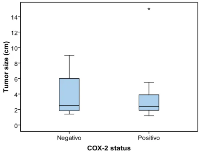
No statistic significance was found between tumor size and COX-2 positivity (p = 0.725). Minimum value for tumor size was the same in both positive and negative COX-2 groups (1 cm). The tumor size mean values for both groups are very similar, 3.95 cm for negative and 3.36 cm for positive COX-2 group (see annex 3). Standard deviation values were also closest, 2.709 cm and 2.952 cm for negative and positive COX-2 group (see figure 13).
Figure 13: Correlation between COX-2 expression and tumor size. For COX-2 negative expression the
minimum value is 1 cm and the maximum value is 9 cm, with a mean of 3.95 cm and a standard deviation of 2.709 cm. For COX-2 positive expression the minimum value is 1cm and the maximum value is 15 cm, with a mean of 3.36 cm and a standard deviation of 2.952 cm.
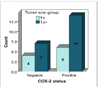
21 Even considering 2 groups of breast cancer (see figure 14), one with better prognosis tumors, with less than 2 cm (T1) and other with worst prognostic disease, with size of 2 or more cm, it was not found statical significance between tumor size and COX-2 imunopositivity (p = 0.510) (see annex 4).
Figure 14: Correlation between COX-2 expression and age, considering 2 groups. For COX-2 negative
expression there are 4 tumors with less than 2 cm and 7 tumors with 2cm or more. For COX-2 positive expression there are 6 cases with less than 2 cm and 14 cases with 2 cm or more.
Tumor histologic grade was considered as low risk for G1 tumors and high risk for G2 and G3 tumors. Of G1 histologic grade was recorded in 22.6% cases and 77.4% was classified as G2 and G3 histologic grade (see annex 5). Six cases (30.0%) with G1 histologic grade and 14 cases (70.0%) with G2 and G3 histologic grade were reported as positive for COX-2 immunostaining. Negative COX-2 expression group was found in 1 case (9.1%) with G1 histologic grade and in 10 cases (90.9%) with high G2 or G3 histologic grade (see figure 15).
There was not found any statistical significance correlation between COX-2 imuno-positivity and tumor hitological grade (p = 0.192).
22
Figure 15: Correlation between COX-2 expression and differentiation grade. For COX-2 negative
expression there is 1 tumor with low differentiation grade and 10 tumors with G2 or G3 histologic grade. For COX-2 positive expression there are 6 tumors with low differentiation grade and 14 tumors with G2 or G3 histologic grade.
Axillary node metastasis were documented in 9 of 31 cases (29.0%) the percentage of cases positive and negative for axillary node metastasis (54.5%) were very similar (45.5%) in those negative COX-2 tumors (see annex 6). In positive COX-2 cases the results are much different; there were 16 cases with no axillary node metastasis (80%) and 4 cases with axillary node metastasis (20%) (see figure 16). However, statistical analysis have not showed significance between COX-2 positivity and axillary node metastasis (p = 0.140).
Figure 16: Correlation between COX-2 expression and axillary node metastasis. In 16 of the 20 women
whose tumors overexpressed COX-2 there was no axillary node metastasis and in 4 of them there was axillary node metastasis. In tumors with negative COX-2 expression, 6 of 11 women had no axillary node metastasis and 5 of them had axillary node metastasis.
23
Correlation between Clinicopathologic Factors and Progesterone Receptor
Expression
Fourteen cases were considered imunopositive for PR and 17 tumors were negative.
The age of the patients was not statistically correlated with positivity for PR (see annex 7). The minimum age of the patients with tumors negative and positive to PR was the same, however, the group of the patients with tumors positive for PR expression, seemed more heterogeneous in considering the age, with a higher mean and higher standard deviation age (see figure 17).
Figure 17: Correlation between PR expression and age. For PR negative expression the minimum value
is 39 years and the maximum value is 81 years, with a mean of 67.59 years and a standard deviation of 11.609 years. For PR positive expression the minimum value is 39 years and the maximum value is 91 years, with a mean of 71 years and a standard deviation of 17.537 years.
Negative PR group had a higher mean (4.60 cm) and higher standard deviation (3.471 cm) than positive PR expression group which has a mean value of 2.32 cm and a standard deviation value of 0.816 cm (see figure 18). Negative PR expression group have tumors with higher size than positive PR expression group and is a more heterogeneous group than positive PR expression group.
The PR positive tumors were statistically significant smaller then PR negative tumors (see annex 8). There is statistical significance (p = 0.032).
24
Figure 18: Correlation between PR expression and tumor size. For PR negative expression the minimum
value is 1 cm and the maximum value is 15 cm, with a mean of 4.60 cm and a standard deviation of 3.471 cm. For PR positive expression the minimum value is 1 cm and the maximum value is 4 cm, with a mean of 2.32 cm and a standard deviation of 0.816 cm.
As reported earlier tumor differentiation grade was considered as low risk (G1) or high risk (G2 or G3) and it was correlated with PR expression groups.
In negative PR group 15 of 17 cases (88.2%) had G2 or G3 differentiation grade and in positive PR group 9 of 14 cases (64.3%) had G2 and G3 histologic grade (see figure 19).
When PR immunostaininig is negative it seems that tumors tend to be a higher risk histologic grade, however, there is no statistical significance (p = 0.124) (see annex 9).
Figure 19: Correlation between PR expression and differentiation grade. For PR negative expression
there are 2 tumors with low differentiation grade and 15 tumors with G2 or G3 histologic grade. For PR positive expression there are 5 tumors with low differentiation grade and 9 tumors with G2 or G3 histologic grade.
25 In tumors with immunopositivity for PR there were 13 of 14 (92.9%) cases without axillary nodes metastasis (see figure 20). PR negative immunostaining was strongly statistically associated with presence of axillary node metastasis (p = 0.018) (see annex 10).
Figure 20: Correlation between PR expression and axillary node metastasis. In 13 of women whose
tumors expressed PR there was no axillary node metastasis and in 1 of them there was axillary node metastasis. In tumors with negative PR expression, 9 women had no axillary node metastasis and 8 of them had axillary node metastasis.
Correlation between Progesterone Receptor Expression and
Clinicopathologic Factors according to Cyclooxygenase-2 Expression
In order to evaluate any association between PR and COX-2 immunoexpression it was constituted 4 groups: positive COX-2 expression with positive PR expression (COX-2+/PR+),
positive COX-2 expression with negative PR expression (COX-2+/PR-), negative COX-2
expression with positive PR expression (COX-2-/PR+) and negative COX-2 expression with
negative PR expression (COX-2-/PR-).
COX-2+/PR+ group is constituted for 11 of 31 cases (35.48%), COX-2+/PR- group is constituted
for 9 of 31 cases (29.03%), COX-2-/PR+ group is constituted for 3 of 31 cases (9.68%) and
COX-2-/PR- group is constituted for 8 of 31 cases (25.81%) (see table 2).
Table 2: Correlation between COX-2 and PR expression.
COX-2 negative expression COX-2 positive expression Total PR negative expression n = 8 (25.81%) n = 9 (29.03%) n = 17 (54.84%) PR positive expression n = 3 (9.68%) n = 11 (35.48%) n = 14 (45.16%) Total n = 11 (35.49%) n = 20 (64.51%) 31 (100%)
26 As it can be seen in figure 21 a) and b), in patient age analysis, the group COX-2+/PR+ is the
more heterogeneous and showed the higher value. Patients in the group COX-2-/PR+ seems to
be the more aged with mean age of 83 years (see annex 11).
Figure 21: Correlation between PR expression and age according to COX-2 expression. a) The COX-2-/PR
-group is more heterogeneous than COX-2-/PR+ group, with a standard deviation of 6.989 years. COX-2
-/PR+ has a higher mean and median than COX-2-/PR- group. b) COX-2+/PR+ group has a higher mean and
standard deviation than COX-2+/PR- group
Evaluating tumor size, the COX-2-/PR- group has shown to be the more heterogeneous (see
figure 22a), ranging the size from 10 mm to 90 mm, and the group COX-2-/PR+ the more
homogeneous (see annex 12). Comparing COX-2 positive expression group it was found that the size of the COX-2+/PR+ were statistically smaller that COX-2+/PR- group (see figure 22b),
there is statistical significance (p = 0.043).
Figure 22: Correlation between PR expression and tumor size according to COX-2 expression. a) The
COX-2-/PR- group is more heterogeneous and has higher mean than COX-2-/PR+ group, with a standard
deviation of 2.941 cm and a mean of 4.60 cm. b) COX-2+/PR+ group has a lower mean and standard
deviation than COX-2+/PR- group.
a)
b)
27 There are 10 cases with high differentiation grade, 8 of them belong to COX-2-/PR- group and
in this group there is no low differentiation grade, the number of high differentiation grade represents 100% (see figure 23 a and b). For COX-2-/PR+ group there are 2 cases with high
differentiation grade and 1 case with low differentiation grade, which represents 66.7% and 33.3%, respectively (see annex 13).
For COX-2+/RP- group there are 2 of 9 cases (22.2%) with low differentiation grade and 7 of 9
cases (77.8%) with high differentiation grade. Similar results were obtained for COX-2+/PR+
group, 4 of 11 cases (26.4%) have low differentiation grade and 7 of 11 cases (63.6%) have high differentiation grade.
All groups have more cases of high differentiation grade, and there is no statistical significance for any group. Negative COX-2 expression group has a P-value = 0.273 and positive COX-2 expression group has a P-value = 0.426.
Figure 23: Correlation between PR expression and differentiation grade according to COX-2 expression.
a) In COX-2-/PR- group all tumors have a high differentiation grade and in COX-2-/PR+ group there is only
one case with low differentiation grade. b) For COX-2 positive expression group tumors with PR positive expression have higher number of cases with low differentiation grade (G1).
For negative COX-2 expression group 6 of 11 (54.55%) women had no axillary node metastasis (see annex 14).
In COX-2-/PR- group 50% of women had axillary node metastasis and in COX-2-/PR+ group there
was only 1 woman with axillary node metastasis (33.3%) (see figure 24a). For negative COX-2 expression group there was not found statistical significance (P-value = 0.576).
For positive COX-2 expression group there are 16 of 20 women (80%) without axillary node metastasis and 4 of 20 women (20%) with axillary node metastasis (see figure 24b).
28
Figure 24: Correlation between PR expression and axillary node metastasis according to COX-2
expression. a) In COX-2-/PR- there is the same number of cases with and without axillary node
metastasis, for PR negative expression there are more cases without axillary node metastasis. b) COX-2+/PR- has a higher number of cases with axillary node metastasis than COX-2+/PR+ group, which has only
cases without axillary node metastasis.
There were nine women whose tumors were COX-2+/PR-, and 5 (55.6%) of them had no
axillary node metastasis. In COX-2+/PR+ group all women (100%) had no axillary node
metastasis. These groups are statistical related (P-value = 0.026).
29
Chapter 4: Discussion
COX-2 is the induced cyclooxygenase isoform, is expressed in most of the tissues and commonly expressed in more than 50% of invasive breast cancers (Haffty et al. 2008; Singh-Ranger et al. 2008). COX-2 is involved in chronic inflammation and is responsible for AA conversion into prostaglandins, which are associated with increased aromatase that in turn, results in high local ER (Mendelson and Hardy 2006; Schetter et al. 2010).
ER upregulation is associated with breast cancer progression and PR has been considered has an estrogen-induced target gene (Cui et al. 2005). However, Hardy et al. found that PR expression is an independent predictor of breast cancer diagnosis (Hardy et al. 2008). PR is a COX-2 and aromatase inhibitor and plays an important anti-inflammatory and protective role in breast cancer (Hardy et al. 2008).
Based on these findings we attempted to determine a relation between COX-2 expression, PR expression and clinicopathologic factors (age, tumor size, histologic grade and axillary node metastasis), which are known to be prognostic factors.
As previously reported, our study was performed in 31 women with invasive ductal breast cancer and COX-2 expression was detected by semiquantitative scoring in 64.52% of tumors. These results are consistent with those of Haffty et al., who found COX-2 expression in 58% of women (n = 504) with breast cancer (Haffty et al. 2008).
When we performed a comparative study between COX-2 expression and age, we observed that the tumors with COX-2 expression had a tendency for incidence in younger ages, once all women with 50 years or less (n=5) had COX-2 expression. However, that results have shown no statistical significance (p = 0.121) probably due to the reduced number of cases. Haffty et al. found similar results: nearly 70% of women with less than 40 years of age expressed COX-2 and these results are consistent with those of Ristmäki et al. and Denkert et al. studies (Ristimäki et al. 2002; Denkert et al. 2003; Haffty et al. 2008).
Previous studies have also reported that tumors with COX-2 expression were higher than 2 cm (Ristimäki et al. 2002; Denkert et al. 2003; Haffty et al. 2008), however, in our study, COX-2 expression and tumor size were not statistical related (p > 0.005), probably due to the low number of cases in the study.
Correlating COX-2 expression with histologic grade and with axillary node metastasis we found no statistical association between them. The group of tumors that expressed COX-2 and the group of tumors that has no COX-2 expression had similar number of cases with high histologic grade. However, other researchers concluded that COX-2 expression is associated with high histologic grade (Ristimäki et al. 2002; Denkert et al. 2003). In our study axillary node