Faculdade de Farmácia
Creatine Protects Against Rotenone-induced Cell
Death of Cerebellar Granule Neurons
Sofia Isabel Almeida Fortalezas
Mestrado em Ciências Biofarmacêuticas
Especialização em Neurociências
iii
Universidade de Lisboa
Faculdade de Farmácia
Creatine Protects Against Rotenone-induced Cell
Death of Cerebellar Granule Neurons
Sofia Isabel Almeida Fortalezas
Tese de Mestrado orientada por:
Professor Carlos Gutiérrez Merino
Professora Cecília Rodrigues
Mestrado em Ciências Biofarmacêuticas
iv
The studies performed in this thesis were carried out in the Oxidative Stress and Bioenergetics in Brain and Muscle research group (CCV008), at the Department of Biochemistry and Molecular Biology and Genetics, Faculty of Sciences, University of Extremadura, Spain, under the supervision of Carlos Gutiérrez Merino, Ph.D.
This work has been funded by ERASMUS fellowship to Sofia Fortalezas (29206-IC-1-2007-1-PT-ERASMUS-EUCX-1, Registo nº 38/SMP/2011) and grant GR10092 of Junta de Extremadura with FEDER cofinanciation.
vi
Acknowledgements
En primer lugar agradezco al Profesor Carlos Gutiérrez Merino por haberme dado la oportunidad en realizar este trabajo en su laboratorio; por la magnifica orientación, transmitiendo a cada día una nueva enseñanza. Gracias también por me confiar elfuturo desarrollo deltemade este trabajo,lo que me permitiráseguir con esta aventura.
À Professora Cecília Rodrigues, por ser a minha tutora interna e por me ter permitido realizar este trabalho além-fronteiras. Agradeço ainda toda a disponibilidade, atenção e ajuda durante este último ano.
A mis compañeros de lab, Dorinda, María, Alex y Ricardo les agradezco la amable y acogedora bienvenida, las enseñanzas transmitidas y la ayuda diaria al hacer este trabajo. Sin ellos no hubiera sido posible. Agradezco aún la amistad y el compañerismo.
Aos meus “amiguinhos”, pilares da minha vida académica que continuam hoje e seguirão sempre, não poderia nunca faltar o meu grande Obrigada.
Aos meus pais por empréstimo agradeço o apoio e o orgulho que nutrem por mim como se de uma verdadeira filha me tratasse.
Ao meu Manel agradeço o orgulho demonstrado, o incentivo, o apoio, a amizade e o amor. Pedro és o meu catalisador, obrigada pela exigência que faz de mim a cada dia uma pessoa melhor.
À minha família. Ao meu mano agradeço-lhe por ser parte da minha vida, por toda a amizade e orgulho. E por fim mas não menos importantes, pelo contrário, agradeço profundamente aos meus pais, por serem os meus melhores amigos, por me apoiarem em todas as minhas decisões e por me proporcionarem mais esta etapa da minha vida académica. Vai para vocês o meu maior Obrigada!
viii
Abstract
Parkinson´s Disease (PD) is the second most prevalent neurodegenerative brain disorder worldwide. Nevertheless, there is lack of certainty on the pathophysiology of the neurodegenerative mechanisms underlying PD. Several neurotoxins, rotenone among them, have been shown to induce parkinsonism-like brain degeneration and are widely used in cellular and animal models of PD.
In spite of an extensive association of PD with dopaminergic neuron degeneration of the substantia nigra pars compacta, other brain areas like the cerebellum have been more recently implicated in the pathology of the disease. Therefore, we used a rotenone/cerebellar granule neurons (CGN) model to study not only the potential of creatine (as ergogenic compound), epicatechin and kaempferol (as antioxidant compounds) to afford neuroprotection against rotenone neurotoxicity but also to better understand the cellular mechanisms underlying this neurotoxicity.
Our results revealed a strong protection by creatine against rotenone-induced CGN death, while kaempferol did not afford a significant protection and epicatechin elicited at most a very weak protection. On the contrary, kaempferol also antagonized the protective effect of creatine. These results lend support to the potential use of creatine in PD therapeutics, and alert for the avoidance of the consumption of foods or infusions with high content in kaempferol.
Furthermore, we noted that rotenone triggered an energetic failure in CGN as a primary event, supported not only by the protection afforded by creatine but also by a deregulation in calcium homeostasis thought voltage operate calcium channels type L and N-Methyl-D-aspartate receptors stimulation and store-operated calcim entry inhibition, by the elevation of AMP-kinase active levels and by mitochondrial membrane depolarization. Rotenone also promoted an enhanced production of reactive oxygen species and a weak nitrosative stress, but only as a later event in the development of CGN death.
In conclusion, our results support a role for creatine in affording neuroprotection against rotenone neurotoxicity, through a mechanim that prevent an energetic failure.
Keywords: Parkinson´s disease; cerebellar granule neurons; rotenone; creatine;
ix
Resumo
A doença de Parkinson é a segunda doença neurodegenerativa mais prevalente a nível mundial. Apesar disso, são ainda pouco conhecidos os mecanismos neurodegenerativos que estão por detrás desta doença. Varias neurotoxinas, nas quais se inclui a rotenona, têm vindo a ser demonstradas como indutoras de degenerescência cerebral de tipo-Parkinsonismo e são, por isso, vastamente utilizadas em modelos celulares e animais da doença de Parkinson.
Apesar da extensiva associação entra a doença de Parkinson e a degenerescência dos neurónios dopaminérgicos da substantia nigra pars compacta, mais recentemente, outras áreas cerebrais, nomeadamente o cerebelo, têm sido implicadas na patogénese da doença. Portanto, neste trabalho, foi utilizado um modelo celular de neurónios granulares do cerebelo expostos à neurotoxina rotenona. Este modelo foi utilizado, não só para estudar a capacidade da creatina (como composto ergogénico) e de epicatequina e kaempferol (como antioxidantes) em proteger contra a neurotoxicidade despoletada pela rotenona, mas também para melhor entender os mecanismos subjacentes a esta neurotoxicidade.
Os resultados obtidos revelaram que a creatina apresenta uma elevada protecção contra a morte celular, induzida pela rotenona nos neurónios granulares do cerebelo, enquanto o kaempferol não ofereceu qualquer protecção e a epicatequina, por sua vez, promoveu uma protecção demasiado fraca. Pelo contrário, o kaempferol demostrou um efeito antagónico relativamente à creatina. Os resultados suportam, assim, o potencial uso da creatina na terapêutica da doença de Parkinson e alertam para que se evite o consumo de comidas e infusões com um elevado conteúdo em kaempferol nesta mesma terapêutica.
Para além disso, os nossos resultados revelaram que a rotenona como evento primário conduz a uma falência energética nos neurónios granulares do cerebelo. Esta conclusão é suportada, não só pela protecção exercida pela creatina, mas também pela observação de uma desregulação nos níveis citosólicos de cálcio através de uma estimulação dos canais de cálcio do tipo L operados por voltagem e dos receptores N-Metil-D-aspartato e pela inibição da entrada capacitativa de cálcio; pelo aumento dos níveis da AMP-quinase activa e ainda pela despolarização da membrana mitocondrial.
x
A rotenona promoveu, por fim, a produção de espécies reactivas de oxigénio e um stresse nitrosativo fraco, no decurso da morte dos neurónios granulares do cerebelo.
Em suma, os resultados suportam a utilização da creatina como composto neuroprotector contra a neurotoxicidade exercida pela rotenona, através da prevenção da ocorrência de uma falência energética.
Palavras-chave: Doença de Parkinson; neurónios granulares do cerebelo; rotenona;
xii
Table of contents
Acknowledgements………... iv
Abstract………... viii
Resumo………... ix
Table of contents………...………... xii
Abbreviations, acronyms and symbols………... xiv
I. Introduction………. 2
1.1. Parkinson´s disease... 3
1.1.1. The involvement of cerebellum in PD………... 4
1.1.2. Neutoxin models of PD……….……….. 6
1.1.2.1. Cerebellar granule neurons as Parkinsonism model…... 6
1.1.3. Cell death in PD... 8
1.2. Brain bioenergetics alterations in PD... 9
1.2.1. Creatine Kinase... 10
1.2.1.1. Creatine... 12
1.2.2. AMP-Kinase……… 13
1.3. Oxidative stress in the brain... 14
1.3.1. Oxidative stress in Parkinson´s Disease... 14
1.3.1.1. Flavonoids in PD: Kaempferol and Epicatechin... 15
1.4. Calcium homeostasis in brain... 16
1.4.1. Alterations of calcium homeostasis in PD... 17
1.5. Aims……… 20
II. Material and Methods……… 23
2.1. Equipment and reagents………... 24
2.2. Buffers and solutions……….... 25
2.3. Biological Material………... 26
2.4. Cell cultures……….. 26
2.5. Viability assays………. 26
2.6. Protein quantification and determination………. 27
2.7. Cell death pathways……….. 28
2.8. Measurement of CGN mitochondrial membrane potential………... 30
2.9. Reactive oxygen and nitrogen species…….………. 30
2.10. Determination of the intracellular free Ca2+ concentration in CGN and cell bioenergetics markers………... 31
2.11. Statistical analysis………... 34
III. Results………... 36
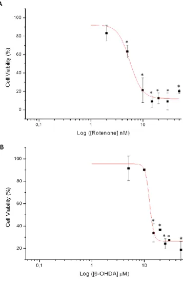
3.1. Sustained exposure (12 h) of CGN to the neurotoxins rotenone and 6-OHDA………... 37
3.1.1. Rotenone and 6-OHDA induce cell death in CGN………... 37
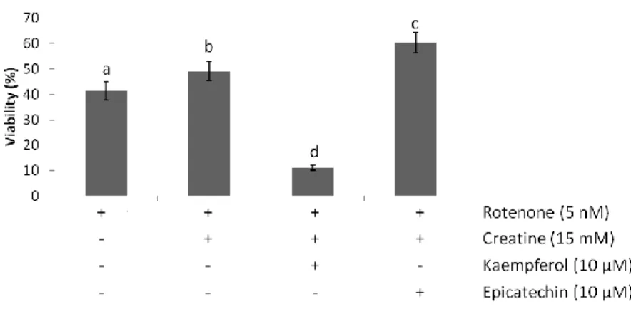
3.1.2. Creatine, but not kaempferol and epicatechin, protects against rotenone-induced cell death in CGN………... 38
3.1.2.1. Creatine in combination with epicatechin present a weak synergistic effect against rotenone-induced cell death in CGN, while kaempferol antagonizes the protective effect of creatine….………... 39
3.1.3. Cathepsin D activation in rotenone-induced cell death in CGN………. 40
3.1.4. Creatine completely attenuates rotenone-induced cathepsin D activation ... 41
3.1.5. Creatine protects against mitochondrial membrane depolarization caused by rotenone in CGN………. 42
xiii
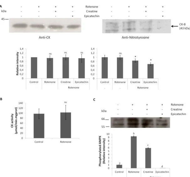
3.1.6. Rotenone causes generalized oxidative stress but only a weak nitrosative
stress in CGN……… 44
3.1.7. Creatine protects against AMPK activation induced by rotenone in CGN……….. 47
3.2. Acute exposure (30 min) of CGN to rotenone……… 50
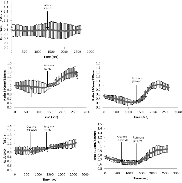
3.2.1. Creatine attenuates Ca2+ homeostasis deregulation promoted by rotenone in CGN………... 50
3.2.2. Epicatechin, but not kaempferol, attenuated Ca2+ homeostasis deregulation promoted by rotenone, and, both flavonoids antagonize the protective effect of creatine………... 52
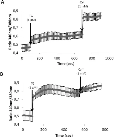
3.2.3. Rotenone leads to Ca2+ homeostasis deregulation through functional alterations of several Ca2+ transport systems of the plasma membrane and ER………... 54
3.2.4. Acute (30 min) exposure of CGN to rotenone did not lead to oxidative stress neither to mitochondrial membrane depolarization………... 58
IV. Discussion………...……… 62
V. Conclusions and Future work………... 69
xiv
Abbreviations, acronyms and symbols
[Ca2+]i Intracellular calcium concentration
∆ψm Mitochondrial membrane potential
6-OHDA 6-hydroxydopamine
ADP Adenosine diphosphate
AMC 7-amino-4-methyl-coumarin
AMPK Adenosine monophosphate-protein kinase
ATP Adenosine triphosphate
BB-CK Cytosolic brain-type creatine kinase
Ca2+ Calcium
CGN Cerebellar granule neurons
CK Creatine kinase
CPA Cyclopiazonic acid
CTC Cerebellothalamocortical
CyCK Cytosolic form of creatine kinase
DA Dopaminergic
DCF 2',7'-Dichlorofluorescein
DIV Days in vitro
DMEM Dulbecco’s modified Eagle’s medium DMSO Dimethyl sulfoxide
ER Endoplasmic reticulum
FBS Fetal bovine serum
FCCP Carbonyl cyanide-4-(trifluoromethoxy)phenylhydrazone
fMRI Functional magnetic resonance imaging
fura-2 AM Fura-2-acetoxymethyl ester
GSH Glutathione
H2DCFDA 2’,7’-Dichlorodihydrofluorescein diacetate
HD Huntington’s disease
L-DOPA Levodopa
L-VOCC Voltage operate calcium channel type L
MCB Monochlorobimane
MPP+ 1-methyl-4-phenylpyridinium
xv
mPTP Mitochondrial permeability transition pore
MtCK Mitochondrial creatine kinase
MTT 3-(4,5-dimethyl-thiazole-2yl)-2,5-diphenyltetrazolium bromide
Na+ Sodium
Na+/K+-ATPase Sodium potassium ATPase
NADP+ Oxidized nicotinamide adenine dinucleotide phosphate
NADPH Reduced nicotinamide adenine dinucleotide phosphate
NCX Na+/Ca2+ exchanger
NMDA N-Methyl-D-aspartate
PBS Phosphate buffered saline
PCD Programmed cell death
PCr Phosphocreatine
PD Parkinson’s disease
PET Positron emission tomography
PI Propidium iodide
PMCA Plasma membrane Ca2+ ATPase
RNS Reactive nitrogen species
ROS Reactive oxygen species
SDS Sodium dodecyl sulfate
SDS-PAGE Sodium dodecyl sulfate-polyacrylamide gel electrophoresis
SERCA Ca2+-ATPase of sarco-endoplasmic reticulum
sMtCK Mitochondrial sarcomeric muscle form of creatine kinase
SNc Substantia nigra pars compacta
SOCE Store-operated calcim entry
SPECT Single-photon emission computed tomography
TG Thapsigargin
TMRE Tetramethylrhodamine, ethyl ester
TMS Transcranial magnetic stimulation
TRPC-1 Transient receptor potential C1
uMtCK Mitochondrial ubiquitous brain form of creatine kinase
VOCC Voltage-operated calcium channels
2
I. Introduction
3
1.1. Parkinson´s disease
Parkinson’s disease (PD) is a neurodegenerative disorder characterized, in part, by motor disturbances, including tremor, rigidity, bradykinesia, postural instability, and rest tremor originating from loss of dopaminergic (DA) neurons of the substantia nigra
pars compacta (SNc) (Dauer and Przedborski, 2003). In the later stages of the disease
autonomic and sensorimotor dysfunction, cognitive decline, depression and sleep disturbances also occur and become clinically relevant (Marsden, 1990). Non-motor functional deficits often precede the major motor symptoms by a number of years and it has been suggested that they are indicative of neurodegeneration that originates in the brain stem and progresses throughout the brain (Braak et al., 2003).
PD is the second most prevalent neurodegenerative brain disorder, affecting 1 to 2% of the population above 65 years of age and its prevalence increases to approximately 4% in individuals above 85 years of age (Bekris et al., 2010; de Rijk et al., 2000).
PD pathology is not restricted to the DA system; degeneration of cholinergic, serotinergic, noradrenergic, peptidergic and DA brainstem nuclei, together with the presence of proteinaceous intraneuronal inclusions in the soma or dendrites (termed Lewy bodies and Lewy neuritis respectively) of neurons in the central, autonomic and enteric nervous system (Braak et al., 2003; Braak et al., 2006; Braak et al., 2007; Hornykiewicz, 1975; Jellinger, 1991; Klos et al., 2006) have been implicated in disease´s pathology. In fact, in the last decades the traditional focus on neurons has been changed. Indeed, it is increasingly recognized that degenerating neurons in PD, such as DA neurons of the nigrostriatal pathway, do not survive after isolation. These neurons receive a variety of afferents and are surrounded by a large number of non-dopaminergic neurons like GABAergic and cholinergic neurons and non-neuronal cells such as astrocytes and microglia. Thus, it is the current belief that the neurodegeneration in PD occurs in response to a mixture of deleterious mechanisms taking place both inside and outside of degenerating neurons.
While a fraction of PD occurrence is related to mutations in genes, over 90% of PD is sporadic, that is, it occurs in the absence of any obvious genetic linkage. Many evidences suggest that there is not much difference between the sporadic and the rare familial forms of PD. Specific mutations in nuclear genes encoding α-synuclein (α-syn), DJ-1, LRRK2, PINK1, and parkin as well as within the mtDNA were identified in familial forms of PD, giving the possibility, by manipulating their expression levels in
4
cellular and animal models, to investigate their physiological function and early pathogenic changes that may lead to neurodegeneration (reviewed in (Cali et al., 2012)).
The most significant pathological features of PD are mitochondrial dysfunction, oxidative stress (Dexter et al., 1994; Jenner and Olanow, 1998), altered protein handling, and inflammatory response (Hirsch et al., 1998; McGeer et al., 1988a; McGeer et al., 1988b), which are considered to lead to cell dysfunction and death mainly by apoptosis or autophagy (Schapira and Jenner, 2011).
1.1.1.
The involvement of cerebellum in PD
As mentioned before, the pathology of PD is not restricted to DA degeneration. Recent studies have implicated other cerebral zones and because of the relevance of this work we highlight the cerebellum.
The cerebellum is a structure located in the posterior fossa of the skull, dorsal to the pons and substantia nigra (Figure 1.1). Although the cerebellum accounts for approximately 10% of the brain’s volume, it contains over 50% of the total number of neurons in the brain (Larsell, 1947). The cerebellum is involved in several functions related to movement such as maintenance of balance and posture; coordination of voluntary movements; motor learning and in certain cognitive functions, such as language. Although motor commands are not initiated in the cerebellum; rather, the cerebellum modifies the motor commands of the descending pathways to make movements more adaptive and accurate (Knierim, 2012).
Recently, the application of medical techniques such as transcranial magnetic stimulation (TMS), single-photon emission computed tomography (SPECT), positron emission tomography (PET) and functional magnetic resonance imaging (fMRI) have demonstrated the occurrence of significative alterations in the cerebellum of PD and Parkinsonism patients (Brockmann et al., 2012; Cao et al., 2011; Kimura et al., 2011; Koch et al., 2009; Ni et al., 2010; Wu et al., 2009a; Wu et al., 2009b).
Ni et al, (2012) applying TMS in the cerebellum verified decreased excitability of the cerebellothalamocortical (CTC) pathway in PD. The authors concluded that the CTC pathway is involved in the generation or transmission of postural tremor in PD.
5
Figure 1.1: Schematic representation of specific anatomy areas of the brain.
Other authors (Kimura et al., 2011) compared the regional cerebral blood flow of patients with different types of Parkinsonism with PD patients and controls. They observed a decreased regional cerebral blood flow in the cingulate gyrus and thalamus in progressive supranuclear palsy patients, whereas Parkinson variant of multiple system atrophy showed decreased regional cerebral blood flow in the cerebellum. These findings suggest that parkinsonian disorders show a distinct SPECT pattern in the frontal cortex, thalamus, and cerebellum. Therefore these measurements may be helpful in screening for the differential diagnosis of parkinsonian syndrome.
Since resting state brain activity in PD can give clues to the pathophysiology of the disorder, Wu et al (2009a) used a regional homogeneity method to investigate PD-related modulations of neural activity in the resting state. The authors verified a decreased regional homogeneity in extensive brain regions, including the putamen, thalamus, and supplementary motor area; and increased in some other areas, including the cerebellum, primary sensorimotor cortex, and premotor area. Later the same authors (Wu et al., 2009b) concluded that PD patients at off-state had significantly decreased functional connectivity in the supplementary motor area, left dorsal lateral prefrontal cortex and left putamen, and had increased functional connectivity in the left
6
cerebellum, left primary motor cortex, and left parietal cortex compared to normal subjects. The authors concluded that a disrupted pattern of functional connectivity of the motor network in PD is an important factor contributing to some motor deficits in PD, such as akinesia.
1.1.2.
Neutoxin models of PD
The possible involvement of oxidative stress/mitochondrial dysfunction as an etiological factor of PD is further supported by studies with specific neurotoxins that are extremely potent inducers of Parkinsonism in humans and animals. The best studied of these toxins are 6-hydroxydopamine (6-OHDA) and 1-methyl-4-phenylpyridinium (MPP+), the active metabolite of 1-methyl-4-phenyl-1,2,3,6-tetrahydropyridine (MPTP), which selectively destroys catecholaminergic neurons. Both toxins have been shown to generate hydroxyl radicals in the caudate of treated animals (reviewed in (Cannon and Greenamyre, 2010)). The rotenone model has also been used to study PD; rotenone is a pesticide and a complex I inhibitor, which induces DA cell loss in cell culture (Testa et al., 2005) and in animal models (Alam and Schmidt, 2002; Betarbet et al., 2000). An inhibition of mitochondrial complex I by rotenone may not only enhance reactive oxygen species (ROS) production but also lead to mitochondrial dysfunction, such as a decrease in adenosine triphosphate (ATP) production and mitochondrial membrane depolarization (Barrientos and Moraes, 1999; Li et al., 2003; Sherer et al., 2003a). Complex I has been suggested as a strong modulator of the mitochondrial permeability transition pore (mPTP), which is responsible for a critical step in the mitochondria-dependent apoptotic pathway (Batandier et al., 2004; Chauvin et al., 2001; Fontaine et al., 1998; Fontaine and Bernardi, 1999). The mPTP is a complex and large conductance channel; its opening provokes mitochondrial membrane depolarization, release of cytochrome c and sequential activation of caspases that eventually lead to apoptotic cell death (Green and Reed, 1998). In addition, rotenone can cause PD type neuropathology and movement abnormalities (Hoglinger et al., 2006; Sherer et al., 2003a; Sherer et al., 2003b).
1.1.2.1. Cerebellar granule neurons as Parkinsonism model
The cerebellum is essential for fine motor control of movement and posture, and its dysfunction disrupts balance and impairs control of speech, limb and eye7
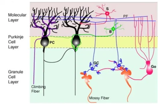
movements. The developing cerebellum consists mainly of three types of neuronal cells: granule cells in the external germinal layer, Purkinje cells, and neurons of the deep nuclei (Figure 1.2).
Figure 1.2: Cellular organization of the cerebellar cortex. The cerebellar cortex has only
three layers. The molecular layer contains two main types of interneurons, the basket (B) and stellate (S) cells. The Purkinje cell layer contains the cell bodies of Purkinje cells (PC), whose dendrites arborize in the molecular layer; these are the only neurons projecting out of the cerebellar cortex. The deepest layer, the granule cell layer, contains the cell bodies of granule cells (GC) and other interneurons, such as Golgi cells (Go), Lugaro cells and unipolar brush cells (not depicted here). Granule cells are glutamatergic interneurons that extend a single axon through the internal granular layer (IGL), and Purkinje cell layer (PCL) that splits into two branches, the parallel fibers (PF), in the molecular layer, forming synaptic contacts on Purkinje cell dendritic spines. There are two main types of extracerebellar axons projecting to the cerebellar cortex: the mossy fibers that contact the granule cells and have several origins in the hindbrain and spinal cord, and the climbing fibers that project to Purkinje cells and originate from the inferior olivary nucleus in the brainstem (adapted from (Chedotal, 2010)).
Granule cells of the cerebellum are the largest homogeneous neuronal population of mammalian brain (Chambers and Sprague, 1955a; Chambers and Sprague, 1955b; Saab and Willis, 2003). Due to their postnatal generation and the feasibility of well characterized primary in vitro cultures, cerebellar granule neurons (CGN) are well accepted as a model for the study of cellular and molecular correlates of neurodegeneration/neuroprotection (Contestabile, 2002).
These glutamatergic neurons are extremely sensitive to calcium (Ca2+) deregulation, in particular mediated by voltage operate Ca2+ channel type L (L-VOCC)
8
activity (Franklin and Johnson, 1992; Franklin and Johnson, 1994). The major role in maintaining cytosolic Ca2+ concentrations within the optimal range for the survival and normal excitability of CGN in culture is by Ca2+ influx through L-VOCC (Franklin and Johnson, 1992; Franklin and Johnson, 1994; Gutierrez-Martin et al., 2005).
CGN have been widely used as PD model. Rotenone has been shown to induce cell death of CGN (Isaev et al., 2004). As little as 5 nM rotenone increased mitochondrial superoxide levels and potentiated glutamate-induced cytosolic Ca2+ deregulation, the first irreversible stage of necrotic cell death (Yadava and Nicholls, 2007).
Although 6-OHDA is a neurotoxin specific for catecholamine neurons in both the central and peripheral nervous systems, cultured rat CGN have been demonstrated to be another useful in vitro system for studying the mechanism of 6-OHDA-induced neurotoxicity as a PD model. Relatively low concentrations (µM) of 6-OHDA induce apoptosis of CGN via activation of a caspase-3-like proteases (Dodel et al., 1999). In parallel, 6-OHDA was also demonstrated to produce free radicals in these neurons (Lin et al., 2003) as well as enhance excitation of CGN by glutamate (Garber-Goldsman et al., 1986).
On these grounds, CGN have acquired a special position in modern neuroscience as one reliable model for the study of neural development, function and, in particular for this work, PD pathology.
1.1.3.
Cell death in PD
There are several mechanisms by which cells could die. Until Kerr first used the term apoptosis (Kerr et al., 1972), all cells were thought to die by necrosis. Necrosis is characterized by cytoplasmic swelling, irreversible plasma membrane damage, and organelle breakdown (Fiers et al., 1999; Grooten et al., 1993). DNA in necrotic cells is usually degraded randomly by extracellular DNAse I present in culture serum that has not been heat-inactivated (Napirei et al., 2004), or by lysosomal DNAse II (Tsukada et al., 2001), giving rise to a smear of DNA (Higuchi, 2003). The cellular contents leak into the extracellular environment, where they may act as a “danger signal”, and consequently necrosis is usually associated with inflammation (Proskuryakov et al., 2003). In addition, there is now sufficient evidence that necrosis, a process traditionally regarded as “passive,” may also be genetically programmed. After signalling- or
9
damage-induced lesions, necrosis can include signs of controlled processes such as mitochondrial dysfunction, enhanced generation of ROS, ATP depletion, proteolysis by calpains and cathepsins, and early plasma membrane rupture. Furthermore, the inhibition of specific proteins involved in regulating apoptosis or autophagy can shift the type of cell death to necrosis (Festjens et al., 2006; Golstein and Kroemer, 2007).
For a long time the term “programmed cell death” (PCD) was synonymous of apoptosis. Today is known that apoptosis (or Type I cell death) is merely one type of PCD. Apoptosis is the major cell death pathway used to remove unwanted and harmful cells in a “clean” manner (Ellis et al., 1991), as during apoptosis the membrane integrity remains intact with the contents of the dying cell enclosed within apoptotic bodies, without releasing them into the extracellular space. DNA in apoptotic cells is degraded specifically, giving rise to a characteristic laddering pattern on the gels (Higuchi, 2003; Napirei et al., 2004; Tsukada et al., 2001).
Another type of PCD, autophagic cell death (or Type II cell death), has received a significant attention from PD researchers in the past few years. Autophagy is recognized by the formation of autophagosomes, double membrane autophagic vacuoles that eventually fuse with lysosomes to form autolysosomes. Swallowed contents and the inner membrane of the autophagosome are subsequently degraded by lysosomal hydrolases (Levine and Klionsky, 2004). Various forms of environmental stress induce autophagy, which eventually results in either caspase-dependent (Guimaraes et al., 2003; Xue et al., 1999) or caspase-independent cell death (Gozuacik and Kimchi, 2004; Xue et al., 1999; Yanagisawa et al., 2003).
Nevertheless the same stimulus can trigger multiple cell death pathways, depending on its intensity and duration, brain region and cell type, and also depending on the bioenergetics state of the cell (Eguchi et al., 1997; Young et al., 2004). All these three types of cell death have been identified in PD, predominating apoptosis (Kostrzewa, 2000) and autophagy (Anglade et al., 1997; Schapira and Jenner, 2011).
1.2. Brain bioenergetics alterations in PD
Cellular mechanisms regulating energy utilization must function properly to sustain the cell’s life. Dysfunction in mitochondria has been widely associated with PD. Therefore, and because mitochondria plays a central role in energy production, bioenergetics alterations have been associated with PD.
10
The shortage of mitochondrial supply of ATP and other energy-rich metabolites could be counterbalanced by cellular enzymes such as creatine kinase (CK) and adenosine monophosphate-protein kinase (AMPK), especially in locations of high energy consumption like the cytosol.
1.2.1.
Creatine Kinase
CK is an enzyme that rapidly catalyses the conversion of creatine and consumes ATP to produce phosphocreatine (PCr) and adenosine diphosphate (ADP), being an important enzyme in producing and buffering energy stores in excitable cells (Schlattner et al., 2006).
In fact, large amounts of energy are required to maintain the signalling activities of the cells in the central nervous system. Energy consumption in the brain is largely due to the maintenance of brain function-related processes, for example, for maintenance of membrane potential by the sodium potassium ATPase (Na+/K+-ATPase), Ca2+ homeostasis by the Ca2+-ATPase, neurotransmitters synthesis, secretion and recycling, intracellular signalling, and axonal as well as dendritic transport (Ames, 2000). Indeed, it has been proposed that mechanisms to facilitate energy transfer within cells that require fluctuating high energy levels, such as brain, include the juxtaposition of intracellular sites of ATP generation with sites of ATP consumption, as well as the transfer of high-energy phosphates between these sites by the CK/PCr system (Burklen et al., 2006).
The PCr/CK energy pathway represents an extremely efficient energy buffering system for two major reasons. First, PCr has a slightly higher diffusion capacity than ATP, allowing for a more efficient energy delivery system to different subcellular locations. Second, the subcellular localization of cytosolic and mitochondrial CK couples areas of high energy consumption with energy production. Thus, the CK/PCr essentially serves as a spatial ‘‘energy shuttle’’ or energy circuit within the cell (Wallimann et al., 1992).
There are four isoforms of CK based on its tissue expression (muscle or brain) and subcellular distribution (cytosolic or mitochondrial). In the brain, the dimeric cytosolic form of CK (CyCK) is called brain-type CK (BB-CK). The octameric mitochondrial CK (MtCK) is classified into two forms: sarcomeric muscle form (sMtCK) and brain form called ubiquitous MtCK (uMtCK) (Booth and Clark, 1978; Schlattner et al., 2006). Both
11
MtCKs are located in the mitochondrial intermembrane space, along the entire inner membrane and also at peripheral contact sites where inner and outer membranes are in close proximity (Biermans et al., 1990). There, MtCK can directly transphosphorylate intramitochondrially produced ATP into PCr (Jacobus, 1985), which is then exported into the cytosol (Figure 1.3).
Figure 1.3: The creatine kinase/phosphocreatine system. Compartment-specific isoenzymes
of creatine kinase (CK) are found in mitochondria (sMtCK, uMtCK, left) and cytosol (BB-CK, right). They are either associated with ATP-delivering processes (oxidative phosphorylation or glycolysis, left) and ATPconsuming processes (ATPases, to maintain local ATP/ADP ratios, right), or occur in soluble form (to maintain global cytosolic ATP/ADP ratios, center right). A large cytosolic phosphocreatine (PCr) pool of up to 30 mM is built up by CK from creatine (Cr), using ATP from oxidative phosphorylation (e.g., in heart) or glycolysis (e.g., in fast-twitch glycolytic muscle). The large phosphocreatine pool is then used as a temporal energy buffer to maintain constant global and local ATP/ ADP ratios over a wide range of workloads. The higher diffusibility of phosphocreatine, as compared to ATP, together with localized CK isoenzymes, is used for spatial energy buffering, i.e., for an energy shuttle between ATP-providing or-consuming processes. The latter seems to be most important for cells that are polarized and/or have very high or localized ATP consumption. Adapted from (Schlattner et al., 2006).
CK isoenzymes are highly susceptible to oxidative stress, an important pathophysiological condition associated with the progress of neurodegeneration in PD (Schlattner et al., 2006; Wang et al., 2001). In this scenario, MtCK has been suggested to be more vulnerable than CyCK due to its mitochondrial localization (Koufen and Stark, 2000). In line with this, it has been demonstrated a compromised CK system in
12
common neurodegenerative diseases (Aksenov et al., 2000; Ferrante et al., 2000; Wendt et al., 2002).
1.2.1.1. Creatine
Creatine is a constituent of a normal diet of protein-based foods, such as meat, milk, and nuts. It is not considered an essential nutrient, as kidneys, liver, pancreas, and possibly brain cells are able to synthesize this compound endogenously from the amino acids arginine, glycine, and methionine (Andres et al., 2008; Beard and Braissant, 2010; Wyss and Kaddurah-Daouk, 2000).
The primary physiological function of creatine is to buffer energy concentrations in tissues with significant and fluctuating energy demands, especially in muscles and brain (Wyss and Schulze, 2002). Until recently, the interest in creatine has been largely centered primarily on its use as an ergogenic aid to enhance sports performance (Benzi, 2000; Froiland et al., 2004). Nevertheless, it is becoming increasingly evident that administration of creatine might be beneficial in diseases with impaired bioenergetics like PD (Andreassen et al., 2001b; Baker and Tarnopolsky, 2003; Bender et al., 2005; Bender et al., 2006; Bender et al., 2008a; Brustovetsky et al., 2001; Burklen et al., 2006; Shefner et al., 2004).
Creatine produced dose-dependent neuroprotective effects against MPTP toxicity, reducing the loss of dopamine within the striatum and the loss of DA neurons in the SNc (Matthews et al., 1999). In addition, it has been shown that creatine may stabilize MtCK, and prevent activation of the mPTP (O'Gorman et al., 1997).
Due to its neuroprotective effects, creatine is now in clinical trials for the treatment of PD and Huntington’s disease (HD) (Hersch et al., 2006; NINDS-NET-PD, 2006). A placebo-controlled randomized pilot trial of creatine supplementation in PD showed that creatine improved patient mood and led to a smaller dose increase of dopaminergic therapy but had no effect on overall Unified Parkinson's Disease Rating Scale scores or dopamine transporter SPECT (Bender et al., 2006). However, a phase II futility trial in PD showed approximately a 50% improvement in Unified Parkinson’s Disease Rating Scale at one year, and the compound was judged to be non futile. Due to this finding, creatine is now under further investigation in a phase III clinical trial by the NET-PD investigators (NINDS-NET-PD, 2006).
13
More recently, oral creatine supplementation showed to attenuate levodopa (L-DOPA)-induced dyskinesia, a motor complication that arises in Parkinson patients after a chronic treatment with L-DOPA, in 6-OHDA-lesioned rats (Valastro et al., 2009). However, the molecular mechanisms underlying the neuroprotective actions of creatine are under current scientific debate. For example, stimulation by creatine of hippocampal Na+,K+-ATPase activity via N-Methyl-D-aspartate (NMDA)–calcineurin pathway is a very recent novel finding (Rambo et al., 2012), suggesting that the role of creatine in neuronal bioenergetics modulation can be more complex than currently assumed.
The safety of creatine supplementation has been reviewed extensively, and it has been concluded that creatine supplementation does not have any deleterious effects in humans (Bender et al., 2008b; Hersch et al., 2006; Mihic et al., 2000). These evidences clearly show the potential of creatine in PD’s therapy and in other neurological diseases.
1.2.2.
AMP-Kinase
AMPK is a heterotrimeric complex of a catalytic subunit (α) and two regulatory subunits (β and γ). AMPK is activated by phosphorylation of the α subunit at Thr172 (Hawley et al., 1996), being the downstream component of a protein kinase cascade. Once activated, AMPK switches-on catabolic pathways that generate ATP, while switching-off energy-consuming anabolic processes, thereby, acting as a key cellular modulator for the maintenance of the energy balance within cells. AMPK activation has been demonstrated to be regulated not only by cellular AMP/ATP ratio, but also by Ca2+ concentration and ROS (Hawley et al., 2005; Jung et al., 2008; Park et al., 2006; Weekes et al., 1994). When activated, AMPK phosphorylates several intracellular protein targets, like acetyl-CoA carboxylase, nitric oxide synthase and peroxisome proliferator-activated receptor gamma coactivator-1-α (Chen et al., 2000; Fryer et al., 2000; Suwa et al., 2003).
It has been reported that AMP-activated protein kinase is also activated by MPTP and other investigators have shown that overexpression of AMPK increased cell viability (Choi et al., 2010; Culmsee et al., 2001). These and other studies suggest that activation of AMPK may prevent neuronal cell death in neurodegenerative disease like PD. This possibility is also supported by recently published experimental data. Wu et al (2011) have demonstrated that AMPK and/ or mammalian silent information regulator 2 are required in resveratrol mediated autophagy induction, leading to neuronal survival
14
on PD cellular models. On the other hand, Arsikin et al (2012) have demonstrated that the neurotoxin 6-OHDA induces cytotoxic autophagy in SH-SY5Y neuroblastoma cells through the oxidative stress-dependent activation of AMPK and subsequent inhibition of the main autophagy repressor mTOR. Thus, these studies reveal that depending on the cellular insult the activation of AMPK can play either a neuroprotector or neurotoxic role.
1.3. Oxidative stress in the brain
The imbalance between normal cellular and environmental-induced production of ROS and reactive nitrogen species (RNS) and the ability of cells to efficiently defend against them (by the natural antioxidant defence system) is called oxidative stress. Oxidative stress produced in the body is toxic, could damage DNA, lipids and proteins leading to necrosis, ATP depletion, and prevention of controlled apoptotic death (Beal, 2005).
Since the brain uses 20% of the inspired oxygen and 80-90% of the consumed oxygen to produce energy during oxidative phosphorylation and the cefaloraquid liquid has lower antioxidant potency than the blood plasma, it is not surprising that neuronal cells are particularly sensitive to oxidative stress. During oxidative phosphorylation, neurons in the brain are extremely vulnerable to oxidative damage because of their high metabolic activity, low antioxidant capacity (Nunomura et al., 2006), non-replicative nature and the presence of high levels of polyunsaturated fatty acids which are readily oxidized (Perry et al., 2002). Additional chalenge is the presence of the blood-brain barrier, which protects the brain from toxins by limiting their diffusion into neurons and glia but also prevents/reduces the uptake of some blood-circulating antioxidants like vitamin E, into the brain. In this regard, it is to be noted that neurons may be exposed to ROS/RNS produced intracellularly and extracellularly, respectively, by neuronal cells and non-neuronal cells, such as microglia and endothelial cells of blood vessels.
1.3.1.
Oxidative stress in PD
The etiology of PD is unknown although mitochondrial dysfunction, oxidative and nitrosative stress have been implicated in the mechanisms associated with PD pathogenesis (Danielson and Andersen, 2008; Dawson and Dawson, 2003; Fahn and Cohen, 1992; Fiskum et al., 2003; Hashimoto et al., 2003; Jenner, 2003; Kanthasamy et
15
al., 2010; Tsang and Chung, 2009). However, it is not clear whether oxidative stress is a consequence of dysfunctional and dying neurons or plays a causal role in this neurodegenerative disease.
Postmortem tissues from PD patients have provided experimental evidences supporting that a deficient function of complex I of the mitochondrial electron-transport chain in SNc, i.e. 30–40% decrease in its activity, may be one of the central causes of sporadic PD (Dawson and Dawson, 2003). The decreased activity could be due to underproduction of certain complex I subunits, complex I disassembly, or self-damage by ROS produced during its function (Beal, 2004; Keeney et al., 2006; Schapira, 2001). More evidences of oxidative stress and PD come from examination of human PD brain showing oxidative damage to DNA and protein (protein carbonyls) besides immunocytochemical evidences for oxidative modifications of proteins by nitration, nitrosylation and glycation (Alam et al., 1997; Beal, 2002; Brown and Borutaite, 2004; Castellani et al., 1996; Floor and Wetzel, 1998; Good et al., 1998; Kikuchi et al., 2002; Zhang et al., 1999), and also by increased levels of lipid peroxidation (Dalfo et al., 2005). Along with the increase in oxidative damage, decreased levels of the antioxidant glutathione (GSH) were also found in the SNc of PD patients (Pearce et al., 1997; Perry et al., 1982; Perry and Yong, 1986).
1.3.1.1. Flavonoids in PD: Kaempferol and Epicatechin
In the last decade flavonoids have been described as important neuroprotectors against PD (Gagne et al., 2003; Pan et al., 2003; Panickar et al., 2009; Tan et al., 2008; Vauzour et al., 2010). This neuroprotection has been mainly attributed to their antioxidant and anti-inflammatory capacities (Aquilano et al., 2008; Dajas et al., 2003; Gutierrez-Merino et al., 2011). In the present study, taking into account the experimental data pointing out the occurrence of oxidative stress in the brain of patients with PD, we have studied the ability of the flavonoids kaempferol and epicatechin to afford protection against rotenone-induced neuronal death.Kaempferol is a natural flavonoid that exists in propolis (Scheller et al., 1990), tea (Tsaknis and Lalas, 2005), Gingko biloba (Smith and Luo, 2003) and other plant sources, which have been considered as a potent antioxidant and anti-inflammatory compound (Comalada et al., 2006; Martini et al., 2004; Wang et al., 2006). Ginkgo biloba extracts, quercetin and kaempferol have been reported to rescue PC12 cells
16
exposed to MPP+-induced oxidative stress and also to reverse neurotoxin effect (Gagne et al., 2003). More recently, the neuroprotective effect of kaempferol against MPTP was confirmed in a mouse model (Li and Pu, 2011). Kaempferol derivatives have also been recognized with protective potential in PD; they have been shown to prevent oxidative stress–induced cell death in a genetic (DJ-1)–dependent manner (Qu et al., 2009). Very recently, kaempferol was implicated in the enhancement of mitochondrial turnover by autophagy, revealing to be antiapoptotic and antioxidant in rotenone models of PD (Filomeni et al., 2012).
Epicatechin is a catechin present in cocoa (Dreosti, 2000; Sanchez-Rabaneda et al., 2003), green tea (Graham, 1992) and red wine (Damianaki et al., 2000). This flavonoid has been widely demonstrated to act as a cellular antioxidant, free radical scavenger and iron chelating (Lee et al., 2003; Pan et al., 2003; Xu et al., 2004), being neuroprotector in PD, not only by itself but also as a constituent of the mentioned sources of polyphenols (Datla et al., 2007; Gao et al., 2012; Levites et al., 2001). For instance, green tea polyphenols have demonstrated to elicit neuroprotection in cell cultures and animal models, such as the prevention of neurotoxin-induced neurodegeneration of the striatum and DA neurons of the SNc (Panickar et al., 2009). Recently, Vauzour et al. (2010) have reported that the flavonoids catechin, epicatechin and quercetin and polyphenols like caffeic acid and p-coumaric acid afford protection against the neurotoxicity of 5-S-cysteinyl-dopamine, which has been proposed to contribute to the progression of the brain neurodegeneration in PD.
1.4. Calcium homeostasis in brain
Intracellular Ca2+ regulates a wide array of cellular processes and is important for signal transduction. Many biological processes of great importance for the neuronal function are extremely dependent on cytosolic Ca2+ concentration, such as secretion of neurotransmitters and synaptic plasticity (Trifaro and Vitale, 1993), intracellular signaling pathways that mediate the metabolic extracellular neuronal stimuli (Berridge et al., 2000) and development of neurites (Benowitz and Routtenberg, 1997). Ca2+ plays a central role not only in normal and healthy neurons but is also involved in the many cellular processes (e.g. oxidative stress, mitochondrial impairment, proteasomal dysfunction, excitotoxicity, neuroinflammation, apoptosis) that can lead to cell death in
17
PD (Berridge et al., 2000; Bueler, 2010; Hegde and Upadhya, 2011; Lau and Tymianski, 2010; Witte et al., 2010).
The concentration of cytosolic free Ca2+ in resting neurons (≈100 nM) is 10,000 fold lower than the concentration of Ca2+ in the extracellular space (≈1.2 mM) (Gleichmann and Mattson, 2011). This concentration gradient leads to a significant increase in cytosolic Ca2+ after depolarization, rendering Ca2+ regulation a critical process in neurons. To maintain Ca2+ homeostasis, Ca2+ entering neurons is rapidly sequestered in intracellular organelles, such as the mitochondria and the endoplasmic reticulum (ER), or pumped back across the plasma membrane concentration gradient, all of which require the consumption of high levels of energy in the form of ATP (Gleichmann and Mattson, 2011). In neurons the Ca2+ transport systems of the plasma membrane most important in the control of the homeostasis of cytosolic Ca2+ are voltage-operated calcium channels (VOCC), some ionotropic receptors (with the NMDA receptors playing an outstanding role in the brain), the plasma membrane Ca2+ ATPase (PMCA) and the Na+/Ca2+ exchanger (NCX) (Gutierrez-Merino, 2008). Ca2+ influx through L-VOCC has a major role in maintaining cytosolic Ca2+ concentrations within the optimal range 70 to 200 nM for the survival and normal excitability of CGN in culture (Gutierrez-Martin et al., 2005).
The sustained impairment of intracellular Ca2+ homeostasis in neurons associated with the oxidative stress induced by an elevated production of ROS is a common metabolic feature in brain neurodegenerative diseases. Moreover, cytosolic Ca2+ concentration can be regarded as a major bioenergetic marker for neuronal activity and neuronal survival, as it has been shown that a sustained low cytosolic Ca2+ concentration elicits apoptosis in neurons in culture (Franklin and Johnson, 1992; Franklin and Johnson, 1994), and also that a steadily elevated cytosolic Ca2+ concentration induces rapid cell death by necrosis mediated by activation of calpains (Choi, 1988; Franklin and Johnson, 1992; Garcia-Bereguiain et al., 2008; Gutierrez-Martin et al., 2005; Orrenius et al., 1989).
1.4.1.
Alterations of calcium homeostasis in PD
In spite of the occurrence of degeneration in other brain areas, DA region is one of the most affect in PD patients. Many characteristics of DA neurons have been point out that these are neurons with high energy requirements (Surmeier et al., 2011a).
18
Unlike the vast majority of neurons in the brain, adult SNc DA neurons are autonomously active, generating broad slow action potentials regularly (2–4 Hz) in the absence of synaptic input (Grace and Bunney, 1983). This pacemaking activity is believed to be important in maintaining ambient DA levels in regions that are innervated by these neurons, particularly the striatum (Romo and Schultz, 1990). Whereas the majority of neurons rely exclusively on monovalent cation channels (like sodium (Na+)) to drive pacemaking, SNc DA neurons also express ion channels that allow extracellular Ca2+ to enter the cytoplasm (Ping and Shepard, 1996; Puopolo et al., 2007), which lead to elevated intracellular Ca2+ concentrations (Chan et al., 2007; Wilson and Callaway, 2000). The use of Ca2+ rather than Na+ ions for pacemaking involves more energy costs for neurons in order to maintain a safe intracellular Ca2+. The Ca2+ channels involved in this pacemaking activity have a distinctive Cav1.3
pore-forming subunit (Chan et al., 2007). Cav1.3 Ca2+ channels are relatively rare
(Sinnegger-Brauns et al., 2009), but they are found at an extraordinary high density in SNc DA neurons (Guzman et al., 2009; Khaliq and Bean, 2010; Puopolo et al., 2007).
It is now accepted that Ca2+ entry through plasma membrane Cav1.3 Ca2+
channels during activity is either pumped back across the plasma membrane (by PMCA and/or NCX) or rapidly sequestered in the ER or mitochondria (Surmeier et al., 2011a) (Figure 1.4). Both processes waste cellular energy stored in the form of ATP. The metabolic demand created by these ATP-dependent steps in Ca2+ homeostasis should increase oxidative phosphorylation in mitochondria and the production of damaging ROS (Guzman et al., 2010). This places SNc DA neurons in a stressful working situation, as ROS damage mitochondrial proteins such as complex I and mtDNA, reducing the efficiency of oxidative phosphorylation (Harman, 1972; Wallace, 2005). In extreme cases, the stress on mitochondria induces mPTP opening, swelling and the release of cytochrome c and other pro-apoptotic proteins such as apoptosis inducing factor (Nicholls, 2002). In parallel, ROS are capable of damaging ER proteins, elevating the concentration of misfolded proteins that need to be degraded by proteasomes and autophagosomes (Kaufman, 1999). The unfolded protein response triggered by this elevation in misfolded proteins should further reduce ER production of proteins and potentially lead to the release of pro-apoptotic factors such as C/EBP homologous protein (CHOP) (Chan et al., 2009; Oyadomari and Mori, 2004). The role of mitochondria in Ca2+ homeostasis could further compromise their ability to generate ATP, leading to a functionally important drop in cytosolic ATP levels (Nicholls, 2002).
19
Figure 1.4: Ca2+ transport in SNc DA neurons. The steep concentration gradient for Ca2+
enables it to cross the plasma membrane readily into cells through open pores such as L-type Ca2+ channels (Cav1.3 VOCC-subtype). Once inside neurons, it is either transported back across the plasma membrane (by PMCA or NCX) or sequestered in intracellular organelles (i.e. mitochondria and ER). The ER uses high-affinity smooth ER Ca2+ (SERCA) pumps that depend upon ATP to take Ca2+ from the cytoplasm into the ER lumen. Ca2+ flows back into the cytoplasm after the opening of inositol trisphosphate receptors (IP3R) and ryanodine receptors (RyR) also located in the ER membrane. Mitochondria are often found in close apposition to the ER and plasma membrane, creating a region of high (but localized) Ca2+ concentration that drives Ca2+ into the matrix of mitochondria through a Ca2+ uniporter. Ca2+ can leave the mitochondrion through a number of mechanisms. The dominant mitochondrial Ca2+-efflux pathway in neurons is through mitochondrial NCXs. Ca2+ release through higher conductance ion channels, such as the mitochondrial permeability transition pore (mPTP), has also been proposed. The mPTP is known to have two conductance states: a low-conductance state that is reversible and participates in physiological Ca2+ handling, and a high conductance state that is irreversible and leads to mitochondrial swelling and loss of molecules such as cytochrome c that trigger apoptosis (adapted from (Chan et al., 2009)).
Genetic mutations or environmental toxins such as rotenone could further compromise mitochondrial or ER function, rendering them more vulnerable to Ca2+ stress (Surmeier et al., 2011b). By rushing the decline in ER and mitochondrial function
20
and the accelerated loss of SNc DA neurons, these genetic and environmental factors could be seen as “causing” PD.
1.5. Aims
The major aim of this work was to assess the potential of the ergogenic molecule creatine and the flavonoids epicatechin and kaempferol (and possible synergism between these compounds) to afford neuroprotection of CGN in culture against rotenone, a toxin used to induce parkinsonism-like neurodegeneration.
Due to its energetic capacity, creatine has been shown to be a neuroprotector in some neurodegenerative diseases such as HD and PD, being at the time in phase III clinical trials (Hersch et al., 2006; NINDS-NET-PD, 2006). Moreover, we wanted to evaluate the benefits/inconveniences of co-administration of creatine with flavonoids like kaempferol and epicatechin that have been proposed to be bioactive compounds accounting, at least in part, for the beneficial effects of green tea against the progress of neurodegeneration in PD treatment.
In addition, as the cellular mechanisms underlying the neurotoxicity exerted by rotenone in CGN are not well understood, a second major goal of this work is to use creatine and the above mentioned flavonoids as tools to shed light on the relative relevance of the different mechanisms proposed to mediate brain neurodegeneration in PD.
To achieve these major goals, we have defined the experimental strategy and partial objectives schematically presented in figure 1.5.
21
Figure1.5: Schematic representation of the experimental strategy and partial objectives of this research work.
23
II. Material and Methods
24
2.1. Equipment and reagents
2.1.1. Scientific equipment
Laminar Flow Cabinet Nüve LN 090 CO2 incubator Heal Force - HF 90
Microcentrifuge Eppendorf 5415R
Ultracentrifuge Beckman Coulter Optima™ L-90K Spectrophotometer PG Intruments T70
Fluorescence SpectrophotometerPerkin Elmer 650-40
Varioskan Flash spectral scanning multimode reader Thermo Scientific Epifluorescence microscope Nikon Diaphot 300
Microscope camera Hamamatsu ORCA-R2
Microscope Wheel Filters Lambda 10-2
Electrophoresis system and western blotting BIO-RAD
2.1.2. Chemicals and reagents
All antibodies were used at a dilution in the range recommended in the product datasheets. The primary antibodies used are described in Table 2.1, and the secondary antibodies labeled with fluorophores are described in Table 2.2.
Western blotting reagents anti-goat IgG horseradish peroxidase anti-rabbit IgG horseradish peroxidase and SuperSignal West Dura Extended Duration Substrate were purchased from Pierce (Rockford, IL, USA).
All other reagents and chemicals were supplied by Sigma-Aldrich, Roche or
25
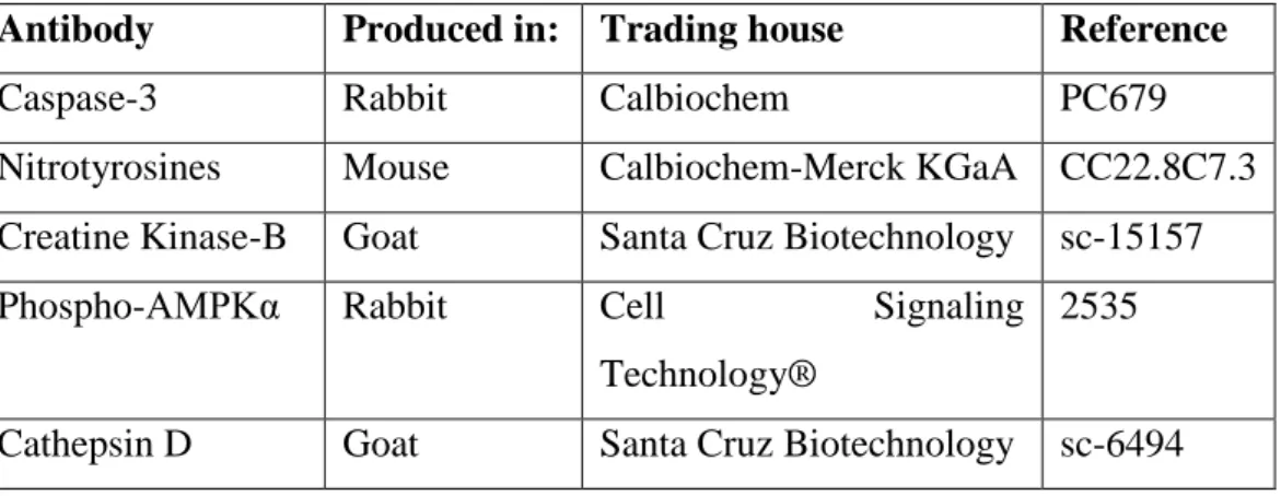
Table 2.1- Informative description of the primary antibodies used in Western blot
experiments.
Antibody Produced in: Trading house Reference
Caspase-3 Rabbit Calbiochem PC679
Nitrotyrosines Mouse Calbiochem-Merck KGaA CC22.8C7.3
Creatine Kinase-B Goat Santa Cruz Biotechnology sc-15157
Phospho-AMPKα Rabbit Cell Signaling
Technology®
2535
Cathepsin D Goat Santa Cruz Biotechnology sc-6494
Table 2.2- Informative description of the secondary antibodies used in Western blot
experiments.
Antibody Trading house Reference
anti-Rabbit IgG-Alexa488 Invitrogen
(Molecular Probes, Eugene, OR, USA)
A11008
anti-Goat IgG-Alexa488 Invitrogen
(Molecular Probes, Eugene, OR, USA)
A11055
anti-Mouse IgG-Alexa488 Invitrogen
(Molecular Probes, Eugene, OR, USA)
A11001
2.2. Buffers and solutions
The composition of MLocke 25 buffer (pH 7.4 at 37° C) used in all experimental work with cerebellar granule cells is: 4 mM NaHCO3, 10 mM N-[tris (hidroxymetil)
metil] glicine, 5 mM glucose, 2.3 mM CaCl2, 1 mM MgCl2, 134mM NaCl and 25 mM
KCl.
The composition of sample buffer used for SDS-PAGE in this work is 26.6 mM Tris-HCl pH 6.8, 0.86% sodium dodecyl sulfate (SDS), 0.43% (v/v) β-mercaptoethanol, 0.014% bromophenol blue prepared in 20 mM Tris pH 7 and 37.3% glycerol.
The composition of the electrophoresis buffer used for sodium dodecyl sulfate-polyacrylamide gel electrophoresis (SDS-PAGE) throughout the experimental work is: 25 mM Tris, 190 mM Glycine, 10% SDS (pH 8.3).
The transfer buffer composition used in Western blotting in all this work is: 25 mM Tris, 190 mM Glycine, 0.1% SDS, 20% methanol (pH 8.3).
26
The composition of phosphate buffered saline (PBS) is 4.3 mM Na, 0.4 mM
KH2PO4, 137 mM NaCl and 27 mM KCl (pH 7).
2.3. Biological Material
Cultures of cerebellar granule cells were prepared from 7 days old Wistar rats, weighing 20-25 g. The rats were supplied and maintained by the Animal Service of the University of Extremadura, where they have been fed and maintained at a constant temperature of 22-23 ⁰ C and humidity ranging between 60 and 80 %.
2.4. Cell cultures
2.4.1. Cerebellar granule cells primary cultures
Cultures of CGN were obtained from dissociated cerebella of 7 day old Wistar rats as described previously (Samhan-Arias et al., 2004). Cells were plated in Dulbecco’s modified Eagle’s medium (DMEM) supplemented with 10% heat inactivated fetal bovine serum (FBS), 5 mM glucose, 19.63 mM KCl, 3.7 ng/mL insulin, 7 µM para-aminobenzoic acid, 50 U/mL penicillin, 25 U/mL streptomycin, 0.91 mM pyruvate and 2 mM L-glutamine on 35 mm dishes coated with poly-D-lysine, at a density of 2.75x10⁶ cells/dish. Cultures were maintained at 37ºC in a humidified atmosphere of 95 % air/5 % CO2. Cytosine arabinofuranoside (10 µM) was added to fresh culture medium 48 h
after plating to prevent replication of nonneuronal cells. The culture medium was replaced with serum-free medium 7 days after plating. Cells were maintained afterward in serum-free F12 medium supplemented with 12.5 mM glucose, 20.82 mM KCl, 5 µg/ml insulin, 0.1 mg/ml apo-transferrin, 20 nM progesterone, 50 U/ml penicillin, 25 U/ml streptomycin, 0.1 mg/ml pyruvate, 2 mM L-glutamine. All experiments were performed using mature CGN at 9–10 days in vitro (DIV).
2.5. Viability assays
2.5.1. MTT assay
Neuronal viability was assessed by the 3-(4,5-dimethyl-thiazole-2yl)-2,5-diphenyltetrazolium bromide (MTT) reduction test as described in (Martin-Romero et al., 1996; Samhan-Arias et al., 2004). Viable cells reduce MTT to formazan which can be determined spectrophotometrically. In brief, the culture medium was replaced with 2 mL of MLocke 25 buffer and incubated for 15 min with 0.3 mg MTT. Thereafter,
27
formazan was dissolved in 1 mL of dimethyl sulfoxide (DMSO) and measured at 490 nm and 700 nm.
2.5.2. Treatment of CGN with Rotenone
To investigate the effect of rotenone on CGN viability, the treatment was performed at 9 DIV in CGN. To this end, the plates containing the CGN were incubated for 12 hours at 37 ⁰ C with 0, 2, 5, 10, 15, 20, 30 and 50 nM of Rotenone prepared in DMSO. After the incubation period the plates were subjected to MTT assay described in section 2.5.1.
2.5.3. Treatment of CGN with 6-OHDA
To investigate the effect of 6-OHDA on CGN viability, cells at 9 DIV were incubated for 24 hours at 37 ⁰ C with 0, 5, 10, 15, 20, 25, 30 and 50 µM of 6-OHDA prepared in DMSO. After the incubation period the viability was evaluated by MTT assay described in section 2.5.1.
2.5.4. Flavonoids protection against rotenone-induced cell death in CGN
To investigate the protective effect of the flavonoids: kaempferol and epicatechin, cells at 9 DIV were incubated for 1 hour with 0, 2, 5 and 10 µM of each. Thereafter, rotenone (IC50 concentration) was added to the culture medium and
incubated for 12 hours. After the incubation period the cell viability was evaluated by MTT assay described in section 2.5.1.
2.5.5. Creatine protection against rotenone-induced cell death in CGN
To investigate the protective effect of creatine, cells at 9 DIV were incubated for 1 hour with 0, 5, 10, 15, 20 and 30 mM. Thereafter, rotenone (IC50 concentration) was
added to the culture medium and incubated for 12 hours. After the incubation period the viability was evaluated by MTT assay described in section 2.5.1.
2.6. Protein quantification and determination
2.6.1. Preparation of cell lysates
CGN cultured for 9 DIV were washed with buffer MLocke 25. Then, the CGN were resuspended and centrifuged at 4 ° C for 2 min at 2000g in a refrigerated
28
Eppendorf microcentrifuge, pellets of lysed cells were re-suspended in 100 µL of buffer (50 mM HEPES, pH 7.4, 100 mM NaCl , 0.1% CHAPS, 1 mM DTT and 0.1 mM EDTA) for colorimetric measurements and in 200 µL of buffer (5 mM NaP, pH 7, 1 mM EDTA, 0.5% Tween 20 supplemented with protease inhibitor cocktail Roche Biochemicals (COMPLETE)) for Western blotting.
2.6.2. Protein concentration
Protein concentration was determined by the method of Bradford (1976) using the Bio-Rad (Hercules, CA, USA) protein assay reagent and bovine serum albumin as standard.
2.6.3. Western blotting
SDS-PAGE were run at a concentration of 15%, 12.5% and 7.5% acrylamide, depending on the molecular weight of the protein of interest, using 20 µg protein of CGN lysates per lane. Gels were transferred to nitrocellulose membranes of 0.2 µm or 0.4 µm average pore size (TransBloTTransfer Medium, BioRad). Nitrocellulose membranes were blocked by 1 h incubation at room temperature with 5% (w/v) non-fat dry milk in PBS. Then, nitrocellulose membranes were washed three times with PBST, e.g. PBS supplemented with 0.05% polyoxyethylenesorbitan monolaurate (Tween 20). Immunodetection of proteins was performed with its specific antibody at recommended dilutions. After incubation with the first antibody overnight, membranes were washed six times with PBST and incubated for 1 h at room temperature with the secondary antibody IgG conjugated with horseradish peroxidase (rabbit, mouse and anti-goat with a dilution of 1:8.000, 1:50.000 and 1:500.000, respectively), then washed six times with PBST, followed by incubation for 3 min with SuperSignal West Dura Substrate (Pierce). Western blots were revealed by exposure to an AmershamHyperfilm MPautoradiography film (GE Healthcare, UK).
2.7. Cell death pathways
2.7.1. Caspase-3 and Cathepsin D activation
Caspase-3 and cathepsin D activations were investigated by Western blotting after SDS-PAGE of lysates as described in 2.6.3. To detect caspase-3 was used a polyclonal rabbit anti-activated caspase-3 antibody (1:200; Calbiochem PC679) against
29
the 17 kDa cleaved (active) fragment of caspase-3. For positive control, activated recombinant human caspase-3 (Calbiochem) was also loaded to the gels. Cathepsin D was revealed using a goat polyclonal anti-cathepsin D antibody (1:100; sc-6994 from Santa Cruz Biotechnology).
2.7.2. Calpain activity
Calpain activity was measured using the fluorogenic substrate Suc-LY-AMC (Calbiochem-Merck KGaA,). An aliquot of lysate with 30 µg of protein was incubated with 2.2 mL of the reaction buffer containing 50 mM Tris–HCl pH 7.5, 50 mM NaCl, 5 mM β-mercaptoethanol, 5 mM CaCl and 50 µM Suc-LY-AMC. The release of 7-amino-4-methyl-coumarin (AMC) was monitored at an excitation wavelength of 380 nm and 460 nm of emission on a fluorescence spectrometer. To detect non-specific protease activity, the reaction rate was also measured in the presence of 10 µM Z-2-LLY-FMK (Calbiochem-Merck KGaA), a specific inhibitor of calpain, and the value was subtracted from that observed in the absence of the inhibitor. Fluorescence units were converted into moles of AMC released using a standard curve obtained with free AMC, and calpain activity was expressed in pmol of AMC cleaved per minute per milligram of protein.
2.7.3. Hoechst 33258 and propidium iodide staining
A direct estimation of disrupted plasma membrane CGN was obtained through a modified double-staining technique as described in (Soares et al., 2008) with modifications. Briefly, culture medium was discarded, attached cells were washed with MLocke 25 buffer, and a stock solution of bisbenzimide (Hoechts 33258 at 10 mg/mL) was added to dishes, yielding final concentration of 20 µg/mL, during 30 min at 37 ⁰ C. To the same dishes, a stock solution of propidium iodide (PI) (10 mg/mL) was added, yielding a final concentration of 20 µg/mL, during the final 5 min of staining. Bisbenzimide- (stains genetic material) and PI-stained (indicates disrupted plasma membrane cells) cells were examined and photographed, using a Nikon Diaphot 300 inverted microscope. Filters for PI fluorescence used a 550 nm wavelength, and for bisbenzimide, a 420–505 nm wavelength.