ARTIGO DE REVISÃO
Recent Surgical Advances in Peyronie’s Disease
Avanços Cirúrgicos Recentes na Doença de Peyronie
1. Department of Urology. Hospital Santa Maria. Centro Hospitalar Lisboa Norte. Lisbon. Portugal. Autor correspondente: Sandro Gaspar. sandrosilvagaspar@gmail.com
Recebido: 19 de fevereiro de 2015 - Aceite: 24 de novembro de 2015 | Copyright © Ordem dos Médicos 2016 Sandro GASPAR1, José Santos DIAS1, Francisco MARTINS1, Tomé LOPES1
Acta Med Port 2016 Feb;29(2):131-138 ▪ http://dx.doi.org/10.20344/amp.6317
RESUMO
Introdução: A doença de Peyronie, uma doença fibrótica da túnica albuginea do pénis tem estado associada a encurtamento peniano
e a algum grau de disfunção eréctil. Afeta a qualidade de vida do doente, levando a stress psicológico, mental e físico. A deformidade peniana perturba a vida sexual do doente, levando a episódios de depressão, disfunções sexuais e a ansiedade associada ao ato sexual. A etiologia da doença de Peyronie permanece por esclarecer.
Material e Métodos: A pesquisa na literatura foi efetuada nas bases de dados da Medline, Embase e Cochrane no mês de Janeiro de
2015 no sentido de identificar artigos relacionados com a doença de Peyronie, nomeadamente o tratamento cirúrgico, técnicas, resul-tados bem como tratamentos complementares. Publicações que não envolvessem humanos não foram consideradas. Identificámos artigos originais, artigos de revisão e editoriais acerca do assunto em questão. Todos os artigos publicados na língua inglesa foram selecionados para screening. Os critérios de elegibilidade para inclusão envolveram a relevância associada ao tema.
Resultados: Existe uma variedade de deformidades penianas associada com a doença de Peyronie que ainda não têm uma solução
não cirúrgica que seja eficaz. Apresentamos uma atualização das técnicas cirúrgicas atuais bem como o algoritmo de tratamento as-sociada a esta doença.
Discussão: Todos os tratamentos cirúrgicos têm como objetivo a correção da curvatura, a preservação da função eréctil e do
compri-mento peniano, bem como o de minimizar a morbilidade.
Conclusão: Até à data não existem estudos de medicina baseada na evidência que determinem o melhor tratamento cirúrgico para a
doença de Peyronie. Após o diagnóstico, a reconstrução cirúrgica deve ter como objetivo um pénis funcional, com uma retificação da sua curvatura que permita o acto sexual.
Palavras-chave: Induração Peniana; Pénis/cirurgia; Procedimentos Cirúrgicos Urológicos Masculinos; Prótese de Pénis. ABSTRACT
Introduction: Peyronie’s disease, a fibrotic disorder of the tunica albuginea of the penis, has been associated with penile shortening
and some degree of erectile dysfunction. It affects patient’s quality of life, leading to severe psychological, mental, and physical stress. Penile deformation hampers sexual life leading to depression, lack of sexual confidence, loss of sexual function and performance anxiety. Peyronie’s disease etiology is yet to be known.
Material and Methods: Literature search was conducted in Medline, Embase, and Cochrane databases in January 2015 in order to
identify papers related to Peyronie’s disease, concerning evolving surgical management, technique, outcomes as well as ancillary treatments. Publications not concerning humans were not considered. We identified original articles, review articles, and editorials addressing the subject. All articles published in the English language were selected for screening. The eligibility criteria for inclusion were based on relevance concerning the subject.
Results: The variety of penile deformities associated with Peyronie’s disease still doesn’t have an effective and reliable non-surgical
therapy. We summarize the updated surgical techniques and management algorithm described for Peyronie’s disease.
Discussion: Surgical management shares similar goals: correcting the curvature, preserving erectile function and penile length, and
minimizing morbidity.
Conclusion: To date there is no high level of evidence-based data to determine the best surgical treatment of Peyronie’s disease. After
proper diagnosis, surgical reconstruction should be based on giving a functional penis, that is, rectifying the penis with rigidity enough to enable sexual intercourse.
Keywords: Penile Induration/surgery; Penile Prosthesis; Penis/surgery; Urologic Surgical Procedures, Male.
INTRODUCTION
Peyronie’s disease (PD), named in 1743 after François Gigot de La Peyronie,1 represents a benign penile disease caused by a fibrotic wound-healing disorder of the tunica albuginea, leading to a characteristic penile deformity during erection.2 It may result from macro or microtrauma to the erect penis in genetically susceptible individuals or with an underlying disease of the elastic fibers and collagen bundles of the penis.3
It dramatically affects patient’s quality of life, causing severe psychological, mental, and physical stress. Sexual intercourse is hampered: painful erections and
penile curvature/deformation make penetration difficult or impossible.2,4 PD’s pathophysiology is still unknown. PD’s reported incidence is 3.2 – 8.9% in the general po- pulation but it may be higher due to underreporting with caucasians living in northern Europe, with an average age of 55 - 65 years being the most affected.5 Prevalence is higher in patients with co-morbidities like diabetes mellitus, concomitant erectile dysfunction (ED) or history of previous radical prostatectomy, hypertension, dyslipidemia, obesity, smoking, low testosterone and Dupuytren’s contractures.5-8 Men with unstable or progressive deformity, or those who
ARTIGO DE REVISÃO refuse surgery, may be offered medical or minimal invasive
treatments. Treatment options include: oral medications, topical agents, intralesional injections, mechanical stretching or vacuum devices, and well as extracorporeal shockwave therapy. Surgical treatment when the condition is its chronic phase, completely stable, is the most effective treatment of PD.9
MATERIAL AND METHODS
The urologist has to perform a thorough anamnesis and physical examination to identify potential surgical candidates. PD’s natural history has two separate stages, an active or progressive one, and one chronic or stable one. The first stage, the inflammatory one, is characterized by penile pain during erection or intercourse and a progressive penile curvature or deformity because of plaque evolving, lasting usually for 6 – 18 months after the first symptoms. Approximately 10% of patients will have spontaneous improvement of their disease, while the majority will progress to the second stage or watch their disease worsen. During this second stage, pain subsides and curvature stabilizes. Plaque stabilization is characterized by fibrosis, dystrophic calcifications, and rarely, ossification.10 It is during this stage that surgical intervention occurs. An history of penile trauma must be sought (reported in 5% – 13% of cases)5 as well as Dupuytren’s contracture (associated risk of 3% – 22 % of PD), Ledderhose disease and tympanosclerosis.5,11,12 Important determinations during consultation are regular measures of stretched penile length, girth measurement, penile sensation to touch and degree of penile curvature.13 Patient self-reporting of penile curvature is unreliable and should not be used as a clinical objective marker and an objective evaluation through a proper clinical examination of the erect penis (induced pharmacologically) is needed.5 Sexual history, including erectile rigidity, concomitant ED and baseline erectile function must be thoroughly assessed using validated questionnaires such as the International Index of Erectile Function (IIEF), the Sexual Health Inventory for Men (SHIM) or the Peyronie’s Disease Questionnaire (PDQ).14 Veno-occlusive dysfunction (scarring of the tunica albuginea leads to failure of occlusion of subtunica venules) may translate clinically as failure to achieve a rigid erection or a difficulty to maintain it. Another cause may be an impaired cavernosal arterial inflow. Co-morbidities like vascular disease, diabetes mellitus, cardiac disease and habits like tobacco use, should also be sought as family history of penile deformities.5
Penile ultrasonography is an inexpensive, non-invasive and universally available technique for that allows for evaluation of intracorporeal fibrosis and fibrotic plaques, as well as to eliminate other differential diagnosis like spongiofibrosis or any malignant disorders. Duplex penile ultrasound following provocative vasoactive medication may be an objective way to assess the degree of penile deformity and establishing baseline erectile function.15 CT and Penile MRI are more expensive and not universally available but may also be used to detect penile lesions and
calcifications.16 These exams, along with biothesiometry are not evidence-based recommendations prior to surgery.11 The restoration of penile and sexual functionality is the primary goal of surgical intervention for the patient to resume normal sexual activity. Reconstructing a perfect straight penis, recovery of substantial penile length or normalized penile sensation, and spontaneously normalized erectile function, may not always be possible.11 One must set realistic expectations and address potential complications like penile shortening, decreased rigidity, residual or recurrent deformity, decreased sensation and ED.5
RESULTS
Major factors that determine surgical approach are the inability to perform penetrative intercourse, an extensive plaque calcification, a failed conservative/medical treatment and patient’s preference for definite results. Factors like the location and degree of curvature, the stretched penile length, other penile deformities, may dictate the ideal surgical option (Fig. 1). Penile rigidity adequate for intercourse enables patients for a tunical shortening or lengthening procedure. Factors associated with post-operative ED are operative ED, age (> 55 years), curvature > 60º, pre-operative venous leak.17
Tunica plication: procedures that shorten the tunica
In case of a penile curvature, under 60 to 70 degrees, adequate penile length and rigidity, good preoperative erectile function and no indentation or hourglass defect, shortening the longer side of the penis (opposite to the curvature) will correct the curvature, equalizing length in both sides of the penis.16 They are generally considered to be easy to perform and an excellent surgical option.18 Several tunica albuginea plications have been described, including the Nesbit procedure, the modified Nesbit, the Yachia technique, the Giammusso technique, the Lemberger technique, the Essed-Schroeder technique, the Duckett-Baskin-Levine technique, and the 16 or 24-dot plication technique.18
The Nesbit procedure was first described by Nesbit in 196519 for a congenital curvature, called chordee: a transverse incision of the tunica albuginea is performed, on the convex side of the shaft, with approximately 1 mm for each 10º of curvature. Only later, in 1979 was it used on PD with satisfying rates.
A number of techniques soon followed, modificating the original classic Nesbit operation. Rehman, in 1997, described a novel technique with partial thickness shaving instead of conventional excision of a wedge of tunica albuginea with lesser bleeding, diminished cavernous tissue damage and improved adhesion of plicated layers, translating in high satisfaction rates.21 The Yachia procedure involves degloving the penis and making a longitudinal incision in the tunica albuginea opposite the plaque. The edges are reapproximated horizontally (according to the Heineke-Mikulicz principle) with a resulting shortening of the side opposite the plaque and overall straightening of the penis.22
ARTIGO DE REVISÃO
Both the Nesbit and Yachia procedures are associated with some loss of penile tactile sensation and ED. It is believed it is probably due to disruption of the neurovascular bundle and cavernosal integrity.23 A less invasive technique was developed by Essed and Schroeder in 1985, consisting in placing tight non-reabsorbable sutures on the convex side of the tunica albuginea to effectively plicate and straighten the penis, not requiring excision or incision of the tunica or mobilization of the neurovascular bundle (Fig. 2) with cosmetic and functional satisfaction rates ranging 81% to 95%.24 Another plication technique was popularized by Gholani and Lue in 2002: the 16 to 24-dot suture. There‘s a distribution of knot tension along a long span of the shaft of the penis, lowering the risk of tunica tearing, with less patient discomfort and less episodes of recurrence.25 In spite of penile shortening (not suited for shorter phallic lengths) and long-term palpable sutures, this approach has minimal risk of ED, as well as of neurovascular damage.3,26
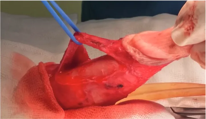
Procedures that lengthen the tunica: plaque incision/ plaque excision and grafting
Tunica lengthening procedures involve incising or
excising the plaque, and adding graft material to cover the defect (Fig. 3). Patients need to have an adequate erectile capacity as these patients have an increased risk of postoperative ED.6 It’s recommended in situations where the curvature is greater than 60º – 70º, if there are complex deformities or the presence of an hourglass or a hinge-destabilizing component.2,21 Excision can be avoided if the surgeon is able to perform relaxing incisions on the plaque with added grafting or no grafting at all. The graft (usually 20% larger than the defect) is then sutured to the tunica
albuginea with separate running suture.3,27
In 1950, Lowsley and Boyce28 were the first authors to perform plaque excision and grafting with fat for the treatment of PD, followed by Horton in Devine, in 1974, using a dermal skin graft.29 It has the advantage of a higher Figure 1 - Surgical Treatment Algorithm
a without progression of symptoms b ED: erectile dysfunction
• Stable disease (> 12 months)a
• Inability to perform penetrative intercourse • Extensive plaque calcification
• Failed conservative / medical / minimally invasive treatment
• Patient’s preference for defenitive and reliable results
Tunica plication 1. Curvature < 60º 2. Absence of hourglass or
indentation deformity 3. Loss of lenght predicted < 20º
Plication or wedge resection Nesbit procedure
Modified Nesbit
Plication variations and corporoplasty
Grafting procedure Plaque incision and grafting Plaque excision and grafting Plaque incision / excision and
vascularized flap Penile desassembly
Penile prosthesis
Penile prosthesis + manual modeling (molding)
Penile prosthesis + Nesbit / plication Penile prosthesis + incision / excision
with grafting
Plaque incision / excision and grafting procedures
1. Curvature > 60º 2. Hourglass or indentation 3. Penile shortening
Inflatable penile prosthesis (IPP) implantation
1. Severe penile curvature 2. Severe deformity 3. Severe penile shortening 4. > 2 cm tunical defect after plaque
incision Adequate pre-operative rigidity
for intercourse
ARTIGO DE REVISÃO tensile strength, and compliance when compared with
fat but due to high rates of ED and graft retraction, they are not so popular nowadays.30 The portuguese urologist Sousa Sampaio described a similar technique consisting in creating multiple or single I-shaped dorsal defects covered with dura-mater with good success, in spite of the initial small series.31 In 1991, Gelbard et al closed the gaps using temporalis fascia grafts. In fact, autologous materials are an excellent option for they are a natural tissue without additional costs for the patient: rectus muscle aponeurosis, tunica vaginalis, saphenous vein, fascia lata, and a variety of other autogenous materials.32-35
Excision of plaque occurs it’s if large or calcified. If not, incisions only are performed. The type of incision on the plaque is also important. Lue et al, in 1998, reported the use of a H-shaped tunica albuginea incision on the plaque in order to release the contracture using a venous path graft.42 Egydio et al published in 2008 a series of patients with a plaque section following the application of geometrical principles to define the precise site and size of tunica
albuginea incision and grafting procedure offering an option
where tunica is not shortened but lengthened, instead, associated with a better penile shape outcome.36
Kuehhas and Egydio later described the STAGE (superficial tunica albuginea geometric-based excision) technique, based on the same geometrical principles: multiple superficial tunica albuginea excisions and the use of absorbable sutures. A more natural healing process occurs in which the integrity of the tunica albuginea is not compromised with a good functional and cosmetic outcome.37
Figure 2 - Artificial erection
ARTIGO DE REVISÃO Austoni in 2005, using a soft prosthesis implant and
relaxing albugineal incisions followed by saphenous grafting, also had very high rates of patient satisfaction.38 More recently, Shaeer et al, in 2011, performed a plaque incision through a trans-corporal approach, restoring straightness and length to the penis, allowing implantation of a longer prosthesis in a straight penis, without the need to mobilize the neurovascular bundle.39,40
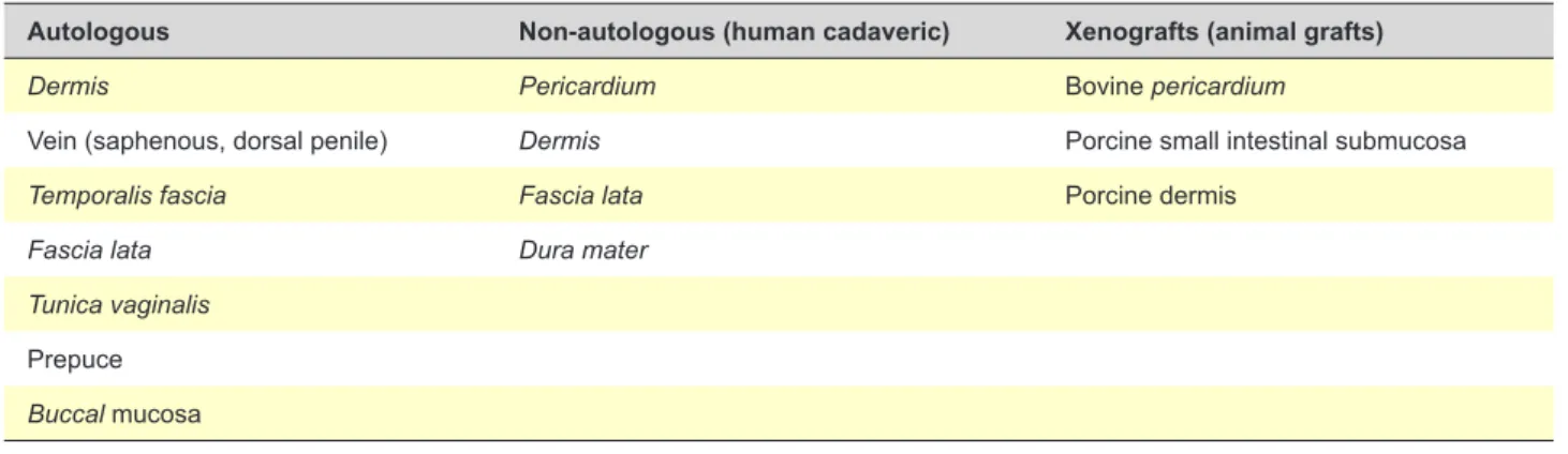
Selecting an adequate graft is still an open discussion. There is no randomized-control trial comparing different grafts and outcomes and no consensus or guideline exists on the best one. The ideal graft has to withstand the elevated corporal pressure in order to preserve erectile capacity, pliable, compliant (without allowing aneurismal dilation) with a low infection rate, non-thrombogenic biologically tolerated with no antigenic activity and little fibrotic reaction, inexpensive e readily available.41 Grafts can be divided into three categories: autologous grafts, nonautologous and synthetic graft (Table 1):
• Autologous grafts include buccal mucosa, tunica
vagi-nalis or albuginea, saphenous vein, lower abdomen dermis, fascia lata, fascia temporalis, rectus sheath.33 They cause less inflammatory reaction with a lower rate of infection. Three out of four patients report a ‘complete straightened’ penile curvature.2,47 Saphenous vein is very good option. In spite of the need for a separate incision and longer operating time, it’s simple to harvest, reliable, with a large surface area, good compliance and with a low fibrotic reaction. But it seems to have a short half-life, since most series published demonstrate deterioration 5 years after surgery.42
• Non-autologous or extracellular matrix tissues (ECM) include both allo- and xenografts - bovine and cadaveric
pericardium, fascia lata, small intestine submucosa-SIS
collagen Xeece and porcine dermis.43-45 Xenografts, isolated from animals, like bovine pericardium and porcine small intestinal submucosal grafts are good options in the surgical treatment of PD. There is no need for a separate incision, a reduced morbidity (and surgery time) and also decreased hypothetical risk of prions transfer and/or infection, with good satisfaction rates.
• Synthetic materials (polyester, polytetrafluoroethylene-PTFE, Dacron) have been used with various results and are no longer recommended nowadays. They have a high
risk for infection, allergic reaction, a high rate of fibrosis and contracture, as well as recurrence of the plaque. 48
Tunica albuginea has not been satisfactory, as it can’t
prevent cavernous insufficiency or guarantee penile straightening on a long follow-up.46 Buccal mucosa looks to be a very attractive substitute for tunica albuginea due to it’s anatomical structure with a high vascularization has also a quick take with a low risk of hypoxia-induced fibrosis while preserving tissue elasticity and lengthening, reducing the risk of penile shortening and de novo ED.47
A novel approach using an acellular human dermis tissue graft (Epiflex®) that exhibits ECM characteristics looks promising.41 In spite of the low number of patients reported and short follow-up some advantages were met: good intraoperative handling and tensile strength, homologous, cell-free and with extremely low residual DNA, good tolerability, no antigenicity and minimum fibrotic reactions, low cost with reduced postoperative complications such as penile hematoma or hypoesthesia. Overall, grafting for PD has been troubled by significative venous leakage with consequent ED. Prophylactic ligation of the dorsal vein is performed with questionable success.48
In this category, patients with a severe penile deviation or with a large plaque in the distal third of the shaft causing curvature of > 60º – 70º, severe indentation or shortening, there is still another option, the technique of penile disassembly, described by Perovic in 1998.49 In this technique the glans and urethra are completely mobilized off of the distal corpora and the pathological tunica
albuginea is excised and the glands repositioned, resulting
in a functional penis with no remarkable shortening. They reported excellent correction of glans tilt and curvature in the distal third of the corporeal bodies.50
Penile prosthesis
Penile prosthesis addresses two problems: ED and the penile shaft deformation. It’s recommended in patients with PD refractory to conservative therapy, harboring cavernosal arterial insufficiency and inadequate preoperative rigidity, a venous leak, or combined vascular abnormality that causes ED.51 Penile prosthesis alone can correct the curvature in almost 70% of patients.52 Both malleable and inflatable penile prostheses (IPP) have been used with worst results Table 1 - Available grafts for Peyronie’s disease
Autologous Non-autologous (human cadaveric) Xenografts (animal grafts)
Dermis Pericardium Bovine pericardium
Vein (saphenous, dorsal penile) Dermis Porcine small intestinal submucosa
Temporalis fascia Fascia lata Porcine dermis
Fascia lata Dura mater
Tunica vaginalis Prepuce Buccal mucosa
ARTIGO DE REVISÃO
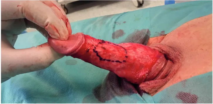
Figure 4 - Plaque excision
from the latter.21 Patients with extended circumferential plaques and severe corporal fibrosis may need additional surgical dilators (Rossello® cavernotomes or the Otis® urethrothomes).53 The ideal IPP is the three-piece inflatable penile prosthesis because it offers more support during modeling than a malleable prosthesis.8 The degree of residual curvature after surgery determines subsequent treatment, with 19% – 42% of patients needing additional straightening procedures.54 If the curvature is less than 30 degrees with concomitant ED, straightening may be achieved with an inflatable prosthesis alone5 in about 6 – 9 months following resume of sexual activity.9 If residual curvature is over 30 degrees patients usually have to go through manual penile modeling, introduced in 199455 by Wilson and Delk: it consists in forcibly bending the penis in the direction opposite the curvature and holding for up to 90 seconds, if possible, leading to plaque rupture. Cylinders have to be insufflated. The objective is to progressively lower the curvature to under 30 degrees. In order to prevent urethral perforation from prostheses dislodgment, bending of the penis must be with a hand on the shaft rather than on the glands, preventing downward pressure on the cylinder tips.2 It has become the predominant method utilized in the United States for penile straightening during IPP placement.56,64,65
If curvature > 30º persists, subsequent plaque-releasing incisions, transcorporeal techniques (transcorporeal incision, scratch procedure) or grafting are also usually needed. Grafting is recommended for tunical defects greater than 2 cm preventing prostheses herniation.10,57 There are currently two companies marketing IPP: American Medical Systems (AMS) and Coloplast. Both the AMS 700 CX® and the Coloplast Titan® seem to be similar when it comes to associated technical problems or patient satisfaction.58 Major side-effects include penile shortening
in about 50% of cases, penile hypoesthesia, paresthesia, infection, erosion, mechanical failure and difficulties using the device.5,9 Kayigil et al published in 2013 an alternative technique to IPP placement: combining penile revascularization and Essed-Schroder plication or plaque excision with venous grafting. In spite of the small sample (nine patients) and short follow-up, complete straightening was obtained with improvement in IIEF-5 scores.59
Finally, severe penile shortening without major curvature in Peyronie’s disease also deserves special consideration, when stretching of the penis is the main therapeutic goal. Levine et al, in 2008, created a new device called FastSizeTM Medical Extender that performs periodic gentle stretching of the penis, through small metal extensions, without the need for invasive surgery.60 Surgically the division of the suspensory ligament, providing lengthening of the free portion of the shaft, provides little satisfaction for the patient.61 A ‘sliding technique’ from Rolle et al, published in 2012, also addresses severe shortening of the penis due to Peyronie’s disease. Two separate transverse incisions with posterior dilatation of the corpora cavernosa, the insertion of the prosthesis and added suture of two grafts seems promising, with an increase in length close to 3 cm.62 Also Egydio and Sansalone reported a length gain of over 3 cm by performing circumferential incision and grafting with subsequent IPP placement.63 A novel report describes preoperative traction therapy for men with a shortened penis, prior to IPP placement that may yet be another alternative to prevent shortening, with even some length gain (0.5 – 2.0 cm).64
DISCUSSION
Post-operative rehabilitation
The purpose of postoperative rehabilitation after non-prosthetic surgery is to promote erectile function, reduce
ARTIGO DE REVISÃO the risk of ED and reduce penile shortening optimizing a
straight healing.2,5 Massage and stretch therapy starts 2 weeks after surgery. Patient must counsel on how to perform it. The patient is instructed to grasp the penis by the glans and gently stretch it and then with his other hand massage the shaft of the penis for 5 min twice per day for 2 - 4 weeks. The massage and stretch can also be performed by the patient’s partner if possible. This will reinitiate the sexual experience for the couple and hopefully diminish the fear of reinjuring the penis, for which the partner may feel responsible.2,9
Penile extenders in the treatment of Peyronie’s disease
Patients in the chronic stage of their disease, where medical treatments have no place, with a mild to moderate degree of curvature (not exceeding 50°) and no concomitant ED, have no recommend treatment. A penile extender device65 (Fig. 4) produces an improvement in penile curvature with acceptable results, probably useful in a multimodal treatment strategy.
CONCLUSION
PD is still is a multifactorial challenge for the urologist weather is it because of the conflicting results of nonsurgical treatment options or the demanding for a experienced surgeon to obtain optimal results in approaching PD. There is to date, no high level of evidence-based data to determine
which is the best surgical treatment of PD. Patients best suited for surgery are ones with a stable disease, usually with 12 months into the disease without progression. Failed medical therapy and patient’s preference for a definitive resolution are also important additional indications for surgical treatment. Debate still exists on the ideal graft material that withstands the elevated intracorporal pressures and natural contracture. Plaque surgery with focus in preventing penile length loss is still a challenge and a few technique modifications have shown up in the last few years. Penile traction therapy prior to prostheses placement or after grafting has shown good promising outcomes in small trials.
PROTECTION OF HUMANS AND ANIMALS
The authors declare that the procedures were followed according to the regulations established by the Clinical Research and Ethics Committee and to the Helsinki Declaration of the World Medical Association.
CONFLICTS OF INTEREST
The authors declare that there are no conflicts of interest.
FUNDING SOURCES
This Project was not sponsored nor did it receive any grant or financial assistance.
REFERENCES
1. Musitelli S, Bossi M, Jallous H. A brief historical survey of ‘Peyronie’s disease’. J Sex Med. 2008;5:37–46.
2. Levine L, Larsen S. Surgery for Peyronie’s disease. Asian J Androl. 2013;15:27–34.
3. Martinez D, Ercole C, Hakky TS, Kramer A, Carrion R. Peyronie’s disease: still a surgical disease. Adv Urol. 2012;2012:206284. 4. Nelson CJ, Mulhall JP. Psychological impact of Peyronie’s disease: a
review. J Sex Med. 2013;10:653–60.
5. Ralph D, Gonzalez-Cadavid N, Mirone V, Perovic S, Sohn M, Usta M, et al. The management of Peyronie’s disease: evidence-based 2010 guidelines. J Sex Med. 2010;7:2359–74.
6. Chung E, Clendinning E, Lessard L, Brock G. Five-year follow-up of Peyronie’s graft surgery, outcomes and patient satisfaction. J Sex Med. 2011;8:594–600.
7. Taylor FL, Levine LA. Peyronie’s disease. Urol Clin North Am. 2007;34:517–34.
8. Gur S, Limin M, Hellstrom WJ. Current status and new developments in Peyronie’s disease: medical, minimally invasive and surgical treatment options. Expert Opin Pharmacother. 2011;12:931-44.
9. Carson CC, Levine L. Outcomes of surgical treatment of Peyronie’s disease. BJU Int. 2014;113:704–13.
10. Garaffa G, Minervini A, Christopher NA, Minhas S, Ralph DJ. The management of residual curvature after penile prosthesis implantation in men with Peyronie’s disease. BJU Int. 2011;108:1152-6.
11. Segal RL, Burnett AL. Surgical management for Peyronie’s disease. World J Mens Health. 2013;31:1-11.
12. Levine LA, Burnett AL. Standard operating procedures for Peyronie’s disease. J Sex Med. 2013;10:230-44.
13. Fausto de Souza D, Micaelo L, Cuzzi T, Ramos-E-Silva M. Ledderhose disease: an unusual presentation. J Clin Aesth Dermatol. 2010;3:45–7. 14. Ohebshalom M, Mulhall J, Guhring P, Parker M. Measurement of penile
curvature in Peyronie’s disease patients: Comparison of three methods. J Sex Med. 2007;4:199–203.
15. Chung E, De Young L, Brock GB. Penile duplex ultrasonography in men with Peyronie’s disease: is it veno-occlusive dysfunction or poor
cavernosal arterial inflow that contributes to erectile dysfunction? J Sex Med. 2011;8:3446-51.
16. Kalokairinou K, Konstantinidis C, Domazou M, Kalogeropoulos T, Kosmidis P, Gekas A. US imaging in Peyronie’s disease. J Clin Imaging Sci. 2012;2:63.
17. Flores S, Choi J, Alex B, Mulhall JP. Erectile dysfunction after plaque incision and grafting: short-term assessment of incidence and predictors. J Sex Med. 2011;8:2031–7.
18. Sherer BA, Warrior K, Levine LA. 2013-2014 updates in Peyronie’s disease management. Curr Urol Rep. 2014;15:459.
19. Nesbit RM. Congenital curvature of the phallus: report of three cases with description of corrective operation. J Urol. 1965;93:230–2. 20. Ralph DJ, Al-Akraa M, Pryor JP. The Nesbit operation for Peyronie’s
disease: 16-year experience. J Urol. 1995;154:1362–3.
21. Rehman J, Benet A, Minsky LS, Melman A. Results of surgical treatment for abnormal penile curvature: Peyronie’s disease and congenital deviation by modified Nesbit plication (tunical shaving and plication). J Urol. 1997;1571288–91.
22. Yachia D. Modified corporoplasty for the treatment of penile curvature. J Urol. 1990;143:80–2.
23. Serefoglu EC, Hellstrom WJ. Treatment of Peyronie’s disease: 2012 update. Curr Urol Rep. 2011;12:444–52.
24. Al-Shaiji TF, Brock GB. Peyronie’s disease: evolving surgical management and the role of phosphodiesterase 5 inhibitors. Scientific World Journal. 2009;9:822–45.
25. Gholami SS, Lue TF. Correction of penile curvature using the 16-dot plication technique: a review of 132 patients. J Urol. 2002;167:2066–9. 26. Langston JP, Carson CC 3rd. Peyronie disease: plication or grafting. Urol
Clin North Am. 2011;38:207–16.
27. Miranda AF, Sampaio FJ. A geometric model of plaque incision and graft for Peyronie’s disease with geometric analyses of different techniques. J Sex Med. 2014;11:1546-53.
28. Lowsley OS, Boyce WH. Further experiences with an operation for the cure of Peyronie’s disease. J Urol. 1950;63: 888–902.
ARTIGO DE REVISÃO 30. Miranda AF, Sampaio FJ. A geometric model of plaque incision and graft dermal graft. J Urol. 1974;111:44–6.
for Peyronie’s disease with geometric analyses of different techniques. J Sex Med. 2014;11:1546-53.
31. Sampaio JS, Passarinho A. Surgical correction of severe Peyronie’s disease without plaque excision. Eur Urol. 1989;16:460-2.
32. Gelbard MK, Hayden B. Expanding contractures of the tunica albuginea due to Peyronie’s disease with temporalis fascia free grafts. J Urol. 1991;145:772–6.
33. Hellstrom WJ, Reddy S. Application of pericardial graft in the surgical management of Peyronie’s disease. J Urol. 2000;163:1445–7. 34. Chun JL, McGregor A, Krishnan R, Carson CC. A comparison of
dermal and cadaveric pericardial grafts in the modified Horton-Devine procedure for Peyronie’s disease. J Urol. 2001;166:185–8.
35. Lue TF, El-Sakka AI. Venous patch graft for Peyronie’s disease. Part I: technique. J Urol. 1998;160:2047–9.
36. Egydio PH, Sansalone S. Peyronie’s reconstruction for maximum length and girth gain: Geometrical principles. Adv Urol. 2008:205739. 37. Kuehhas FE, Egydio PH. The STAGE technique (Superficial Tunica
Albuginea Geometric-Based Excision) for the correction of biplanar congenital penile curvature. J Sex Med.204;11:299–306.
38. Austoni E, Colombo F, Romanò AL, Guarneri A, Kartalas Goumas I, Cazzaniga A. Soft prosthesis implant and relaxing albugineal incision with saphenous grafting for surgical therapy of Peyronie’s disease: a 5-year experience and long- term follow-up on 145 operated patients. Eur Urol. 2005;47:223–9.
39. Shaeer O. Trans-corporal incision of Peyronie’s plaque. J Sex Med. 2011;8:589–93.
40. Kovac JR, Brock GB. Surgical outcomes and patient satisfaction after dermal, pericardial, and small intestinal submucosal grafting for Peyronie’s disease. J Sex Med. 2007;4:1500–8.
41. Adamakis I, Tyritzis SI, Stravodimus KG, Migdalis V, Mitropoulos D, Constantinides CA. A novel approach for the surgical management of Peyronie’s disease using an acellular, human dermis tissue graft: preliminary results. World J Urol. 2011;29:399–403.
42. Montorsi F, Salonia A, Maga T, Bua L, Guazzoni G, Barbieri L, et al. Five-year follow-up of plaque incision and vein grafting for Peyronie’s disease. J Urol. 2000;163:1704-8.
43. Edygio PH, Lucon AM, Arap S. Treatment of Peyronie’s disease by incomplete circumferential incision of the tunica albuginea and plaque with bovine pericardium graft. Urology. 2002;59:570–4.
44. Breyer BN, Brant WO, Garcia MM, Bella AJ, Lue TF. Complications of porcine small intestine submucosa graft for Peyronie’s disease. J Urol. 2007;177:589–91.
45. Kalsi JS, Christopher N, Ralph DJ, Minhas S. Plaque incision and fascia lata grafting in the surgical management of Peyronie’s disease. BJU Int. 2006;98:110–4.
46. Teloken C, Graziottin T, Rhoden E, Da Ros C, Fornari A, Soares FC, et al. Penile straightening with crural graft of the corpus cavernosum. J Urol. 2000;164:107–8.
47. Cormio L, Zucchi A, Lorusso F, Selvaggio O, Fioretti F, Porena M, et al. Surgical treatment of Peyronie’s disease by plaque incision and grafting with buccal mucosa. Eur Urol. 2009;55:1469–76.
48. Dalkin BL, Carter MF. Venogenic impotence following dermal graft repair for Peyronie’s disease. J.Urol. 1991;146:849–51.
49. Perovic SV, Vukadinovic V, Djordjevic ML, Djakovic N. The penile disassembly technique in hypospadias repair. Br J Urol. 1998;81:479– 87.
50. Vo J, Wessells H. Partial penile disassembly and corporeal resection for Peyronie’s disease with distal narrowing. J Urol. 2000;164:449–50. 51. Taylor FL, Abern MR, Levine LA. Predicting erectile dysfunction
following surgical correction of Peyronie’s disease without inflatable penile prosthesis placement: Vascular assessment and preoperative risk factors. J Sex Med. 2012;9:296–301.
52. Garaffa G, Minervini A, Christopher NA, Minhas S, Ralph DJ. The management of residual curvature after penile prostheses implantation in men with Peyronie’s Disease. BJU Int. 2011;108:1152–6.
53. Mulcahy JJ, Wilson SK. Management of Peyronie’s disease with penile prostheses. Int J Impot Res. 2002;14:384–8.
54. Wilson SK, Carson CC. Surgical straightening with penile prosthesis. In: Levine LA, Tolowa NJ editors. Peyronie’s disease. A guide to clinical management. New York: Humana Press; 2007. p. 249-58.
55. Wilson SK, Delk JR. A new treatment for Peyronie’s disease: modeling the penis over an inflatable penile prosthesis. J Urol. 1994;152:1121–3. 56. Levine LA, Benson J, Hoover C. Inflatable penile prosthesis placement
in men with Peyronie’s disease and drug-resistant erectile dysfunction: a single-center study. J Sex Med. 2010;7:3775–83.
57. Mulhall J, Ahmed A, Anderson M. Penile prosthetic surgery in Peyronie’s disease: defining the need for intraoperative adjuvant maneuvers. J Sex Med. 2004;1:318–21.
58. Chung E, Solomon M, DeYoung L, Brock GB. Comparison between AMS 700 CX and coloplast titan inflatable penile prosthesis for Peyronie’s disease treatment and remodeling: clinical outcomes and patient satisfaction. J Sex Med. 2013;10:2855-60.
59. Kayigil O, Okulu E. The surgery with penile corrective techniques as an alternative to pros- thesis implantation in patients with Peyronie’s disease having ED: preliminary results. Int J Impot Res. 2013;25:166– 71.
60. Levine LA, Newell MM. FastSizeTM medical extender for the treatment of Peyronie’s disease. Expert Rev Med Devices. 2008;5:305–10. 61. Li CY, Kayes O, Christopher N, Minhas S, Ralph DJ. Penile suspensory
ligament division for penile augmentation: Indications and results. Eur Urol. 2006;49:729–33.
62. Rolle L, Ceruti C. A new, innovative, lengthening surgical procedure for Peyronie’s disease by penile prosthesis implantation with double dorsal-ventral patch graft: the ‘sliding technique’. J Sex Med. 2012;9:2389–95. 63. Egydio PH, Kuehhas FE, Sansalone S. Penile length and girth
restoration in severe Peyronie’s disease using circular and longitudinal grafting. BJUI. 2012;111:E213–9.
64. Levine LA, Rybak J. Traction therapy for men with shortened penis prior to penile prosthesis implantation: a pilot study. J Sex Med. 2011;8:2112– 7.
65. Rahman NU, Carrion RE, Bochinski D, Lue TF. Combined penile placation surgery and insertion of penile prosthesis for severe penile curvature and erectile dysfunction. J Urol. 2004;171:2346.