Universidade de Lisboa
Faculdade de Ciências
Departamento de Biologia Vegetal
Epithelial Cells from Lung: Production,
Cultivation and Characterization
Usage for Validation of Compounds Efficacy in Rescuing Mutant CFTR
Marta Augusta Moreira Fortio da Palma
Dissertação
Mestrado em Biologia Molecular e Genética
Universidade de Lisboa
Faculdade de Ciências
Departamento de Biologia Vegetal
Epithelial Cells from Lung: Production,
Cultivation and Characterization
Usage for Validation of Compounds Efficacy in Rescuing Mutant CFTR
Marta Augusta Moreira Fortio da Palma
Mestrado em Biologia Molecular e Genética
Dissertação orientada pela
Professora Doutora Margarida D. Amaral
(Departamento de Química e Bioquímica, FCUL)
ii Marta Augusta Moreira Fortio da Palma é bolseira de investigação ao abrigo do projecto "Identification of Novel Targets Involved in CFTR Traffic", financiado pela CFF-US Cystic Fibrosis Foundation (Refª 7207534)
Acknowledgments / Agradecimentos
iii
Acknowledgments / Agradecimentos
No fim de mais esta jornada gostaria, antes de mais, agradecer à Professora
Doutora Margarida Amaral por ter acreditado em mim e nas minhas capacidades e
me ter aceite como sua orientanda. Agradeço a sua supervisão, o seu rigor e o seu
entusiasmo que é uma inspiração para mim.
Gostaria também de agradecer à minha segunda “familia” que são todos os meus
colegas que partilharam e partilham o dia a dia comigo no laboratório e que foram os
principais responsáveis pela minha decisão na realização deste trabalho: ao Simão,
o meu “irmãozinho”, pela sua boa disposição que não acaba nunca e à sua
capacidade de ajudar em qualquer circunstância; ao Francisco e ao seu humor que
nos proporcionaram momentos delirantes; à Joana Almaça pelo seu carinho; à
Joana Martins, Diana e Inna pela sua amizade; à Filipa Mendes, Anabela Ramalho,
Carlos Farinha e Luka Clarke -os “pilares”-pelos seus ensinamentos e partilha de
experiências; aos meus “meninos” Verónica, João Fernandes, João Coelho e Sara
Canato, sempre prontos à ajudar; e finalmente um grande obrigada à Marisa pela
sua amizade inesgotável, pelo companheirismo, pelos ensinamentos, pelos
momentos bons e menos bons que passámos juntas.
À minha amiga Susana, pela sua amizade constante, pelo seu entusiasmo e
coragem.
Aos meus pais por sempre me apoiarem e acreditarem em mim: à minha mãe pelo
seu suporte de “mãe” e ao meu pai, que me conhece, que me ouve e que me
aconselha. Finalmente, ao Pedro, o meu “descomplicómetro”, a minha âncora e meu
companheiro de aventuras.
Table of contents
iv
Table of Contents
ACKNOWLEDGMENTS / AGRADECIMENTOS ... III SUMMARY ... V RESUMO... VI ABBREVIATIONS ... XI
CHAPTER I - GENERAL INTRODUCTION ... 1
1. CYSTIC FIBROSIS - OVERVIEW ... 1
1.1. Clinical description ... 1
1.2. The CFTR Gene ... 2
1.3. CFTR Mutations and Functional Classification ... 3
2. CFTR PROTEIN ... 4
2.1. CFTR Biogenesis and Traffic ... 4
2.2. CFTR Function ... 5
2.3. Small-Molecules Rescuing CFTR Mutants ... 5
3. CELLULAR SYSTEMS AS MODELS TO STUDY CFTR ... 6
3.1. Cell Lines ... 6
3.2. Polarized Monolayers of Human Airway Epithelial Cells ... 7
4. OBJECTIVES OF THE PRESENT WORK ... 10
CHAPTER II - MATERIALS AND METHODS ... 11
1. BIOLOGICAL MATERIALS ... 11
2. METHODS ... 11
2.1. Protocols for the Isolation and Cultivation of Primary Human Bronchial Epithelial (HBE) Cells and Establishement of Polarized Culture Systems ... 11
2.1.1. Protocol 1 – UNC Method for Primary HBE Cultures ... 11
2.1.2. Protocol 2 – Vertex Method for Primary HBE Cultures ... 13
2.2. Transepithelial Electric Resistance (TEER) ... 14
2.3. Immunofluorescence Assays ... 14
2.4. Western blot Technique ... 15
2.4.1. Preparation of total protein extracts ... 15
2.4.2. Western blot ... 15
CHAPTER III - RESULTS AND DISCUSSION ... 16
1. COMPARISON OF PROTOCOLS FOR THE ISOLATION AND CULTIVATION OF PRIMAY HBE CELLS ... 16
2. CHARACTERIZATION OF THE PRIMARY HBE CULTURES BY IMMUNOFLUORESCENCE AND WESTERN BLOT OF EPITHELIAL DIFFERENTIATION AND POLARIZATION MARKERS ... 21
3. SUITABILITY OF PRIMARY HBE MONOLAYERS FOR THE EVALUATION OF COMPOUND EFFICACY IN RESCUING MUTANT CFTR TRAFFIC ... 23
CHAPTER IV - CONCLUSIONS AND PERSPECTIVES ... 26
REFERENCES ... 28
APPENDIX 1 – CELL LINES ... 32
Summary
v
Summary
Cystic Fibrosis (CF) is the most common lethal genetic disease among Caucasians. It is caused by mutations in the gene encoding the CF transmembrane conductance regulator (CFTR) protein, a cAMP-regulated chloride (Cl-) channel that functions at the apical membrane of epithelial cells. F508del is the most common disease-causing mutation occurring in ~90% of CF patients leading to a traffic/ processing defect. Lung disease is the main cause of CF mortality.
The majority of studies on wt- and F508del-CFTR proteins have been conducted in heterologous, non-epithelial or non-polarized/epithelial cellular systems. However, the efficiency of CFTR processing is cell-type and polarization dependent, being endogenous epithelial CFTR maturation significantly different from those in overexpressing systems. It is, thus important to generate models of epithelial, polarized cells so as to resemble more closely the in vivo situation. Primary cultures of human bronchial epithelial (HBE) cells are therefore instrumental for studying basic and applied aspects of CF and other lung diseases. The goals of the present work were: 1) to compare two different protocols for the isolation and cultivation of primary HBE cells regarding the isolation and dissociation of the epithelial layers; 2) to characterize the established primary HBE cultures regarding epithelial differentiation and polarization; and 3) to assess suitability of HBE monolayers to investigate efficacy of small-molecule compounds in rescuing F508del-CFTR traffic.
Our data showed no significantly differences between the two protocols tested. When cultured as air-liquid interface (ALI) cultures, HBE cells are able to polarize, thus closely recapitulating their physiological morphology and epithelial characteristics. Finally, we tested these cells by Western blot for the maturation of F508del-CFTR protein following treatment with different small-molecule correctors, showing the suitability of these HBE monolayers as a valuable pharmacology model for CFTR modulator drug discovery.
Keywords: Cystic Fibrosis, production of human epithelial cells, cell differentiation, efficacy of small-molecule compounds to rescue CFTR.
Resumo
vi
Resumo
A Fibrose Quística (FQ) é a doença autossómica recessiva letal mais comum na população Caucasiana com uma incidência de cerca de 1 em 2.500-6.000 nascimentos e com uma frequência de portadores de 1 em 25-40 indivíduos.
Esta doença é caracterizada pela grave disfunção pulmonar causada pela acumulação de muco que tende a obstruir as vias respiratórias, resultando em infecções bacterianas recorrentes em >95 % dos pacientes. É causada por mutações no gene CFTR (do inglês "Cystic Fibrosis Transmembrane Conductance Regulator") localizado no braço longo do cromossoma 7. Desde a sua identificação em 1989 até à data, mais de 1.900 mutações causadoras de doença foram já identificadas neste gene. No entanto, uma única mutação denominada F508del (representando a deleção do aminoácido fenilalanina na posição 508 da proteína) é responsável por 70% dos cromossomas FQ mundiais e ocorre em 90% dos doentes, pelo menos num alelo. As mutações no gene CFTR estão divididas em 6 classes definidas de acordo com as consequências que as alterações moleculares provocam, nomeadamente a alteração de função ou inibição da produção de CFTR. Esta classificação funcional é da maior importância para o desenvolvimento de estratégias terapêuticas já que mutações da mesma classe vão provavelmente utilizar a mesma abordagem visando corrigir o seu defeito básico.
A proteína CFTR é expressa na membrana apical das células epiteliais sendo a sua principal função o transporte de iões Cl- na membrana apical das células epiteliais das vias respiratórias, intestino, pâncreas e glândulas de suor. No entanto, a principal causa de morbilidade e mortalidade é a doença respiratória crónica, caracterizada pela colonização bacteriana das vias aéreas principalmente por Pseudomonas aeruginosa. Uma vez instaladas estas infecções bacterianas, o transporte mucociliar torna-se ineficiente e as infecções recorrentes originam uma resposta inflamatória exacerbada contribuindo para insuficiência respiratória e, em ultimo caso, à morte. O transplante pulmonar constitui assim, o último recurso para estes doentes em estados avançados. Para além dos ciclos de infecção característicos, os sintomas incluem frequentemente insuficiência pancreática (~85 % dos pacientes), ileus meconial (5-10% dos pacientes), infertilidade masculina quase universal e elevadas concentrações salinas no suor. Esta última característica era já a base do principal método de diagnóstico – o teste do suor, mesmo antes de ser conhecida a causa genética da doença.
A maioria dos estudos sobre o tráfego e processamento da proteína normal (ou "wild-type", wt) e F508del-CFTR têm sido realizados em sistemas heterólogos, nomeadamente células não-epiteliais/ não-polarizadas. No entanto, tem sido demonstrado que a eficiência de processamento da CFTR está largamente dependente do tipo de célula e do grau de
Resumo
vii polarização, sendo significativamente diferente o processamento da CFTR expressa endogenamente ou em sistemas de subexpressão. Assim, torna-se importante produzir células primárias epiteliais polarizadas de forma a reproduzir tanto quanto possível as condições in vivo. Alguns laboratórios têm envidado esforços para optimizar técnicas de isolamento e cultura de células epiteliais das vias respiratórias a partir de tecido nativo.
Assim, o primeiro objetivo deste trabalho foi o de comparar dois protocolos (um proveniente da Universidade da Carolina do Norte-UNC e o outro proveniente da Vertex Pharmaceuticals) de produção e isolamento de células epiteliais primárias a partir de pulmões humanos resultantes de transplantes pulmonares, diferentes em relação ao isolamento e à dissociação do epitélio pulmonar. Pretendíamos desta forma obter um protocolo optimizado a ser utilizado no nosso laboratório. Tendo concretizado este primeiro objectivo, o segundo objetivo foi o de caracterizar as culturas de células primárias polarizadas quanto à respetiva diferenciação epitelial e polarização. Por fim, em ensaios bioquímicos e funcionais, testámos a adequabilidade destas monocamadas de células primárias, para investigar a eficácia de compostos (pequenas-moléculas) no resgate do tráfego da proteína F508del-CFTR, o que constituiu o terceiro objetivo deste trabalho.
Os resultados obtidos mostraram não haver diferenças significativas entre os dois protocolos usados para estabelecer as culturas primárias. Foi importante também observar que não houve diferenças significativas entre a viabilidade celular. As principais diferenças encontradas entre os dois protocolos prendem-se com o revestimento ("coating") dos insertos de membrana porosa ("Snapwell inserts"), bem como com a formulação dos dois "Meios de Diferenciação" que foram usados. Assim, observou-se que células HBE ("Human
Bronchial Epithelial") que crescem em membranas pré-revestidas com o "NIH-3T3 coating"
(protocolo Vertex) não atingiram confluência, isto é, as células não foram capazes de cobrir a membrana (ao fim de 7 dias em cultura). Por outro lado, as células HBE plaqueadas em membranas com revestimento de "colagénio IV" (protocolo UNC) foram capazes de cobrir completamente a superfície da membrana após 24 h, após o qual se diferenciam num período que oscila entre as 3 e as 5 semanas, isto é, independentemente dos "Meios de Diferenciação" utilizados.
Informação obtida a partir de relatórios do laboratório da Vertex, aponta para algumas diferenças na função de CFTR a partir de células que foram mantidas com "Meio de Diferenciação" com extrato de cérebro bovino (BBE, usado em "Meio Diferenciação 2"-DM2) vs extrato de pituitária bovina (BPE, usado em "Meio de Diferenciação 1"-DM1). Os mesmos autores relataram também diferenças na função de CFTR quando usam
"Ultroser-G" que está no DM2, defendendo que a presença deste substituto do soro bovino fetal (mas
mais concentrado) no meio está correlacionada com uma maior função de CFTR. No entanto, não pudemos comprovar estas disparidades, pois enquanto o DM2 tem na sua
Resumo
viii composição BBE e "Ultroser-G", o meio DM1 contém BPE e, em vez de "Ultroser-G", contém albumina de soro bovino (BSA) e factor de crescimento epidérmico (EGF). Assim, seria útil testar futuramente a produção de células HBE primárias com meios iguais mas apenas modificando a composição com BBE/BPE e também alternadamente com
"Ultroser-G"/BSA+EGF.
O segundo objectivo deste trabalho foi o de caracterizar as células epiteliais brônquicas humanas (HBE) em termos de diferenciação/polarização usando técnicas de imunofluorescência e de Western blot usando marcadores epiteliais já conhecidos. Culturas primárias de células a partir de brônquio ou de traqueia (HBE/HTE), mantidas em ALI (interface ar-liquido), conseguem imitar as condições fisiológicas in vivo e conduzem a um estado de diferenciação celular e a um fenótipo de epitélio mucociliar exibindo muitas das características das vias respiratórias humanas, incluindo a secreção de muco, motilidade ciliar e formação de junções celulares. É pois considerado um modelo fisiologicamente mais interessante quando comparado com outros modelos in vitro.
No presente trabalho demonstrámos que células HBE apresentam coloração específica nas junções intercelulares para ambos os marcadores de adesão celular (ZO-1 e E-caderina). Para além disso, as células totalmente diferenciadas mostram coloração positiva para os cílios e valores de resistência elétrica transepitelial (TEER) superiores a 600Ω.cm2 o que indica serem adequadas para ensaios bioquímicos e funcionais.
No terceiro objectivo deste trabalho, pretendíamos avaliar se estas células primárias polarizadas eram adequadas para testar a eficácia de pequenas moléculas no resgate da função da CFTR. Com efeito, nos últimos anos, têm vindo a ser identificadas, em "screens" de alto rendimento ("high-throughput screens"), diversas pequenas moléculas com potencial terapêutico para corrigir o defeito básico na FQ estando já algumas aprovadas para uso clínico, como exemplo o VX-661 e o VX-809 (Lumacaftor), ambos em ensaio clínicos de fase IIb, considerados "correctores". Estes compostos têm como objectivo corrigir os defeitos básicos de folding e tráfego da proteína F508del-CFTR. Por outro lado, compostos denominados "potenciadores" pretendem restabelecer o defeito de abertura ("gating") do canal de cloreto. Entre estes conta-se o VX-770 (Ivacaftor) recentemente aprovado pela
Food and Drug Administration (FDA), nos Estados Unidos da América e pela European Medicines Agency (EMEA) na Europa.
Neste trabalho, utilizando estas células primárias HBE, testámos por Western blot o efeito de pequenas moléculas correctoras e potenciadoras da maturação e função da proteína CFTR mutada. Em geral, os resultados combinados dos ensaios bioquímicos e funcionais (dados não mostrados) revelaram que os ensaios com o VX-809/Lumacaftor apresentaram consistentemente melhores resultados no resgate da F508del-CFTR do que os reagentes de laboratório VRT-325 (C3) ou composto 4a (C4a). Os ensaios realizados
Resumo
ix com os melhores compostos ("lead compounds") obtidos através de screens realizados pelo consórcio TargetScreen2 (União Europeia) não evidenciaram efeitos claros de resgate nestas células primárias de epitélio brônquico. No entanto, estes resultados carecem de ser repetidos em diferentes células primárias originárias de doentes com FQ.
Por último, a capacidade de armazenamento de stocks congelados destas células permite, não só uma maior disponibilidade deste tipo de células, bem como a produção de diferentes estágios de maturação usando amostras do mesmo paciente, mas também replicados com amostras de diferentes indivíduos.
No geral, o trabalho realizado e apoiado por outros relatórios sobre
o
VX-770/Ivacaftor e VX-809/Lumacaftor, juntamente com dados clínicos, vêm dar suporte à utilização do sistema HBE como um modelo farmacológico útil na validação pré-clínica dos moduladores do canal CFTR.Palavras-chave: Fibrose Quística, produção de células epiteliais humanas, diferenciação
Abbreviations
xi
Abbreviations
aa Amino acid
ABC ATP-binding cassette
ACTV Amphotericin, ceftazidime, tobramycin, vancomycin ALI Air-liquid interface
ATP Adenosine 5’Triphosphate BBE Bovine brain extract
BEGM Bronchial epithelial growth medium BPE Bovine pituitary extract
BSA Bovine serum albumin
C4a N-[2-(5-Chloro-2-methoxy-phenylamino)-4’-methyl-[4,5’]bithiazolyl-2’-yl]-benazmide
cAMP cyclic Adenosine 5’Monophosphate CF Cystic Fibrosis
CFTR Cystic Fibrosis Transmembrane Conductance Regulator CK Cytokeratin
Cl- Chloride ion
DM Differentiation medium
DMEM-H Dulbecco’s Modified Eagle’s Medium - high glucose DMSO Dimethyl sulfoxide
DPBS Dulbecco's Phosphate Buffered Saline DSC Differential scanning fluorimetry DTT Dithiothreitol
EGF Epidermal grow factor ER Endoplasmic reticulum FBS Fetal bovine serum
HBE Human bronchial epithelial
HBSS HBSS - Hank's Balanced Salt Solution HTE Human tracheal epithelial
J-MEM Joklik's minimal essential medium LHC LHC serum-free media
MSD Membrane-spanning domain Na+ Sodium ion
NBD Nucleotide binding domain
Abbreviations
xii PBST Phosphate Buffered Saline with Tween 20 at 0.1% (v/v)
PFA Paraformaldehyde PK Protein kinase RD Regulatory domain SD Standard deviation SDS Sodium dodecyl sulfate
TEER Transepithelial electric resistance UNC University of North Carolina UPP Ubiquitin Proteasome Pathway UPP Ubiquitin Proteasome Pathway v/v Volume per volume
VRT-325 4-cyclohexyloxy-2-{1-[4-(4-methoxy-benzenesulfonyl) Piperazin-1-yl]ethyl}quinazoline VX-770 N-(2,4-di-tert-butyl-5-hydroxyphenyl)-4-oxo-1,4-dihydroquinoline-3-carboxamide VX-809 3-(6-(1-(2,2-difluorobenzo[d][1,3]dioxol-5-yl)cyclopropanecarboxamido)-3-methylpyridin-2-yl)benzoic acid
w/v Weight per volume WB Western blot wt Wild type ZO Zona occludens
Chapter I General Introduction
1
Chapter I - General Introduction
1 Cystic Fibrosis - Overview
Cystic fibrosis (CF), also known as mucoviscidosis, is one of the most widespread life-shortening autosomal recessive diseases among Caucasians affecting about 1 in 2500-6000 live births and a carrier frequency of 1 in 25 individuals (Collins, 1992).
Although known for centuries, as shown in many early reports dating back to the 17th century, the first medical descriptions of CF date from 1936 with the work of Fanconi, who termed it as "Mukoviszidose", a German term meaning "thickened mucus". The complete description of CF as a "cystic fibrosis of the pancreas" in 1938 by Dorothy Anderson in the United States was the first comprehensive pathophysiological characterization of the disease (Anderson 1938).
During the 1950’s, a heat wave in the east coast of the United States, lead to the identification of an increased salt content in the sweat of cystic fibrosis patients (Di Sant’Agnese, Darling, Perera, & Shea, 1953). Based on this physiological dysfunction, the sweat chloride test was developed, which remains a reliable, cheap diagnosis test for CF.
In the first half of 1980’s, it became evident that CF was related to a malfunction of epithelial tissues, described to be impermeable to chloride (Cl-). These findings were followed by a major effort to map the gene responsible for CF, that was cloned in 1989, and to identify the protein that it encodes, named cystic fibrosis transmembrane conductance regulator (CFTR) (Kerem et al., 1989; Riordan et al., 1989; Rommens et al., 1989), and later shown to function as a Cl- channel (Bear et al., 1992; Welsh & Smith, 1993). At the same time, the F508del mutation was identified as a prevalent disease-causing mutation (Riordan et al., 1989).
Due to the complexity of its pathophysiology and modest success of pharmacological therapies, CF is still a life-threating disease with reduced life expectancy (in the mid thirties, in the USA) (Cystic Fibrosis Foundation Patient Registry, 2008). Furthermore, after the identification of the CF gene, it was thought that the cure would be easily achievable by gene therapy. Nevertheless given the major difficulty associated with this strategy, gene therapy is at a standstill and it is currently believed that pharmacological approaches will be a faster method to correct the basic defect in CF (Amaral & Kunzelmann, 2007).
1.1 Clinical description
Clinically, CF is a multisystemic disorder. It is mainly characterized by elevated sweat Cl- and sodium (Na+) concentrations, exocrine pancreatic insufficiency (~85%), intestinal
Chapter I General Introduction
2 obstruction called meconium ileus (15-20% of CF newborns) and male infertility (~95%) (Welsh & Smith, 1995; Zielenski & Tsui, 1995). Other manifestations include cirrhosis of the liver and diabetes mellitus (Welsh & Smith, 1995). However, the major cause of morbidity and mortality is a chronic respiratory disease, characterized by colonization of the conducting airway mainly by Pseudomonas aeruginosa (Collins, 1992). Once bacterial infections set in, the mucociliary clearance becomes inefficient and recurrent infections originate an exacerbated inflammatory response contributing to respiratory failure and ultimately to death (Amaral & Kunzelmann, 2007).
Lung transplantation is currently the only definitive treatment for advanced CF. Double-lung or triple (heart-lungs) transplant is usually required, having a 55% rate of survival - 3 years following the transplant (McPhee, Papadakis, & Rabow, 2010).
1.2 The CFTR Gene
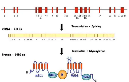
The CFTR gene or ABCC7 is a large (~190 kb) gene located on the long arm of chromosome 7, band 31-32 (7q31-q32). It contains 27 exons that after splicing result in an mRNA of about 6.2 kb that directs the synthesis of a protein with 1480 aa residues (Riordan et al., 1989) (Figure 1.1).
Figure 1.1 – Scheme representing the CFTR gene, mRNA and protein. N – N-terminus; MSD – membrane-spanning domain; NBD – nucleotide-binding domain; R – regulatory domain; C – C-terminus. [Adapted from Zielenski and Tsui 1995].
Chapter I General Introduction
3
1.3 CFTR Mutations and Functional Classification
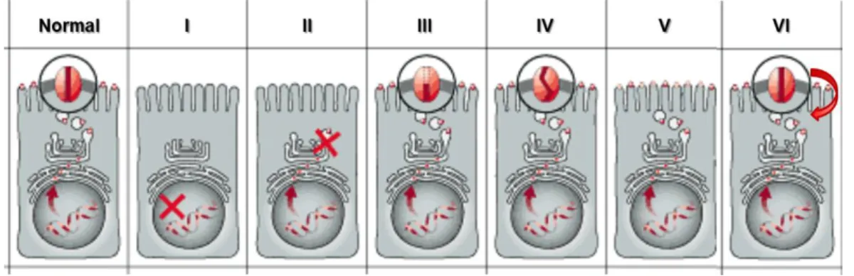
To date, more than 1,900 disease-causing mutations have been reported to the CFTR Mutation Database (http://www.genet.sickkids.on.ca/cftr/) and most are presumably disease-causing. Thus, it is difficult to establish how each of these mutations affects processing and/or function of CFTR protein and how to repair each single defect. This led Tsui (Tsui, 1992) and subsequently Welsh and Smith (Welsh & Smith, 1993) and later Lukacs (Haardt, Benharouga, Lechardeur, Kartner, & Lukacs, 1999) to classify the different mutations according to the mechanism by CFTR Cl- channel function is disrupted (Figure 1.2). This classification is important for the development of therapeutic strategies, as mutations in the same class will likely need the same approach aimed at correcting their basic defect.
Figure 1.2 – Classes of CFTR mutations. (Normal) CFTR protein in the plasma membrane of cells functioning as a Cl- channel; (I) class I mutation: prevent translation; (II) class II: defective processing; (III) class III: defective regulation; (IV) class IV: defective conductance; (V) class V: reduced synthesis; (VI) class VI: decreased stability. [Adapted from Zielenski and Tsui 1995].
Class I mutations cause premature termination of CFTR mRNA translation in the nucleus, resulting in severely decreased or absent CFTR protein production (such as G542X mutation. Most mutations in this class are nonsense, frameshift or aberrant splicing mutations.
Class II mutations, such as F508del or A561E, lead to a retention of protein in the endoplasmic reticulum (ER) followed by a premature targeting for degradation, resulting in reduced amounts or no functional protein at the cell membrane.
Class III mutants (eg.G551D) are located correctly at the cell membrane but are unable to function as a cAMP-activated Cl- channel.
Class IV mutants are also properly located but have altered conductance or gating properties causing a reduced Cl- efflux rate through the CFTR channel, in general associated with milder CF disease.
Class V mutants cause a reduction in the levels of functional CFTR often due to alternative splicing or impaired protein recycling to the cell membrane. In this case, disease severity correlates with the levels of normal transcripts.
Chapter I General Introduction
4 Class VI mutants lead to reduced stability of CFTR at the cell surface (with normal folding, trafficking and Cl- channel function), caused in general by C-terminal truncations, but also includes F508del-CFTR when rescued from the ER.
2. CFTR Protein
CFTR is a multidomain protein containing 1,480 amino acid (aa) residues and functioning as a cAMP-activated and phosphorylation-regulated Cl- channel at the apical membrane of exocrine epithelial cells. CFTR is a member of the ATP-binding cassette (ABC) transporter superfamily, being also named ABCC7.
CFTR structure resembles that of a typical ABC transporter with two membrane-spanning domains (MSD1 and 2) and two nucleotide-binding domains (NBD1 and NBD2). In contrast to most ABC transporters, CFTR also contains a unique and large regulatory domain (RD) with multiple phosphorylation sites (Sheppard and Welsh, 1999).
2.1 CFTR Biogenesis and Traffic
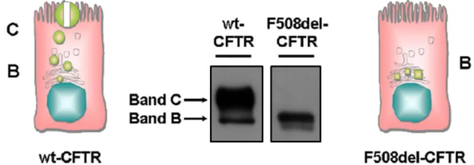
CFTR assembly begins with its co-translational insertion into the ER membrane synthesis where it is core-glycosylated (Cheng et al., 1990). This CFTR immature form, called band B, has a molecular mass of about 140 kDa (Figure 1.3). After passing through the ER quality control, that assesses its folding status, the core-glycosylated form of wild type (wt)-CFTR traffics to the Golgi complex where it undergoes further glycosylation and gradually reaches its mature form, known as band C (170-180 kDa, figure 1.3) (Cheng et al., 1990).
Figure 1.3 – Western blot CFBE41o- cells expressing wt- and F508del-CFTR. Cartoons with permission, Amaral M.D., unpublished; own blot images.
In contrast, the F508del-CFTR protein fails to acquire a native conformation being retained within the ER and targeted to degradation via the ubiquitin (Ub)-proteasome pathway (UPP) (Farinha and Amaral 2005).
Chapter I General Introduction
5
2.2 CFTR Function
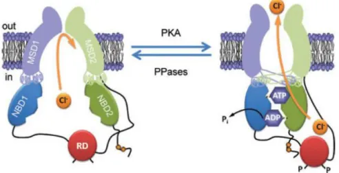
CFTR functions mainly as a Cl- channel but has been described to be involved in several other cellular activities, among which being a regulator of other ion conductances is the most relevant (Amaral et al., 2001). The current model for the mechanism of channel function and regulation (Figure 1.4) envolves firstly the phosphorylation of RD by cAMP-dependent protein kinase A (PKA), and also PKC, that allows ATP binding to the NBDs, which then dimerize (Hwang & Sheppard, 2009; Sheppard & Welsh, 1999). Consequently, the MSDs alter their conformation, allowing the opening of the channel pore and passive flow of ions. It has also been shown that intramolecular interaction between the RD and either the N-terminal or NBD1 are required to regulate CFTR function (Baker et al., 2007).
Although the mechanism of the CFTR channel gating is not fully understood, opening and closing of this Cl- channel is tightly regulated by the cellular balance of kinase and phosphatase activity, by ATP levels and also by the extent of RD phosphorylation (Hwang & Sheppard, 2009).
Figure 1.4 – Conformational changes of the CFTR Cl− channel during channel gating. The simplified model shows a CFTR Cl- channel under quiescent (left) and activated (right) conditions. Communication between the NBDs and MSDs via the intracellular loops is either or orthogonal (e.g. NBD1–MSD2) according to the most recent CFTR structural models based on Sav168834. P (phosphorylation of the RD); Pi (inorganic phosphate); PKA (cAMP-dependent protein kinase); PPase (protein phosphatase). In and Out denote the intra- and extracellular sides of the membrane, respectively. [Reproduced from Hwang and Sheppard, 2009].
2.3 Small-Molecules Rescuing CFTR Mutants
One of the major challenges facing the pharmaceutical industry today is the identification and validation of novel, high-quality "druggable" targets common to distinct diseases which, despite being clinically very diverse (i.e., with different symptoms), share the same basic molecular defect(s).
There are two groups of pharmacological approaches to rescue CFTR dysfunction: the first group of pharmacological chaperones are those acting at the protein trafficking level
Chapter I General Introduction
6 and are termed correctors (Pedemonte et al., 2005). The second group includes compounds that enhance the conductance of the channel and are termed potentiators (Van Goor et al., 2009). Thus, pharmacological rescue relies mostly on mutation-specific therapies and can be envisaged according to the class of CFTR mutation involved (Amaral & Kunzelmann, 2007) (Figure 1.2-Classes of CFTR mutations).
Correctors are the most promising compounds to be used for the repair of class II mutations. Both the most common CFTR mutation, F508del, as well as the "Portuguese mutation", A561E (the second most common mutation in Portugal (Mendes et al., 2003)) fall in this class. Currently, VX-809/Lumacaftor and VX-661, have been identified as good correctors (Van Goor et al. 2009; Accurso et al. 2010), being VX-809/Lumacaftor currently in clinical trials.
As refered to above, potentiators are aimed to activate CFTR protein present at the membrane but with gating/conductance defects (class III/IV mutations). Compounds such as genistein and other flavonoids can activate Cl- conductance and overcome mutational defects in those classes. The potentiator VX-770/Ivacaftor/KalydecoTM (Van Goor et al., 2009) has been shown to increase CFTR activity in CF patients carrying the G551D mutation and it is the first modulator drug, aimed at correcting the basic defect of a CFTR-mutant, to be released to patients (recently approved as a drug by FDA and EMEA). Additionally, VX-770/Ivacaftor/KalydecoTM has also been shown to correct other gating defect CFTR mutants located at the cell surface (Yu et al., 2012), thereby opening new possibilities for the treatment of the CF disease. Moreover, interim results from phase IIb clinical trials showed improvement in lung fuction in patients homozygous for the F508del mutation taking both VX-809 and VX-770.
3. Cellular Systems as Models to Study CFTR
Establishment of the mechanism of action (MoA) of novel compounds and their introduction into clinical trials usually requires studies in cellular systems with heterologous expression of CFTR. Comparative efficacy assessment between heterologous expression systems and airway primary cultures/native human tissues is thus of the ultimate importance before CFTR modulators reach the clinical setting (Ashlock & Olson, 2011).
3.1 Cell Lines
Systems for the culture of human epithelial cells have evolved significantly in recent years. Culture of epithelial cells from organs like bronchus, trachea, nose, skin, esophagus, mammary gland and kidney are possible due to continuous technical improvement (Gruenert et al. 1990).
Chapter I General Introduction
7 Due to the very low levels of endogenous CFTR expression, restricted availability and amount of native epithelial cells, the characterization of the biochemical and genetic mechanisms that causes CF has relied greatly on data obtained in immortalized cell lines stably expressing the CFTR protein or heterologous expression systems (Da Paula et al., 2005). Thus, immortalized epithelial cells from different tissues and species (Appendix 1 - Table 1) have contributed to advance in our understanding of CF disease. Accordingly, the airway epithelium has been the primary tissue target for the development of immortalized cells due to its ability to mimic the molecular events that are involved in CF morbidity and mortality (Gruenert et al. 2004). However, the efficiency of CFTR processing is cell-type and polarization dependent, being endogenous epithelial CFTR maturation significantly different from overexpressing systems. Moreover, it is important to generate models of epithelial, polarized cells so as to resemble more closely the in vivo situation. Primary cultures of human bronchial epithelial (HBE) cells are thus instrumental for studying basic and applied aspects of CF and other lung diseases.
3.2 Polarized Monolayers of Human Airway Epithelial Cells
A limitation in the understanding of CF pathogenesis is the difficulty in studying the link between the mutant CF gene (CFTR) expression and the associated molecular and cellular effects in the absence of an identified animal model for this disease. Although cell lines derived from CF patients airways have been established, they exhibit great heterogeneity, making it difficult to relate their properties exclusively with the CF phenotype. Therefore, all advances in increasing the capacity of human airway epithelial cell cultures to mimic the in vivo epithelium allow the production of better tools to assess native airway function and thus understand CF pathogenesis (Wiszniewski et al., 2006).
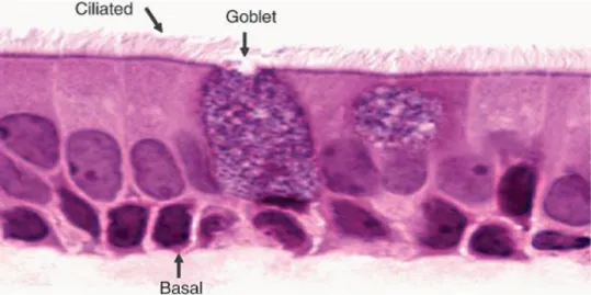
The normal human bronchial epithelium (HBE) is formed by ciliated columnar cells (that constitute ~50% of the epithelial cell population in all airways), basal cells (the second most common cell type in the larger airways) and secretory mucous - Goblet cells (Figure 1.5) (Gruenert et al. 1995; Vaughan et al. 2006).
Epithelia display two main features that distinguish them from other tissues, namely polarity and tightness. Polarity is generated by the asymmetric distribution of proteins between the apical or basolateral membrane. Assemblies of proteins forming "tight junctions" separate the two membrane domains and ensure epithelial tightnes. The formation and permeability of tight junctions is also responsible for the resistance and integrity of the tissue (Brown and Stow 1996).
Chapter I General Introduction
8 Figure 1.5 – Normal Human Bronchial epithelium contains ciliated cells, goblet cells and basal cells. Reproduced from Vaughan et al. 2006)
Thus tight junctions, but also adherens junctions and desmosomes, play a fundamental role in maintaining the polarized phenotype and provide the characteristic tightness of epithelia (Figure 1.6).
Figure 1.6 – Schematic representation of polarized epithelial cells. The junctional complexes and the apical or basolateral distribution of several polarity markers are shown. [Reproduced from Mendes et al. 2004].
In fact, tight junctions selectively regulate the passage of molecules across the paracellular pathway (gating function) of epithelia and passively separate molecules into the apical and basolateral plasma domains (fencing function). To monitor the gating function of tight junctions a simple assay can be used – measuring the transepithelial resistance (TEER) measurements. TEER measurement allows the evaluation of the degree of tightness of the epithelial cells growing on coated filters. Epithelial cells thus asymmetrically distribute
Chapter I General Introduction
9 receptors, transporters, ion channels, and lipids between the apical and basolateral membranes to establish and maintain polarity and function (Mendes et al., 2004).
ALI cultures of primary HBE/HTE cells mimic the in vivo environment and drive differentiation towards a mucociliary phenotype. This culture system is superior to other in
vitro models for many research applications as it configures a model that is physiologically
more relevant (BéruBé, Prytherch, Job, & Hughes, 2010). Submerged culture of primary HBE or HTE cells is possible; however, cells in this system fail to undergo mucociliary differentiation. Notwithstanding, ALI cultures of HBE cells, exhibit many of the characteristics of the human airways, including mucus secretion, ciliary motility and formation of cellular junctions (BéruBé et al., 2010).
Several antigens can be used as biomarkers to assess the correct polarization of epithelial cells: ion channels, for instance, such as the sodium-potassium (Na+/K+) ATPase which is present in the basolateral membrane of most epithelia; cytokeratins which are related to the expression of epithelial specific intermediate filaments and distinguish types of epithelia (simple, pseudo-stratified, stratified, squamous). Other molecules present in junctional complexes such as Calcium-dependent adhesion protein (E-cadherin) or zona
occludens (ZO-1) proteins are a powerful tool to characterize cells in term of their fully
Chapter I General Introduction
10
4. Objectives of the present work
Some laboratories (Gruenert, Randell, Van Goor) have made efforts to optimize techniques for isolation and culture of airway epithelial cells from native tissues. Freshly harvested airway epithelial cells cultured on plastic dishes acquire a poorly differentiated, squamous phenotype and can be repeatedly passaged (primary cultures). These passaged primary airway epithelial cells, when cultured under appropriate conditions, undergo differentiation and polarization, which enables them to more closely recapitulate their normal
in vivo morphology and epithelial characteristics (BéruBé et al., 2010). However, there are
differences in the protocols developed by those researchers and at present it is not established which one is superior for studies involving CFTR.
Lungs explanted from CF patients during transplantation are a potentially useful source of cells for primary cultures. Also, lungs from potential (non-CF) organ donors which are frequently unsuitable for transplantation as a result of age, smoking history, or acute injury such as aspiration, pulmonary edema, or pneumonia, are still very useful for research. This requires establishment of working relationships with surgeons and pathologists and compliance with appropriate regulations. Our research group has established such collaborative research protocol with transplant centres, both in Lisboa (Hospital de Santa Marta, Prof José Fragata) and Universitario y Politécnico La Fe, Valencia, Spain (Dr. Amparo Solé), so as to receive the explanted lungs of CF patients and of other conditions or donors (controls).
The objectives of the present work were the following:
1) To compare two different protocols for the establishment of polarized cultures of CF and non-CF primary HBE cells;
2) To characterize the above two types of primary HBE cells by immunofluorescence and Western Blot using known markers of epithelial differentiation and polarization;
3) To assess the suitability of the established HBE cultures to investigate efficacy of small-molecule compounds in rescuing the traffic of F508del-CFTR.
Chapter II Materials and Methods
11
Chapter II - Materials and Methods
1
Biological Materials
The Membrane Protein Disorders Unit of the BioFIG (University of Lisboa/Faculty of Sciences; head: Margarida D Amaral) has established collaborations with the Cardio-Thoracic Surgery Department at Hospital Santa Marta, Lisboa (Prof. José I Fragata) and with Unidad de Trasplante Pulmonar y Fibrosis Quística - Hospital Universitario y Politécnico La Fe, Valencia, Spain (Dra. Amparo Solé), in order to receive explanted lungs from CF and non-CF individuals, which received approval from the Ethics Committee of both Hospitals to be used in the establishment of primary cultures of human bronchial epithelial (HBE) cells.
2
Methods
Here, we tested two protocols for the establishment of primary cultures of human bronchial/tracheal epithelial (HBE/HTE) cells: one from Scott Randell’s lab in University of North Carolina (UNC), Cystic Fibrosis Center at Chapell Hill-USA (M. Fulcher, Gabriel, Burns, Yankaskas, & Randell, 2005) and another from Vertex Pharmaceuticals Incorporated (San Diego-USA) (Neuberger, Burton, Clark, & Van Goor, 2011), with the aim of obtaining an optimized protocol.
2.1 Protocols for the Isolation and Cultivation of Primary Human Bronchial
Epithelial (HBE) Cells and Establishement of Polarized Culture Systems
Whole lungs were obtained from non-CF, CF and donor subjects following lung transplantation. After explanted, lungs were packed in Dulbecco’s phosphate buffered saline (DPBS) maintained at 4ºC, transported to the laboratory on ice and processed within 24 h. All procedures were performed in a class II, biological safety cabinet and standard precautions were followed. The first step (common for both protocols and described below) consisted in dissecting the airways by removing the excess of connective tissue and slicing them into small segments (Figure 2.1-A).
2.1.1. Protocol 1 – UNC Method for Primary HBE Cultures
As referred above, tissue segments were cleaned by removal of any additional connective tissue and lymph nodes through rinsing in DPBS solution (Figure 2.1-A). Segments were slit longitudinally and cut into 2-5 cm portions with a scalpel (Figure 2.1-B). Finally, they were transferred to specimen cups containing chilled DPBS solution. Because human tissue samples are likely to contain microbial contaminants (yeasts, bacteria or fungi),
Chapter II Materials and Methods
12 exposure to antibiotics should begin as soon as possible using a cocktail of common antibiotics – such as amphotericin, ceftazidime, tobramycin, and vancomycin (ACTV cocktail). Dissected tissues were then washed three times in "Wash Medium" prepared by adding the above mentioned antibiotics to Joklik's minimal essential medium (J-MEM). Tissues from chronically infected patients containing abundant secretions are further treated to remove thick mucus and other debris with a "Soak Solution 1" containing supplemental antibiotics, dithiothreitol (DTT, 0.5mg/mL) and DNase (10µg/mL) and washed again in the "Wash Medium" (Figure 2.1-C) (See Appendix 2 for media and solutions composition).
Figure 2.1- CF Lung Dissection Steps: (A) removing all excess of connective tissue and cleaning of mucus (arrows show mucus); (B) segmenting the bronchi in small portions; (C) rinsing tissue in "wash medium" to remove mucus and any debris.
After washing, tissue segments were transferred into 50-mL conical tubes containing 30 mL wash medium plus 4 mL Protease/DNase solution - "Dissociation Solution" (Figure 2.2-A). The tubes were placed on a rocking platform (at 60 cycles/min), in the cold room (4°C), for 18-24h. To stop tissue dissociation, we then transferred the contents of 50-mL tubes into 150-mm tissue-culture dishes, added fetal bovine serum (FBS) to a final concentration of 10% (v/v) to neutralized protease and scrapped epithelial surface with a convex surgical scalpel (Figure 2.2-B). Tissue surfaces were then rinsed and cells collected from the dish with J-MEM (Figure 2.2-C).
Figure 2.2- Dissociation and Isolation of Airway Epithelial Cells: (A) tissue segments are transferred into 50mL conical tubes containing "Dissociation Solution"; (B) scrapping epithelial surface with a convex surgical scalpel; (C) collecting dissociated cells from the dish.
Chapter II Materials and Methods
13 Solutions containing dissociated cells were pooled into 50-mL conical tubes and centrifuged at 500g for 5 min at 4°C. Additionally, whenever the pellet contained too much red blood cells, a cell lysis treatment was performed (for more protocol details, see Appendix 2). After this procedure, cells were resuspended in "Declump Solution 1" in order to disperse the epithelial sheets and incubated for 15-30 min at 37ºC. FBS was added to a final concentration of 10% (v/v) and the suspension was centrifuged at 500g for 5 min. Finally, the pellet was resuspended in a volume calculated in order to achieve a cell suspension with approximately 5 x 10⁶ cells/mL, being cells counted using a hemocytometer (Neubauer chamber). Primary HBE cells were then plated in bronchial epithelium growth medium (BEGM) and seeded on fibronectin-collagen-coated plastic dishes at a density of 2-6 x 10⁶ per 100mm dish. 24h later, the attachment of cells to the plastic dish was evaluated by observation under the inverted microscope. If the cells were well attached and the dish contained few clumps of floating epithelial cells, cells were washed with DPBS and fed with BEGM plus antibiotics. Large floating clumps of cells can be "rescued" to increase cell yield. For this purpose, medium was collected in 50-mL conical tubes, the dishes washed with DPBS and added to the harvested clumps. Cells were pelleted at 500g for 5 min and followed the previously described protocol with "Declump Solution 1" and cells were replated into new 100mm dishes at a density of 2-6 x 10⁶.
Primary cell media (BEGM) was supplemented with additional antibiotics (ACTV) for the first 3 days after plating, and changed every 2-3 days to prevent acidification. After reaching 70-90% confluency, cells were trypsinized (passage 1) and seeded on collagen IV coated porous membranes (Snapwell Inserts Ø12mm, from Corning-Costar®, New York, NY, USA) at a density of 2–3 x 10⁵ and grown submerged in ALI-Air-Liquid Interface
("Differentiation medium"-DM1) medium for 72h. After 72h, the DM1 medium was removed from the apical side of the filter to establish an ALI culture. This results in fully differentiated HBE cell cultures in 3-5 weeks, exhibiting the desired characteristics of differentiated airway epithelia. (See Appendix 2 for media and solutions composition).
2.1.2. Protocol 2 – Vertex Method for Primary HBE Cultures
Similarly to the previous protocol, lungs were placed in a tray containing DPBS (4◦C) and primary and secondary bronchial tubes were removed and cleaned from the connective tissue by blunt dissection. Bronchial tubes were cut into 2-5cm lenghs and placed into 50mL tubes containing the same "Wash Medium" as in Protocol 1. Bronchial tubes were then placed in a "Soak Solution 2" containing DTT (0.5mg/mL) and DNase (62.5µg/mL). After this procedure, "Soak Solution 2" was removed and replaced with the "Dissociation Solution" (same as in protocol 1) and placed on a roker plate for 36h at 4ºC at 60cycles/min. To separate the epithelial cell layer from the underlying connective tissue, the epithelial surface
Chapter II Materials and Methods
14 was gently scrapped with a convex surgical scalpel, as in protocol 1. The epithelial sheets were then transferred into 15-ml conical tubes containing J-MEM. To disperse the epithelial sheets, J-MEM was replaced with 14 ml of AccutaseTM ("Declump Solution 2") and incubated at 37ºC for 20 min (similarly to what was done in protocol 1). The material was collected by spinning the conical tubes for 5 min at 500g and ressuspended in BEGM plus antibiotics (ACTV) for cell counting. Cells were then plated at a density of 2–6 × 10⁶ into pre-treated
NIH-3T3 conditioned media coated 100-mm dishes (see Appendix 2 for the detailed protocol of NIH-3T3 conditioned media coating). BEGM medium was replaced every 2-3 days to prevent acidification. After reaching ~80% confluency, cells were trypsinized to NIH-3T3 conditioned media coated porous membranes (Snapwell Inserts Ø12mm, from Corning-Costar®, New York, NY, USA) at a density of 2–3 × 10⁵ and grown submerged in
"Differentiation Medium"-DM2 for 72h. After 72h, the "Differentiation Medium" was removed from the apical side of the filter to establish an ALI culture resulting in fully differentiated HBE cell cultures in 3-5 weeks (See Appendix 2 for media composition).
2.2 Transepithelial Electric Resistance (TEER)
The transepithelial electric resistance (TEER) was measured every 3 days in differentiating cells using an volt-ohm meter (Millicell-ERS, Millipore) over 23 days at ALI, to confirm development of tight junctions. Briefly, 0.4mL of media was placed in the apical compartment. Cultures were equilibrated in the incubator for 30 min before measurement of TEER. Apical medium was then aspirated to restore ALI. TEER of insert and medium alone was substracted from measured TEER and Ω.cm2 calculated by multiplying by the insert area.
2.3 Immunofluorescence Assays
HBE cells were expanded in 8-well chamber slides and/or grow in Snapwell inserts. Cells were firstly washed for three times, 5 min each, with PBS and fixed with 4% (w/v) paraformaldehyde (PFA), both at the apical and basolateral sides. Cells were permeabilized with Triton-X (0.5% v/v) for 10-15 min and washed again with PBS as previously. Next, cells were incubated with 1% (w/v)(bovine serum albumin (BSA) as blocking solution for 20 min. For the polarized cells, membranes were removed from the Snapwell support with the help of a scalpel, split ion four and placed on a 12-well plate with PBS. Following this procedure, cells were incubated with primary antibodies (dilution 1:100) diluted in 1% (w/v) BSA for 1h (Table 2.1). Cells were then washed three times with PBS supplemented with Triton-X (0.05%) for 5 min and incubated with fluorescent conjugated secondary antibodies (Alexa Fluor 488 donkey anti-mouse- dilution 1:500 - Invitrogen, Carlsbad, CA, USA), diluted in BSA 1% (w/v) for 1h. Final washes were performed as before following by post-fixation with 4% PFA for 10 min. Preparations were mounted in DAPI Fluoromount-G to label nuclei
Chapter II Materials and Methods
15 (SouthernBiotech, AL-USA). Immunofluorescence staining was observed and images acquired using a confocal microscope Leica TCS SPE (Leica, Jehna, Germany) for polarized cells and a Zeiss Axiovert 200M (Carl Zeiss AG,Oberkochen, Germany) for chamber-slides.
Table 2.1 – Primary antibodies for immunofluorescence
Antibody Cell structure Host Supplier (ref. #)
Cytokeratin 18
Intermediate filaments (fully differentiated epithelial cells: ciliated and secretory
epithelial cells)
Mouse Santa Cruz
sc-32329 Cytokeratin 14 Intermediate filaments (basal and poorly
differentiated epithelial cells) Mouse
Santa Cruz sc-53253
E-cadherin Adherens junctions Mouse BD (610181)
zonula occludens
protein (ZO-1) Tight Junctions Mouse Invitrogen (339100)
Acetylated Tubulin Microtubulos, ciliary axonemas Mouse Sigma T6793
2.4 Western blot Technique
2.4.1 Preparation of total protein extracts
Protein extracts from HBE cells were prepared by lysis in Laemmli sample buffer: 1.5 % (w/v) Sodium-dodecyl-sulfate (SDS); 10 % (v/v) glycerol; 0.001 % (w/v) bromophenol blue; 0.5 mM dithiothreitol (DTT); 31.25 mM Tris (pH 6.8). DNA was sheared by treatment with 1 μl (5U) benzonase®
nuclease (Sigma-Aldrich, St Louis, MI, USA). All protein extracts were quantified by modified micro-Lowry method (Farinha et al. 2004).
2.4.2 Western blot
After protein quantification, 30-100µg of total protein per sample were separated by SDS-polyacrylamide gel electrophoresis (PAGE) on 7 % (w/v) mini-gels, followed by transfer onto PVDF membrane (Immobilon-P Membrane, PVDF, 0.45 µm, Merck Millipore, MA-USA). Prior to CFTR detection, the membrane was blocked with 5% (w/v) skimmed milk in PBS containing 0.1% (v/v) Tween 20 (PBST) for 2h to prevent non-specific adsorption of the antibodies. After blocking, the membrane was incubated with primary anti-CFTR monoclonal antibody 596 (Cystic Fibrosis Foundation, Bethesda, MA, USA diluted 1:3000), anti-zonula
occludens protein (ZO-1, 1:100 dilution), anti-E-Cadherin (1:100 dilution) or anti-acetylated
tubulin (1:20.000 dilution) for 2h at room temperature (RT) in 5% (w/v) milk-PBST. Membranes were washed then 3 times for 10 min with PBST and incubated with a secondary goat peroxidase-conjugated anti-mouse IgG antibody at 1:3,000 (Bio-Rad
,
CA-USA) for 1h at RT in 5% (w/v) milk-PBST. After 3 washes (10 min) with PBST, blots were developed using the Chemiluminescent Substrate ImmobilonTM Western detection system (Merck Millipore) and Super RX Fujifilm or using Immun-StarTM Western CTM Cheluminescent Kit (Bio-rad, CA-USA) and ChemidocTM XRS+ Image System (Bio-rad, CA-USA).Chapter III Results and Discussion
16
Chapter III - Results and Discussion
1
Comparison of Protocols for the Isolation and Cultivation of
primay HBE Cells
In vitro cultures of HBE cells are instrumental for studying basic and applied aspects
of CF and other lung diseases. Lungs removed from CF patients during transplantation are a potentially useful source of cells for culture. Therefore, it is necessary to have optimal methods allowing large-scale isolation and production of cells from a single lung. Bearing in mind this objective, we compared two similar protocols (Fulcher et al. 2005; Van Goor et al. 2011) in order to achieve an optimal protocol to perform our studies.
Both methods use the same dissection procedure. Subsequently, the small tubes (bronchi and bronchioles) are soaked with similar solutions in both protocols, but containing DNase in different concentrations, namely 10µg/mL (protocol 1 - UNC) or 62,5µg/mL (protocol 2 - Vertex) (Table 3.1). The purpose of DNase treatment is to remove mucus and other debris, especially when tissues are from chronically infected patients. The next step, for both protocols is dissociation by a protease (proteolytic digestion). The only difference between the two protocols is the incubation time with the "Dissociation Solution". In the first protocol (UNC), tissues are incubated for 18-24h, while in the second protocol (Vertex) incubation is for 36h (Table 3.1). Based in our data, there was no significant difference resulting from these different incubation times. Actually, we observed that the epithelial layer is completely removed from the underlying connective tissue after 18h.
Originally, in the second protocol (Vertex) there was no step concerning the elimination of red blood cells. However, we observe that lungs from CF patients (especially the ones with severe disease) usually have larger amounts of mucus and red blood cells than non-CF or even donors. Moreoever, we verified that it is crucial to remove these for epithelial cell viability. So, we have adopted to include this step to remove red blood cells also in protocol 2.
To prevent cell clumping, both protocols use solutions to desagregate cells. The first protocol uses a "homemade" solution – "Declump solution 1" - which basically contains DTT, collagenase and DNase. The second protocol uses a commercial enzyme-Accutase™ which coomonly replaces Trypsin/EDTA
for
cell detachment and dissociation ("Declump Solution 2") (Table 3.1).Chapter III Results and Discussion
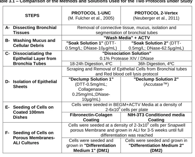
17 Table 3.1 – Comparison of the Methods and Solutions Used for the Two Protocols under Study
STEPS PROTOCOL 1-UNC (M. Fulcher et al., 2005)
PROTOCOL 2-Vertex (Neuberger et al., 2011) A- Dissecting Bronchial
Tissues
Removal of connective tissue, mucus, isolation and segmentation of bronchial tubes
B- Washing Mucus and Cellular Debris
"Wash Media" + ACTV "Soak Solution 1"
(DTT-0.5mg/L; DNase-10µg/mL)
"Soak Solution 2" (DTT-0.5mg/L; DNase-62.5µg/mL) C- Dissociatiating the
Epithelial Layer from Bronchia Tubes
"Dissociation Solution" 0.1% Protease XIV / DNase
18-24h Digestion, 4ºC 36h Digestion, 4ºC
D- Isolation of Epithelial Sheets
Scraping and Removal of Epithelial Cells from Bronchial tubes and Red blood cell lysis protocol
"Declump Solution 1" (DTT-0.5mg/mL; Collagenase- 0.25mg/mL;DNase-10µg/mL) "Declump Solution 2" (Accutase™) E- Seeding of Cells on Coated 100mm Dishes
Cells were seeded in BEGM+ACTV Media at a density of 2-6x10⁶ cells per plate
Fibronectin-Colagen Coating
NIH-3T3 Conditioned media Coating
F- Seeding of Cells on Porous Membranes- ALI Cultures
Cells were seeded at a density of 2-3x10⁵ cells per Snapwell porous Membrane and grown in ALI for 3-5 weeks until full
differentiation was reached Cells were seeded and
grown in "Differentiation Medium 1" (DM1)
Cells were seeded and grown in "Differentiation Medium 2"
(DM2)
We did not observe a significant difference in cell dispersion between the two protocols. Importantly, there was no significant difference in terms of cell viability (~74%), as we can observe in Figure 3.1.
Figure 3.1 – Cell viability obtained with the 2 protocols. Comparative cell viability (in %) assessed by staining with 0.4% trypan blue for protocols using Declump solution 1 and 2 in HBE cells from donors, CF and Non-CF individuals. Results are mean + standard deviation (SD); n= number of experiments.
Chapter III Results and Discussion
18 Cells were then plated in different pre-treated 100-mm dishs: fibronectin-collagen coated for protocol 1 and NIH-3T3 condioned media coated dishes for protocol 2 (Table 3.1). At this time, we can still observe beating cilia. Although we were able to observe that after 24h the number of cells attached to the surface of 100-mm dish was greater in NIH-3T3 coating (protocol 2) than fibronectin- collagen coating (protocol 2, Figure 3.2), both cultures ended up growing at the same rhythm (data not shown).
Figure 3.2 – HBE cells from CF individual 24h after plating in 100-mm dish. (A) in collagen-fibronectin coating; (B) NIH-3T3 coating.
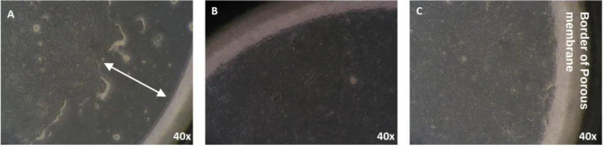
About 7-12 days after seeding, cells reach 80-90% confluency and are trypsinized (passage 1), counted and seeded on porous membranes - Snapwell inserts Ø12mm-pre-coated with collagen IV and supplemented with DM1 (protocol 1) or with NIH-3T3 coating and DM2 (protocol 2). We observed that cells (from donors and CF patients) in inserts pre-coated with NIH-3T3 coating and fed with either DM1 or DM2, did not reach confluency, i.e., the cells were not able to cover the membrane. As observed in Figure 3.3-A, there were gaps at the borders of the membrane. On the other hand, HBE cells (from both CF patients and donors) plated in collagen IV coating inserts with DM1 or DM2, were able to completely cover the membrane surface after 24h (Figure 3.3-B-C). Cells remained submerged with media for at least 3 days and after this period, media from apical side was removed in order to establish an ALI culture.
Figure 3.3 – HBE donor cells plated on Snapwells Inserts. (A) in pre-coated inserts with NIH-3T3 coating and feed with DM2 (arrow indicates the lack of cells at the border of the porous membrane); (B) in pre-coated inserts with collagen IV and feed with DM1; (C) in pre-coated inserts with collagen IV and feed with DM2.
B order of P or ous m e m bra ne
Chapter III Results and Discussion
19 Every 3 days, TEER measurements of ALI cultures were performed with an Electric Resistance System-Millicell-ERS (Millipore) (Figure 3.4), in order to assess the degree of epithelial tightness and the polarization status. Cells were then washed apically with Hank’s Balanced Salt Solution (HBSS) and media was replaced on the basolateral side.
Figure 3.4 – Monitoring Transepithelial Resistance (TEER) of ALI cultures growing on Snapwell Inserts.
TEER measurements of ALI cultures from different donors (Figure 3.5) and different CF patients (Figure 3.6), seeded and mantained according to protocol 1 or 2, were performed. As described above, we did not succeed in polarizing HBE cells in NIH-3T3 coated membranes, so we proceeded with the comparison of the ALI cultures establishment in DM1 or DM2 in collagen IV coated membranes. Figure 3.5 shows TEER data from HBE cells from two different donors, fed with DM1 or DM2 during 23 days. After this period, cells generally exhibited resistance values exceeding 600Ω.cm2
, a value previously (Fulcher et al. 2009; Neuberger et al. 2011) and recently (Tian et al., 2012) shown to be suitable to perform functional or biochemical assays. HBE cells from Donor 1 (when supplemented with DM2), did not achieve high TEER values and at day 19 it was observed that some cells were actually detaching, thus creating gaps in the porous membrane (Figure 3.5, Donor1 + DM2, green diamonds). Also HBE cells from the same donor but growing on DM1 (Figure 3.5, Donor1 + DM1, blue triangles), did not achieve as high TEER values as the other cultures from Donor 2 (Figure 3.5, Donor 2 + DM1, red squares), although they were able to reach ~500Ω.cm2 and no gaps were observed. Regarding Donor 2, both cultures showed to be
successfully differentiated and reached TEER values of 1500Ω.cm2 for DM1 (Figure 3.5, Donor 2 + DM1, red squares) or ~2000Ω.cm2 for DM2 (Figure 3.5, Donor 2 + DM2, black
Chapter III Results and Discussion
20 Figure 3.5 – – Transepithelial Resistance (TEER) measurements plotted as a function of the number of days the monolayers were cultured in ALI with either DM1 or DM2. TEER measurements were obtained from HBE cells derived from two different donors and seeded on collagen IV porous membranes. The values are means ± SD on each time point. Where error bars are not visible, the symbol has obscured them.
n indicates number of cultures of the same individual.
Regarding the CF monolayers, we can observe in Figure 3.6 that the ALI cultures fed with DM1, derived either from CF1 or CF2 subjects, originated very tight monolayers at day 23 (TEER values ranging from 900-2000Ω.cm2). On the other hand, the ALI cultures fed with
DM2 started to detach at day 8 and were not able to recover nor to reach TEER values higher than 200Ω.cm2. Thus, in this case, we cannot conclude which of the two differentiation
media give better results for CF monolayers. Importantly, Figure 3.6 also shows that we are able to establish successful ALI cultures from cells directly seeded on porous membranes after being expanded on plastic (Figure 3.6, CF1 + DM1, red circles) or from thawed cells (Figure 3.6, CF1 + DM1, black squares).
Figure 3.6 – Transepithelial Resistance (TEER) measurements plotted as a function of the number of days the monolayers were cultured in ALI with either DM1 or DM2. TEER measurements were obtained from HBE cells derived from two different CF patients (CF1: A561E homozygous- and CF2: F508del homozygous). Other details as in Figure 3.5.
Chapter III Results and Discussion
21 Notwithstanding, a modification in the cell seeding protocol was required when thawing cells. For cells directly seeded on membranes from 100mm dishes, we routinely seed cells at a cell density of 2 - 2,5 x 10⁵ cells per membrane and allow this culture submerged in "Differentiation Media" for 3 days prior to ALI establishement. Regarding the thawed cells, and because cell viability after thawing greatly impairs attachment of healthy cells, we had to seed more cells per membrane (2,5 - 3 x 10⁵ cells) and to submerge cultures for 5 days prior to ALI establishment. This modification in the protocol allowed cells to better recover from the thawing procedure and prevented their detachment during ALI, which we previously observed before this optimization (data not shown).
2.
Characterization
of
the
primary
HBE
Cultures
by
Immunofluorescence
and
Western
Blot
of
Epithelial
differentiation and polarization markers
Labelling of cells for given biomarkers by immunofluorescence is one of the most powerful tools to characterize distinct cellular types. Thus, here the primary HBE cells were characterized using different epithelial markers so as to visualize the expression and localization of specific proteins. These experiments were performed on cells growing on plastic as well as cells growing on porous membrane supports in ALI.
Figure 3.7 – Immunolocalization of several organelle markers in non-polarized HBE cells (Donor 2 - WT and CF2 patient - F508del/F508del). Cells were grown in 8-well chamber slides and analysed by immunocytochemistry using the following Abs: anti-cytokeratins for intermediate filaments, anti-acetylated-α- tubulin for microtubules, anti-ZO-1 and anti-E-cadherin for cell adhesion proteins. Bar=50 μm.