Carla Bernardete
Rodrigues Barradas
Alterações climáticas e doenças associadas a
Botryosphaeriaceae em Eucalyptus em Portugal
Climate changes and Botryosphaeriaceae diseases
of Eucalyptus in Portugal
2016
Carla Bernardete
Rodrigues Barradas
Alterações climáticas e doenças associadas a
Botryosphaeriaceae em Eucalyptus em Portugal
Climate changes and Botryosphaeriaceae diseases
of Eucalyptus in Portugal
Tese apresentada à Universidade de Aveiro para cumprimento dos
requisitos necessários à obtenção do grau de Doutor em Biologia, realizada sob a orientação científica do Doutor Artur Jorge da Costa Peixoto Alves, Investigador Principal do Departamento de Biologia da Universidade de Aveiro, do Professor Doutor António Carlos Matias Correia, Professor Catedrático do Departamento de Biologia da Universidade de Aveiro e do Doutor Alan John Lander Phillips, Investigador Principal da Faculdade de Ciências e Tecnologia da Universidade Nova de Lisboa
Apoio financeiro da FCT e do FEDER através do programa COMPETE no âmbito do projeto de investigação PANDORA. Bolsas com referência:
PTDC/AGR-FOR/3807/2012 FCOMP-01-0124-FEDER-027979
Apoio financeiro da Fundação para a Ciência e Tecnologia e do Fundo Social Europeu no âmbito do III Quadro Comunitário de Apoio.
Bolsa de Doutoramento: SFRH/BD/77939/2011
Ao Professor Doutor António Correia (in memoriam) Esta tese ficará para sempre incompleta sem o seu último contributo.
o júri
presidente Doutor Artur da Rosa Pires
professor catedrático do Departamento de Ciências Sociais, Políticas e do Território da Universidade de Aveiro
Doutora Maria Helena Mendes da Costa Ferreira Correia de Oliveira professora associada do Instituto Superior de Agronomia, Universidade de Lisboa
Doutor José Manuel Moutinho Pereira
professor auxiliar da Escola de Ciências da Vida e do Ambiente da Universidade de Trás-os-Montes e Alto Douto
Doutora Maria Helena Pires Bragança
investigadora auxiliar do Instituto Nacional de Investigação Agrária e Veterinária de Oeiras
Doutora Glória Catarina Cintra da Costa Pinto
professora auxiliar convidada do Departamento de Biologia da Universidade de Aveiro
Doutor Artur Jorge da Costa Peixoto Alves
agradecimentos Em primeiro lugar, gostaria de agradecer ao meu orientador Doutor Artur Alves pelo muito que me ensinou, por ter contribuído para o meu crescimento científico e intelectual, pela confiança, dedicação, compreensão e, acima de tudo, pela paciência e amizade. Foi um prazer partilhar contigo esta viagem. Aos meus co-orientadores, o Doutor António Correia e o Doutor Alan Phillips, muito obrigada pela ajuda, disponibilidade e pelos conhecimentos transmitidos. À minha “co-orientadora” Doutora Glória Pinto pela dedicação e pela ajuda imprescindível sem a qual não seria possível fazer a avaliação fisiológica das plantas.
Aos co-autores do trabalho científico: Barbara Correia, Catarina Moreirinha, Cláudia Jesus, Eugénio Diogo, Helena Bragança e Ivonne Delgadillo pelo seu valioso contributo.
Aos funcionários do departamento de Biologia, em especial à Dª Helena e Engº Armando, pela disponibilidade e por facilitarem tantas vezes o meu trabalho.
As colegas do laboratório (Carina, Anabela, Liliana, Eliana, Forough, Laura, Susana, Marta, Martinha, Cátia, Jaque, Nádia, Fernanda) pelo apoio, espírito de ajuda, companheirismo, amizade e pelos momentos bem passados. Um obrigada muito especial à minha “irmã mais nova” Carina por seres a pessoa que és e por estares sempre ao meu lado.
Às minhas amigas de sempre; Ana Catarina, Isabel, Tété e Cristina (a Galega) pelos momentos bem passados (pelos menos bons também). Obrigada por tudo! Ana Catarina, a ti tenho de te agradecer um pouco mais do que tudo, mas tu sabes bem isso;)
À minha grande família, em especial aos meus pais, pelo apoio que sempre me deram, por acreditarem em mim e, principalmente, por terem feito de mim a pessoa que hoje sou.
Ao Vítor pelo apoio incondicional, pelo amor e dedicação que tanto me ajudaram a ultrapassar os momentos difíceis. Obrigada por me deixares sonhar e me fazeres sorrir!
Por fim gostaria de agradecer à Pipeta, minha companheira durante estes últimos anos, o teu olhar os teus abraços fazem-me superar os problemas.
palavras-chave Botryosphaeriaceae, eucaliptos, alterações climáticas, stress hídrico,
patogenicidade, stress combinado, espetrospopia de infravermelho, fisiologia vegetal.
resumo O eucalipto é das espécies florestais mais plantadas devido à sua importância económica. Em Portugal, (principalmente E. globulus) representa atualmente 26% da área total, sendo a espécie florestal mais abundante no país. Membros da família Botryosphaeriaceae podem ocorrer como endofíticos ou patogénios latentes numa variada gama de hospedeiros lenhosos. Várias espécies têm sido associadas a eucaliptos em todo o mundo. Apesar da sua importância económica, não existem estudos relacionados com a ocorrência de espécies de Botryosphaeriaceae associadas a eucaliptos em Portugal, nem sobre o impacto que as alterações climáticas possam ter no desenvolvimento de doenças.
Foi estudada a comunidade de espécies de Botryosphaeriaceae que ocorre tanto em plantações saudáveis como doentes de E. globulus por todo o país. Nove espécies pertencentes a três géneros (Botryosphaeria, Diplodia e Neofusicoccum) foram identificadas, sendo o género Neofusicoccum claramente dominante tanto em plantas doentes como saudáveis. Neofusicoccum algeriense, D. corticola e D. seriata foram descritos pela primeira vez em E. globulus, enquanto N. algeriense, N. kwambonambiense e N. eucalyptorum correspondem aos primeiros registos em Portugal.
É fundamental detetar precocemente estes fungos de modo a evitar surtos de doenças que possam resultar em elevadas perdas económicas. A sua
identificação, de modo geral, baseia-se em técnicas moleculares que embora muito poderosas na discriminação de espécies podem ser demoradas e dispendiosas. A técnica de espectroscopia de infravermelho médio (MIR) permitiu a discriminação de espécies de Botryosphaeriaceae com base no seu perfil de “fingerprint” de infravermelho. Esta técnica revelou potencial para ser uma alternativa eficaz, rápida e económica aos métodos de identificação convencionais.
Em ensaios de inoculação artificial foram encontradas diferenças claras na agressividade destes fungos. Neofusicoccum kwambonambiense e D. corticola foram as espécies mais virulentas, em contraste com B. dothidea e D. seriata. Apesar de algumas diferenças nos parâmetros morfo-fisiológicos não foi encontrada qualquer relação direta entre o tamanho da lesão (agressividade) e as respostas fisiológicas da planta. Considerando o perfil fisiológico global e as dimensões das lesões notou-se uma clara variação na suscetibilidade entre os diferentes genótipos de plantas testadas.
As alterações climáticas influenciam a ocorrência e gravidade das doenças nas plantas. Os nossos resultados indicam que as plantas em stress hídrico são mais suscetíveis a espécies de Botryosphaeriaceae. Esta resposta foi
particularmente relevante quando os fungos foram inoculados em plantas que já se encontravam em privação de água. Além disso, o pré-condicionamento das plantas a condições de seca levou a um ligeiro aumento da resistência à infeção fúngica.
keywords Botryosphaeriaceae, eucalypts, climate changes, drought stress, pathogenicity, stress interaction, infrared spectroscopy, plant physiology.
abstract Eucalypts are one of the most widely planted forest trees due to their economic importance. In Portugal, they (mostly E. globulus) represent currently 26% of the total forest area, being the most abundant forest tree in the country. Botryosphaeriaceae species occur as endophytes or latent pathogens on a diverse range of woody hosts, including eucalypts. Despite the economic importance of these plants, there are no studies related to the occurrence of Botryosphaeriaceae species associated with them in Portugal or the impact of climate changes in the triggering of diseases.
The community of Botryosphaeriaceae species occurring on diseased and healthy E. globulus trees was studied on several plantations throughout the country. Nine species from three different genera (Botryosphaeria, Diplodia and Neofusicoccum) were identified being the genus Neofusicoccum clearly
dominant on both diseased and healthy trees. Neofusicoccum algeriense, D. corticola and D. seriata were reported for the first time on E. globulus, while N. algeriense, N. eucalyptorum and N. kwambonambiense correspond to the first reports in Portugal.
The early detection of Botryosphaeriaceae species could allow preventing disease outbreaks that may result in significant economic losses. The identification of these fungal species is, in general, based on molecular
techniques, that although being very powerful in discriminating species, can be time consuming and still quite expensive. Mid-infrared spectroscopy (MIR) technique allowed the discrimination of species of Botryosphaeriaceae based on their infrared fingerprint profile, being a powerful, cost-effective and faster alternative method to conventional identification techniques.
In artificial inoculation trials, marked differences in aggressiveness between these fungi were reported. Neofusicoccum kwambonambiense and D. corticola were the most virulent species while B. dothidea and D. seriata were the less ones. Despite some differences in morpho-physiological parameters no direct relation was found between lesion sizes (aggressiveness) and plant morpho-physiological responses. Considering the global morpho-physiological profile and lesion sizes, a clearly variation in susceptibility between different genotypes of
eucalypts in study was shown.
It is known that climate changes influence the occurrence and severity of plant diseases. Our results indicate that water stressed plants are more susceptible to Botryosphaeriaceae diseases. This response was particularly relevant when the plant was inoculated while water deprivation was already occurring. Moreover, drought primed plants presented a slightly increased resistance to fungal infection.
Our results reinforce the fact that management strategies for plantations should not overlook the impact that Botryosphaeriaceae diseases can have in a climate change scenario.
Table of contents
List of figures ... v
List of tables ... vii
Thesis outline ... ix
CHAPTER 1. Introduction 11
The host: Eucalyptus species ... 13
Characteristics and importance ... 13
Eucalyptus globulus plantations in Portugal ... 13
Pests and diseases of Eucalyptus plants ... 14
Pests and diseases of Eucalyptus plants in Portugal ... 15
The pathogen: Botryosphaeriaceae species ... 16
Characteristics and importance ... 16
Diversity of genera and species ... 17
How to identify species? ... 18
Pathogenicity and hosts range ... 20
Host-pathogen interactions ... 21
Plants response to biotic and abiotic stresses ... 21
Fungal effect on plant response ... 21
Climate changes ... 23
Botryosphaeriaceae diseases in Eucalyptus sp. ... 23
Aims of the work ... 30
References ... 31
CHAPTER 2. Diversity and potential impact of Botryosphaeriaceae species associated with Eucalyptus globulus plantations in Portugal 45
Abstract ... 47
Introduction ... 47
Materials and Methods ... 48
Fungal isolation and morphological characterization ... 48
Molecular characterization ... 49
ii
Results ... 50
Fungal isolation and morphological characterization ... 50
Molecular characterization ... 50
Pathogenicity trials ... 63
Discussion ... 63
Acknowledgments ... 68
Reference ... 69
CHAPTER 3. Mid-infrared spectroscopy (MIR) as a tool to differentiate species in the family Botryosphaeriaceae 75
Abstract ... 77
Introduction ... 77
Materials and Methods ... 78
Sample preparation ... 78
Mid-infrared spectroscopy ... 79
Data analysis ... 80
Results and Discussion ... 80
Acknowledgments ... 85
References ... 85
CHAPTER 4. Effects of Botryosphaeria, Diplodia and Neofusicoccum species on two Eucalyptus species and one hybrid: from pathogenicity to physiological performance 91 Abstract ... 93
Introduction ... 93
Materials and Methods ... 95
Plant material ... 95
Fungal culture and plant inoculation ... 95
Trial conditions and monitoring of the infection ... 96
Length of lesions ... 96
Evaluation of plant morpho-physiological performance ... 96
Growth and water status ... 96
Leaf gas-exchange measurements... 97
Total soluble sugars (TSS) content ... 97
Statistical analyses ... 98
Results ... 98
Monitoring of the infection ... 98
Length of lesions ... 99
Evaluation of plant morpho-physiological performance ... 100
Growth and plant water status ... 100
Leaf gas-exchange measurements... 101
Photosynthetic pigments and chlorophyll a fluorescence analysis... 102
Total soluble sugars (TSS) content ... 103
Multivariate approach of physiological profile ... 103
Discussion ... 105
Acknowledgements ... 110
References ... 110
CHAPTER 5. Drought-Disease interaction on Eucalyptus globulus under Neofusicoccum eucalyptorum infection 117
Abstract ... 119
Introduction ... 119
Materials and Methods ... 121
Plant material ... 121
Fungal culture and plant inoculation ... 121
Experimental design ... 122 Plant treatments ... 122 Monitoring of infection ... 123 Morphological parameters ... 123 Physiological parameters ... 123 Water status ... 123 Photosynthetic pigments ... 123 Lipid peroxidation ... 124 Statistical analyses ... 124 Results ... 124
iv Monitoring of infection ... 124 Morphological parameters ... 126 Physiological parameters ... 127 Water status ... 127 Photosynthetic pigments ... 128 Lipid peroxidation ... 129 Discussion ... 130 Acknowledgements ... 133 References ... 133
CHAPTER 6. General Discussion 141
General discussion ... 143
Future work ... 146
Figure 1.1: Distribution of the species plants for total area in Portugal. ... 14
Figure 2.1: Phylogenetic relationships of Botryosphaeria species.………….………….……….51
Figure 2.2: Phylogenetic relationships of Diplodia species. ... 51
Figure 2.3: Phylogenetic relationships of Neofusicoccum species. ... 54
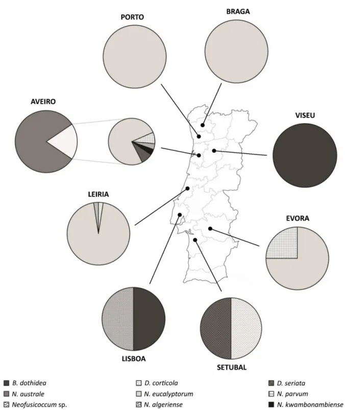
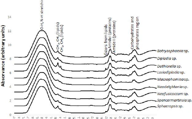
Figure 2.4: Map of Portugal indicating the distribution of Botryosphaeriaceae species.55 Figure 3.1: MIR spectra representative of each genus studied...80
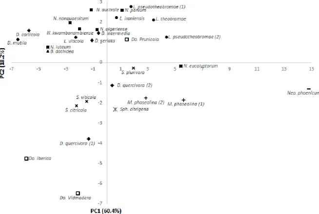
Figure 3.2: Scores scatter plot of the principal component analysis (PC1 vs PC2). ... 81
Figure 3.3: Loadings plot profile of the principal component analysis (PC1 vs PC2). .... 82
Figure 3.4: HCA of spectra obtained from fungal mycelium. ... 84
Figure 4.1: Internal and external lesions……….……….100
Figure 4.2: Growth rate, relative water content and midday shoot water potential...101
Figure 4.3: Leaf gas-exchange. ... 102
Figure 4.4: ɸPSII and Fv/Fm in leaves……….103
Figure 4.5: Total chlorophyll, carotenoid content and total soluble sugars……….104
Figure 4.6: PCA biplot of the morpho-physiological data ... 105
Figure 5.1: Representative pictures of lesions and symptoms……….125
Figure 5.2: Lesion lengths (cm) and growth rate (cm) in E. globulus plants………126
Figure 5.3: Relative water content and water potentialin E. globulus plants…………...128
Table 1.1: Botryosphaeriaceae species associated to eucalypts plants……… 25
Table 2.1: Relative abundance of Botryosphaeriaceae species (in E. globulus)……….50
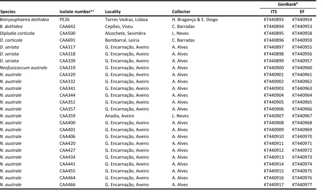
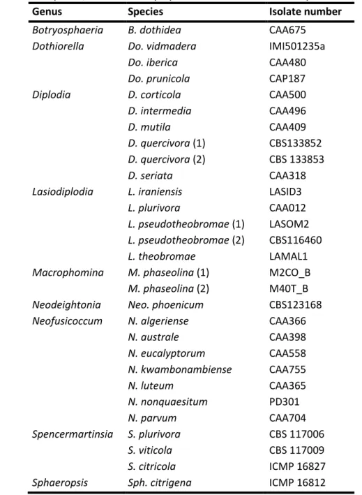
Table 2.2: List of isolates obtained from E. globulus and used in this study. ... 56
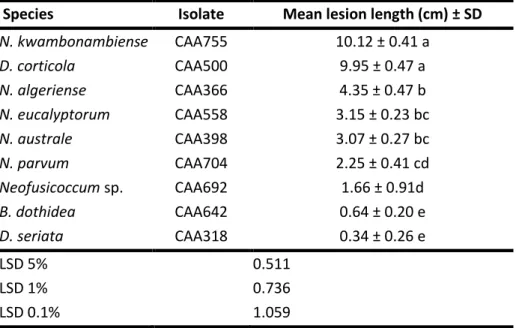
Table 2.3: Back-transformed means ± SD of lesion lengths………63
Table 4.1: Fungal isolates used in pathogenicity tests………..95
Table 4.2: One-way ANOVA summary table for morpho-physiological parameters. .... 99
Table 4.3: One-way ANOVA summary table for PC1 and PC2………104
Table 4.4: Dunnett multiple comparison tests……….104
Table 5.1: One-way ANOVA summary table for lesion length (experiment A)…………..125
Table 5.2: One-way ANOVA summary table (experiment B)………. 126
The present thesis documents the research work carried out in the scope of evaluating the potential impact that climate changes, especially drought, would have on the development of Botryosphaeriaceae diseases on Eucalyptus.
In Portugal, the forestry sector has a great economic relevance and Eucalyptus species are one of the most widely planted and commercially exploited forest species in this country. Members of the family Botryosphaeriaceae are known to cause diseases in a wide range of plants including eucalypts and mostly when the host is exposed to physiological stress. Thus, it is expected that these pathogens will be favored by climatic changes, especially drought. However, virtually nothing is known regarding the occurrence and severity of Botryosphaeriaceae species associated with eucalypts in Portugal, as well as the effect of climate changes on diseases of eucalypts.
This document is organised in six chapters:
The first chapter comprises the general review of literature to provide background for the work carried out, and to state the objectives of the research.
In chapter 2, the diversity and potential impact of Botryosphaeriaceae species associated with Eucalyptus globulus plantations in Portugal are presented. In this chapter the fungal species associated with these plants as well as their aggressiveness to them were identified. The fungal species obtained in this chapter were used in the subsequent work.
In chapter 3, an alternative method for fungal identification is presented based on infrared spectroscopy (MIR) coupled with multivariate analysis. Molecular techniques, although very powerful in discriminating species, still have some limitations and MIR have proved to be a good alternative.
In chapter 4, the pathogenicity of the Botryosphaeriaceae species identified in chapter 2 was evaluated towards three Eucalyptus species.
In chapter 5, the concepts of multiple stress (fungal disease and drought), priming and predisposition were tested. This information allows a better understanding of fungi-drought interaction to improve the establishment and productivity of
Eucalyptus species in Portugal.
Finally, Chapter 6 presents the main conclusions of this work, as well as some ideas for future work.
CHAPTER 1
Introduction
The host: Eucalyptus species
Characteristics and importance
Eucalyptus sensu lato (Myrtaceae) is a species-rich group that includes the genera Eucalyptus, Angophora, and Corymbia (Paine et al. 2011) and according to The Plant List
(2013) more than 800 species are accepted. These plants are mostlyAustralian natives (Du
et al. 2015; Wingfield et al. 2015) but they can also be found as native trees in Indonesia, Philippines, and New Guinea (Paine et al. 2011).
In spite of their origin, eucalypts are the most widely planted hardwood trees and the total area planted is estimated at 20 million ha (Du et al. 2015; Wingfield et al. 2015). For instance, Brazil (21%), India (19%), and China (13%) are the three non-native countries with the largest planted area (Paine et al. 2011; Wingfield et al. 2015).
Eucalypts are an important economic resource for the forest-related industries (Paine et al. 2011; Slippers et al. 2004b) since they exhibit fast-growth rates, excellent pulp properties, short rotation, easy vegetative propagation and wide adaptability to soils and climates (Brondani et al. 2012; Old et al. 2003). Furthermore, eucalypts are also planted as ornamental trees namely in North America (Paine et al. 2011).
Currently, more than ten species and their hybrids are well established in commercial plantations around the world (Wingfield et al. 2015). Eucalyptus grandis W. Hill
is more commonly exploited in tropical/subtropical areas while Eucalyptus globulusLabill.
is widely planted in temperate zones (Carocha et al. 2015), including Mediterranean climates. For instance, in the Southern Hemisphere tropics and subtropical regions, eucalypts plantations are an important source of fibre (Slippers et al. 2004b). In China, eucalypts plantations have been increasing, and currently there are about 2-6 million ha of established plantations that have an important role in paper and structural timber industries (Chen et al. 2011). In Colombia, Eucalyptus species are used by private companies and in government projects to produce timber and pulp, and protect soils from erosion (Rodas et al. 2009). In Ethiopia, eucalypt plantations represent around 100 000 ha and supply wood for fuel, construction and production of poles and posts (Gezahgne et al. 2004). Several species and hybrids of Eucalyptus have been extensively planted in Mexico where they cover around 25 000 ha of commercial plantations (de la Mora-Castañeda et al. 2014). Eucalyptus globulus is well adapted to the Mediterranean climate where it is exploited mainly for the production of pulp and currently occupies approximately 812 000 ha in Portugal (ICNF 2013) and 760 000 ha in Spain (Gominho et al. 2014).
Eucalyptus globulus plantations in Portugal
In Portugal, the forest sector is one of the pillars of the economy since this activity generates numerous jobs (3 % of the total employment) and contributes widely to the national exports (10 % of the Portuguese exports values) (Louro et al. 2014).
14 Chapter 1 Eucalyptus species were introduced in Portugal in the middle of the 19th century
(Águas et al. 2014; Bragança et al. 2015) and, nowadays, are the most abundant forest trees
in the country (Figure 1.1), representing approximately 26% of the total forest area (ICNF
2013).
Figure 1.1:Distribution of the species plants for total area in Portugal (adapted from ICNF (2013)). Eucalyptus globulus or blue gum, which is well adapted to Mediterranean climate
(Granda et al. 2014), is the dominant species in Portugal, both in commercial and
non-commercial plantations. Since these plants have high productivity of about 16 m3 ha–1 year–
1 (reaching 30 m3 ha–1 year–1), short rotation (12 years), and excellent pulp properties, they
are widely exploited by pulp industries (Águas et al. 2014; Pita et al. 2011). In spite of the dominance of E. globulus, other Eucalyptus species have been introduced in Portugal also. For instance, E. camaldulensis is more common in urban parks or growing as arboreta and
roadside trees (Pessoa et al. 2014). According to Decreto-Lei no565/99 there are more 21
species and hybrids in Portugal whose occurrence is infrequent or even rare. Pests and diseases of Eucalyptus plants
Eucalypts are distributed worldwide and their total plantation area has increased in the last centuries (Paine et al. 2011; Roux et al. 2005). Despite their high adaptability to different environments, the incidence of pests and pathogens associated with these hosts is increasing (Naidoo et al. 2014; Paine et al. 2011; Roux et al. 2005).
The distribution of Eucalyptus pests currently is not restricted to Australia and it is possible to find Australian insect herbivores colonizing new environments around the world, although the patterns of colonization are not yet clear (Paine et al. 2011; Phillips 2008; Zhou and Wingfield 2011).
Among pathogens, fungal species are the most frequently found associated with
Eucalyptus trees, although some bacteria (Coutinho et al. 2002) and oomycetes (Wingfield
and Knox-Davies 1980) have also been reported. Some fungal species are endophytes,
Eucalypts 26% Other hardwoods 6% Cluster pine 23% Pine 6% Other softwoods 2% Cork oak 23% Holm-oak 11% Other oaks 2% Sweet chestnut 1%
living inside the plants without causing any symptoms, whereas others are important pathogens that cause diseases on these plants (Old et al. 2003; Slippers and Wingfield 2007; Slippers et al. 2004c). Diseases can appear in different stages of the plant growth, from seedlings in nurseries to trees after out-planting (Old et al. 2003). Moreover, some fungi spend part of their life-cycles as endophytes within healthy plant tissue making the detection very difficult and increase the possibility of their transmission to different areas through introduction of germplasm (Old et al. 2003; Slippers et al. 2004c, 2009).
Pests and pathogens found in eucalypt plantations may come from their native environment (through seeds and plants) or can be introduced as result of host shifts (Naidoo et al. 2014; Paine et al. 2011). For instance, host jumps of fungal species between native Myrtaceae and Eucalyptus have previously been reported (Pérez et al. 2010; Slippers et al. 2005) as well as host shifts of native pests (Paine et al. 2011; Wingfield et al. 2008). In their native environment, the variability of host phenotype provides significant protection against pests and pathogens, but in commercial plantations the genetic uniformity of the plants increases the risk of diseases leading to high economic losses (Chen et al. 2011; Naidoo et al. 2014; Old et al. 2003; Slippers et al. 2009).
Several authors have focused on the most frequent and severe pests and pathogens that globally affect eucalypts plants (Armengol et al. 2008; Crous et al. 1989; de la Mora-Castañeda et al. 2014; Roux et al. 2005; Silva et al. 2014; Zhou and Wingfield 2011). Thus, bacterial wilt Ralstonia solanacearum (Smith) Yabuuchi et al., bacterial dieback
Xanthomonas eucalypti (Coutinho et al. 2002), root rot Phytophthora cinnamomi Rands
(Oomycete) (Naidoo et al. 2014; Wingfield and Knox-Davies 1980), myrtle rust Puccinia
psidii G. Winter (Coutinho et al. 1998; Naidoo et al. 2014), and stem canker Chrysoporthe
austroafricanaGryzenhout & M.J. Wingf. (Naidoo et al. 2014; Wingfield et al. 1989) and Botryosphaeriaceae (Rodas et al. 2009; Smith et al. 1994), are some examples of the most frequently reported pathogens. Concerning insect pests, blue gum psyllid Ctenarytaina
eucalypti Maskell, psyllid Glycaspis brimblecombei Moore, snout beetle Gonipterus scutellatus Gyllenhal, galling wasp Leptocybe invasa Fisher & LaSalle, longhorned borer Phoracantha semipunctata Fab. (Naidoo et al. 2014) are also recognized as a threat to
eucalypts.
Pests and diseases of Eucalyptus plants in Portugal
When eucalypts were introduced in Portugal, as it was observed in other countries where these plants are exotic species, they have been almost free from pests and diseases (Bragança et al. 2015; Wingfield et al. 2008). However, in the last decades, it has been verified an increasing tendency in damage caused by pests and pathogens in Portuguese eucalypt forests (Bragança et al. 2015; Reis et al. 2012; Silva et al. 2012) and this is correlated with the marked increase in occupied area by Eucalyptus species (Branco et al. 2014).
16 Chapter 1
Concerning insect pests, most of them are invasive species which are native to Australia (Branco et al. 2014). This is the case of Gonipterus plantensis Marelli, the major problem in Portuguese eucalypt plantations and the species responsible for tree growth losses in the Centre and North of Portugal (Reis et al. 2012). Ctenarytaina eucalipti Maskell, which feeds on the buds of the juvenile leaves, constitutes a problem in nurseries, however, in the field they have low impact because of the presence of many native predators (Azevedo and Figo 1979). Phoracantha semipunctata Fab. mainly attacks weakened trees under water stress, leading to plant death (Caldeira et al. 2002), and P. recurva Newman, which causes less severe injuries, is more restricted to the southern areas (Valente and Ruiz 2002).
There are some other Australia natives pest species, but their entrance in Iberian Peninsula was not through a direct pathway, for example, Ctenraytaina spatulata Taylor,
Glycaspis brimblecombei Moore, Blastopsylla occidentalis Taylor (Pérez-Otero et al. 2011;
Valente et al. 2004), Rhombacus eucalypti Ghosh & Chakrabarti (Ferreira et al. 2006),
Leptocybe invasa Fisher & La Salle, Ophelimus maskelli Ashmead (Branco et al. 2006), and Thaumastocoris peregrinus Carpintero & Dellapé (Garcia et al. 2013).
Although, eucalypts plantations were almost free from fungal diseases for a long period after their introduction in Portugal, Botryosphaeriaceae and Mycosphaerella spp. were considered the most important agents in earlier studies. In recent decades, other pathogens were also associated with dieback, canker and mortality observed on eucalypt stands, namely Phomopsis spp., Teratosphaeria spp., Cytospora spp., Pestalotiopsis spp.,
Phytophthora spp., Sporothrix spp., Phoma sp., Harknessia sp., Cylindrocarpon sp., Biscogniauxia mediterranea (De Not.) Kuntze, Teratosphaeria gauchensis (M.N. Cortinas,
Crous & M.J. Wingf.) M.J Wingf. & Crous, and Quambalaria eucalypti (M.J. Wingf., Crous & W.J. Swart) J.A. Simpson (Bragança et al. 2015; Branco et al. 2014; Silva et al. 2014). However, studies related with the importance of these species to Eucalyptus diseases as well as their distribution remain scarce in Portugal (Branco et al. 2014).
Considering the objective of this thesis, the following sections will focus on the Botryosphaeriaceae.
The pathogen: Botryosphaeriaceae species
Characteristics and importance
Members of the family Botryosphaeriaceae have a cosmopolitan distribution,
apparently with the exception of the polar regions, and are reported as occurring on
monocotyledonous, dicotyledonous and gymnosperm hosts (Barr 1987; Crous et al. 2006; Phillips et al. 2013). This family is a comprehensive group that includes pathogens, endophytes and saprophytic species that globally infect a wide range of woody plants
(Phillips et al. 2013; Slippers et al. 2007; Smith et al. 1996; von Arx 1987). Opportunistic human infections have also been reported for some species (Saha et al. 2012).
Botryosphaeriaceae species are often referred to as weak or opportunistic pathogens because they cause diseases mainly on stressed or wounded plants after drought, hail, wind, frost or insect damage (Chen et al. 2011; Mohali et al. 2007; Slippers and Wingfield 2007). Nevertheless, they can penetrate healthy plants through lenticels, open stomata, wounds, or other openings on twigs, stems, roots and leaves, and can remain in a latent stage (Old et al. 1990; Rodas et al. 2009). Consequently, they can live as endophytes for long periods without apparent damage or disease symptoms (Burgess et al. 2005; Chen et al. 2011; Smith et al. 1996) until some stress triggers the infection, so they become active and cause serious diseases (Old et al. 1990; Rodas et al. 2009). Several species can be found as saprophytes on dead wood or other material (Fisher et al. 1993; Old et al. 1990; Rodas et al. 2009).
Despite the efforts of several authors (Gezahgne et al. 2004; Lynch et al. 2013; Mohali et al. 2009; Mullerin 2013; Pérez et al. 2010; Rodas et al. 2009; Slippers et al. 2004c; van Niekerk et al. 2004), the interactions between Botryosphaeriaceae species and their hosts are not yet completely clear. In other words, some Botryosphaeriaceae species can be isolated both from healthy and diseased tissues, even for the same host (Pavlic et al. 2007; Piškur et al. 2011) and it is known that these fungi are able to perform a switching of their lifestyle, from endophytic to parasitic and vice versa, in response to hosts or environmental factors (Rai and Agarkar 2014). Diplodia sapinea, contrary to what happens for almost of these species, its ecological role to Pinus trees is well studied (Bihon et al. 2011; Swart and Wingfield 1991) and understood (Slippers et al. 2013). Considering the current knowledge about the contribution of these species on plant diseases (Dakin et al. 2010; Piškur et al. 2011) and predicting an increase in their importance as result of climate change (Desprez-Loustau et al. 2006), the need to better understand the diversity, distribution and pathogenicity of the species should be reinforced.
Diversity of genera and species
The family Botryosphaeriaceae was firstly introduced as a sub-family in the Pseudosphaeriaceae by Theissen and Sydow (1918). After almost a century of controversy of revisions it was finally established by Schoch et al. (2006) as the unique family within the order Botryosphaeriales (Dothideomycetes, Ascomycota). However, based on recent extensive phylogenetic studies, several species were excluded or allocated to other families within the same order (Liu et al. 2012; Phillips et al. 2013; Slippers et al. 2013). Even so, this family is rich in genera/species and is the largest family of the order Botryosphaeriales (Trakunyingcharoen et al. 2015).
When Phillips et al. (2013) reviewed the taxonomy of this family, at least 78 genera were legitimate according to MycoBank (http://www.mycobank.org). In this study, in the
18 Chapter 1
light of the abolished dual nomenclature, many genera were reduced to synonyms, some new genera were introduced and some old genera were resurrected. Currently, according to more recent studies (Crous et al. 2013; Jami et al. 2014; Liu et al. 2012; Phillips et al. 2013) at least 19 genera (Alanphillipsia, Aplosporella, Barriopsis, Botryosphaeria,
Botryobambusa, Cophinforma, Diplodia, Dothiorella, Endomelanconiopsis, Lasiodiplodia, Macrophomina, Neodeightonia, Neofusicoccum, Neoscytalidium, Phaeobotryon, Pseudofusicoccum, Spencermartinsia, Sphaeropsis, Tiarosporella) are accepted in this family that includes more than 100 species.
How to identify species?
The identification of Botryosphaeriaceae species was, for many years, based on the
morphological features of the spores (size, shape, septation, wall thickness and texture of
the conidia as well as details of conidiogenesis) (Denman et al. 2000; Jacobs and Rehner 1998; Slippers et al. 2009). In certain species, culture morphology, coloration, and growth rates at different temperatures can also be helpful to species identification (Denman et al. 2000; Pennycook and Samuels 1985).
Despite microscopy upgrade in the last century (Crous et al. 2015), the identification of closely related or cryptic species remained complicated based only in the morphological characters, because species exhibit extensive morphological plasticity and fungal morphology can be influenced by the substrate on which it is grown (Alves et al. 2007; Denman et al. 2000; Pennycook and Samuels 1985). The use of host association is also not a valid taxonomic character while a single species can be found associated to a wide range of hosts and the same host can be colonized by various fungal species (Alves et al. 2007; Crous et al. 2015).
Since the emergence of DNA based techniques several methodologies have been successfully applied to fungi identifications (Alves et al. 2007; Crous et al. 2015). Nowadays, molecular tools are used, in combination with morphological characters, to distinguish members of Botryosphaeriaceae (Crous et al. 2006; Phillips et al. 2013; Slippers et al. 2009). Sequences of ITS region (internal transcribed spacer region) of ribosomal DNA are successfully used to discriminate fungi at different taxonomic level (Crous et al. 2015; White et al. 1990) and was proposed by Schoch et al. (2012) as the official DNA barcode for fungi. However, for some cryptic species ITS sequence data seems not to be enough. In this cases, different protein coding genes (translation elongation factor 1-alpha (tef1), beta-tubulin (tub2) gene, glyceraldehyde-3-phosphate dehydrogenase (GAPDH), actin (act) and histone H3 (his3) and more conserved gene regions (large subunit (LSU), small subunit (SSU), and RNA polymerase II (RPB2) gene) might help the identification (Crous et al. 2015; Phillips et al. 2013).
In spite of DNA sequencing being the broadly accepted methodology for fungal identification, other techniques as micro-satellite loci (de Wet et al. 2003), PCR-RFLPs
(Dreaden et al. 2014; Slippers et al. 2004c), species-specific primers (Luchi et al. 2005), ISSRs (Zhou et al. 2001), RAPDs (Smith and Stanocz 1995), SSRs (Burgess et al. 2004) and ARDRA (Alves et al. 2005) have been successfully applied for characterization and identification of Botryosphaeriaceae species. In addition, fingerprinting techniques can be powerful tools to rapidly and reliably screen a large number of isolates when species have been identified from a particular host or area (Alves et al. 2007).
Molecular approaches, for instance, allowed to separate and characterize important closely related or cryptic species such as Diplodia sapinea and D. scrobiculata (de Wet et al. 2003) Neofusicoccum eucalyptorum and N. eucalypticola (Slippers et al. 2004c), N. parvum and N. ribis (Slippers et al. 2004a), N. luteum and N. australe (Slippers et al. 2004b), and resolve species complexes in Diplodia (Alves et al. 2014; Alves et al. 2004; Phillips et al. 2012). Taking this into account, molecular tools have greatly contributed to species separation and geographical distribution, which are very important to understand the evolution and ecology of Botryosphaeriaceae species (Slippers et al. 2009).
DNA-based techniques, although very powerful in discriminating species, still have some limitations: they can be sensitive to mutations or do not reflect the phenotypic diversification, the choices of specific primers can be a hard task, protocols are complex and time consuming and reagents are expensive (Mancini et al. 2013; Santos et al. 2010). In the recent years some alternative techniques have increased their importance in order to overcome these limitations.
Matrix-assisted laser desorption ionization-time of flight mass spectrometry (MALDI-TOF MS) is presently the most promising method for routine identification, differentiation and classification of microorganisms (Chalupová et al. 2014; Mancini et al. 2013), including filamentous fungi (Chalupová et al. 2014; Lecellier et al. 2015) even though these are one of the most challenging ones (Mancini et al. 2013). This is a reliable, fast, easy, low labor and consumable cost methodology and allows a direct identification of phytopathogenic fungi without need to isolate them from their hosts (Chalupová et al. 2014; Lecellier et al. 2015). The technique relies on the principle that metabolites composition is characteristic for each species, since the samples were maintained under the same experimental conditions (Chalupová et al. 2014). For instance, Mancini et al. (2013) found that spectra are clearly dependent on mycelial age when used MALDI-TOF to distinguish three monophyletic species of Diplodia (D. sapinea, D. seriata and D.
scrobiculata).
Mid-infrared spectroscopy (MIR) is a powerful technique that is fast, effective, needs only small sample quantities, is reagent-free, and normally do not require sample pre-treatment (Schmitt and Flemming 1998). Moreover, the recent infrared spectrometers are quite inexpensive, unlike MALDI-TOF equipments. It is an analytical technique based on the vibrations between atoms in a molecule (Stuart 2004) and such vibrations are likely to conform to a ‘fingerprint’ of the molecule as a whole, rather than a specific group within the molecule (Smith 2011). In terms of the electromagnetic spectrum, MIR comprises the
20 Chapter 1
infrared radiation between 4000-200 cm-1 (Santos et al. 2010), which gives information
about some important cell macromolecules like as proteins, lipids, nucleic acids and carbohydrates (Lecellier et al. 2015). Thus, MIR is a perfect tool to assess the overall molecular composition of the microbial cells in a fast and non-destructive manner. Hence, it is one of the most promising techniques in microbiology since it allows the detection, identification, characterization and authentication of several microorganisms including filamentous fungi (Erukhimovitch et al. 2005; Fischer et al. 2006; Lecellier et al. 2015; Naumann et al. 2005; Santos et al. 2010). However, these works are focused manly in fungi with clinical, food and industry importance and no studies were addressed to Botryosphaeriaceae species.
MALDI-TOF MS and MIR, in order to allow species identification as molecular
methodologies, depend on a good and complete database. Although, spectral libraries for filamentous fungi are not yet completely build (Lecellier et al. 2014, 2015).
Pathogenicity and hosts range
Botryosphaeriaceae species are frequently associated with disease symptoms observed in plants with high agricultural, forestry, ecological and economic value (Barr, 1972, 1987; Alves et al. 2005). Diseases caused by these species include fruit rots, leaf spots, seedling damping-off and collar rot, cankers, blight of shoots and seedlings, gummosis, blue-stain of the sapwood, dieback, and tree death (Rodas et al. 2009; Slippers et al. 2007). Further, their pathogenicity is typically associated with biotic and abiotic stresses, in which drought stress is the most frequently reported (Chen et al. 2011; Mohali et al. 2007; Old et al. 1990; Phillips et al. 2013).
Members of Botryosphaeriaceae can be found associated with a wide range of woody hosts, namely eucalypts (Chen et al. 2011; Mohali et al. 2009; Pérez et al. 2010; Rodas et al. 2009; Slippers et al. 2004c), pines (Alves et al. 2013; Mohali et al. 2007), grapevines (Urbez-Torres et al. 2010; van Niekerk et al. 2004), oaks (Alves et al. 2004; Linaldeddu et al. 2014), mango (Marques et al. 2013), and Rosaceae (Phillips et al. 2012) Even though, some species seem to be specific to the host (Neofusicoccum eucalyptorum appears to be almost exclusive to Eucalyptus spp. among other Myrtaceae plants (Pillay et al. 2013) and Diplodia corticola is specialized on cork oak and other oak species (Alves et al. 2004; Linaldeddu et al. 2014; Lynch et al. 2013)), a consistent pattern of pathogen-host association has not been verified for the majority of the species (Pillay et al. 2013).
Recent studies demonstrate that the distribution of Botryosphaeriaceae species has been highly influenced by anthropogenic activity. Owing to their endophytic nature, these species were introduced trough the germplasm into new environments (Slippers et al. 2004c). For instance, species that were common on eucalypts in their native environment, as N. parvum and N. eucalyptorum, were also found on Myrtaceae plants growing nearby
eucalyptus plantations. Host jumps between native Myrtaceae and non-native Eucalyptus plantations has also been reported (Pérez et al. 2010).
Host-pathogen interactions
Plants response to biotic and abiotic stresses
Plant hosts, owing to their sessile lifestyle, are continually exposed to a broad range of environmental stresses, namely abiotic (drought, salinity, heat, cold, chilling, freezing,
nutrient, high light intensity, ozone (O3) and anaerobic stresses) and biotic (bacteria,
oomycetes, fungi, viruses, nematodes, and attack by herbivore pests) stresses (Atkinson and Urwin 2012; Naidoo et al. 2014; Suzuki et al. 2014). So, they have developed strategies that allow them to recognize stress source minimizing the possible damage which compromise growth and reproduction. Thus, plants respond through activation of a complex biochemical and molecular response systems (Atkinson and Urwin 2012;
Ramegowda and Senthil-Kumar 2015) and adjust their physiological mechanisms to
preserve homeostasis during the stress occurrence (Bostock et al. 2014).
In the field, plants are exposed not only to an isolated stress but to a set of them. Therefore, the plant response to multiple and simultaneous stresses is unique and cannot be directly extrapolated from the response to each individual stress alone (Atkinson and Urwin 2012; Suzuki et al. 2014). Moreover, combined stresses can have either a negative (i.e., susceptibility) or positive (i.e., tolerance) effect depending on various factors as the abiotic stress type, the pathogen in cause, the severity of stresses, and which one starts first (Jactel et al. 2012; Ramegowda and Senthil-Kumar 2015). In this respect, it is difficult to anticipate which would be the plants response considering only the pathogen species, while pathogen-host interaction depends on various factors. Even so, some studies were addressed in order to understand which modifications occur in plants physiology as well as in biochemical and molecular response system in case of fungal infection (Alves et al. 2011; Bettucci and Alonso 1997; Desprez-Loustau et al. 2006; Luque et al. 1999; Mayek-Pérez et al. 2002; Pinkard and Mohammed 2006). Further, it is recognized that water stress can influence the appearance and development of fungal diseases by disturbing the physiological status of the host plants and, consequently, their capacity to resist fungal infection, being this the case of Botryosphaeriaceae diseases (Desprez-Loustau et al. 2006).
Fungal effect on plant response
Plants have to deal permanently with environmental challenges and are not able to escape physically. Even so, during fungal attack, plants activate several defense strategies with the objective to limit or prevent the pathogen entry and spread (Atkinson and Urwin 2012; Naidoo et al. 2014; Torres 2010). These strategies are divided into constitutive and inducible defences (Freeman and Beattie 2008). Constitutive defences are the first line of defense against fungal invasions and include many preformed barriers such as outer bark,
22 Chapter 1
leaf cuticle, plant cell walls composition (pectin and lignin) (Freeman and Beattie 2008; Kovalchuk et al. 2013; Naidoo et al. 2014), and secretory cells, glands and ducts (where defensive substances are produce, transport and storage) (Naidoo et al. 2014). While constitutive defences are continuous, preformed and non-specific barriers, inducible defences are activated during pathogens invasion and consist in the production of chemical compounds, as antimicrobial and toxic secondary metabolites (Freeman and Beattie 2008; Kovalchuk et al. 2013; Naidoo et al. 2014), antioxidant, low-molecular- weight (LMW) compounds (such as phenolic compounds, terpenoids, and alkaloids) (Kovalchuk et al. 2013), pathogen-degrading enzymes, and apoptosis induction (Freeman and Beattie 2008; Z. Li et al. 2014).
Considering eucalypts, beyond the mechanisms described for plants in a global way, these plants present secretory cavities, mainly located on leaves, which produce and store essential oils and resinous substance with unknown function (Naidoo et al. 2014). Further, the formation of barrier zones or reaction zones to prevent fungal spread is also described for some species of eucalypts (Naidoo et al. 2014; Tippett and Shigo 1981).
Inducible defences normally are only activated when constitutive mechanism fail because chemicals production and maintenance have high energy costs (Freeman and Beattie 2008; Naidoo et al. 2014). The success of these responses are related with plants ability to differentiate self from non-self. Hormone signalling pathways, phenolic compounds production, pathogen associated molecular patterns (PAMPs) and reactive oxygen species (ROS) play a very important role in plant responses at this level (Atkinson and Urwin 2012; Kovalchuk et al. 2013; Naidoo et al. 2014; Torres 2010).
Plant physiological parameters are also frequently affected by fungal activity. For instance, some endophytic fungi are able to promote the plants growth by production of enzymes that degrade substances which have negative impacts on the growth of the plants (e.g. phenolic acid allelochemicals), secretion of plant growth promoters (e. g. gibberellins (GAs) and indoleacetic acid) or contribution to nitrogen uptake (Zhou et al. 2014). On the other hand, several pathogenic fungi, including some Botryosphaeriaceae species, produce phytotoxins and secondary metabolites which are spread in the whole plant and have a negative effect on photosynthesis and transpiration rate (Berger et al. 2007; Linaldeddu et al. 2009; Luque et al. 1999). The decreased photosynthesis rate leads to a decline in enzymes and proteins production that play an important role in the plant defense against the pathogen (van Niekerk et al. 2011). Conversely, some fungi do not produce these compounds, being restricted to the infection zone and, consequently, do not affect the leaf
gas exchange parameters (Linaldeddu et al. 2009).Li et al. (2014) observed a decrease of
total chlorophyll amount in plants infected with fungi as a result of down-regulation of the gene expression for the chlorophyll a,b binding proteins. Modifications in the accumulation of soluble sugars depend on the fungal species, while in some cases occur an increment in the accumulation of sugars, in other situations sugar content presents no alterations or even decreases (Berger et al. 2007). During fungal infection, the amount of ROS, which play
a crucial role in signalling, is higher (Berger et al. 2007). Some fungal species are able to successfully colonize xylem leading to vessel obstruction and consequently plant wilt (Polle and Luo 2014). Unfortunately, neither the same plant responds in a similar way facing different fungi nor the same fungi induces the same plant response. Thus, the lack of patterns in plants responses difficults our understanding about plant-fungi interaction. Recently, the application of omics technologies have been improved our knowledge about this subject (Kovalchuk et al. 2013).
Climate changes
Climate has a great influence not only on plant and animal distribution but also on their parasites and pathogens. Thus, the distribution patterns and the severity of pathogens can be directly or indirectly affected by climate, since this factor also affects host physiology and parasite-associated organisms (Desprez-Loustau et al. 2007).
According to the report released by IPCC (2013), it is expected an increase in global temperature and frequency of extreme events (higher or lower precipitation events in different areas and extended dry periods) in the 21st century. Thus, the occurrence of abiotic stresses will be intensified by climate changes which will also lead to shifts in the distribution of species, expand the host range of pathogens and increase in pathogens aggressiveness (Desprez-Loustau et al. 2007; Ramegowda and Senthil-Kumar 2015). Consequently, the frequency and severity of plant diseases should be favoured in a scenario of climate changes (Lindner et al. 2010; Ramegowda and Senthil-Kumar 2015; Sturrocka et al. 2011). Among abiotic factors, drought stress is the one which is most frequently associated with emergence of fungal diseases, namely Botryosphaeriaceae diseases (Pérez et al. 2010; Slippers and Wingfield 2007; Smith et al. 1994).
However, as mentioned above, it should not be overlooked that the response of plants to multiple simultaneous stresses is complex and depends on the source of stress and the pathogen involved (Atkinson and Urwin 2012; Jactel et al. 2012; Ramegowda and Senthil-Kumar 2015; Suzuki et al. 2014). Moreover, the reason why some host-pathogen interactions resulted in tolerance while others lead to susceptibility is not already clear and no general trend can be assumed (Desprez-Loustau et al. 2007; Jactel et al. 2012; Ramegowda and Senthil-Kumar 2015).
Botryosphaeriaceae diseases in Eucalyptus sp.
Eucalypt plantations have increased in extent and economic relevance, so the study of their pathogens is crucial.
In early studies, Botryosphaeriaceae species have been associated to eucalypts in their native environment (Burgess et al. 2006b; Old et al. 1990; Slippers et al. 2004c). Nowadays, these fungi are considered to be a threat to the sustainability and production of eucalypts both in native and non-native plantations (Chen et al. 2011; de la
Mora-24 Chapter 1
Castañeda et al. 2014; Mohali et al. 2009; Rodas et al. 2009). The genetic uniformity of the commercial plantations increases the risk of diseases (Chen et al. 2011; Old et al. 2003; Slippers et al. 2009) and the presence of these plants close to native plants may have a negative effect, considering that host jumps are common in the Botryosphaeriaceae species (Pavlic et al. 2007; Pérez et al. 2009, 2010).
Some members of Botryosphaeriaceae family can occur as endophytes in healthy eucalypts for a long period without producing any symptoms (Pavlic et al. 2009; Smith et al. 1996). On the other hand, they may also occur as pathogenic and cause different disease symptoms, such as dieback of shoots and branches, cankers on the stems followed by kino exudation, coppice failure and even host death (Chen et al. 2011; Gezahgne et al. 2004; Slippers et al. 2009). Further, Rodas et al. (2009) verified that young eucalypts are more susceptible than older ones. When these authors observed plants aged between 6 to 36 months they found that the prevalence of the symptoms were most common on plants aged between 18 to 26 months.
In early studies, investigators believed that Botryosphaeria dothidea and
Neofusicoccum ribis were the most abundant species in eucalypts growing in temperate
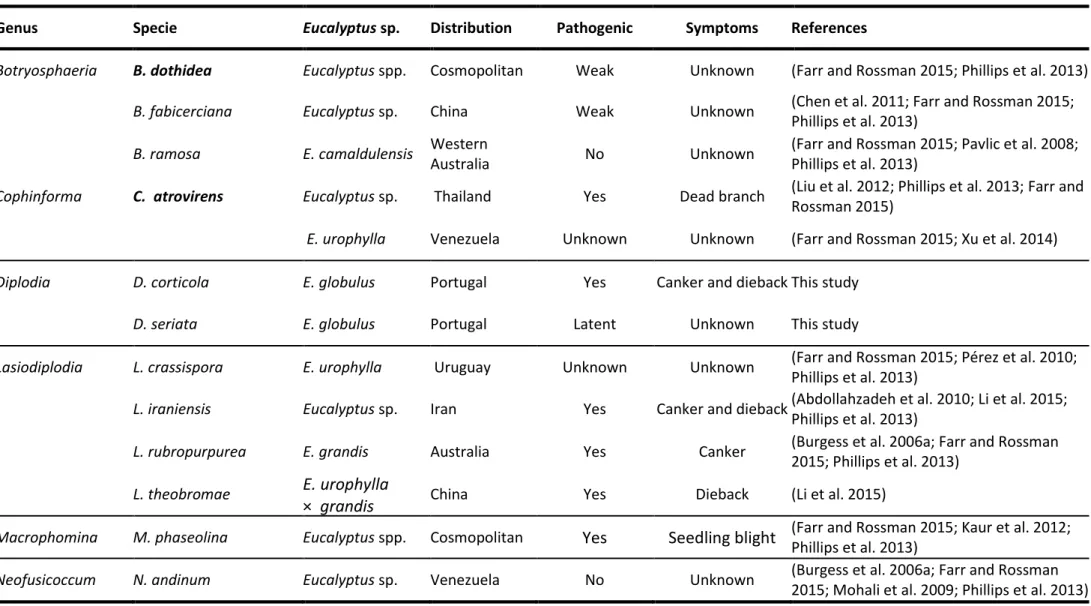
areas, causing stem cankers and dieback (Chen et al. 2011; Pérez et al. 2010; Slippers et al. 2009). However, in the light of the most recent studies, it is known that these species are not common on Eucalyptus plants. Nowadays, at least 29 Botryosphaeriaceae species have been confirmed as occurring in eucalypts (Phillips et al. 2013; Pillay et al. 2013; Barradas et. al. 2016) as shown on table 1.1.
Although efforts have been made to comprehensively understand which species are associated with eucalypts, little is known about their pathogenicity. Thus, pathogenicity tests conducted both in greenhouses and field trial allowed to identify some of the causal agents of diseases. For instance, pathogenicity tests confirmed the ability of N. parvum to cause stem canker on Eucalyptus in Ethiopia (Gezahgne et al. 2004) and N. eucalyptorum on E. grandis in Uruguay (Pérez et al. 2009). Pavlic et al. (2007) inoculated Eucalyptus
grandis × camaldulensis clones and found that N. ribis, N. parvum and L. theobromae, were
the most aggressive while B. dothidea was less pathogenic to the plants. Pathogenicity tests performed in E. grandis also found N. ribis as the most aggressive and B. dothidea as the less one (Rodas et al. 2009). The species N. parvum and N. ribis were the most aggressive species in a study conducted in Venezuela, in which differences in clones tolerance were also verified (Mohali et al. 2009). Pérez et al. (2010) inoculated E. grandis plants with species isolated both from introduced eucalypts and native Myrtaceae trees and found that
L. pseudotheobromae, N. eucalyptorum and the N. parvum-N. ribis complex are the most
aggressive to the plants. In artificial inoculation trials conducted in Portugal it was found that D. corticola and N. kwambonambiense were the most aggressive fungi, while B.
dothidea and D. seriata were the least aggressive ones. Further, differences in plant
tolerance were also observed among the Eucalyptus species (Mohali et al. 2009; Chapter 4).
Table 1.1:Botryosphaeriaceae species confirmed to be associated to eucalypts plants, distribution and associated symptoms. Ex-type strains in bold face.
Genus Specie Eucalyptus sp. Distribution Pathogenic Symptoms References
Botryosphaeria B. dothidea Eucalyptus spp. Cosmopolitan Weak Unknown (Farr and Rossman 2015; Phillips et al. 2013)
B. fabicerciana Eucalyptus sp. China Weak Unknown (Chen et al. 2011; Farr and Rossman 2015;
Phillips et al. 2013)
B. ramosa E. camaldulensis Western
Australia No Unknown
(Farr and Rossman 2015; Pavlic et al. 2008; Phillips et al. 2013)
Cophinforma C. atrovirens Eucalyptus sp. Thailand Yes Dead branch (Liu et al. 2012; Phillips et al. 2013; Farr and
Rossman 2015)
E. urophylla Venezuela Unknown Unknown (Farr and Rossman 2015; Xu et al. 2014)
Diplodia D. corticola E. globulus Portugal Yes Canker and diebackThis study
D. seriata E. globulus Portugal Latent Unknown This study
Lasiodiplodia L. crassispora E. urophylla Uruguay Unknown Unknown (Farr and Rossman 2015; Pérez et al. 2010;
Phillips et al. 2013)
L. iraniensis Eucalyptus sp. Iran Yes Canker and dieback(Abdollahzadeh et al. 2010; Li et al. 2015;
Phillips et al. 2013)
L. rubropurpurea E. grandis Australia Yes Canker (Burgess et al. 2006a; Farr and Rossman
2015; Phillips et al. 2013)
L. theobromae E. urophylla
× grandis China Yes Dieback (Li et al. 2015)
Macrophomina M. phaseolina Eucalyptus spp. Cosmopolitan Yes Seedling blight (Farr and Rossman 2015; Kaur et al. 2012; Phillips et al. 2013)
Neofusicoccum N. andinum Eucalyptus sp. Venezuela No Unknown (Burgess et al. 2006a; Farr and Rossman
Chapter 1 26
Genus Specie Eucalyptus sp. Distribution Pathogenic Symptoms References
Neofusicoccum N. australe Eucalyptus spp. Australia Weak Unknown (Farr and Rossman 2015; Phillips et al. 2013;
Taylor et al. 2009)
E. globulus Spain Yes Canker and dieback(Armengol et al. 2008; Farr and Rossman
2015)
E. globulus Portugal Yes Canker and diebackThis study
N. algeriense E. globulus Portugal Yes Canker and diebackThis study
N. eucalypticola Eucalyptus spp. Eastern
Australia Unknown Unknown
(Farr and Rossman 2015; Phillips et al. 2013; Slippers et al. 2004c)
N. eucalyptorum Eucalyptus spp. Australia Yes Canker and dieback(Phillips et al. 2013; Slippers et al. 2004c)
Eucalyptus spp. South Africa Yes Canker and dieback(Farr and Rossman 2015; Phillips et al. 2013;
Smith et al. 2001)
Eucalyptus spp. Uruguay Yes Bark lesions (Pérez et al. 2010; Phillips et al. 2013)
E. grandis Zimbabwe Yes Stem canker (Jimu et al. 2015)
E. globulus Portugal Latent Unknown This study
N. kwambonambiense Eucalyptus spp. Australia Yes Unknown (Pillay et al. 2013; Sakalidis et al. 2013)
Eucalyptus spp. Uganda Yes Canker (Sakalidis et al. 2013)
E. globulus Portugal Yes Canker This study
N. luteum Eucalyptus spp. Eastern
Genus Specie Eucalyptus sp. Distribution Pathogenic Symptoms References
Neofusicoccum N. macroclavatum Eucalyptus spp. Western
Australia Yes yes (Burgess et al. 2005; Phillips et al. 2013)
N. mediterraneum Eucalyptus sp. (California) USA Unknown Unknown (Farr and Rossman 2015; Inderbitzin et al.
2010; Phillips et al. 2013)
Eucalyptus sp. Greece Unknown Unknown (Farr and Rossman 2015; Inderbitzin et al.
2010; Phillips et al. 2013)
N. occulatum Eucalyptus spp. Australia yes Unknown (Farr and Rossman 2015; Phillips et al. 2013;
Sakalidis et al. 2011a)
Eucalyptus sp. (Hawaii) USA Unknown Unknown (Sakalidis et al. 2013)
Eucalyptus sp. Uganda Unknown Unknown (Sakalidis et al. 2013)
E. grandis Uruguay Unknown Unknown (Sakalidis et al. 2013)
N. parvum Eucalyptus spp. Australia Unknown Unknown (Barber et al. 2005; Phillips et al. 2013;
Sakalidis et al. 2013)
Eucalyptus spp. China Unknown Unknown (Sakalidis et al. 2013)
Eucalyptus sp. Colombia Unknown Unknown (Sakalidis et al. 2013)
Eucalyptus spp. Ethiopia Yes Dieback and death (Gezahgne et al. 2004; Phillips et al. 2013;
Sakalidis et al. 2013)
Eucalyptus sp. (Hawaii) USA Unknown Unknown (Sakalidis et al. 2013)
Eucalyptus spp. Indonesia Unknown Unknown (Sakalidis et al. 2013)
Chapter 1 28
Genus Specie Eucalyptus sp. Distribution Pathogenic Symptoms References
Neofusicoccum N. parvum E. globulus Portugal Yes Canker and dieback This study
Eucalyptus spp. Spain Yes Canker and dieback(Farr and Rossman 2015; Iturritxa et al.
2011; Sakalidis et al. 2013)
E. smiithi South Africa Yes Unknown (Farr and Rossman 2015; Sakalidis et al.
2013; Xu et al. 2015)
E. grandis Swaziland Unknown Unknown (Sakalidis et al. 2013)
E. obliqua Thailand Unknown Unknown (Farr and Rossman 2015; Sakalidis et al.
2013; Trakunyingcharoen et al. 2015)
E. grandis Uganda Unknown Unknown (Sakalidis et al. 2013)
E. urophylla Venezuela Unknown Unknown (Farr and Rossman 2015; Mohali et al. 2007;
Phillips et al. 2013)
E. grandis Zambia Unknown Unknown (Sakalidis et al. 2013)
E. grandis Zimbabwe Yes Stem canker (Jimu et al. 2015)
N. ribis E. camaldulensis Australia Unknown Unknown (Farr and Rossman 2015; Sakalidis et al.
2011b)
E. grandis Australia Unknown Unknown (Farr and Rossman 2015; Urbez-Torres et al.
2012) E. camaldulensis x
E. grandis Australia Unknown Unknown
(Farr and Rossman 2015; Inderbitzin et al. 2010)
E. pellita Australia Unknown Unknown (Farr and Rossman 2015; Xu et al. 2015)
Genus Specie Eucalyptus sp. Distribution Pathogenic Symptoms References
Neofusicoccum N. vitifusiforme Eucalyptus spp. Australia Yes Leaf lesions (Phillips et al. 2013; Taylor et al. 2009)
Neoscytalidium Ne. hyalinum Eucalyptus spp. worldwide Yes gummosis, dieback,
wilt and cankers (Phillips et al. 2013)
Pseudofusicoccum P. adansoniae Eucalyptus sp. Australia Unknown Unknown (Pavlic et al. 2007; Phillips et al. 2013;
Sharma et al. 2013)
P. ardesiacum Eucalyptus sp. Western
Australia Unknown Unknown
(Pavlic et al. 2008; Phillips et al. 2013; Sharma et al. 2013)
P. kimberleyense Eucalyptus sp. Western
Australia Unknown Unknown
(Pavlic et al. 2008; Phillips et al. 2013; Sharma et al. 2013)
P. stromaticum Eucalyptus spp. Venezuela Unknown Unknown (Mohali et al. 2006; Phillips et al. 2013; Sharma et al. 2013)
Sphaeropsis S. eucalypticola Eucalyptus sp. Thailand Unknown Collected from a
30 Chapter 1
Aims of the work
Climate change is a major challenge for forest health and productivity since it is expected to impose a drastic modification of growth conditions, and probably an increase in the occurrence of diseases. In fact, environmental conditions are recognized to strongly influence plant diseases, affecting the host, the pathogen, and the interactions between them, however these subjects are not completely understood. Hence, researches carried out in order to identify and clarify the interaction between the different factors of decline (biotic/abiotic) should be privileged.
In Portugal, forest activity is an important economic sector, generating many jobs. Indeed, Portugal extracts more benefits from one hectare of forest than any other country in the Mediterranean area. To maintain these economic values and ensure competitiveness, forest research in Portugal must focus on the most representative (both in area and economy) forest species, namely Eucalyptus globulus, Pinus pinaster and Quercus suber. Eucalyptus globulus is at present the most abundant tree species in Portuguese forests. It is known that water availability limits the growth and survival of these plants and consequently, Botryosphaeriaceae diseases can be favoured. Taking this into account, the main goal of this thesis was to evaluate the potential impact of climate change on Botryosphaeriaceae-related diseases of eucalypts in Portugal.
For that, this work aimed to answer the following questions:
1. Which species of Botryosphaeriaceae occur in association with Eucalyptus species in Portugal?
2. Which species are actually pathogenic to Eucalyptus species?
3. Is there variation in susceptibility to these pathogens among Eucalyptus species and clones?
4. How does water stress affect Botryosphaeriaceae-related diseases of eucalypts?
References
Abdollahzadeh, J., Javadi, A., Goltapeh, E.M., Zare, R., & Phillips, A.J.L. (2010). Phylogeny and morphology of four new species of Lasiodiplodia from Iran. Persoonia, 25, 1– 10.
Águas, A., Ferreira, A., Maia, P., Fernandes, P.M., Roxo, L., Keizer, J., et al. (2014). Natural establishment of Eucalyptus globulus Labill. in burnt stands in Portugal. Forest
Ecology and Management, 323, 47–56.
Alves, A.A., Guimarães, L.M. da S., Chaves, A.R. de M., DaMatta, F.M., & Alfenas, A.C. (2011). Leaf gas exchange and chlorophyll a fluorescence of Eucalyptus urophylla in response to Puccinia psidii infection. Acta Physiologiae Plantarum, 33(5), 1831– 1839.
Alves, A., Barradas, C., Phillips, A.J.L., & Correia, A. (2013). Diversity of Botryosphaeriaceae species associated with conifers in Portugal. European Journal
of Plant Pathology, 135(4), 791–804.
Alves, A., Correia, A., Luque, J., & Phillips, A.J.L. (2004). Botryosphaeria corticola, sp. nov. on Quercus species, with notes and description of Botryosphaeria stevensii and its anamorph, Diplodia mutila. Mycologia, 96(3), 598–613.
Alves, A., Linaldeddu, B.T., Deidda, A., Scanu, B., & Phillips, A.J.L. (2014). The complex of Diplodia species associated with Fraxinus and some other woody hosts in Italy and Portugal. Fungal Diversity, 67(1), 143–156.
Alves, A., Phillips, A.J.L., Henriques, I., & Correia, A. (2005). Evaluation of amplified ribosomal DNA restriction analysis as a method for the identification of
Botryosphaeria species. FEMS Microbiology Letters, 245(2), 221–229.
Alves, A., Phillips, A.J.L., Henriques, I., & Correia, A. (2007). Rapid differentiation of species of Botryosphaeriaceae by PCR fingerprinting. Research in Microbiology,
158(2), 112–121.
Armengol, J., Gramaje, D., Perez-Sierra, A., Landeras, E., Alzugaray, R., Luque, J., Martos, S., et al. (2008). First report of canker disease caused by Neofusicoccum australe on
Eucalyptus and pistachio in Spain. Plant Disease, 92(6), 980.
Atkinson, N.J., & Urwin, P.E. (2012). The interaction of plant biotic and abiotic stresses: from genes to the field. Journal of Experimental Botany, 63(10), 3523–43.
Azevedo, F., & Figo, M.L. (1979). Ctenarytaina eucalyptii mask. (Homoptera, Psyllidae).
Boletin del Servicio de Defensa contra Plagas, 5, 41–46.
Barber, P.A., Burgess, T.J., Hardy, G.E.S.J., Slippers, B., Keane, P.J., & Wingfield, M.J. (2005). Botryosphaeria species from Eucalyptus in Australia are pleoanamorphic, producing Dichomera synanamorphs in culture. Mycological Research, 109(12),