ENCAPSULATION OF ROSMARINIC ACID INTO
BIOPOLYMER-BASED MICROPARTICLES FOR
TOPICAL DELIVERY
DISSERTATION FOR MASTER DEGREE IN BIOENGENEERING
SPECIALIZATION IN BIOLOGICAL ENGINEERING
FRANCISCA CASANOVA CERQUEIRA BASTOS
Developed within the discipline of Dissertation
Conducted at Laboratory for Process Engineering, Environment, Biotechnology and Energy
Department of Chemical Engineering, Faculty of Engineering, University of Porto SUPERVISOR: DR. LÚCIA SANTOS
“Caminante, son tus huellas el camino y nada más; Caminante, no hay camino, se hace camino al andar. Al andar se hace el camino, y al volver la vista atrás se ve la senda que nunca se ha de volver a pisar. Caminante no hay camino sino estelas en la mar.”
i
ACKNOWLEDGMENTS
I would like to formally express my gratitude to the following people and institutions – who (and which) have made this master’s thesis possible:
My supervisor, Dr. Lúcia Santos, for proposing a theme that really interests me and for accepting me as a master student. I am grateful for her guidance and support, availability, criticism and comprehensive advice.
The Laboratory for Process Engineering, Environment, Biotechnology and Energy (LEPABE) and the Department of Chemical Engineering, Faculty of Engineering of the University of Porto, for providing the facilities, equipment and materials employed in this work.
The Fundação para a Ciência e a Tecnologia (FCT) for funds provided by FEDER through the Operational Programme for Competitiveness Factors – COMPETE, ON.2 - O Novo Norte - North Portugal Regional Operational Programme and National Funds through FCT under the projects: PEst-C/EQB/UI0511, NORTE-07-0124-FEDER-000025 - RL2- Environment & Health.
The whole 201 Lab Group for welcoming me in the group and for the excellent working environment and leisure times, and constant support, supervision and encouragement. A special thank you to Berta Estevinho, Marzieh Moeenfard and Vera Homem, for their availability, guidance and dedication.
Bruna Barrias, my dear friend and lab companion, for sharing this academic phase with me and for the unconditional support, encouragement and friendship. Thank you for always being there. My parents, Maria José e Vítor, and my close friends and family, for the patience, love and encouragement, and for always believing in me.
“I can no other answer make but thanks, and thanks, and ever thanks.”
iii
ABSTRACT
Antioxidants constitute important cosmetic active ingredients capable of both protecting skin cells against the damaging effects of reactive species, as well as protecting cosmetic formulations against oxidative degradation. Recently interest has increased in finding natural antioxidants for cosmetic applications due to their strong activities and non-toxicity. Rosmarinic acid (RA) is a naturally occurring phenolic compound with a number of interesting biological activities, including antioxidant, anti-carcinogenic, anti-inflammatory, amongst others, and is therefore a compound of interest for cosmetic applications. However transdermal delivery of RA through cosmetic formulations is a challenge due to various reasons, including instability, poor solubility in water and low partition coefficient, constraining the transport across biological barriers, the inclusion in a cosmetic formulation and the efficacy of the antioxidant. To circumvent these drawbacks microencapsulation technology has been proposed as a delivery system to increase stability, protect against degradation and to direct and control the release of active ingredients. Many encapsulation methods are described in the literature, but one of the most used in industrial applications is the spray-drying process, due to its low cost, availability of equipment and efficiency. Biopolymers and biodegradable polymers, such as chitosan, are the encapsulating materials with greater interest for cosmetic applications. The purpose of this study was to prepare and characterize chitosan and modified chitosan microparticles encapsulating RA by a spray drying technique, as means to overcome its limitation in cosmetic formulations, as well as to study the controlled release of RA from the particles under cosmetic formulation conditions (water and oil) and evaluate the antioxidant capacity. High performance liquid chromatography (HPLC) and UV-Vis spectrometry methods for RA determination were developed and validated. The methods presented good linearity with correlation coefficients greater than 0.999, and good precision results with coefficients of variation values lower than 5%. The limits of detection were 0.02 mg.L-1 for HPLC and 0.38 mg.L-1 and 0.25 mg.L-1 for UV-Vis spectrometry
in water and oil, while the limits of quantification were 0.08 mg.L-1, 1.27 mg.L-1 and 0.83 mg.L-1,
respectively. We concluded that is possible to encapsulate RA using different types of chitosan, through a spray-drying process. Satisfactory product yields of 42.6% and 39.8%, and association efficiencies of 92.6% and 59.6% were obtained for chitosan and modified chitosan particles. Spherical particles with mean diameters of 4.2 and 7.7 μm were obtained for chitosan and modified chitosan. Controlled release studies of RA showed a fast release (100% at 30 min) for modified chitosan particles in water, and a slower release (75% at 2 h) in oil. Chitosan particles showed a fast release (90% at 45 min) in both mediums. Antioxidant assessment showed that chitosan-based microparticles did not compromise RA good antioxidant activity performance. As preliminary tests, the results in this study are significant and prove the success of RA microencapsulation for a topical delivery, but further studies need to be performed to obtain a more controlled and sustained release system.
iv
RESUMO
Antioxidantes constituem importantes ingredientes ativos cosméticos capazes de proteger as células da pele contra os efeitos nocivos de espécies reativas, e de proteger as formulações cosméticas da degradação oxidativa. Atualmente o interesse em encontrar antioxidantes naturais para aplicações cosméticas tem aumentado, devido à sua forte atividade e não-toxicidade. O ácido rosmarínico (RA) é um composto fenólico natural com diversas propriedades de interesse: antioxidante, anti-cancerígena, anti-inflamatória, etc., sendo um composto de interesse para aplicações cosméticas. No entanto, o transporte transdérmico do RA através de formulações cosméticas é um desafio, devido à instabilidade, baixa solubilidade em água e baixo coeficiente de partição, restringindo o seu transporte através de barreiras biológicas, a inclusão em formulações cosméticas e a eficácia do antioxidante. Para contornar estas questões têm sido propostas tecnologias de microencapsulação, que permitem melhorar a estabilidade, proteger contra a degradação e controlar e dirigir a libertação de compostos bioativos. Têm sido descritos diversos métodos de encapsulação, mas um dos mais usados em aplicações industriais é o processo de secagem por atomização, devido ao baixo custo, disponibilidade de equipamento e eficiência. Biopolímeros e polímeros biodegradáveis, como o quitosano, são os materiais encapsulantes de maior interesse para aplicações cosméticas.
O objetivo deste estudo foi preparar e caracterizar micropartículas de RA encapsulado por quitosano e quitosano modificado pela técnica de secagem por atomização, como forma de transpor as suas limitações em aplicações cosméticas, assim como estudar a libertação controlada do RA sob condições de formulações cosméticas (água e óleo) e avaliar a capacidade antioxidante. Métodos de HPLC e espectroscopia UV-Vis para determinação do RA foram desenvolvidos e validados. Estes métodos apresentaram boa linearidade, com coeficientes de correlação superiores a 0.999, e boa precisão, com coeficientes de variação inferiores a 5%. Os limites de deteção foram 0.02 mg.L-1 para HPLC e
0.38 mg.L-1 e 0.25 mg.L-1 para espectroscopia, em água e óleo, enquanto os limites de quantificação
foram 0.08 mg.L-1, 1.27 mg.L-1 e 0.83 mg.L-1, respetivamente. Concluiu-se que é possível encapsular
RA usando diferentes tipos de quitosano por secagem por atomização. Foram obtidos rendimentos de 42.6% e 39.8%, e eficiências de associação de 92.6% e 59.6%, para as partículas de quitosano e quitosano modificado. Produziram-se partículas esféricas com diâmetros médios de 4.2 e 7.7 μm. Os estudos de libertação controlada mostram uma rápida libertação (100% após 30 min) para as partículas de quitosano modificado em água, e uma libertação mais lenta (75% após 2 h) em óleo. As partículas de quitosano mostraram uma rápida libertação (90% aos 45 min) nos dois meios. A avaliação da atividade antioxidante mostrou que a encapsulação em partículas de quitosano não comprometeu a boa capacidade antioxidante do RA. Como testes preliminares os resultados obtidos são significativos e provam o sucesso da encapsulação de RA para aplicação tópica, mas mais estudos devem ser realizados para se obter um sistema de libertação mais controlado.
v
CONTENT LIST
ACKNOWLEDGMENTS i ABSTRACT iii RESUMO iv GLOSSARY ix CHAPTER 1: INTRODUCTION 1 1.1. Background 1 1.2. Cosmetic Ingredients 2 1.2.1. Antioxidants in Cosmetics 21.2.2. Natural sources of compounds of cosmetic interest 4
Rosemary - Rosmarinus officinalis 6
Rosmarinic acid 7
1.3. Microencapsulation 12
1.3.1. Microencapsulation techniques 14
1.3.2. Microencapsulation in cosmetics 16
1.3.3. Delivery systems for topical application 17
1.3.4. Encapsulating materials 20
1.3.5. Controlled release 23
CHAPTER 2: STATE OF THE ART 24
CHAPTER 3: WORK OUTLINE 31
3.1. Aims of the thesis 31
3.2. Thesis Organization 31
CHAPTER 4: MATERIALS AND METHODS 32
4.1. Materials 32
4.1.1. Standards and Reagents 32
vi
4.2. Methods 33
4.2.1. Analytical methodology for rosmarinic acid quantification 33
4.2.2. Preparation of RA-loaded microparticles 36
4.2.3. Characterization of RA-loaded microparticles 37
4.2.4. Controlled Release study 37
4.2.5. Antioxidant activity Assessment 38
4.2.6. Quality assurance and control 38
4.2.7. Waste treatment 38
CHAPTER 5: RESULTS AND DISCUSSION 39
5.1. Analytical Methods Validation 39
5.1.1. HPLC 40
5.1.2. UV-Vis Spectrometry 41
5.2. Rosmarinic acid encapsulation and characterization of the microparticles 43
5.2.1. Product yield 43
5.2.2. Association efficiency 44
5.2.3. Particle morphology and particle size distribution 45
5.3. Controlled Release 47
5.4. Antioxidant activity 50
CONCLUSIONS 52
LIMITATIONS AND FUTURE WORK 53
REFERENCES 54
APPENDIX 61
A. Biosynthetic pathway of rosmarinic acid 61
B. Analytical Methods Validation 62
B.1. High Performance Liquid Chromatography 62
B.2. UV-Vis Spectrometry 63
C. Antioxidant Activity 64
vii
FIGURES LIST
Figure 1. Example of the mechanism of the antioxidant activity of phenols. 3
Figure 2. Rosemary (Rosmarinus officinalis). 6
Figure 3. (a) Scheme of a microcapsule. (b) Morphology of microcapsules. 13
Figure 4. Scheme and photography of the spray-drying process/equipment. 15
Figure 5. Schematic representation of a) human skin; b) stratum corneum. 18
Figure 6. Chemical structure of chitosan. 21
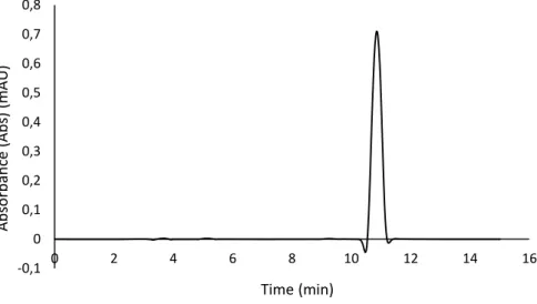
Figure 7. Chromatogram of rosmarinic acid (20 mg.L-1) analysed by HPLC-DAD at 330 nm. 40
Figure 8. Absorption spectra of RA (0.50 mg.L-1) between 200 and 600 nm for a) water and
b) coconut oil. 42
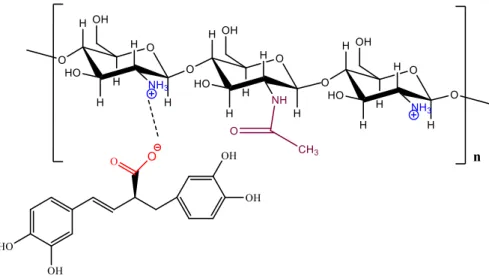
Figure 9. Possible reversible ionic interactions between COO- group of RA and NH
3+ of
chitosan. 44
Figure 10. SEM micrographs of RA loaded chitosan (A, C) and modified chitosan (B, D)
microparticles. 45
Figure 11. Size distribution in volume of RA microparticles produced with chitosan (a) and
modified chitosan (b). 46
Figure 12. Size distribution in number of RA microparticles produced with chitosan (a) and
modified chitosan (b). 46
Figure 13. Chromatogram of RA-chitosan (a) and RA-modified chitosan (b) microparticles
after 10 min of release in water, analysed by HPLC-DAD at 330 nm. 47
Figure 14. Hydrolysis reaction of rosmarinic acid. 48
Figure 15. Comparison of the RA release profile from chitosan (a) and modified chitosan (b)
microparticles in different mediums: water and coconut oil. 49
Figure 16. Comparison of the RA release profile from microparticles in water (a) and
coconut oil (b) using different biopolymers: chitosan and modified chitosan. 49
Figure 17. Oxidation of ABTS by potassium persulfate to generate radical cation ABTS•+ and
viii
TABLES LIST
Table 1. Properties and biological activities of rosemary (Rosmarinus officinalis) antioxidants. 8
Table 2. Chemical and physical properties of rosmarinic acid. 10
Table 3. Usual methods for microencapsulation and respective particle size produced. 14
Table 4. Studies on the encapsulation of rosmarinic acid or rich sources thereof for
industrial and medical applications. 25
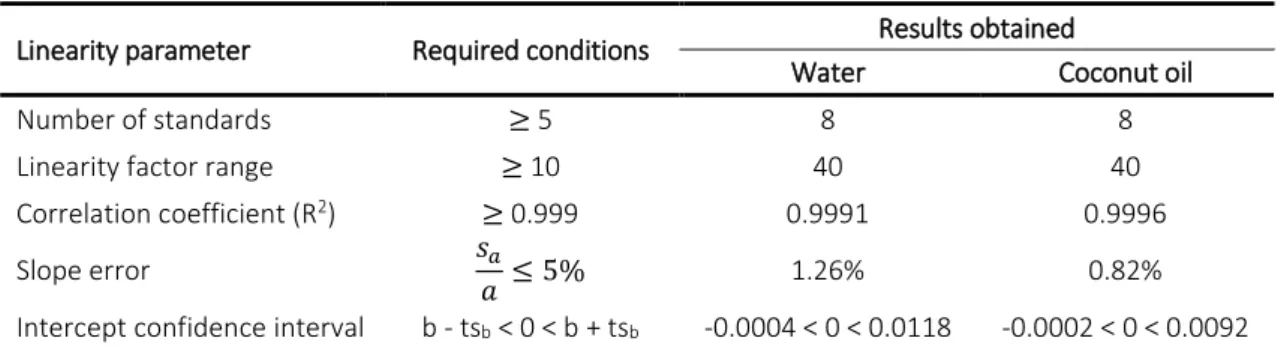
Table 5. Quantification parameters of the HPLC method for rosmarinic acid quantification. 41
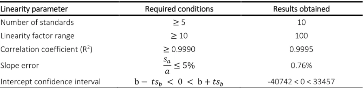
Table 6. Linearity conditions for the validation of the HPLC calibration curve. 41
Table 7. Reliability parameters of the HPLC method for RA quantification. 41
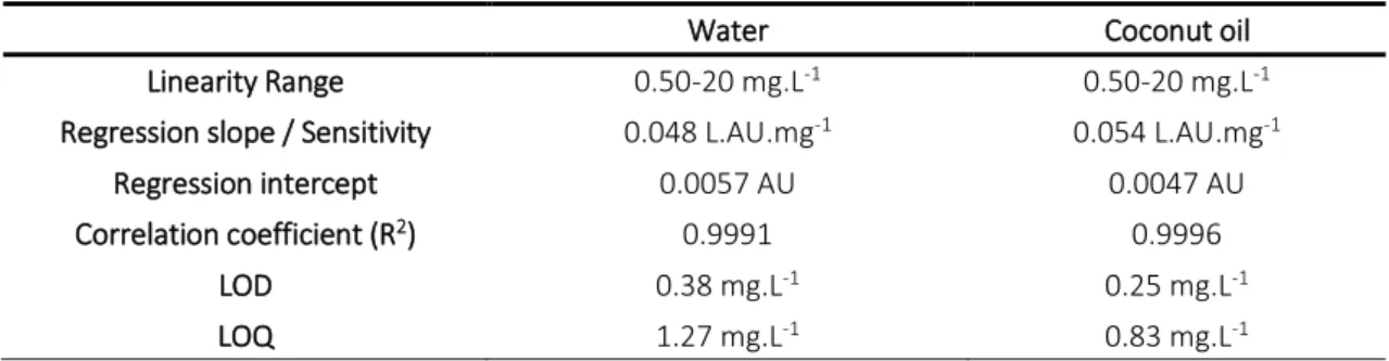
Table 8. Quantification parameters of the UV-Vis spectrophotometer for RA quantification in
water and coconut oil. 42
Table 9. Linearity conditions for the validation of the UV-Vis spectrometry calibration curves. 43
Table 10. Reliability parameters of the UV-Vis spectrophotometer for RA quantification in
water and coconut oil. 43
Table 11. Particle size distribution results by laser granulometry analysis. 46
Table 12. Antioxidant activity estimated by ABTS of RA free in solution and encapsulated
ix
GLOSSARY
A Peak area
Abs Absorbance
ABTS 2,2-azinobis (3-ethyl-benzothiazoline-6-sulfonic acid)
AE Association efficiency
AHA Alpha hydroxy acid
BHA Butylated hydroxyanisole
BHT Butylated hydroxytoluene
C Concentration
CA Caffeic acid
CAS Chemical Abstracts Service
CD Cyclodextrin
CoA Coenzyme A
CO2 Carbon dioxide
CV Coefficient of variation
DAD Diode-array detection
DMF Dimethyl formamide
DMSO Dimethyl sulfoxide
DNA Deoxyribonucleic acid
GSH Glutathione
HIV-1 Human immunodeficiency virus type one
HPLC High-Performance Liquid Chromatography
IUPAC International Union of Pure and Applied Chemistry
LM Light microscope
LOD Limit of detection
LOQ Limit of quantification
MS Mass Spectrometry
NLC Nanostructured lipid carriers
NP Nanoparticles
PB Phosphate buffer
PBS Phosphate buffered saline
PCL Polycaprolactone
PEG Polyethylene glycol
PLA Poly(lactic acid)
PLGA Poly(lactic co-glycolic acid)
QSPR Quantitative structure-permeability relationships
RA Rosmarinic acid
ROS Reactive oxygen species
SEM Scanning electron microscope
SLN Solid lipid nanoparticles
TE Trolox equivalents
UV Ultraviolet radiation
INTRODUCTION
1
CHAPTER 1: INTRODUCTION
1.1. Background
Microencapsulation has been widely explored by the pharmaceutical, food, cosmetic, textile, personal care, chemical, biotechnology and biomedical industries. There are numerous possibilities to use microencapsulation as a technique to obtain products with high added value and therefore widespread interest has developed in microencapsulation technology (Hammad et al., 2011; Dubey et al., 2009; Wesselingh et al., 2007; Ghosh, 2006; Benita, 2005).
The sector of cosmetics and personal care products has been evaluated in multi-billion dollars internationally and has shown great expansion in the global market. To have success in such a competitive and demanding sector, the products must differentiate (Euromonitor, 2011; Michael, 2009). The skin-care and cosmetic formulators are being challenged to develop efficacious and clearly distinctive topical formulations and also include new bioactive ingredients, which can be achieved by means of using emergent technologies, such as microencapsulation. Numbers of materials as cosmetic active ingredients are being investigated such as antioxidants, vitamins, sun filters and natural plant extracts, namely essential oils and natural compounds (Kim et al., 2010). Meanwhile many of these cosmetic and personal care active ingredients are unstable and sensitive to temperature, pH, light and oxidation, and may undergo reactions that lead to the reduction or loss of its effectiveness or even to the degradation of the product. Thus, microencapsulation technologies have been proposed to increase stability, to protect against degradation, to direct and control the release of active ingredients used in cosmetic products, or even to mask undesired properties of the active components, such as their odour. In addition, the topical and transdermal delivery of cosmetic active ingredients requires safe, non-toxic and effective means to reach the target destination sites in the body. Therefore microencapsulation has been used in the development of cosmetic formulations that are more stable, more effective and with improved sensory properties, having found an increasing number of applications in this market (Pardeike et al., 2009; Soest, 2007; Sinko, 2006; Lumsdon et al., 2005; Rosen, 2005; Gallarate et al., 1999). Published patents in the area of microencapsulation suggest that both industrial and academic sectors are urging to explore this area, namely in the fields of cosmetics and personal care products for topical application (Conopco Inc. and D/B/A Unilever, 2010; Capsutech Ltd., 2009; Coreana Cosmetics Co. Ltd, 2001; Shaklee Corporation, 2001; Maybelline Intermediate Company, 1999; L’Oreal, 1998; Sunsmart, Inc. and Sibmicro Encapsulation Technologies, Inc., 1998; R.P. Scherer Corporation, 1996; Durand, 1995).
INTRODUCTION
2
1.2. Cosmetic Ingredients
1.2.1. Antioxidants in Cosmetics
Antioxidants are molecules capable of inhibiting the oxidation of other molecules (Zheng et al., 2001). Although oxidation reactions are essential for life, they can also be damaging, leading to cell injury and death. Topically applied antioxidants constitute an important group of cosmetic active ingredients capable of preventing the occurrence and reducing the severity of skin damage and aging (Oresajo et al., 2012; Wang et al., 2008).
Skin, being the outermost barrier of the body, is frequently exposed to oxidative stress from exogenous sources (ultraviolet (UV) radiation, air pollutants, toxins) leading to the generation of reactive oxygen species (ROS) and free radicals. Owing to the presence of one unpaired electron in the outermost shell of the atomic nucleus, ROS and free radicals are highly reactive and have the affinity to either donate or obtain electrons from another species to achieve stability. This may lead to the oxidation and damage of biomolecules, including lipids, proteins and deoxyribonucleic acid (DNA). To counteract the harmful effects of free radicals, the skin possesses an antioxidant network responsible for maintaining the balance between these reactive species and antioxidants. The antioxidant network of the skin contains antioxidant species such as vitamin E, ubiquinones, carotenoids, vitamin C, uric acid and glutathione (Abla et al., 2013; Psotova et al., 2006). Intrinsic and extrinsic free radical formation cause oxidative stress, the major and most important cause of skin damage. Intrinsic free radical formation occurs when the ability of human skin cells to repair DNA damage steadily reduces with years and the antioxidative defence becomes less effective (Poljsak et al., 2013). On the other hand, antioxidants present in the skin are susceptible to UV exposure and a single suberythemal dose1 can deplete their concentration in half, resulting in extrinsic free radical formation (Abla et al., 2013). Ionizing radiations, including UV radiation, are major environmental factors that dramatically alter skin homeostasis, causing a massive generation of cytotoxic reactive oxygen species and inducing DNA damage. Their cytotoxicity increases by interaction with membrane phospholipids, inducing peroxidative processes and the generation of lipoperoxy radicals (Sánchez-Campillo et al., 2009). Free radical formation and oxidative stress may lead to skin damage, premature skin aging, inflammatory reactions and malignant skin lesions (skin cancer) (Abla et al., 2013; Oresajo et al., 2012). Under these oxidative conditions the endogenous antioxidant system may be insufficient and exogenous agents with a strong antioxidant capacity could be beneficial (Sánchez-Campillo et al., 2009).
INTRODUCTION
3
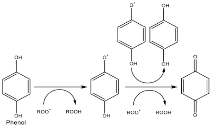
Antioxidants are compounds that can delay or inhibit the oxidation of other molecules by inhibiting the initiation or propagation of oxidizing chain reactions (Zheng et al., 2001). Antioxidants act either by an indirect mechanism where they form chelates or by a direct mechanism where they donate an electron to the reactive oxygen species (Abla et al., 2013). Inhibition of propagating radicals during the oxidative chain reaction is believed to be the dominant mechanism by which phenolic antioxidants operate. A mechanism proposed to account for this behaviour is given in Figure 1 (Servili et al., 2014; Zheng et al., 2001; Basaga et al., 1997).
Figure 1. Example of the mechanism of the antioxidant activity of phenols.
Antioxidants protect skin cells against the damaging effects of reactive species, such as singlet oxygen, superoxide, peroxyl and hydroxyl radicals, thus preventing oxidative stress and skin aging and damage. Several studies concerning topically applied antioxidants have recently confirmed that antioxidant compounds can scavenge oxidative radicals, decrease lipid peroxidation and thus provide protection against oxidative stress. Therefore supplementing skin with antioxidants may strengthen the antioxidant capacity and reduce ROS induced skin damage (Abla et al., 2013). Moreover cosmetic products, which commonly comprise fats, oils and lipid containing matrices, are often prone to oxidation reactions, which generate compounds that cause rancidity, off-aromas and activity losses (Carrillo et al., 2006; Basaga et al., 1997). Oxidative degradation of fats, oils and lipids is a major factor responsible for the deterioration of cosmetics and thereby for limiting the shelf life of these products. The use of antioxidants during the manufacturing process can provide protection against oxidative degradation of cosmetic products caused by free radicals (Wang et al., 2008; Aruoma et al., 1996).
A variety of antioxidants can be used in topical cosmetic products. These include vitamin C (ascorbic acid and its derivatives), vitamin E (tocopherol and its derivatives), ubiquinones and its derivatives, and glutathione (GSH) and its precursors. In addition, activators of several enzyme systems that regenerate these antioxidants, such as GSH peroxidases and GSH reductase, and enzymes that neutralize ROS, such as superoxide dismutases, catalase, and quinone reductase,
INTRODUCTION
4
are also used topically to potentiate antioxidant systems within the skin. Plant-derived compounds such as carotenoids and phenols are an important group of compounds showing antioxidant properties. Dozens of polyphenols have been shown to have antioxidant and anti-inflammatory activities on the skin, and are currently used in skin care products (Oresajo et al., 2012). In general there are three basic categories of antioxidants: natural, synthetic and semi-synthetic. Recently, interest has increased considerably in finding naturally occurring antioxidants for cosmetic applications to replace synthetic ones, which are being restricted due to their carcinogenic effects (Zheng et al., 2001).
Nevertheless, the efficacy and benefit of an antioxidant is very much dependent on the delivery of the antioxidant to the organism. When formulating with antioxidants, compatibility is a major concern. Care must be taken to protect the antioxidant from neutralizing other cosmetic active ingredients or from being neutralized under the conditions of the formulation. Product stabilization is also crucial. Since antioxidants can be very unstable, they may become oxidized and inactive before reaching the target body site. Antioxidants must also be absorbed into the skin, reach their target tissue in the active form, and remain there long enough to exert the desired effects (Oresajo et al., 2012).
These issues can be solved by applying the microencapsulation technology, which provides the required technique for conversion of the antioxidant to an effective functional ingredient. The application of microencapsulated bioactive compounds as functional ingredients in cosmetic applications exhibits a significant potential, since it could enable the enrichment of various cosmetic products with natural antioxidants and its effective permeation through the skin. Owing to microencapsulated ingredients, the production of many products that were considered technically unfeasible has been enabled (Belščak-Cvitanović et al., 2011).
1.2.2. Natural sources of compounds of cosmetic interest
Herbs have been used for a large range of purposes including medicine, pharmaceuticals, nutrition, food preservation, flavourings, beverages, repellents, fragrances and cosmetics. Since prehistoric times, herbs were the basis for medicinal therapy until synthetic drugs were developed in the nineteenth century (Okoh et al., 2010; Zheng et al., 2001). In recent decades, the use of herbs and plants has been of great interest, as they have been the sources of natural products, commonly named as bioactive compounds (Zibetti et al., 2013; Wang et al., 2008). Bioactive compounds are natural active ingredients produced in plants as secondary metabolites that have an effect on a living organism, tissue or cell (Zibetti et al., 2013). The use of bioactive compounds from various herbs as functional ingredients in food, beverage and cosmetic
INTRODUCTION
5
applications is gaining growing interest in the last few years (Belščak-Cvitanović et al., 2011). Natural matrices represent a rich source of biologically active compounds and are an example of molecular diversity, with recognized potential for the development of cosmetics or cosmeceuticals (Barroso et al., 2014).
Bioactive compounds of plant origin have been shown to have several beneficial properties. Nowadays the use of natural compounds is also increasing around the world due to their mild features and low side effects (Belščak-Cvitanović et al., 2011; Sánchez-Campillo et al., 2009; Erkan et al., 2008). Cosmetic preparations from herbal origin are popular among consumers, as these agents are typically non-toxic and possess strong activities (Barroso et al., 2014).
Herbs provide a wide spectrum of bioactive compounds with beneficial activities, namely polyphenols, vitamins, polysaccharides and minerals (Belščak-Cvitanović et al., 2011). Bioactive properties of various plants are connected with the presence of phenolic compounds, especially flavonoids. The biological, pharmacological and medicinal properties of this group of compounds have been extensively reviewed and related to their antioxidant properties by preventing UV induced oxygen free radical generation and lipid peroxidation. Many of these phytochemicals possess significant antioxidant capacities that have been associated with lower incidence and lower mortality rates of cancer (Zheng et al., 2001). Since oxidative stress is one of the major mechanisms for skin aging and damage, phytochemicals such as phenolic compounds could be useful for treating or preventing those conditions (Barroso et al., 2014; Luis et al., 2007). In order to prolong the storage stability of cosmetics and foods, synthetic antioxidants are used for industrial processing. But the toxicity effects of some synthetic antioxidants, such as butylated hydroxytoluene (BHT) and butylated hydroxyanisole (BHA) have already been documented, and they include carcinogenic effects in living organisms (Türkoğlu et al., 2007). For this reason, governmental authorities and consumers are concerned about the safety and the potential effects of synthetic additives on health, and are demanding for natural ingredients. Preliminary studies demonstrated that some herbs extracts are as efficient as the synthetic antioxidants (Hernández-Hernández et al., 2009; Erkan et al., 2008; Wang et al., 2008). Phenolic compounds are also known to possess antimicrobial activity and are generally recognized as safe substances, therefore they are used to prevent post-harvest growth of native and contaminant bacteria (Okoh et al., 2010). For all the described reasons in recent years a lot of interest has been devoted to exploring antioxidants from natural sources (Psotova et al., 2006; Basaga et al., 1997).
Recently numerous reports have described antioxidants and compounds with beneficial activities present in fruits, vegetables, herbs and cereals extracts (Wang et al., 2008). Rosemary
INTRODUCTION
6
(Rosmarinus officinalis), sage (Salvia officinalis), thyme (Thymus vulgaris) and lavender (Lavendula angustifolia) are native to the Mediterranean region and cultivated world-wide, and balm (Melissa officinalis) and spearmint (Mentha spicata) are common plants in Britain and other european countries. Researchers have found that these plants are a source of compounds possessing high antioxidant, anti-inflammatory, antimicrobial and anti-carcinogenic activities. In particular rosemary extracts possess very useful antioxidant properties, which appear to be related to their content of phenolic compounds, amongst which rosmarinic acid was found to be one of the most important (Hernández-Hernández et al., 2009; Erkan et al., 2008; Wang et al., 2004; Aruoma et al., 1996). Gachkar et al. (2007) reported the chemical composition, antibacterial, antioxidative and radical-scavenging properties of the essential oils of Rosmarinus officinalis and Cuminum cyminum obtained by steam distillation. The antimicrobial activity of essential oils obtained from oregano, thyme, sage, rosemary, clove, coriander, garlic and onion against both bacteria and fungi has also been reported (Okoh et al., 2010).
The inclusion of antioxidants, or rich sources thereof (including herb extracts), in cosmetics is becoming a common procedure of this industry, namely in the production of various cologne waters, body lotions and creams, sunscreens, bathing lotions, hair lotions, shampoos and as components of disinfectants and insecticides (Campos et al., 2012; Okoh et al., 2010; Sánchez-Campillo et al., 2009; Wang et al., 2004).
Rosemary - Rosmarinus officinalis
Rosmarinus officinalis (Figure 2), commonly known as rosemary, is a perennial herb that belongs to the Lamiaceae family (Sui et al., 2012; Okoh et al., 2010; Kim et al., 2001). It is native to the Mediterranean region in countries like Portugal, Spain, Morocco, Tunisia and Italy, but is now widely distributed and has been cultivated in many regions (Fernandes et al., 2013; Liu et al., 2011). Rosemary extracts have been used for thousands of years as flavourings, pharmaceuticals, alternative medicine and natural therapies (Couto et al., 2012; Zu et al., 2012; Fadel et al., 2011).
INTRODUCTION
7
Recently rosemary has been largely studied as a source of natural products with diverse biological activities (Visentin et al., 2012). In addition to being used as a food flavouring, several extracts, essential oils and chemical constituents isolated from Rosmarinus officinalis demonstrated a number of interesting biological activities, including antioxidant, antimicrobial, anti-inflammatory, anti-tumorigenic, anti-allergic, metal chelating, chemo-preventive and cyto-protective activities (Couto et al., 2012; Sui et al., 2012; Liu et al., 2011; Wang et al., 2004), which makes them suitable candidates as bioactive ingredients to design functional cosmetic, pharmaceutical and food products (Visentin et al., 2012; Zu et al., 2012; Psotova et al., 2006).
R. officinalis has been widely accepted as one of the spices with highest antioxidant activities of
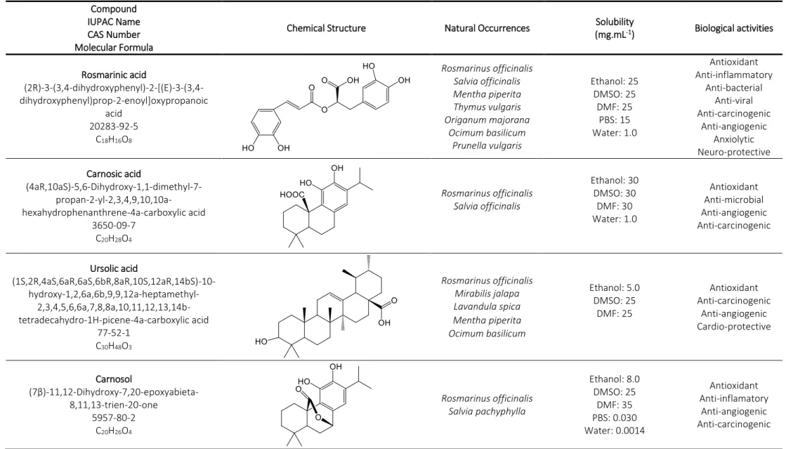
all the herbs and spices that have been investigated (Wang et al., 2008). The antioxidant activity of rosemary is related to rosemary’s content of polyphenolic compounds (Couto et al., 2012; Del Bano et al., 2003). The compounds associated with the antioxidant activity of the herb are the phenolic acids such as rosmarinic acid, ursolic acid and caffeic acid, and the phenolic diterpenes such as carnosoic acid, carnosol, rosmanol, epirosmanol and methyl carnosate (Table 1) (Zibetti et al., 2013; Carvalho et al., 2005¸ Del Bano et al., 2003). In addition, several flavonoids, such as genkwanin, scutellarein, cirsimaritin and luteolin have been identified (Table 1) (Del Bano et al., 2003). Several studies have indicated that the most active antioxidant compounds in rosemary are the diterpene, carnosic acid, and the phenolic acid, rosmarinic acid, which are the most abundant constituents and also well known for their various biological properties (Sui et al., 2012; Liu et al., 2011; Luis et al. 2007). Rosmarinic acid is the major and most important phenolic component of the plant and is therefore considered a chemical marker of this species. It exhibits a wide spectrum of biological activities, mainly antioxidant and anti-carcinogenic (Couto et al., 2012; Psotova et al., 2006; Wang et al., 2004).
Rosmarinic acid
Rosmarinic acid (RA) is a naturally occurring phenolic compound commonly found in plants belonging to the Boraginaceae family and the subfamily Nepetoideae of the Lamiaceae family (Campos et al., 2014; Sánchez-Campillo et al., 2009). RA was originally isolated in 1958 from the rosemary plant (Rosmarinus officinalis). Some of the species from which this compound has been reported include rosemary (Rosmarinus officinalis), sage (Salvia officinalis), peppermint (Mentha piperita), thyme (Thymus vulgaris), marjoram (Origanum vulgare), basil (Ocimum basilicum) and Prunella (Prunella vulgaris) (Hossan et al., 2014; Kim et al., 2010).
8
Table 1. Properties and biological activities of rosemary (Rosmarinus officinalis) antioxidants. Compound
IUPAC Name CAS Number Molecular Formula
Chemical Structure Natural Occurrences Solubility
(mg.mL-1) Biological activities Rosmarinic acid (2R)-3-(3,4-dihydroxyphenyl)-2-[(E)-3-(3,4-dihydroxyphenyl)prop-2-enoyl]oxypropanoic acid 20283-92-5 C18H16O8 Rosmarinus officinalis Salvia officinalis Mentha piperita Thymus vulgaris Origanum majorana Ocimum basilicum Prunella vulgaris Ethanol: 25 DMSO: 25 DMF: 25 PBS: 15 Water: 1.0 Antioxidant Anti-inflammatory Anti-bacterial Anti-viral Anti-carcinogenic Anti-angiogenic Anxiolytic Neuro-protective Carnosic acid (4aR,10aS)-5,6-Dihydroxy-1,1-dimethyl-7- propan-2-yl-2,3,4,9,10,10a-hexahydrophenanthrene-4a-carboxylic acid 3650-09-7 C20H28O4 Rosmarinus officinalis Salvia officinalis Ethanol: 30 DMSO: 30 DMF: 30 Water: 1.0 Antioxidant Anti-microbial Anti-angiogenic Anti-carcinogenic Ursolic acid (1S,2R,4aS,6aR,6aS,6bR,8aR,10S,12aR,14bS)-10- hydroxy-1,2,6a,6b,9,9,12a-heptamethyl- 2,3,4,5,6,6a,7,8,8a,10,11,12,13,14b-tetradecahydro-1H-picene-4a-carboxylic acid 77-52-1 C30H48O3 Rosmarinus officinalis Mirabilis jalapa Lavandula spica Mentha piperita Ocimum basilicum Ethanol: 5.0 DMSO: 25 DMF: 25 Antioxidant Anti-carcinogenic Anti-angiogenic Cardio-protective Carnosol (7β)-11,12-Dihydroxy-7,20-epoxyabieta-8,11,13-trien-20-one 5957-80-2 C20H26O4 Rosmarinus officinalis Salvia pachyphylla Ethanol: 8.0 DMSO: 25 DMF: 35 PBS: 0.030 Water: 0.0014 Antioxidant Anti-inflamatory Anti-angiogenic Anti-carcinogenic
9 Rosmanol (6β,7β)-7,11,12-Trihydroxy-6,20-epoxyabieta-8(14),9(11),12-trien-20-one 80225-53-2 C20H26O5 Rosmarinus officinalis n.d. Antioxidant Anti-inflamatory Anti-carcinogenic Methyl carnosate methyl (4aR,10aS)-5,6-dihydroxy-1,1-dimethyl- 7-propan-2-yl-2,3,4,9,10,10a-hexahydrophenanthrene-4a-carboxylate 82684-06-8 C21H30O4 Rosmarinus officinalis Salvia officinalis n.d. Antioxidant Anti-bacterial Genkwanin 5-hydroxy-2-(4-hydroxyphenyl)-7-methoxychromen-4-one 437-64-9 C16H12O5 Rosmarinus officinalis Alnus glutinosa Notholaena bryopoda Asplenium normale Teucrium ramosissimum
Soluble in: DMSO, DMF
Antioxidant Anti-carcinogenic Scutellarin 5,6,7-trihydroxy-2-(4-hydroxyphenyl)-4H-chromen-4-one 27740-01-8 C21H18O12 Rosmarinus officinalis Scutellaria barbata Scutellaria lateriflora
Soluble in: alkali hydroxides, glacial acetic acid
DMSO: 1.0 Antioxidant Anti-carcinogenic Cirsimaritin 5-hydroxy-2-(4-hydroxyphenyl)-6,7-dimethoxychromen-4-one 6601-62-3 C17H14O6 Rosmarinus officinalis Teucrium ramosissimum Lemon verbena Cirsium vulgare n.d. Antioxidant Anti-carcinogenic
INTRODUCTION
10
Rosmarinic acid (Table 2) is formally known as (2R)-3-(3,4-dihydroxyphenyl)-2-[(E)-3-(3,4 dihydroxyphenyl)prop-2-enoyl]oxypropanoic acid (Hossan et al., 2014). It is a white-yellow powder slightly soluble in water, but well soluble is most organic solvents (Kim et al., 2010).
Table 2. Chemical and physical properties of rosmarinic acid.
Chemically, RA is an ester of caffeic acid (CA) and 3,4-dihydroxyphenyllactic acid, being a CA dimer formed by esterification (ester condensation) of CA and its hydrated derivative 3,4-dihydroxyphenyllacetic acid. Biologically, the biosynthesis of RA starts with precursor molecules L-phenylalanine and L-tyrosine and uses 4-coumaroyl-CoA (coenzyme A) from the general phenylpropanoid pathway as hydroxycinnamoyl donor (Appendix A: Biosynthetic pathway of rosmarinic acid) (Petersen et al., 2013, 2009, 1993; Sundaram et al., 2010). In plants, RA acts as a preformed constitutively accumulated defence compound (Sánchez-Campillo et al., 2009). Rosmarinic acid was reported to have a number of interesting biological activities (Campos et al., 2014). RA has anti-inflammatory and anti-allergic properties, inhibiting several inflammatory processes by direct effects on T cells (Stansbury et al., 2012; Sánchez-Campillo et al., 2009). This compound possesses antiviral, including against herpes simplex virus and human immune-deficiency virus type one (HIV-1), and antibacterial activities (Gudzenko, 2013; Fecka et al., 2007).
Compound (Acronym) Rosmarinic acid (RA)
IUPAC Name
(2R)-3-(3,4-dihydroxyphenyl)-2-[(E)-3-(3,4-dihydroxyphenyl)prop-2-enoyl]oxypropanoic acid
CAS Number 20283-92-5
Molecular Formula C18H16O8
Chemical Structure
Molar mass (g.mol-1) 360.31
Melting point (°C) 173 Density 1.55 Vapor pressure (mm Hg) 1.10 x 10⁻¹³ log Kow 1.82 pKa 3.57 UV absorption max (nm) 221, 291, 330 Solubility (mg.mL-1) Ethanol: 25 DMSO: 25 DMF: 25 PBS: 15 Water: 1.0
INTRODUCTION
11
Rosmarinic acid was reported to possess notable activity against Bacillus subtilis and Escherichia coli. It has also been reported to possess activity against Pseudomonas aeruginosa, Shigella sp, Staphylococcus aereus, Enterobacter, Candida albicans and Aspergillus niger. In vivo studies have shown that RA also exhibits anti-angiogenic, anxiolytic, anti-atherosclerotic, anti-fibrotic, chemo-protective and neuro-chemo-protective activities, as well as reduction of atopic dermatitis and prevention of Alzheimer’s disease (Bhatt et al., 2014; Silva et al., 2014b; Gudzenko, 2013; Kelsey et al., 2010; Swarup et al., 2007; Sanbongi et al., 2004, Hooker et al., 2001). However the most important activities of RA are the antioxidant and anti-carcinogenic effects (Campos et al., 2014). RA has a potent antioxidant activity, stronger than that of vitamin E, by scavenging oxygen free radicals, delaying vitamin E depletion, preventing the oxidation of low density lipoproteins and providing photo-protection of keratinocytes (Kim et al. 2010; Sánchez-Campillo et al., 2009). RA helps to prevent cell damage caused by free radicals, thereby reducing the risk of cancer (Kim et al., 2010). RA has been found to be effective against a number of cancer cell lines, including skin cancer and melanoma, colon cancer, leukaemia and breast cancer (Osakabe et al., 2004). The effects of skin cancer have been reversed by topical application of RA, which acted against skin tumours by exerting an inflammatory and antioxidant effect. RA was found to have dual anti-carcinogenic capability, namely that of protecting normal cells against radiation but also of sensitizing melanomas to radiation (Alcaraz et al., 2014; Hossan et al., 2014).
Rosmarinic acid is therefore a compound of interest for cosmetic applications. However, transdermal delivery of this antioxidant through cosmetic formulations, such as emulsions and creams, is a challenge due to instability, discoloration, poor solubility in water and low partition coefficient, constraining the transport across biological barriers. The technological handling of such type of compounds is sometimes limitative due to their reactivity, so their incorporation in a cosmetic formulation becomes difficult if they are not duly protected from the interactions to which they are exposed when incorporated in cosmetic matrices. The solubility characteristics of the antioxidant in relation to the site of action must also be considered (Campos et al., 2014, 2012; Silva et al., 2014b; Visentin et al., 2012; Kim et al., 2010). In view of the above mentioned drawbacks, encapsulation with an appropriate carrier material for RA delivery, protection and release is necessary to obtain an effective product (Silva et al., 2014b). Encapsulation will protect the antioxidant during manufacturing processes, improve its long-term stability, its permeability characteristics through the skin and the retention within the skin, and increase the efficacy of the compound (Budhiraja et al., 2014; Campos et al., 2012; Visentin et al., 2012). Among different approaches explored so far, microparticles, especially those made of biocompatible polymers, such as chitosan, seem to assure active and safe delivery of this compound (Silva et al., 2014b).
INTRODUCTION
12
1.3. Microencapsulation
Microencapsulation is a process of encapsulating a material containing an active ingredient (core material) in a shell of a second material (encapsulating/wall material) for the purpose of shielding active ingredients from the surrounding environment permanently or temporarily. This results in small capsules of many useful properties, termed microcapsules or microparticles. Such particles have diameters between one micron and a few millimetres. Particles whose diameter is in the nanometre range are referred to as nanoparticles (Kaur et al., 2013; Jyothi et al., 2010, Benita, 2005). The small size of these particles provides a large surface area that is available for sites of adsorption/desorption, chemical reactions, light scattering, etc. (Arshady, 1999; Gutcho, 1976).
The development of microencapsulation began with the preparation of capsules containing dyes in 1950s by Green and Schleicher. These were incorporated into paper for copying purposes and replaced carbon paper. Nowadays this approach has been widely explored by the pharmaceutical, food, cosmetic, textile, agricultural, veterinary, chemical and biomedical industries (Hammad et al., 2011; Ghosh, 2006). The field with the highest level of microencapsulation applications is the drug sector (68%), followed by food (13%) and cosmetics (8.0%) (Martins et al., 2014). There are numerous possibilities to use microencapsulation as a technique to obtain products with high added value and therefore widespread interest has developed in microencapsulation technology (Martins et al., 2014; Dubey et al., 2009).
Core materials in microcapsules may exist in the form of a solid, liquid or gas. Depending on the application, a wide variety of core materials can be encapsulated, including pigments, fragrances, flavours, enzymes and cells. The size of the core material plays an important role for diffusion, permeability and controlled release. The shell material can be permeable, semi-permeable or impermeable. Compatibility of the core material with the shell is an important criterion for enhancing the efficiency of the microencapsulation, and for this pre-treatment of the core material is often carried out (Estevinho et al., 2013a; Hammad et al., 2011; Ghosh, 2006). The morphology of the internal structure of a microcapsule (Figure 3) depends largely on the selected shell material and the encapsulation method employed. Microcapsules can be classified as mononuclear, polynuclear or matrix type. Mononuclear microcapsules contain the shell around the core and have a single hollow chamber within the capsule. Microcapsules can also be mononuclear with multiple shells. The polynuclear microcapsules have many cores enclosed within the shell, so they have a number of different sized chambers. The matrix type microparticle has the active ingredients integrated within the matrix of the shell material and distributed homogeneously (Chhotalal et al., 2013; Dubey et al., 2009; Ghosh, 2006).
INTRODUCTION
13
Figure 3. (a) Scheme of a microcapsule. (b) Morphology of microcapsules. (Adapted from Ghosh, 2006) Microcapsules are usually characterized by parameters such as particle size, zeta potential, encapsulation efficiency and drug loading (Estevinho et al., 2013a). The size and shape of the microparticles can be determined by light or scanning electron microscope (LM or SEM). Encapsulation or association efficiency (content of core material effectively encapsulated) depends on several variables. The retention of the active agent inside the shell is determined by the chemical nature of the core (molecular weight, chemical functionality, polarity and volatility), shell material properties and the encapsulation technique (Selvaraj et al., 2012; Jyothi et al., 2010; Martins et al., 2010; Gander et al., 1995). Microcapsule solvation, density, compressibility index, Hausnner’s ratio2 and angle of repose can also be determined (Hammad et al., 2011).
Microcapsules have a number of interesting advantages, thus microencapsulation technology is used for several purposes: to combine properties of different materials (e.g. organic and inorganic); to protect sensitive, unstable and reactive materials from their environments and prevent the degradation of active compounds (e.g. from reactions like oxidation and dehydration); to protect the immediate environment of the microcapsules from the active components; to increase stability; for controlled, delayed or sustained release; to reduce dosing frequency; for enzyme and microorganism immobilization; to mask undesired properties of the active components (such as odour, taste and activity); for a targeted release of encapsulated materials; for better processability, as it allows to improve solubility, dispersibility and flowability; for a safe and convenient handling of toxic materials; and to separate incompatible components for functional reasons. Some application examples of the use of microencapsulation technology are isolating vitamins from the deteriorating effects of oxygen, retarding evaporation of a volatile core, improving the handling properties of a sticky material or isolating a reactive core from chemical attack (Ramu et al., 2014; Ghosh, 2006; Lopéz et al., 1997).
2 Index of flow ability of microcapsules.
INTRODUCTION
14 1.3.1. Microencapsulation techniques
Although a variety of techniques have been reported for microencapsulation, no single process is adaptable to all core materials or product applications. The choice of the most suitable method depends on the application of the microsystem, particle size required, physical and chemical properties of the core and the shell, release mechanism intended, production scale and costs. An appropriate combination of starting materials and synthesis methods can be chosen to produce encapsulated products with a wide variety of compositional and morphological characteristics, and the encapsulation process must be optimized in order to provide a satisfactory outcome, considering the intended application. Microencapsulation techniques can be broadly divided into two main categories, namely chemical and physical, with the latter being further subdivided into physico-chemical and physico-mechanical techniques (Silva et al., 2014a; Estevinho et al., 2013a; Wilson et al., 2007). Table 3 outlines common methods used to encapsulate ingredients and the size of particles they produce. Such methods have been described by Silva et al. (2014a), Dubey et al. (2009), Fairhurst et al. (2008), Wilson et al. (2007) and Ghosh (2006).
Table 3. Usual methods for microencapsulation and respective particle size produced.
Spray drying is one of the most common methods used for microencapsulation. It is a continuous and single-cycle process that involves the conversion of a material from a fluid state (solution, emulsion or suspension) into a dried particulate form by atomization in a hot gas medium (generally air) (Estevinho et al., 2014b, 2013a, 2013b). The ingredient to be encapsulated is added to the carrier (the ratio of core to carrier can be optimized for each individual ingredient) and the
Type of Method Method Particle size
(μm) References Chemical process Emulsion Polymerization 0.5–1000 Fairhurst et al., 2008; Ghosh, 2006; Hirech, 2003 Suspension Polymerization 0.5–1000 Interfacial Polymerization 0.5–1000 Physico-chemical process
Coacervation/Phase separation 1–1000 Fidalgo et al., 2013; Di
Marco et al., 2010; Cocero et al., 2009; Martins et al., 2009; Nguyen-Ngoc et al., 2007; Ghosh, 2006; Freitas et al., 2003 Solvent evaporation/extraction 0.5–1000 Sol-gel encapsulation 2–20 Supercritical fluid-assisted microencapsulation 0.5-500 Layer-by-layer assembly 0.5–20 Physico-mechanical process Spray-drying 1–500 Fernandes et al., 2014, 2013; Shinde et al., 2014; Teodoro et al., 2014; Anwar et al., 2010; Fairhurst et al., 2008; Ghosh, 2006; Senuma et al., 2000 Spray-cooling 20 – 500 Co-extrusion 250–2500 Spinning disk 5–1500 Fluidized-bed coating 20–1500 Melt solidification 5-1000 Polymer precipitation 5-1000
INTRODUCTION
15
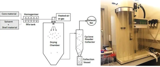
mixture is homogenized. An emulsifier or cross-linking agent may also be added at this stage. This mixture is then fed into the spray dryer with circulating hot air and atomized, which can be made by different types of atomizers: pneumatic atomizer, pressure nozzle, spinning disk, fluid nozzle and sonic nozzle. The solvent is evaporated by the hot air and the shell material encapsulates the core. Small particles are deposited in the collection vessel where they are collected (Fernandes et al., 2014, 2013; Teodoro et al; 2014; Chávarri, 2012; Wilson et al., 2007). A representation of the spray drying process and an image of the equipment are presented in Figure 4.
Figure 4. Scheme (Adapted from Chávarri, 2012) and photography of the spray-drying process/equipment. The properties of the final microparticles depend on the nature of the feeding flow as well as the operating parameters, such as flow rate and inlet temperature. Hot air inlet temperature is typically 150-220 °C and after evaporation the temperature decreases typically to moderate values between 50 and 80 °C. Spray drying can produce, depending on the starting feed material and operating conditions, particles with a size between 1 and 500 µm. Increasing the energy provided to the atomizer decreases the size of the formed particles. For the same energy amount, the size of the particles increases with increasing feed rate. On the other hand, the size of particles also increases with the viscosity and surface tension of the feeding liquid. The microcapsules produced are normally of the polynuclear or matrix type and the mechanisms of release involved are typically controlled by solvents action and diffusion (Estevinho et al., 2014, 2013a; De Vos et al., 2010; Gharsallaoui et al., 2007; Azeredo, 2005).
The shell material, which is hydrated in water, should therefore be soluble in water, have good emulsifying properties, be a good film former, have low viscosity and provide good protection to the encapsulated ingredient. Water-soluble polymers are mainly used as shell materials due to the fact that solvent-borne systems produce unpleasant odours and environmental problems.
INTRODUCTION
16
Typical shell materials used in spray drying include chitosan, sodium alginate, gum arabic, maltodextrins, modified starch, and mixtures of these. The use of encapsulating agents with low solubility in water implies an increase in the amount of water to be evaporated, and thereby a decrease in the dry matter content and in the amount of active ingredient, resulting in a costly process (Estevinho et al., 2013a; Wilson et al., 2007; Ghosh, 2006).
Spray drying has advantages over other encapsulation methods: large equipment availability, possibility of employing a wide variety of encapsulating agents, easy control of microparticle properties by changing the operational parameters, easy scale-up and large-scale production, low process cost and it is a rapid methodology, reproducible and cost-effective, justifying its use in industrial applications (Estevinho et al., 2014, 2013a, 2013b, 2012; Fernandes et al., 2014; Jafari et al., 2008, 2007). Moreover the process is adaptable to a wide range of feedstock and product specifications, as it can be used with solutions, suspensions, slurries, melts and pastes. Microencapsulation by spray drying is thus a simple and low cost commercial process that has been used to encapsulate mainly flavours (Estevinho et al., 2013a), oils and pigments, but also thermo-sensitive products, such as microorganisms, enzymes and essential oils, due to the very short heat contact time and the high rate of evaporation, resulting in high quality, stable, functional and low moisture content products. Biologically active materials have been successfully spray dried without appreciable activity losses (Estevinho et al., 2014a, 2014b, 2013a, 2013b, 2012; Fernandes et al., 2013).
1.3.2. Microencapsulation in cosmetics
Delivery systems and microcapsules play an important and growing role in the cosmetics and personal care industries nowadays. They offer an ideal and unique carrier system for active ingredients, allowing the controlled and targeted release, isolation and protection of the active compounds, improved stability and efficacy, safe administration, to mask undesirable properties of the active components, such as their odour, and also the improvement of the tactile and visual appearance of a variety of cosmetic and personal care products. In these industries there is a constant look for new and novel delivery systems to safely incorporate many of the new and sensitive active ingredients of the cosmetic products. Finding new delivery systems can allow an easier and simpler use and development of critical emulsion systems, where temperature and water/oil content are very important and sensitive active ingredients must be added in a special and sometimes very difficult way or under very controlled conditions. Microencapsulation has the potential of delivering active ingredients in some difficult systems, e.g. containing glycolic acid, alpha hydroxy acids (AHAs), salicylic acid, high alcohol content or critical water-in-oil or
INTRODUCTION
17
silicone emulsions. They can be used to deliver active ingredients into the skin, in a safe, targeted, effective and not painful manner, to protect fragrances or volatile compounds from evaporation, to protect compounds such as antioxidants from oxidation, to protect from degradation caused by heat, light and moisture, or to control the release rate (Martins et al., 2010; Pardeike et al., 2009; Soest, 2007; Rosen, 2005). Recent published patents in the area of microencapsulation suggest that both industrial and academic sectors are urging to explore this area, namely in the fields of cosmetics and personal care products (Martins et al., 2014).
Microencapsulation can be used in cosmetic applications for shower and bath gels, lotions and creams, hair products, sunscreens and tanning creams, makeup, perfumes, soaps, tooth pastes and more. Its study and development may help in the improvement of the cosmetic and personal care industries, as microencapsulation technology brings innovation and allows the production of high added value products, in response to human needs and desires (Barel et al., 2001).
1.3.3. Delivery systems for topical application
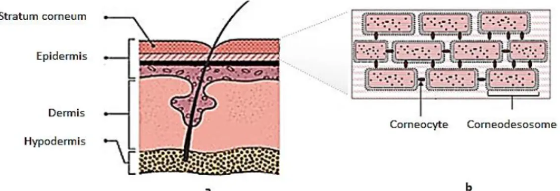
The skin is the largest and most complex human organ, with at least five different cell types contributing to its structure and representing 16% of total body weight (2 m2 of surface). The main function of the skin is the protection of the body, which includes physical, chemical and immunological protection, protection against pathogens, and UV radiation and free radical defences. It is also the major participant in thermoregulation, it functions as a sensory organ and it performs endocrine functions. Human skin is comprised of several distinct layers, namely the stratum corneum (epidermis), the remaining layers of the epidermis, the dermis and the sub-cutaneous adipose tissue (hypodermis). The epidermis is the outer layer and acts as a protective coating against environmental and external influences. It consists of several layers starting with the stratum corneum to the basal cell layer, and is continually being regenerated. The dermis lies beneath the epidermis and is where collagen and elastin are synthesized. It contains the blood vessels, nerves, sweat glands, hair follicles and sebaceous glands. The hypodermis contains the adipose tissue (sub-cutaneous fat) and provides a thermal barrier. The structure of the skin is represented in Figure 5a. The stratum corneum is the upper 10–20 µm layer, which is a lipid-rich matrix composed primarily of ceramides, cholesterol and fatty acids that are assembled into a multi-lamellar bilayer structure. The structure of the stratum corneum comprises protein rich cells (corneocytes) embedded into the intercellular lipid domains. Corneodesmosomes act to link neighboring corneocytes together and provide structural integrity to the stratum corneum. The structure of the stratum corneum is represented in Figure 5b (Ammala, 2013; Harding, 2004).
INTRODUCTION
18
Figure 5. Schematic representation of a) human skin; b) stratum corneum. (Adapted from Harding, 2004) A delivery system is a method of holding, carrying and transporting an active ingredient. It is any type of vehicle that takes an active to a target site. A delivery system can control the release rate of an active ingredient from a formulation at an optimal rate. A number of pathways are possible for the transportation of molecules through the skin. The intercellular route occurs at the interface between cells through the lipid bilayers, following a tortuous permeation pathway. In contrast, transcellular pathways can occur directly through the cells. Transportation via the hair follicles or sweat ducts is also possible (Wiechers, 2008; Flynn, 2002; Moghimi et al., 1999). Topical application of cosmetic formulations often requires the successful delivery of active ingredients through the skin’s barriers to reach the target sites in the body. The main resistance to transdermal transport is in a layer of cells joining the epidermis to the stratum corneum, which itself also limits the transport. The stratum corneum has a highly impermeable nature, which has remained one of the major challenges in effective transdermal delivery. While the lipophilic stratum corneum contains about 13% water, the inner skin epidermis layers become significantly more hydrophilic, containing 50% water, while the dermis contains 70%. It is of extreme importance to understand these properties to design carriers to achieve the desired delivery of cosmetic active substances. It should also be noted that there are wide variations in permeability at different body sites (e.g. face vs. legs vs. palms) which together with factors such as age and external environment can influence the skin’s barrier function (Lam et al., 2014; Forster et al., 2009; Wiechers, 2008; Elias, 2004; Harding, 2004; Rein, 1924).
It is generally reported that the transport of molecules through the epidermis is restricted to molecules of low molecular mass (<500 Da) and moderate lipophilicity (partition coefficients log Kow values between 2 and 3), having enough solubility in the lipid domain of the stratum corneum, while still having sufficient hydrophilic nature to allow partitioning into the skin inner layers. As some active cosmetic substances are too hydrophilic to pass through the stratum corneum or too lipophilic to partition into the epidermis, encapsulation techniques with the right shell materials can overcome this problem by delivering the level of lipophilicity needed for the
INTRODUCTION
19
desired application. Mathematical models can be used to predict skin permeability, which are generally based on quantitative structure-permeability relationships (QSPR), diffusion mechanisms or combinations of both. At the same time the compound should still have the lipophilic or hydrophilic characteristics that allow its solubilisation in the cosmetic itself and ensure its stability during formulation, storage and application of the product (Ammala, 2013; Forster et al., 2009; Jain et al., 2006; Wiechers, 2005; Barry, 2002; Muller et al., 2002, 2000).
Critical aspects should be considered when delivering a cosmetic active ingredient through the skin, such as the right site of action of the cosmetic ingredient, the right concentration of the components, and the correct application time of the product on the skin. It should be considered the influence of the formulation type, formulation polarity, stratum corneum polarity, skin lipid organization and the influence of particle size on skin delivery. The cosmetic delivery should also avoid undesirable transdermal delivery, such as leakage into systemic circulation, and keep the functional molecule in a specific skin layer (Abla et al., 2013; Wiechers, 2008; Fu et al., 2005). Common functional ingredients used in topical application cosmetics are UV filters, antioxidants, moisturizers, skin lightening ingredients, and molecules with anti-aging properties, acting either on the surface of the skin or in specific skin layers (Ammala, 2013; Wiechers, 2008; Elias, 2004). Several skin delivery systems used in cosmetic products have been reported. Vesicles and niosomes (non-ionic surfactant-based vesicles) are much used as skin delivery vehicles for pharmaceutical and cosmetic purposes. Elastic vesicles showed superior properties in comparison with the conventional, rigid vesicles regarding the function potential and the interactions with the human skin. Solid lipid nanoparticles (SLN) and nanostructured lipid carriers (NLC) are also much investigated for cutaneous administration. Flexible liposomes are used as delivery systems for topical applications in cosmetics as well. Since the shell material comprises triglycerides and phospholipids, when used in topical delivery, liposomes can facilitate absorption of the active ingredient into the epidermis and they also have the ability to transport active substances into the deeper skin layers. Micellar nanoparticles, which are liposome-like multi-lamellar structures, have also been reported as skin delivery systems. Encapsulation techniques are also much used as skin delivery systems in topical cosmetic products. Encapsulation of actives for cosmetic and pharmaceutical applications have showed a wide range of applications, as the advantages of different encapsulation technologies were made evident. Other delivery systems, such as phytosomes, transferosomes, nanocrystals and cubosomes have also been reported (Chanchal et al., 2008; El Maghraby et al., 2008; Fairhurst et al., 2008; Wiechers, 2008; Kaur et al., 2007; Rosen, 2005; Barry, 2002; Flynn, 2002).
INTRODUCTION
20 1.3.4. Encapsulating materials
The efficacy of topically applied active ingredients clearly depends on the design of appropriate carriers and the type of vehicle used for their delivery, protection and release. The correct choice of the encapsulating material according to the intended application is essential, as it influences the encapsulation efficiency and the stability of the microparticles. Factors to be considered while selecting a wall material for topical applications include toxicity, biocompatibility, stability, viscosity and mechanical properties, compatibility between the active ingredient and the wall material, release of the active ingredient from the vehicle into the skin, enhancement of active penetration into the stratum corneum, intended particle size and microscopic properties of the surface of the microparticles and processing and economic factors. Since most encapsulating materials do not have all the required properties, a common practice involves a combination of wall materials (Silva et al., 2014b; Abla et al., 2013; Estevinho et al., 2013a).
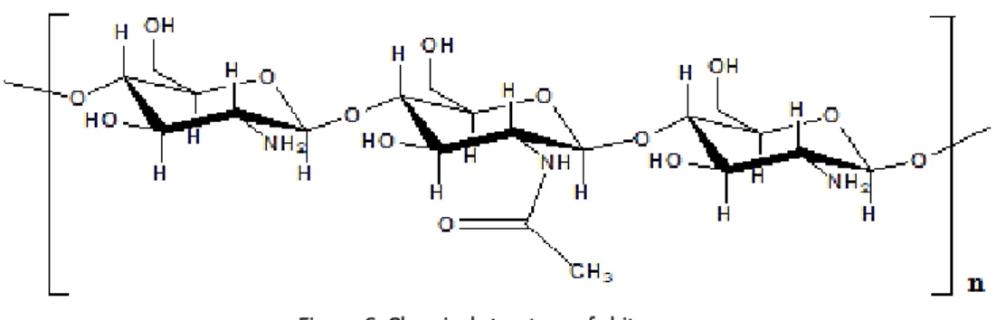
Encapsulating materials can be selected from a wide variety of natural and synthetic polymers. The most commonly used encapsulating materials include polysaccharides (gums, starches, celluloses, alginates, cyclodextrins, maltodextrins, chitosan), proteins (gelatin, casein, soy proteins), lipids (waxes, paraffin, oils), aliphatic polyesters (e.g. poly(lactic acid) – PLA), copolymers of lactic and glycolic acids (e.g. poly(lactic co-glycolic acid) - PLGA) and synthetic polymers (acrylic polymers, polyvinyl alcohol, poly(vinylpyrrolidone)). Inorganic materials, such as silicates, clays and polyphosphates, can also be used as second polymers (Lee et al., 2012; Tarimci, 2011; Cattaneo, 2010; Pawar et al., 2010; Pedro et al., 2009; Wille, 2006; Matsuda et al., 1999). Biopolymers (natural polymers) and biodegradable polymers are the encapsulating materials with greater interest for applications in the field of skin delivery systems. These materials are natural, non-toxic, non-reactive when in contact with the human tissues and can be broken down or metabolized and removed from the body via normal metabolic pathways, while other compounds can potentially accumulate in body tissues and cause irritation. Their properties are strongly defined by structural characteristics, including composition, molecular weight and nature of the chain end groups, and these polymers can be chemically functionalized to obtain improved properties (Ammala, 2013; Haddadi et al., 2008; Mishra et al., 2008; Stevanovic et al., 2007). Among numerous entrapping materials used for the encapsulation of pharmaceutical, cosmetic or food active ingredients, chitosan has received much attention for its excellent biocompatibility (Belščak-Cvitanović et al., 2011). Chitosan is a natural and biodegradable linear co-polymer polysaccharide consisting of β-(1–4)-linked D-glucosamine (2-amino-2-deoxy-D-glucose) and N-acetyl-D-glucosamine (2-acetamido-2-deoxy-D-glucose) (Figure 6) (Silva, 2014; Ammala, 2013).
INTRODUCTION
21
Chitosan is obtained by partial alkaline deacetylation of chitin (a N-acetyl-glucosamine polymer), which is the second most abundant natural polymer in nature. Chitin is synthesized by an enormous number of living organisms and is found predominantly in the exoskeletons shells of crustaceans, insects and other invertebrates (Silva 2014; Estevinho 2013a).
Figure 6. Chemical structure of chitosan.
Chitin is insoluble in aqueous solutions, while chitosan is soluble in acidic solutions due to free amino groups in protonated D-glucosamine units (Silva et al., 2014b; Sashiwa, et al., 2002). Chitosan can be produced with different deacetylation degrees (40–98%) and molecular weights (10–2000 kDa). These two characteristics have a significant role in chitosan properties (e.g. solubility, viscosity) and are extremely important when drug delivery is thought. The higher the deacetylation degree the more chitosan is soluble in water, and the higher the molecular weight more sustained is the release (Estevinho et al., 2013, 2012; Aranaz et al., 2009; Desai et al., 2006). Chitosan has unique and exceptional biological properties that promote its use as drug carrier. Moreover it is a renewable and inexpensive material with reactive amino functional groups, having the potential to be used in many different applications (Silva, 2014; Estevinho et al., 2013a). Chitosan has been widely reported for use in topical and transdermal delivery systems largely due to its non-toxicity, biocompatibility and biodegradability (Silva, 2014; Ammala, 2013; Aranaz et al., 2009). It has also been reported that chitosan has the ability to enhance permeation across the skin by altering the structure of keratin and that it increases the water content of the stratum corneum and cell membrane fluidity. Further, due to its positive charge under slightly acidic conditions, it can depolarize the negatively charged cell membrane and in doing so, it decreases the membrane potential and drives the active component through the skin (Ammala, 2013, Pawar et al., 2010; He et al., 2009). Previous studies also indicated that chitosan possesses various biological activities, such as antitumor and antioxidant effects, anti-cholesterolemic properties and antimicrobial activity against several pathogen and spoilage bacteria (Estevinho e al., 2013a; Fernandez-Saiz et al., 2009; Garcia et al., 2009; Sashiwa et al., 2004; Muzzarelli, 1998; Tokoro et al., 1988).