Artigo de Investigação Médica Mestrado Integrado em Medicina
DNA METHYLATION PROFILE IN PATIENTS WITH
HEPATOCELLULAR CARCINOMA
Sílvia Sousa Neves
Orientadores:
Professor Doutor José Manuel Borges Nascimento Costa Professor Doutor Rui Manuel Ferreira Henrique
ii
Artigo de Investigação Médica Mestrado Integrado em Medicina
DNA METHYLATION PROFILE IN PATIENTS WITH
HEPATOCELLULAR CARCINOMA
Sílvia Sousa Neves
Professor Doutor José Manuel Borges Nascimento Costa Professor Catedrático
Clínica Universitária de Oncologia, Faculdade de Medicina da Universidade de Coimbra Departamento de Oncologia, Centro Hospitalar e Universitário de Coimbra (CHUC)
Professor Doutor Rui Manuel Ferreira Henrique Professor Catedrático
Departamento de Patologia e Imunologia Molecular - Instituto de Ciências Biomédicas Abel Salazar - Universidade do Porto
Diretor do Serviço de Anatomia Patológica
Instituto Português de Oncologia Francisco Gentil, Porto
iii
AGRADECIMENTOS
Ao concluir esta etapa, são muitos a quem gostaria de agradecer por tornaram possível a conclusão desta dissertação, seja pela disponibilização de meios, troca de conhecimento e palavras de amizade, incentivo e motivação.
Ao Professor José Manuel Borges Nascimento Costa, agradeço por ter aceitado orientar esta dissertação e pela confiança depositada em mim para a realização deste projeto.
Ao Professor Rui Manuel Ferreira Henrique, um agradecimento por ter aceitado orientar esta dissertação e pela revisão desta dissertação.
Um agradecimento muito especial à Professora Doutora Ana Bela Sarmento-Ribeiro, não sendo orientadora oficial desta dissertação, orientou e supervisionou este trabalho. Agradeço ainda, pela oportunidade e confiança depositada em mim e no meu trabalho, por toda a ajuda dispensada, pela troca de conhecimento e disponibilidade,
Às minhas colegas de laboratório de Biologia Molecular Aplicada e Oncobiologia da Faculdade de Medicina da Universidade de Coimbra, pela partilha, auxílio e camaradagem durante o trabalho laboratorial.
Aos meus companheiros de curso, agradeço pela amizade, camaradagem e entreajuda que foi essencial para ultrapassar todos os obstáculos durante este percurso.
Ao Quim, pelo apoio incondicional ao longo desta etapa, pela paciência nas minhas más disposições e inquietações, pelo carinho e compreensão que teve pela minha ausência.
Por último, um agradecimento aos meus pais, por nunca questionarem as minhas escolhas, pela alegria com que receberam a minha decisão de me tornar médica, e disponibilizarem tudo o que têm ao seu alcance para que prossiga e lute pelo futuro que ambiciono.
iv
ABBREVATION LIST
AFP – Alpha-Fetoprotein
AASLD – American Association for the Study of Liver Disease
BCLC – The Barcelona Clinic Liver Cancer
COL – Cholangiocarcinoma
CT – Computed tomography
DAPK – Death Associated Protein Kinase DMEM – Dulbecco’s Modified Eagle’s Medium
DNMT – DNA Methyltransferase
EASL – European Association for the Study of the Liver
ESMO – European Society for Medical Oncology
FBS – Fetal Bovine Serum
GSTP1 – Glutathione S-Transferase π 1 HBV – Hepatitis B Virus
HCV – Hepatitis C Virus
HCC – Hepatocellular Carcinoma
HAT – Histone acetylase
HDAC – Histone Deacetylase
M - Methylated
MS-PCR – Methylation specific polymerase chain reaction
MRI – Magnetic resonance imaging
NAFLD – Non Alcoholoic Fatty Liver Disease
PDGFR – Platelet-derived growth factor receptor
PTEN – Phosphatase and Tensin homologue deleted on chromosome 10 RASSF1A – Ras associated domain-containing protein 1A
ROR – Registos Oncológicos Regionais
TSG – Tumor Suppressor Gene
U – Unmethylated
v
ABSTRACT
Hepatocellular carcinoma is the second most frequent cause of cancer related deaths, with the heaviest burden on Southeast Asian and African countries, due to high rates of chronic Hepatitis B Virus infection. The incidence of this tumor on Occidental countries is rising, essentially related to chronic liver diseases as the alcoholic cirrhosis and the chronic Hepatitis C Virus infection.
Epigenetics refers to heritable and reversible alterations on gene expression by regulatory mechanisms such as CpG island methylation, Histone deacetylation and non-coding RNAs interference. Lately, epigenetic modifications have been pointed as being involved in HCC development through Tumor suppressor gene silencing, oncogene activation and chromosomal instability.
The main objective of this work is to investigate the gene promoter methylation status of tumor suppressors genes involved in cell cycle, apoptosis and cell adhesion regulators, in 12 patients with HCC and 5 patients with cholangiocarcinoma, and to correlate the methylation profile with clinical and pathological patient’s characteristics.
By using a methylation-specific PCR protocol, we observed that the hypermethylation frequency of RASSF1A and p15 were significantly higher in HCCs compared with corresponding non-neoplasic tissue, but differences in the hypermethylation status of p21, PTEN, DAPK and GSTP1 were not statistically significant between both tissues. In cholangiocarcinoma, the level of methylation of p15 was significantly higher in tumor tissue than in non-cancerous tissue, but no significant difference in methylation patterns was found between hepatocellular carcinoma and cholangiocarcinoma.
No apparent correlation between the methylation level of the gene promoter in the HCC samples and the analyzed clinical parameters, including patient age, TNM stage, tumor differentiation, was observed. The methylation frequency of genes tested is higher in tumor and non-tumor samples from cirrhotic patient than from non-cirrhotic, being significant for RASSF1A in the tumor tissue, and p15 in non-neoplasic liver but, these significant differences were not observed in the plasma samples. In fact, the inconsistent results of MS-PCR for the paired samples of tissue (both tumoral and non-tumoral) and plasma suggested that plasma DNA could not stand for tissue DNA in our study.
vi In conclusion, our results suggest that methylation of RASSF1A and p15 may have an important role in hepatocarcinogenesis. Aberrant methylation of genes was observed, not only in HCC, but can also be found in non-tumor tissue, mainly in the presence of cirrhosis, and examination of the methylation status in the plasma samples might have limited usage for HCC diagnosis.
KEYWORDS
Hepatocellular Carcinoma, Epigenetics, DNA methylation, Methylation-specific PCR, Tumor suppressor genes
vii
RESUMO
O Carcinoma hepatocelular é a segunda causa mais frequente de mortes relacionadas com o cancro, com prevalência mais elevada no Sudeste Asiático e nos países africanos devido a taxas elevadas de infecção crónica pelo vírus da Hepatite B. A incidência desta neoplasia está a aumentar nos países Ocidentais, essencialmente relacionado com doenças hepáticas crónicas como a cirrose alcoólica e a infecção crónica pelo vírus da Hepatite C.
A epigenética refere-se a alterações na expressão génica reversíveis e hereditárias que são reguladas por mecanismos como a hipermetilação das ilhas CpG, a desacetilação das histonas e ARNs de interferência. Nos últimos anos, as alterações epigenéticas têm sido associadas ao desenvolvimento do carcinoma hepatocelular, nomeadamente através da inibição de genes supressores tumorais, da activação de oncogenes e da instabilidade cromossómica.
O objectivo deste trabalho foi investigar o perfil de metilação de promotores de gene supressores tumorais envolvidos na regulação do ciclo celular, apoptose e adesão celular em 12 doentes com carcinoma hepatocelular e 5 doentes com colangiocarcinoma, e correlacionar o perfil de metilação com as características clínicas e patológicas dos doentes.
Recorrendo à técnica de PCR específica para a metilação, observamos que a frequência de hipermetilação dos genes RASSF1A e p15 demonstrou ser significativamente maior no carcinoma hepatocelular comparando com o tecido não tumoral correspondente, mas o perfil de hipermetilação dos genes p21, DAPK, PTEN e GSTP1 não foi significativamente diferente entre os dois tecidos. No colangiocarcinoma, o nível de metilação do gene p15 revelou ser significativamente maior no tecido tumoral comparando com o tecido não tumoral correspondente. Não foram encontradas diferenças significativas entre o perfil de metilação no carcinoma hepatocelular e colangiocarcinoma.
Não foi encontrada correlação entre o perfil de metilação dos genes de supressão tumoral estudados e os parâmetros clínicos analisados, tais como a idade do doente, o estadio TNM e grau de diferenciação tumoral. A frequência de metilação dos genes testados foi superior nas amostras de tumor e não tumoral dos doentes cirróticos comparando com os não cirróticos, sendo a associação significativa para os genes
viii RASSF1A nos tumores e o gene p15 nos tecido tumoral não adjacente, no entanto, estas diferenças não se reflectiram nas amostras de sangue periférico. De facto, no nosso estudo, a inconsistência dos resultados do PCR de metilação sugerem que a metilação do ADN do sangue não suporta os resultados obtidos nos tecidos quer tumoral quer não- tumoral.
Em conclusão, os nossos resultados sugerem que a metilação dos genes RASSF1A e p15 poderão ter um papel importante na hepatocarcinogénese. A metilação aberrante dos genes de supressão tumoral estava presente no tecido tumoral do carcinoma hepatocelular mas também no tecido tumoral adjacente, nomeadamente na presença de cirrose, e a análise do perfil de metilação nos amostras de plasma parecem ter um papel limitado no diagnóstico do carcinoma hepatocelular,
PALAVRAS-CHAVE
Carcinoma hepatocelular, Epigenética, Metilação do ADN, PCR específico de metilação, Genes de supressão tumoral,
1
INDEX
Agradecimentos ... iii Abbrevation List ... iv Abstract ... v Resumo ... vii 1 – Introduction ... 2 2 – Objectives ... 63 – Material and Methods ... 7
3.1 – Cell lines ... 7
3.2 – Patient sample ... 7
3.3 – Methylation specific PCR ... 7
3.4 – Data Analysis ... 10
4 – Results ... 11
4.1 – Evaluation of gene methylation pattern on different HCC cell lines ... 11
4.2 – Characterization of patient sample ... 12
4.3 – Study of methylation pattern in tumors and corresponding non-cancerous tissue from patient ... 12
4.4 – Correlation between methylation status of the tumor suppressor genes in HCCs and clinicopathological features of patients... 13
4.5 – Correlation between methylation status in HCC patient with or without cirrhosis ... 13
4.6 – Comparison of the hypermethylation status of plasma DNA and paired tissue samples ... 14
5 – Discussion and Conclusions ... 16
2
1 – INTRODUCTION
Liver cancer is the second most frequent cause of cancer related deaths, with the heaviest burden on Southeast Asian and African countries, due to high rates of chronic Hepatitis B Virus (HBV) infection and Aflatoxin 1 ingestion (1, 2). In more developed countries, it is the sixth leading cause of cancer death among men. Most (70% to 90%) of primary liver cancers occurring worldwide are hepatocellular carcinoma. Cholangiocarcinomas that arise primarily from the epithelial lining of the bile duct are rare in most parts of the world, but have high incidence rates in Thailand and other parts of Asia due to the high prevalence of liver fluke infection (1, 2). In developed Countries the incidence of HCC is relatively low, but is rising due to chronic liver disease caused by Hepatitis C Virus infection, cirrhosis related to heavy alcohol consumption, nonalcoholic fatty liver disease (NAFLD) (associated with obesity), type 2 diabetes, and smoking (1, 2)
Population-based studies show that the incidence rate of HCC continues to approximate the death rate, indicating its poor prognosis. Five-year survival rates in the United States have improved modestly due to a better surveillance in identifiable high-risk patients (ie, those with hepatitis B and C viruses) and surgical intervention (resection or transplant) in particular in patients with early-stage disease (3). For diagnosis, invasive biopsy and imaging tools such as ultrasonography, spiral computed tomography (CT) and magnetic resonance imaging (MRI) are used (4, 5).
Many treatment options are available, depending on staging of disease: surgical resection, liver transplant, trans-arterial embolization, ethanol Injection, cryoablation, radiofrequency ablation and chemotherapy. Liver resection remains the gold standard for patients with ressecable HCC and normal liver (5). Unfortunately, many patients are diagnosed in a late stage, when surgery is not an option and the survival expectancy is very low. To date, systemic chemotherapy for HCC is limited to one drug, Sorafenib, an oral multikinase inhibitor of the VEGF, PDGFR and RAF pathways. This drug, showed prolonged median survival and the time to progression by nearly 3 months in patients with advanced hepatocellular carcinoma (6).
Besides the connection between some risk factors and HCC, much is still left to learn about hepatocarcinogenesis. It is generally accepted that the progression from a
3 normal cell to a neoplastic cell involves the loss of tumor suppressor genes and the activation of proto-oncogenes caused by genetic instability (7). Beyond the already known genetic mutations found on HCC cells, it has recently been accepted that epigenetic gene expression modifications may play a pivotal role on hepatocarcinogenesis, causing liver tumors molecular heterogeneity (8).
Epigenetic alterations are reversible and heritable changes in histone modifications, DNA methylation, and non-coding RNA-mediated gene silencing that influences the expression levels of several genes, namely tumor suppressor genes and proto-oncogenes (9).
Histones are the proteins responsible for the basic morphology of DNA in nucleosomes and also responsible for regulation on gene transcription by managing the condensation status of chromatin. These processes are dependent on post-translational modifications such as methylation, acetylation, phosphorylation and others as ubiquitination and sumoylation (10). One of the most studied processes is histone acetylation/deacetylation, performed by the enzymes histone acetyl transferase (HAT) and histone deacetylase (HDAC), respectively. Histone Acetylation leads to a conformational change in DNA, in which the electrostatic repulse “opens” the DNA sequence and allows the interaction with transcription factors (11). The opposite occurs when histone deacethylation occur, leading, in most cases, to gene silencing.
DNA methylation is an important physiological mechanism associated with gene transcription silencing. It is related to several processes, like X chromosome inactivation, genome imprinting and repetitive sequences silencing (12). DNA methylation refers to the addition of a methyl group to the Cytosine base pair of DNA, turning it to methyl-Cytosine promoted by a family set of enzymes: DNA Methyltranferases (DNMT). DNA methylation occurs preferentially in clusters of CpG dinucleotides called CpG islands. About 60% of human gene promoters are associated with CpG islands and are usually unmethylated in normal cells, although some of them (~6%) become methylated in a tissue-specific manner during early development or in differentiated tissues. Hypermethylation of CpG island in promoter sequence is associated with silencing of tumor suppressor genes by subsequent downregulation of mRNA transcript expression, mainly by inhibiting the binding of transcription factors (12). Moreover, hypomethylation is also associated with carcinogenesis conditions since
4 global DNA hypomethylation leads to genomic instability, affects repeat DNA sequences, tissue-specific gene and proto-oncogenes or causes loss of imprinting with a biallelic expression (13).
Finally, microRNAs (miRNA) are non-coding sequences transcribed in the nucleus and later exported to the cytoplasm (ref). They are involved in Epigenetics through their interaction with messenger RNAs (mRNAs), inhibiting their translation (14). All these epigenetic processes are known to interact between each other in a complex balance and close crosstalk (14).
Changes in DNA methylation patterns, including global hypomethylation, thought to be related to chromosomal instability, transposon elements activation (7) and activation of protooncogenes, like c-MYC, and promoter gene supressor hypermethylation, are thought to be early events in hepatocarcinogenesis (13) Hypermethylation of genes promoters have been associated with tumor progression and also etiological risk factors (HBV or HCV infection and alcohol consumption), and correlated with survival after cancer therapy (15).
Some studies have shown that abnormal methylation of tumor suppressor genes, such as p16INK4a, E-cadherin, SFRP1, GSTP1 and RASSF1A was observed in the promoter regions of HCC samples (7) and have been linked to hepatocarcinogenesis. RASSF1A, a gene related to DNA repair was found to be hypermethylated in 85% of 83 tumor samples and was related to aflatoxin B1 exposure (16). Differences have been found in DNMT3B expression between HCC samples, cirrhotic liver samples and normal tissue samples, suggesting tumor suppressor hypermethylation as an early event in hepatocarcinogenesis (17). All these data reinforce the interest on Epigenetics as a new target on Hepatocellular carcinoma, studying the possibility of using aberrant methylated genes as biomarkers for early detection, tumor classification, and response to treatments as well as for therapeutic approach with the use of epigenetic drugs (8).
Since HCC was frequently diagnosed in late stage, when curative treatment are not yet available, early detection, accurate distinction of HCC from benign hepatocellular lesions and improved monitoring of HCCs are urgently needed. The reliable detection of altered methylation patterns of cancer cell, via non-invasive approach in biological fluids has been demonstrated in various types of cancer (18).
5 A multicentric cohort demonstrated that urinary levels of a biomarker panel was able to discriminate patients with bladder cancer and controls, and the levels of biomarker subsets were associated with advancing tumor grade and stage (19).
In HCC, the identification of blood markers is a potential approach for early detection. To date, alpha-Fetoprotein (AFP) has been the unique classic reliable blood tumor marker in HCCs for a long period, but have a variable sensitivity and specificity; so, markers with high sensitivity and specificity should be developed (20).
Abnormal methylation of several tumor gene suppressors was observed in plasma DNA from HCC patients (21-23). These findings indicated that DNA serum sequences that correspond to cancer-related epigenetic events in tumors could potentially be valuable biomarker for detection of malignant lesions that can be used as a non-invasive approach to early detection of hepatocellular carcinoma.
6
2 – OBJECTIVES
The main objective of this research work is to investigate the gene promoter methylation status of tumor gene suppressors involved in cell cycle, apoptosis and adhesion regulators, in patients with HCC, and to correlate the methylation profile with clinical and pathological patient’s characteristics. We intend to investigate:
1) The difference of hypermethylation status of the tumor suppresser genes in HCCs and non-cancerous corresponding tissue;
2) The difference of hypermethylation status of the tumor suppresser genes between HCCs and Cholangiocarcinoma
3) The correlation between hypermethylation status of the tumor suppresser genes in HCCs and clinicopathological features of patients (age, stage, differentiation of tumor)
4) The link between hypermethylation status of the tumor suppresser genes in HCCs and liver cirrhosis
5) If the methylation status of plasma DNA from HCC patients could reflect the methylation status of tumors
7
3 – MATERIAL AND METHODS
3.1–CELL LINES
In our studies we used 3 HCC cell lines, the HUH-7, HepG2 and HepG3 cells, obtained from different HCC samples with different etiologies and with different p53 levels. These cell lines were also used to optimize MSP-PCR conditions.
HUH-7 cell line is an immortalized well differentiated epithelial-like tumorigenic cell
line with overexpressed p53 (24), originally taken from a liver tumor (HCC) of a 57 years old Japanese male in 1982 (25), Recently, Vecchi et al showed that HUH-7 cell line carries a HFE mutation that, as occurs in human C282Y-HFE Hemochromatosis (26). HepG-2 is a continuous cell line that was first obtained from the liver tissue of a fifteen years old Caucasian American male well-differentiated hepatocellular carcinoma that presents normal expression of p53 (25). Hep3-B has been isolated from a liver tumor biopsy of 8 years old boy in 1976, contains an integrated hepatitis B virus genome and does not express p53 due to partial deletion in the p53 gene locus (25).
Cell lines were maintained in DMEM medium (Gibco – Life Technologies, CA, USA) supplemented with 10% heat inactivated Fetal Bovine Serum (FBS) (Gibco – Life Technologies, CA, USA), L-glutamine 2mM, NaHCO3, penicillin 100U/mL and
streptomycin 100μg/mL (Sigma, St. Louis, MO, USA) at 37°C in a humidified incubator containing 5% CO2.
3.2–PATIENT SAMPLE
The study will be performed in DNA extracted from paired fresh frozen tumor, adjacent normal tissues and blood sample from HCC patients undergoing liver resection or biopsy, collected from 2012 at the Bank Tumor ate the Coimbra University Hospital. Also, histological characterization of liver biopsies specimens will be performed. Patient material will be collected according to the principles in the Helsinki II Declaration and protocols approved by the Local Ethical Committee.
3.3– METHYLATION SPECIFIC PCR
Aberrant promoter methylation of these genes was determined by method of methylation-specific polymerase chain reaction (MS-PCR). MS-PCR distinguishes
8 unmethylated alleles of a given gene based on DNA sequence alterations after bisulfite treatment of DNA, which converts unmethylated but not methylated cytosines to uracils. Subsequent polymerase chain reaction using primers specific to sequences corresponding to either methylated or unmethylated DNA sequences was then performed. DNA methylation of CpG islands was then determined by PCR using specific primers for either methylated or unmethylated DNA. Two sets of primers were used to amplify each region of interest: one pair recognized a sequence in which CpG sites were unmethylated (bisulfite-modified to UpG), and the other recognized a sequence in which CpG sites were methylated (unmodified by bisulfite treatment) (27).
3.3.1–DNA EXTRACTION
DNA extraction from tissues and cells was performed by illustra DNA extraction kit HT® from GE Healthcare as indicated by the manufacturer.
The DNA concentration was measured using a NanoDrop 1000 spectrophotometer (Thermo Fisher Scientific Inc, Wilmington, DE) at 260 nm absorbance and the purity of DNA was determined by OD260/OD280.
3.3.2–DNA BISULFITE MODIFICATION
DNA modification was performed using the EZ Gold DNA modification kit (Zymo-Research, CA, USA) according to the manufacturer’s instructions. 500ng of extracted DNA was mixed in 130µl with CT conversion reagent sodium and incubated at 96ºC during 4h. The DNA samples were, then transferred into Zymo-Spin IC columns treated with 600 µl M-binding buffer and centrifuged at 14,000 rpm for 30s. After discarding the flow-through, 200 µl M-wash buffer was added into the columns and centrifuged at 13,000 rpm for 30s. After washing, 200 µl M-desulphonation buffer was added to the sample and incubated at room temperature for 15 min, and then centrifuged at 14,000 rpm for 30 s. Each column was washed twice with 200µl M-wash buffer. The modified genomic DNA was eluted with 15 µl elution buffer.
3.3.3–METHLYATION-SPECIFIC POLIMERASE CHAIN REACTION
DNA methylation of p15, GSTP1, p21, PTEN, RASSF1A and DAPK genes was determined by MS-PCR using specific primers for methylated and unmethylated DNA
9 promoter sequence as previously described (27) (see next section for primers and PCR conditions). Universal unmethylated and methylated DNAs (Millipore, Billerica, MA, USA) were used as internal controls
The PCR was performed in a total of 20 µl according to the manufacturer’s protocol and then optimized using HCC cell lines (28). The PCR reactants was Supreme NZytech taq polymerase (NZytech, Lisboa, Portugal) (1U), 1 mM nucleotides, 6,7 mM MgCl2 and 0,25 mM of each primer (foward and reverse). The PCR primers and
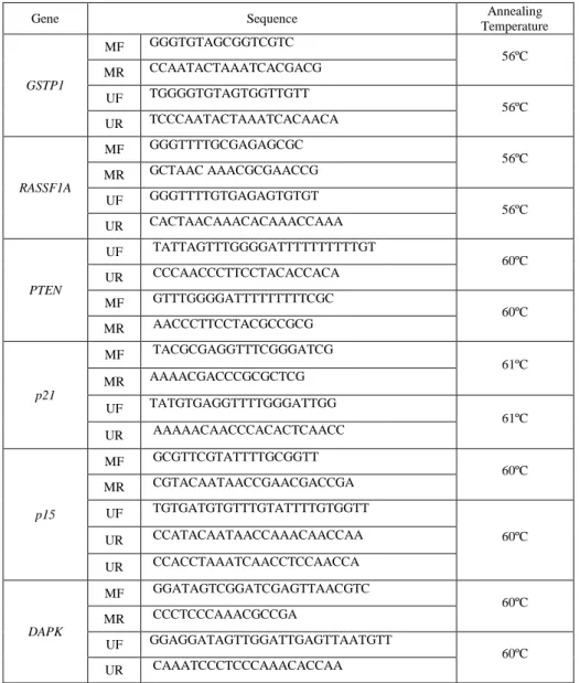
Temperatures used in Methylation Specific PCR are presented in table I.
Table I – MS-PCR primer sequences. All PCR primers were acquired from Sigma Aldrich (St. Louis, MO, USA)
Gene Sequence Annealing
Temperature GSTP1 MF GGGTGTAGCGGTCGTC 56ºC MR CCAATACTAAATCACGACG UF TGGGGTGTAGTGGTTGTT 56ºC UR TCCCAATACTAAATCACAACA RASSF1A MF GGGTTTTGCGAGAGCGC 56ºC MR GCTAAC AAACGCGAACCG UF GGGTTTTGTGAGAGTGTGT 56ºC UR CACTAACAAACACAAACCAAA PTEN UF TATTAGTTTGGGGATTTTTTTTTTGT 60ºC UR CCCAACCCTTCCTACACCACA MF GTTTGGGGATTTTTTTTTCGC 60ºC MR AACCCTTCCTACGCCGCG p21 MF TACGCGAGGTTTCGGGATCG 61ºC MR AAAACGACCCGCGCTCG UF TATGTGAGGTTTTGGGATTGG 61ºC UR AAAAACAACCCACACTCAACC p15 MF GCGTTCGTATTTTGCGGTT 60ºC MR CGTACAATAACCGAACGACCGA UF TGTGATGTGTTTGTATTTTGTGGTT 60ºC UR CCATACAATAACCAAACAACCAA UR CCACCTAAATCAACCTCCAACCA DAPK MF GGATAGTCGGATCGAGTTAACGTC 60ºC MR CCCTCCCAAACGCCGA UF GGAGGATAGTTGGATTGAGTTAATGTT 60ºC UR CAAATCCCTCCCAAACACCAA
Polymerase chain reaction was performed at 95°C hot start for 2 minutes followed by 35 repetitive cycles consisting of denaturation at 95°C for 45 seconds,
10 annealing at specific temperature for 45 seconds, and extension at 72°C for 45 seconds, then finished with a final 10-minute extension. PCR products were run on a 2% agarose gel and visualized by staining with Midori green DNA stain (Nippon Genetics Europe, Deuren, Germany) and visualized under UV illumination (Geldoc XR, Biorad, California, USA)
3.4–DATA ANALYSIS
Statistical analyses were performed using SPSS, version 21.0 (IBM, NY, USA). Hypermethylation of each gene was treated as a binary variable (methylation versus no methylation) by dichotomizing each gene at zero. Proportions of gene methylation between groups were compared using Fisher's exact test. Concordance of DNA methylation pattern in plasma and tumor tissue DNA was assessed by McNemar test comparing each pair matched blood and tumor or non-tumor tissue. All statistical tests were two sided with P ≤ 0.05 considered statistically significant.
11
4 – RESULTS
4.1–EVALUATION OF GENE METHYLATION PATTERN ON DIFFERENT HCC CELL LINES
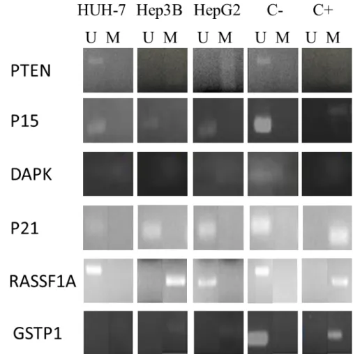
In order to optimize MS-PCR conditions, we studied gene methylation patterns of Tumor Suppressor Genes: RASSF1A, GSTP1, DAPK, PTEN, p15 and p21, in 3 HCC cell lines, HUH-7, HepG2 and Hep3B.
Our results represented in Figure 1 show different patterns of methylation between the different HCC cells lines used. While HepG2 and Hep3B cell lines showed methylation of PTEN, DAPK, GSTP1 and RASSF1A, HUH-7 showed methylation of DAPK and GSTP1. In all cell lines, p21 and p15 were unmethylated.
Figure 1 – Gene Methylation pattern in the HCC cell lines, performed by MS-PCR
12
4.2–CHARACTERIZATION OF PATIENT SAMPLE
Our sample is constituted by 17 patients (3 female and 14 male), with a median age of 66 years old (43-84 years old). 12 patients were diagnosed with carcinoma hepatocellular and 5 patients with cholangiocarcinoma. 64% of patient was in Stage I/II (TNM/AJCC) and 35% present liver cirrhosis concomitantly (Figure 2).
Figure 2 – Characterization of patient samples.
4.3–STUDY OF METHYLATION PATTERN IN TUMORS AND CORRESPONDING NON
-CANCEROUS TISSUE FROM PATIENT
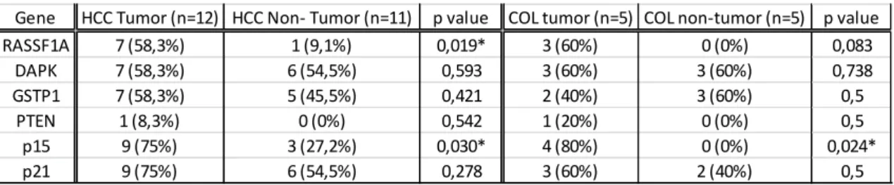
The methylation status level of the RASSF1A, DAPK, GSTP1, PTEN, p15 and p21 gene promoters in tumors and corresponding non-tumoral liver tissues, were evaluated using Methylation Specific Polimerase Chain Reaction (MS-PCR). Methylation frequencies of each promotor gene are presented in Table III.
Table III- Methylation frequency for five genes in hepatocellular carcinoma (HCC) and cholangiocarcinoma (COL) (The association between gene methylation and different groups was assessed by Fisher´s exact test, * p<0,050)
Gene HCC Tumor (n=12) HCC Non- Tumor (n=11) p value COL tumor (n=5) COL non-tumor (n=5) p value RASSF1A 7 (58,3%) 1 (9,1%) 0,019* 3 (60%) 0 (0%) 0,083 DAPK 7 (58,3%) 6 (54,5%) 0,593 3 (60%) 3 (60%) 0,738 GSTP1 7 (58,3%) 5 (45,5%) 0,421 2 (40%) 3 (60%) 0,5 PTEN 1 (8,3%) 0 (0%) 0,542 1 (20%) 0 (0%) 0,5 p15 9 (75%) 3 (27,2%) 0,030* 4 (80%) 0 (0%) 0,024* p21 9 (75%) 6 (54,5%) 0,278 3 (60%) 2 (40%) 0,5
13 Our data showed that the levels of methylation of the promoter gene of RASSF1A, p15 and p21 were higher in HCC tumor tissues compared to their adjacent non-cancerous liver tissues, but only with statistical significance to RASSF1A and p15 gene. Methylation of DAPK and GSTP1 occurred approximately in 50% of tumor and non-tumor tissue. In cholangiocarcinoma, the level of methylation of p15 was significantly higher in tumor tissue than in non-cancerous tissue. No significant difference in methylation pattern was found between hepatocellular carcinoma and cholangiocarcinoma (p=0,855).
4.4–CORRELATION BETWEEN METHYLATION STATUS OF THE TUMOR SUPPRESSOR GENES IN HCCS AND CLINICOPATHOLOGICAL FEATURES OF PATIENTS
Correlations between methylation status and some clinicopathological parameters are summarized in Table IV.
Table IV- Clinical information of HCC samples and their gene promoter methylation level detected by MS-PCR. The association between gene methylation and tumor characteristics was assessed by Fisher´s exact test, * p<0,050 (it was not possible to realize statistical analysis for PTEN gene)
There was no apparent relationship between the methylation level of the gene promoter in the HCC samples and the analyzed clinical parameters, including TNM stage, tumor differentiation. Interestingly, in the unique case of tumor with histological differentiation grade G3, all promotor gene studied are methylated.
No statistically significant association was found between gene promoter methylation and patient's age (data not shown).
4.5–CORRELATION BETWEEN METHYLATION STATUS IN HCC PATIENT WITH OR WITHOUT CIRRHOSIS
It is known that about 80% of HCCs coexist with cirrhosis which is one of the major risk factor and an early event in hepatocarcinogenesis. We intend to verify if the hypermethylation status of five genes in HCCs is different in cirrhotic and non-cirrhotic
Parameter number of cases Tumor p value Tumor p value Tumor p value Tumor p value Tumor p value
TNM stage I/II 86 (75%) 0,533 4 (50%) 0,333 6 (75%) 0,133 6 (75%) 0,533 5 (62,5%) 0,75 III/IV 21 (50%) 2 (100%) 0 (0%) 1 (50%) 2 (100%) Tumor Diferentiation G2 106 (60%) 0,345 6 (60%) 0,345 6 (60%) 0,345 7 (70%) 0,87 7 (70%) 0,87 G3 11 (100%) 1 (100%) 1 (100%) 1 (100%) 1 (100%) RASSF1A DAPK GSTP1 p15 p21
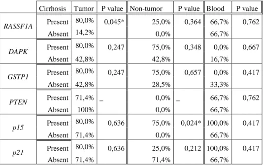
14 patients. The results were shown in Table V. We observed that the methylation frequency of all genes tested is higher in tumor and non-tumor samples from cirrhotic patient than from non-cirrhotic (except p21 gene in non-tumor samples). The hypermethylation of RASSF1A showed significant difference between cirrhotic and non-cirrhotic patient in the tumor group (P=0.045). On the other hand p15, showed significant difference between cirrhotic and non-cirrhotic patient in the non-tumor group (P=0.024). No significant association was observed between methylation status of promotor genes in DNA serum and corresponding tissue (both tumor and non-tumor, in the presence of cirrhosis.
Table V- Frequency of gene promoter methylation detected by MS-PCR in cirrhotic and non-cirrhotic patients with HCC. The association between gene methylation and tumor characteristics was assessed by Fisher´s exact test, * p<0,050 (it was not possible to realize statistical analysis for PTEN gene).
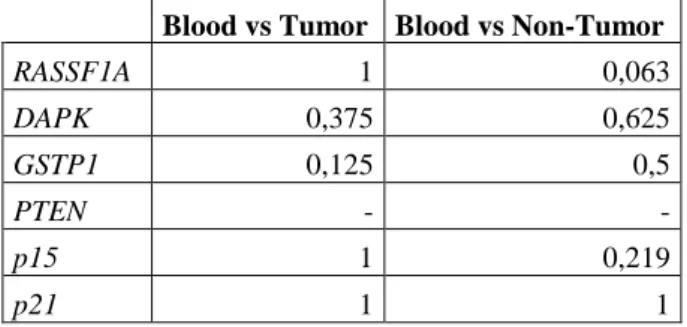
4.6–COMPARISON OF THE HYPERMETHYLATION STATUS OF PLASMA DNA AND PAIRED TISSUE SAMPLES
In order to evaluate if the methylation status of plasma DNA from HCC patients could reflect the methylation status of tumors, we applied a pair-matched non parametric test, the McNemar test, to corresponding samples. Results of p value of applied test are presented in table VI. There is no significant correlation between methylation status of promotor gene in blood samples and tumor or non-tumor matched samples.
Cirrhosis Tumor P value Non-tumor P value Blood P value
RASSF1A Present 80,0% 0,045* 25,0% 0,364 66,7% 0,762 Absent 14,2% 0,0% 66,7% DAPK Present 80,0% 0,247 75,0% 0,348 0,0% 0,667 Absent 42,8% 42,8% 16,7% GSTP1 Present 80,0% 0,247 75,0% 0,657 0,0% 0,417 Absent 42,8% 28,5% 33,3% PTEN Present 71,4% _ 0,0% _ 66,7% 0,762 Absent 100% 0,0% 66,7% p15 Present 80,0% 0,636 75,0% 0,024* 100,0% 0,417 Absent 71,4% 0,0% 66,7% p21 Present 80,0% 0,636 25,0% 0,212 100,0% 0,417 Absent 71,4% 71,4% 66,7%
15
Table VI - Consistency of correlation of the hypermethylation status between tissue DNA and plasma DNA from paired samples using pair matched non-parametric test McNemar (p value) (It was not possible to realize statistical analysis for PTEN gene)
Blood vs Tumor Blood vs Non-Tumor
RASSF1A 1 0,063 DAPK 0,375 0,625 GSTP1 0,125 0,5 PTEN - - p15 1 0,219 p21 1 1
16
5 – DISCUSSION AND CONCLUSIONS
Hepatocellular Carcinoma (HCC) is one of the most common tumors and is often associated with poor prognosis because patients are diagnosed at very late stage. Liver transplantation and resection are the only two curative therapies available; however, in order to qualify for such therapies, HCC patients need to be diagnosed early (5). Apart from AFP level and tumor staging classification such as the Barcelona Clinic Liver Cancer (BCLC) staging system, there is no good prognostic marker that can classify patients and predict survival outcome (20, 29). The large number of HCC associated deaths clearly reflects the limitations of current diagnostic and prognostic tools, showing the importance of novel and effective biomarkers that can improve overall clinical management of HCC.
There has been increasing evidence of a multistep process in human hepatocarcinogenesis. In addition to genetic mutations and chromosomal instability, the aberrant methylation of tumor suppressors plays an important role throughout the process of HCC carcinogenesis. Many studies demonstrated that the DNA methylation based mechanism can contribute to inactivation of tumor suppressor genes, key event in tumorgenesis of a wide spectrum of human tumors, including HCC (15, 30, 31).
In our study, we evaluate methylation profile of several tumor suppressor genes in HCC samples, their corresponding non-cancerous tissue and peripheral blood, from tumor bank of Coimbra University Hospital, and correlate with available clinicopathological parameters.
Our sample is composed in majority by male with advanced age (median age 66 years old) which is in concordance with HCC epidemiology. The incidence of HCC increases with age, reaching its highest prevalence among those aged over 65 years (2). Our sample is only constitute by 12 HCC, which in part, due to the low incidence rate in Portugal (in 2009, 78 new liver tumors cases in Centre region – ROR Centro) (32). Also, most of the patients in this study were in stage I (solitary tumor < 5cm) or II (solitary tumor < 5cm with vascular invasion or multiple small tumors <5cm) at diagnosis which are usually operable and the samples can be stored at tumor bank. Tumors in higher stages normally are not suitable to resection and are treated by radiofrequency ablation, transarterial chemoembolization or with systemic chemotherapy with sorafenib (5). Furthermore, HCCs are diagnosed by invasive
17 methods, such as biopsy, and non-invasive methods, including imaging and tumor markers, but, since percutaneous biopsy can cause several problems, non-invasive methods are preferred in the diagnosis of HCC. Diagnosis Guideline recommend that non-invasive diagnosis of HCC (using multiple-phase multidetector CT scan and/or dynamic contrast-enhanced MRI) is only indicated in cirrhotic patients with identification of the typical vascular hallmark of HCC (hypervascular in the arterial phase with washout in the portal venous or delayed phases). Also, there is no indication for biopsy of a focal lesion in a cirrhotic liver when the patient is not a candidate for any form of therapy because of serious co-morbidity or there is a clear diagnosis by imaging techniques in tumors > 2cm (5).
In our sample, only 41,6% of HCC patient present cirrhosis. It is known that about 80 % of HCC also present cirrhosis. The guidelines for treatment decisions are based on The Barcelona Clinic Liver Cancer (BCLC) Staging System, approved by many American and European Societies (AASLD, EASL, ESMO) that combines tumor burden, hepatic function, and performance status with an evidence-based treatment algorithm. So patients with impaired liver function because of cirrhosis are usually not amenable to resection. Also, as referred previously, HCC diagnosis in cirrhotic patient are mainly realized by imaging techniques avoiding biopsy, so most of non-resecable tumors have no sample in tumor bank.
Regarding to the methylation status, we proposed to evaluate the hypermethylation of tumor suppressor genes RASSF1A, GSTP1, DAPK, PTEN, p21 and
p15 in HCC patient tumor samples, their corresponding non-tumor tissue and peripheral
blood. Our results showed that methylation of the studied promoter genes occurs more frequently in HCC tumor tissues compared to their adjacent noncancerous liver tissues, being more significant to RASSF1A and p15 gene.
PTEN is a tumor suppressor gene that codifies a phosphoinositide-3-phosphatase that inhibits cellular proliferation, survival and growth by inactivating phosphoinositide-3-kinase-dependent signaling (PI3K/AKT pathway). It also suppresses cellular motility through mechanisms that may be partially independent of its phosphatase activity (33). PTEN is silenced by DNA methylation at high frequencies in HCC, and promoter methylation and silencing of PTEN is reported to be associated with HCC (34, 35). In our study, PTEN don´t seem to have a role in hepatocarcinogenesis since it does not
18 appears methylated in almost all cases, which in concordance with studies performed by Um et al (36). In other study, the presence of PTEN methylation also appears to be modest, in16% of tumors (37).
GSTP1 gene encodes glutathione S-transferase π, which protects normal hepatocytes against a number of mutation inducing processes, such as reactive oxygen species linked with chronic hepatic inflammation and reactive electrophilic compounds linked with the hepatic metabolism of dietary and other carcinogens (38). The hypermethylation of the promoter of the GSTP1 gene has been associated with HCC in several studies (39, 40). Death Associated Protein Kinase (DAPK) codifies a protein with the same name which is a calmodulin regulated and cytoskeleton-associated serine/threonine kinase(41). DAPK is thought to be a TSG for its potential to promote apoptosis through p53 pathway and for its ability to inhibit E2F and c-MYC dependent oncogenic transformation (42). In our study, no significant differences in GSTP1 and DAPK methylation were observed between tumor and non-tumor tissue, being methylated in almost 50% of the cases. Harder et al, also observed an elevated level of GSTP1 and DAPK methylation in non-malignant cirrhotic liver and in normal liver (43). P21 is a Cyclin-dependent kinase inhibitor 1A also known as CDKN1A that directly inhibits the activity of cyclin E/CDK2 and cyclin D/CDK4 complexes. P21 functions as a regulator of cell cycle progression at S phase. The expression of p21 is controlled by the tumor suppressor protein p53 (44). P21 have shown to be methylated in 70% of HCC being more common in cirrhotic tissues, which suggest that these genes play an important role in early hepatocarcinogenesis. Our results showed that p21 was more often methylated in tumors than in non-cancerous tissue but the results were not significant.
RASSF1A isa member of the RAS association domain family, a cell cycle-related tumor suppressor protein, which inhibits cyclin D1 and hence induces G1 phase arrest (45). RASSF1A also associates with microtubules mediating its growth inhibitory effects. Loss or altered expression of this gene has been associated with the pathogenesis of a variety of cancers, which suggests the tumor suppressor function of this gene (46). Several studies have shown the role of hypermethylation of RASSF1A in HCC as shown in a metanalysis performed by Li et al (47).
19
P15 is a Cyclin-dependent kinase inhibitor gene transcriptionally activated by transforming growth factor (TGF)-β (48). The p15 protein participates in the cell cycle regulation by binding to the CDK4–cyclin D complex, required for entry into the S phase (49). P15 has been postulated to be a tumor suppressor gene modulating pRb phosphorylation (49). P15 inactivation via hypermethylation could possibly abrogate cell cycle control and confer resistance to the growth-inhibitory effect of TGF-β that is usually overexpressed in HCC cells (50).
In our study, both RASSF1A and p15 have shown a significant increase in gene promoter methylation in tumor compared with non-neoplasic tissue, even the small sample, indicating the possible involvement of these genes in hepatocarcinogenesis. These results are in concordance with other studies genes in which RASSF1A and p15 seems to be more methylated in HCC tumor samples than in their corresponding non-tumor liver tissue (14, 50, 51). Furthermore, RASSF1A gene hypermethylation, have shown to be the better discriminator between HCC and tumor-free liver tissue in several studies (47, 51). The high methylation rate in non-tumor liver tissues observed suggested that epigenetic changes are involved in the early stage of liver carcinogenesis.
With our results, it was not possible to correlate methylation frequencies with clinical characteristics of patients, except for cirrhosis. In other studies, Zhong et al. indicated that no association was apparent between methylation of the RASSF1A gene promoter and patient age (38), on the other hand, Um et al., found that the methylation level of the RASSF1A promoter gene in the HCC samples was correlated with tumor size, being the higher RASSF1A methylation levels for larger tumors (>6 cm) (36). Comparing the methylation profile in cirrhotic and non-cirrhotic patients, we observed a significant difference in hypermethylation frequency of RASSF1A gene in the tumor group, and of p15 in the non-tumor group, cirrhotic patient comparing with non-cirrhotic patient. In several studies, methylation of the RASSF1A promoter gene has been demonstrated in HCCs, and also in cirrhotic livers and chronic hepatitis (17, 52).
In our study, it would be important to assess the etiology of tumor (HCV, HBV, alcohol consumption or NAFLD) in order to correlate etiology and methylation pattern.
The detection of the promoter hypermethylation of tumor suppressor gene in serum DNA could be a valuable biomarker for early detection of both pre-neoplastic lesions and early cancer development among high-risk populations that are at a high risk of HCC. Unlike seen in other studies (8), our data did not correlate methylation profile
20 of blood samples with tumor nor non-tumor tissue. Other studies also showed the limitation in the use of plasma samples methylation for HCC diagnosis (53).
Among the vast quantity of DNA methylation studies in HCC, reported methylation frequencies of identical genes in hepatocellular carcinoma widely varied (13, 34), probably due to HCC heterogeneity and to different techniques employed. Although methylation-specific PCR is a very sensitive assay to assess DNA hypermethylation, the results are dependent on the number of PCR cycles, the amount of input DNA, and the PCR reaction mixture conditions and are qualitative. Some others techniques, quantitative, can be used to study DNA methylation like Real-time PCR-based methylation-specific PCR (MethyLight) (54), Allele-specific bisulfite sequencing, Bisulfite PCR followed by restriction analysis (COBRA) and more recently, Microarray-based genome-wide analysis (55).
In sum, despite the small sample size, our results suggested that methylation of RASSF1A and p15 gene may have an important role in hepatocarcinogenesis. Aberrant methylation of genes was not only present in HCC but can also be found in non-tumor tissue and cirrhosis, which suggests the epigenetic alterations emerge at early stages in the development of the disease. The studied genes modulated several pathways that interfere with apoptosis (56), cellular growth and survival or cellular motility, which indicates epigenetic as a potential target for HCC therapy.
21
6 – REFERENCES
1. Torre LA, Bray F, Siegel RL, Ferlay J, Lortet-Tieulent J, Jemal A. Global cancer statistics, 2012. CA: a cancer journal for clinicians. 2015;65(2):87-108.
2. Ferlay J, Soerjomataram I, Dikshit R, Eser S, Mathers C, Rebelo M, et al. Cancer incidence and mortality worldwide: sources, methods and major patterns in GLOBOCAN 2012. International journal of cancer Journal international du cancer. 2015;136(5):E359-86.
3. Maluccio M, Covey A. Recent progress in understanding, diagnosing, and treating hepatocellular carcinoma. CA: a cancer journal for clinicians. 2012;62(6):394-9.
4. Song DS, Bae SH. Changes of guidelines diagnosing hepatocellular carcinoma during the last ten-year period. Clinical and molecular hepatology. 2012;18(3):258-67.
5. Verslype C, Rosmorduc O, Rougier P. Hepatocellular carcinoma: ESMO-ESDO Clinical Practice Guidelines for diagnosis, treatment and follow-up. Annals of oncology : official journal of the European Society for Medical Oncology / ESMO. 2012;23 Suppl 7:vii41-8.
6. Llovet JM, Ricci S, Mazzaferro V, Hilgard P, Gane E, Blanc JF, et al. Sorafenib in advanced hepatocellular carcinoma. The New England journal of medicine. 2008;359(4):378-90.
7. Huang J. Current progress in epigenetic research for hepatocarcinomagenesis. Science in China Series C, Life sciences / Chinese Academy of Sciences. 2009;52(1):31-42.
8. Mah WC, Lee CG. DNA methylation: potential biomarker in Hepatocellular Carcinoma. Biomarker research. 2014;2(1):5.
9. Esteller M. Epigenetics in Cancer. New England Journal of Medicine. 2008;358(11):1148-59.
10. Liu WR, Shi YH, Peng YF, Fan J. Epigenetics of hepatocellular carcinoma: a new horizon. Chinese medical journal. 2012;125(13):2349-60.
11. Kondo Y, Shen L, Suzuki S, Kurokawa T, Masuko K, Tanaka Y, et al. Alterations of DNA methylation and histone modifications contribute to gene silencing in hepatocellular carcinomas. Hepatology research : the official journal of the Japan Society of Hepatology. 2007;37(11):974-83.
12. Vecchio L, Seke Etet PF, Kipanyula MJ, Krampera M, Nwabo Kamdje AH. Importance of epigenetic changes in cancer etiology, pathogenesis, clinical profiling, and treatment: what can be learned from hematologic malignancies? Biochimica et biophysica acta. 2013;1836(1):90-104.
13. Calvisi DF, Ladu S, Gorden A, Farina M, Lee JS, Conner EA, et al. Mechanistic and prognostic significance of aberrant methylation in the molecular pathogenesis of
22 human hepatocellular carcinoma. The Journal of clinical investigation. 2007;117(9):2713-22.
14. Anwar SL, Lehmann U. DNA methylation, microRNAs, and their crosstalk as potential biomarkers in hepatocellular carcinoma. World journal of gastroenterology : WJG. 2014;20(24):7894-913.
15. Hernandez-Vargas H, Lambert M-P, Le Calvez-Kelm F, Gouysse G, McKay-Chopin S, Tavtigian SV, et al. Hepatocellular Carcinoma Displays Distinct DNA Methylation Signatures with Potential as Clinical Predictors. PLoS ONE. 2010;5(3):e9749.
16. Zhang YJ, Ahsan H, Chen Y, Lunn RM, Wang LY, Chen SY, et al. High frequency of promoter hypermethylation of RASSF1A and p16 and its relationship to aflatoxin B1-DNA adduct levels in human hepatocellular carcinoma. Molecular carcinogenesis. 2002;35(2):85-92.
17. Oh BK, Kim H, Park HJ, Shim YH, Choi J, Park C, et al. DNA methyltransferase expression and DNA methylation in human hepatocellular carcinoma and their clinicopathological correlation. International journal of molecular medicine. 2007;20(1):65-73.
18. Shivapurkar N, Gazdar AF. DNA Methylation Based Biomarkers in Non-Invasive Cancer Screening. Current Molecular Medicine. 2010;10(2):123-32.
19. Chen L-M, Chang M, Dai Y, Chai KX, Dyrskjøt L, Sanchez-Carbayo M, et al. External Validation of a Multiplex Urinary Protein Panel for the Detection of Bladder Cancer in a Multicenter Cohort. Cancer Epidemiology Biomarkers & Prevention. 2014;23(9):1804-12.
20. Farinati F, Marino D, De Giorgio M, Baldan A, Cantarini M, Cursaro C, et al. Diagnostic and prognostic role of alpha-fetoprotein in hepatocellular carcinoma: both or neither? The American journal of gastroenterology. 2006;101(3):524-32.
21. Iyer P, Zekri A-R, Hung C-W, Schiefelbein E, Ismail K, Hablas A, et al. Concordance of DNA methylation pattern in plasma and tumor DNA of Egyptian hepatocellular carcinoma patients. Experimental and Molecular Pathology. 2010;88(1):107-11.
22. Huang W, Li T, Yang W, Chai X, Chen K, Wei L, et al. Analysis of DNA Methylation in Plasma for Monitoring Hepatocarcinogenesis. Genetic testing and molecular biomarkers. 2015.
23. Yeo W, Wong N, Wong W-L, Lai PBS, Zhong S, Johnson PJ. High frequency of promoter hypermethylation of RASSF1A in tumor and plasma of patients with hepatocellular carcinoma. Liver International. 2005;25(2):266-72.
24. Nakabayashi H, Taketa K, Yamane T, Miyazaki M, Miyano K, Sato J. Phenotypical stability of a human hepatoma cell line, HuH-7, in long-term culture with chemically defined medium. Gan. 1984;75(2):151-8.
25. Bressac B, Galvin KM, Liang TJ, Isselbacher KJ, Wands JR, Ozturk M. Abnormal structure and expression of p53 gene in human hepatocellular carcinoma.
23 Proceedings of the National Academy of Sciences of the United States of America. 1990;87(5):1973-7.
26. Vecchi C, Montosi G, Pietrangelo A. Huh-7: A human “hemochromatotic” cell line. Hepatology. 2010;51(2):654-9.
27. Herman JG, Graff JR, Myöhänen S, Nelkin BD, Baylin SB. Methylation-specific PCR: a novel PCR assay for methylation status of CpG islands. Proceedings of the National Academy of Sciences of the United States of America. 1996;93(18):9821-6.
28. Fan X, Inda MM, Tunon T, Castresana JS. Improvement of the methylation specific PCR technical conditions for the detection of p16 promoter hypermethylation in small amounts of tumor DNA. Oncology reports. 2002;9(1):181-3.
29. Llovet JM, Fuster J, Bruix J. The Barcelona approach: diagnosis, staging, and treatment of hepatocellular carcinoma. Liver transplantation : official publication of the American Association for the Study of Liver Diseases and the International Liver Transplantation Society. 2004;10(2 Suppl 1):S115-20.
30. Gao W, Kondo Y, Shen L, Shimizu Y, Sano T, Yamao K, et al. Variable DNA methylation patterns associated with progression of disease in hepatocellular carcinomas. Carcinogenesis. 2008;29(10):1901-10.
31. Nishida N, Kudo M, Nagasaka T, Ikai I, Goel A. Characteristic patterns of altered DNA methylation predict emergence of human hepatocellular carcinoma. Hepatology. 2012;56(3):994-1003.
32. centro R. Registo Oncológico Da região centro 2009. In: Gentil–EPE IPdOdCF, editor. 2014.
33. Leslie NR, Downes CP. PTEN: The down side of PI 3-kinase signalling. Cellular signalling. 2002;14(4):285-95.
34. Yu J, Ni M, Xu J, Zhang H, Gao B, Gu J, et al. Methylation profiling of twenty promoter-CpG islands of genes which may contribute to hepatocellular carcinogenesis. BMC cancer. 2002;2:29.
35. Lee S, Lee HJ, Kim JH, Lee HS, Jang JJ, Kang GH. Aberrant CpG island hypermethylation along multistep hepatocarcinogenesis. The American journal of pathology. 2003;163(4):1371-8.
36. Um TH, Kim H, Oh BK, Kim MS, Kim KS, Jung G, et al. Aberrant CpG island hypermethylation in dysplastic nodules and early HCC of hepatitis B virus-related human multistep hepatocarcinogenesis. Journal of hepatology. 2011;54(5):939-47.
37. Wang L, Wang WL, Zhang Y, Guo SP, Zhang J, Li QL. Epigenetic and genetic alterations of PTEN in hepatocellular carcinoma. Hepatology research : the official journal of the Japan Society of Hepatology. 2007;37(5):389-96.
38. Zhong S, Tang MW, Yeo W, Liu C, Lo YM, Johnson PJ. Silencing of GSTP1 gene by CpG island DNA hypermethylation in HBV-associated hepatocellular carcinomas. Clinical cancer research : an official journal of the American Association for Cancer Research. 2002;8(4):1087-92.
24 39. Tchou JC, Lin X, Freije D, Isaacs WB, Brooks JD, Rashid A, et al. GSTP1 CpG island DNA hypermethylation in hepatocellular carcinomas. International journal of oncology. 2000;16(4):663-76.
40. Lambert MP, Paliwal A, Vaissiere T, Chemin I, Zoulim F, Tommasino M, et al. Aberrant DNA methylation distinguishes hepatocellular carcinoma associated with HBV and HCV infection and alcohol intake. Journal of hepatology. 2011;54(4):705-15.
41. Deiss LP, Feinstein E, Berissi H, Cohen O, Kimchi A. Identification of a novel serine/threonine kinase and a novel 15-kD protein as potential mediators of the gamma interferon-induced cell death. Genes & development. 1995;9(1):15-30.
42. Raveh T, Droguett G, Horwitz MS, DePinho RA, Kimchi A. DAP kinase activates a p19ARF/p53-mediated apoptotic checkpoint to suppress oncogenic transformation. Nature cell biology. 2001;3(1):1-7.
43. Harder J, Opitz OG, Brabender J, Olschewski M, Blum HE, Nomoto S, et al. Quantitative promoter methylation analysis of hepatocellular carcinoma, cirrhotic and normal liver. International journal of cancer Journal international du cancer. 2008;122(12):2800-4.
44. Gartel AL, Radhakrishnan SK. Lost in transcription: p21 repression, mechanisms, and consequences. Cancer research. 2005;65(10):3980-5.
45. Hu L, Chen G, Yu H, Qiu X. Clinicopathological significance of RASSF1A reduced expression and hypermethylation in hepatocellular carcinoma. Hepatology international. 2010;4(1):423-32.
46. Dallol A, Agathanggelou A, Fenton SL, Ahmed-Choudhury J, Hesson L, Vos MD, et al. RASSF1A interacts with microtubule-associated proteins and modulates microtubule dynamics. Cancer research. 2004;64(12):4112-6.
47. Li YS, Xie Q, Yang DY, Zheng Y. Role of RASSF1A promoter methylation in the pathogenesis of hepatocellular carcinoma: a meta-analysis of 21 cohort studies. Molecular biology reports. 2014;41(6):3925-33.
48. Hannon GJ, Beach D. p15INK4B is a potential effector of TGF-beta-induced cell cycle arrest. Nature. 1994;371(6494):257-61.
49. Sherr CJ. The INK4a/ARF network in tumour suppression. Nature reviews Molecular cell biology. 2001;2(10):731-7.
50. Wong IH, Lo YM, Yeo W, Lau WY, Johnson PJ. Frequent p15 promoter methylation in tumor and peripheral blood from hepatocellular carcinoma patients. Clinical cancer research : an official journal of the American Association for Cancer Research. 2000;6(9):3516-21.
51. Moribe T, Iizuka N, Miura T, Kimura N, Tamatsukuri S, Ishitsuka H, et al. Methylation of multiple genes as molecular markers for diagnosis of a small, well-differentiated hepatocellular carcinoma. International journal of cancer Journal international du cancer. 2009;125(2):388-97.
25 52. Di Gioia S, Bianchi P, Destro A, Grizzi F, Malesci A, Laghi L, et al. Quantitative evaluation of RASSF1A methylation in the non-lesional, regenerative and neoplastic liver. BMC cancer. 2006;6:89.
53. Chang H, Yi B, Li L, Zhang HY, Sun F, Dong SQ, et al. Methylation of tumor associated genes in tissue and plasma samples from liver disease patients. Exp Mol Pathol. 2008;85(2):96-100.
54. Eads CA, Danenberg KD, Kawakami K, Saltz LB, Blake C, Shibata D, et al. MethyLight: a high-throughput assay to measure DNA methylation. Nucleic acids research. 2000;28(8):E32.
55. Shen L, Waterland RA. Methods of DNA methylation analysis. Current opinion in clinical nutrition and metabolic care. 2007;10(5):576-81.
56. Udali S, Guarini P, Ruzzenente A, Ferrarini A, Guglielmi A, Lotto V, et al. DNA methylation and gene expression profiles show novel regulatory pathways in hepatocellular carcinoma. Clinical epigenetics. 2015;7(1):43.