UNIVERSIDADE DE LISBOA
FACULDADE DE CIÊNCIAS
DEPARTAMENTO DE BIOLOGIA VEGETAL
Novel human Central Nervous System 3D in vitro models: useful tools for evaluation of viral vector-mediated gene delivery
Ana Catarina Pereira Pinto
Dissertação
Mestrado em Biologia Molecular e Genética
UNIVERSIDADE DE LISBOA
FACULDADE DE CIÊNCIAS
DEPARTAMENTO DE BIOLOGIA VEGETAL
Novel human Central Nervous System 3D in vitro models: useful tools for evaluation of viral vector-mediated gene delivery
Ana Catarina Pereira Pinto
Dissertação
Mestrado em Biologia Molecular e Genética
Orientadores:
Orientador externo: Doutora Catarina Brito, Instituto de Biologia Experimental e
Tecnológica (IBET) e Instituto de Tecnologia Química e Biológica – Universidade
Nova de Lisboa (ITQB-UNL), Oeiras
Orientador interno: Prof. Doutor Jorge Marques da Silva, Departamento de Biologia
Vegetal da Faculdade de Ciências da Universidade de Lisboa, Lisboa
Acknowledgements
I would like to acknowledge all the people directly or indirectly involved in this thesis. To Dr. Paula Alves, for giving me the opportunity to do my master thesis at Animal Cell Technology Unit at ITQB/IBET, for the good working conditions offered and for being a strong example of leadership.
To Dr. Catarina Brito, for her guidance, encouragement and support. For all the good discussions and for being a strong example in my path to becoming a scientist.
To Prof. Dr. Jorge Marques da Silva for accepting to be my internal advisor and for being available to help during my master thesis work.
To Marcos Sousa for the constant availability and help with the stirred-tank bioreactors. To Dr. Margarida Serra, for always being there to help.
To Daniel Simão, for the constant support and indispensable help throughout the year. To all the ACTU colleagues, for the good working environment, friendship and help during this year. A special thanks to Ana Paula, Sofia Rebelo, Marta Estrada and Marta Silva, for all the scientific discussions and encouragement during the good and the bad moments.
Aos meus amigos, pela partilha e pelo apoio. Ao Pedro, pelo carinho, pela paciência e pela força que me dás, para sempre. Às Joanas, esta jornada não teria sido mesma sem vocês; pela amizade e confidencialidade até hoje e para sempre.
À minha família, por me terem acompanhado e ajudado sempre. Por me terem proporcionado todas as condições para que fosse possível atingir cada etapa da minha vida, obrigada.
Preface
This work was performed in the Animal Cell Technology Unit of IBET and ITQB-UNL, within the scope of the EU project “BrainCAV -gene transfer for brain therapy” (FP7-222992) and the FCT funded project “Human central nervous system in vitro models for preclinical research: new tools for studying viral vector-mediated gene” (PTDC/EBB-BIO/119243/2010).
Part of the work described has been included in communications presented in scientific meetings:
Oral Presentations
Brito, C., Simão, D., Pinto, C., Fernandes, P., Serra, M., Schwarz, J., Schiavo, G., Kremer, E. J., Alves, P. M. 3D cultures of human neural progenitor cells: dopaminergic differentiation and genetic modification. TERM STEM 2012, Guimarães, Portugal (October 9 to 13, 2012)
Simão, D., Pinto, C., Fernandes, P., Serra, M., Schiavo, G., Kremer, E, Alves, P. M., Brito, C. Novel Strategies for 3D neural culture & gene delivery: towards human CNS in vitro models for preclinical research. Bioimaging 2012 - 1st International Symposium in Applied Bioimaging, Porto, Portugal (September 20 to 21, 2012);
Poster Communications
Simão, D., Pinto, C., Fernandes, P., Serra, M., Schiavo, G., Kremer, E, Alves, P. M., Brito, C. Towards human Central Nervous System in vitro models for Preclinical Research: strategies for 3D neural culture and gene delivery. 1st SEURAT Summer School, Oeiras, Portugal (June 4 to 8, 2012);
Brito, C., Simão, D., Pinto, C., Fernandes, P., Serra, M., Schiavo, G., Kremer, E, Alves, P. M. Novel strategies for 3D neural culture and gene delivery: human CNS in vitro models for preclinical research. SBE's 3rd International Conference on Stem Cell Engineering, Seattle, USA (April 29 to May 2, 2012);
Brito, C., Simão, D., Pinto, C., Fernandes, P., Serra, M., Schiavo, G., Kremer, E, Alves, P. M. Novel human Central Nervous System 3D in vitro models: useful tools for preclinical evaluation of viral vectors. Cell Culture Engineering XIII, Scottsdale, Arizona, USA (April 22 to 27, 2012);
Brito, C., Simão, D., Pinto, C., Fernandes, P., Serra, M., Schiavo, G., Kremer, E, Alves, P. M. Novel strategies for 3D neural culture and gene delivery: towards human central nervous system in vitro models for preclinical research. Scale-Up and Manufacturing of Cell-Based Therapies I, San Diego, California, USA (January 11 to 13, 2012);
Resumo
A prevenção e tratamento de doenças neurodegenerativas, como a doença de Parkinson, estão ainda longe de se tornarem realidade. Embora as estratégias farmacológicas convencionais se tenham revelado pouco eficazes, resultados preliminares indicam que a terapia génica poderá ter grande potencial. Os vectores adenovirais baseados no serotipo canino 2 (CAV-2) transduzem preferencialmente neurónios em modelos animais, tendo o potencial de ser transportados a longa distância no cérebro via transporte axonal. A expressão epissomal a longo prazo permite uma entrega eficiente do material genético especificamente em neurónios. A aplicação da terapia génica está, no entanto, dependente da resolução de uma série de questões ainda em aberto - em particular, a necessidade de avaliar a sua exequibilidade, eficácia e segurança.
O projecto europeu BrainCAV (www.braincav.eu), no qual este plano de trabalhos de mestrado está integrado, propõe-se avaliar o potencial de vectores CAV-2 para gerar modelos celulares geneticamente modificados que permitam estudar os mecanismos moleculares da doença de Parkinson (por introdução de genes com mutações associadas à doença) e como abordagem terapêutica para doenças neurodegenerativas. Tendo em vista a potencial aplicação dos vectores CAV-2 em ensaios clínicos, será necessário, numa primeira fase, avaliar estes efeitos em células humanas de cérebro. Dada a complexidade do sistema nervoso central (SNC), nomeadamente a importância da interacção neurónio-astrócito, é difícil encontrar modelos celulares adequados. Os modelos bi-dimensionais tradicionais de cultura têm-se mostrado ineficazes na recapitulação da fisiologia dos tecidos vivos. Assim, torna-se necessário recorrer a modelos tridimensionais, que apresentem os diferentes tipos celulares do SNC e que recapitulem as interações celulares no contexto tridimensional (3D) em que ocorrem.
Visto isto, o principal objectivo deste trabalho era o desenvolvimento de um modelo celular 3D do SNC humano, com aplicação no estudo da doença de Parkinson. Para tal foram utilizadas células percursoras neurais derivadas do mesencéfalo fetal (hmNPCs), que apresentam capacidade de diferenciação nas 3 principais linhagens neurais (neurónios, astrócitos e oligodendrócitos), com forte potencial de diferenciação na linhagem dopaminérgica. Estas células são derivadas da região cerebral que, no adulto, é particularmente afectada com a doença de Parkinson, uma patologia que envolve, entre outros sintomas, a perda progressiva de neurónios dopaminérgicos na zona sub-ventricular do cérebro.
Uma metodologia para a geração de neurosferas de hmNPCs diferenciadas tinha sido previamente desenvolvida na unidade de tecnologia de células animais (ITQB-UNL/IBET) [1]. Este processo recorre a sistemas de agitação orbital – erlenmeyers – que são mantidos numa incubadora com atmosfera controlada. Tendo em vista as potenciais aplicações
referidas, neste trabalho procedeu-se a uma caracterização detalhada, nomeadamente em termos de fenótipo dopaminérgico e em termos funcionais.
A caracterização fenotípica com recurso a técnicas de análise de expressão génica (qRT-PCR) e análise proteica (microscopia de fluorescência e Western blot) revelou que no final do protocolo de diferenciação (uma fase de agregação das células, durante 7 dias, seguida de uma etapa de diferenciação, durante 14 dias) a cultura era composta por uma elevada percentagem de células neuronais. Estas apresentavam marcadores típicos de neurónios maturos (βIII-Tubulin+/MAPS+/Sinaptofisina+, este último num padrão de marcação vesicular, indicativo de vesículas pré-sinápticas).
Considerando o objectivo futuro de desenvolver um modelo celular de doença de Parkinson, a diferenciação na linhagem dopaminérgica nesta cultura foi também analisada. No final da diferenciação verificou-se um enriquecimento em neurónios dopaminérgicos nas neurosferas. No entanto, os níveis atingidos mantiveram-se abaixo dos valores obtidos quando estas células são diferenciadas em sistemas bi-dimensionais. A reduzida expressão do factor de transcrição Nurr1, envolvido na manutenção e diferenciação terminal de neurónios dopaminérgicos, sugeriu que as culturas 3D diferenciadas se encontravam num estádio de maturação anterior ao observado nas culturas 2D diferenciadas. Assim, com o objectivo de aumentar a eficiência da diferenciação dopaminérgica, o período de diferenciação foi prolongado; após os 14 dias de diferenciação a cultura foi mantida em condições de maturação (sem morfogéneos), o que permitiu a manutenção de neurosferas diferenciadas em cultura por mais 18 dias, com elevada viabilidade. Adicionalmente foi observado um aumento significativo na expressão de TH, o gene que codifica para a enzima tirosina hidroxilase, da via de produção de dopamina, indicando que a diferenciação dopaminérgica foi favorecida pelas condições de maturação.
O terceiro objectivo consistia na integração das etapas de agregação e diferenciação de neurosferas de hmNPC num sistema em biorreactor controlado. Este sistema de cultura tem importantes vantagens em relação à cultura em erlenmeyer, tais como permitir o controlo e monitorização em tempo real dos parâmetros de cultura (pH, pO2 e temperatura), mantendo-os estáveis ao longo da cultura. Para tal, biorreactores de tanque agitado foram inoculadmantendo-os com uma suspensão celular, permitindo um processo de agregação eficiente durante 7 dias. No final deste período, a caracterização fenotípica das neurosferas revelou que permaneciam num estado proliferativo e indiferenciado. Seguiu-se um período de 14 dias de diferenciação e, no final do mesmo, as neurosferas revelaram uma baixa eficiência de diferenciação neuronal. Esta discrepância relativamente ao processo de diferenciação implementado em erlenmeyer poderá resultar nas diferenças entre os dois sistemas, nomeadamente em relação à dinâmica de agitação. Esta está directamente relacionada como a força hidrodinâmica a que as células estão sujeitas, o que pode influenciar a
diferenciação das mesmas. Além disto, tanto o pH como também os níveis de pO2 ao longo da cultura poderão contribuir para este resultado, pelo que estudos relativamente aos efeitos dos mesmos terão de ser feitos com vista à optimização da cultura em bioreactor.
Assim, a cultura em erlenmeyer foi o sistema escolhido para, numa primeira fase, proceder à avaliação da transdução de neurosferas diferenciadas com vectores CAV-2. Tendo em conta os últimos avanços no campo de engenharia de vectores para terapia génica, a eficiência de transdução e impacto na sobrevivência celular e composição das neurosferas foram avaliados recorrendo a um vector CAV de terceira geração (hd-CAV-2) codificando a proteína repórter eGFP. Estes são vectores CAV-2 aos quais foi retirado a totalidade dos genes virais, o que lhes confere uma maior capacidade de clonagem e uma incapacidade de replicação, propriedades vantajosas em cenários de terapia génica.
Por último, e no sentido de desenvolver o potencial deste sistema de cultura como modelo de doença de Parkinson, procedeu-se à transdução de neurosferas diferenciadas com um vector hd-CAV que codificava para uma proteína LRRK2 com a mutação G2019S que foi associada à etiologia da doença). Para tal, foram utilizadas as condições de transdução optimizadas utilizando o vector hd-eGFP. Os resultados revelaram níveis baixos de expressão do transgene nas culturas transduzidas. Isto poderá indicar problemas com a preparação viral, nomeadamente no que respeita ao rácio entre partículas totais e partículas com capacidade infecciosa. No caso desta razão apresentar valores baixos, poderá em parte justificar os elevados níveis de toxicidade observados, assim como os baixos níveis de expressão de transgene. Portanto, testes mais rigorosos à qualidade da preparação viral terão de ser efectuados previamente ao prosseguimento dos estudos; adicionalmente, outros stocks virais serão testados.
Em resumo, esta tese contribuiu para o melhoramento do processo de diferenciação de hmNPCs em sistemas de agitação orbital, nomeadamente em termos de duração do tempo de cultura e de maturação neuronal. Foi ainda possível implementar um processo em biorreactores de tanque agitado para a agregação de hmNPCs, ainda que a integração da diferenciação das neurosferas no mesmo sistema necessite ser completamente optimizada. Por último, contribuiu para a caracterização do processo de transdução com recurso a vectores CAV de terceira geração como veículos de manipulação genética, com potencial aplicação tanto em terapia génica como para o desenvolvimento de modelos de doença.
Assim, o processo estabelecido nesta tese contribuiu para o desenvolvimento de um modelo celular robusto e reprodutível do SNC, o que poderá contribuir para o aumento da relevância de ensaios pré-clínicos, tanto no desenvolvimento de novos fármacos como na avaliação de vectores de terapia génica.
Palavras chave: hmNPC, modelos celulares 3D, diferenciação dopaminérgica, biorreactores de tanque agitado, vectores hd-CAV.
Abstract
Central Nervous System (CNS) disorders remain a formidable challenge for the development of new and efficient therapies. Gene therapy has emerged as a promising alternative in treating the causes of the disease, instead of merely ameliorating symptoms. For this, hd-CAV vectors have arised as suitable tools, due to the higher safety and lack of immunological memory. Preclinical research of CNS disorders, such as Parkinson’s disease (PD), has traditionally relied on animal models and 2D in vitro cell models, which fail in mimicking in vivo human phenotype. This represents the main obstacle for the translation of CNS drugs into clinical trials.
Thus, the main aim of this thesis was the development a human CNS cellular model using human midbrain-derived neural precursor cells (hmNPCs).
A protocol for differentiation of hmNPC neurospheres using stirred cultured systems with orbital shaking was already implemented. In this thesis, further characterization showed that the neurospheres were enriched in dopaminergic neurons and were functional, as assessed by detection of synaptic vesicle trafficking using FM1-43 probe. Moreover, this culture system was improved in terms of dopaminergic differentiation. By the end of differentiation, higher values the dopaminergic neuron markers of Nurr1 and TH were detected both at mRNA and proteins levels. Importantly, the improved protocol allowed neurosphere viability for long periods of time by adding a maturation period of 18 days, in which differentiation factors were removed from the culture.
Also, a long-term 3D culture system for neural differentiation using controlled stirred-tank bioreactors was implemented, which allowed for aggregation of hmNPCs and is currently under optimization.
Furthermore, the potential of third generation canine adenoviral vectors for genetic manipulation of this cellular model was evaluated. This can be of great value as a powerful tool for disease modeling, namely PD through overexpression of Leucine-rich repeat kinase 2 gene (LRRK2) carrying the PD-associated mutation G2019S. Additionally, these hd-CAV vectors could have important applications in gene therapy.
Key words: hmNPC, 3D in vitro models, dopaminergic differentiation, stirred-tank bioreactors, hd-CAV vectors.
Index
Acknowledgements ... i Preface ... ii Resumo ... iii Abstract ... vi 1. Introduction ... 1 1.1. Parkinson´s Disease ... 1 1.2. Gene therapy ... 11.3. Parkinson´s Disease models ... 2
Animal models... 2
Human cellular models – cell sources ... 3
Human cellular models – Three-dimensional approach ... 4
1.4. Aims ... 5
2. Methods ... 6
2.1. Cell source ... 6
2.2. hmNPC expansion in 2D culture systems ... 6
2.3. hmNPC differentiation in 2D culture systems ... 6
2.4. hmNPC differentiation in 3D culture systems ... 7
Shake flask culture system ... 7
Stirred-tank bioreactor culture system ... 7
2.5. Cell concentration and viability determination ... 8
Trypan Blue exclusion assay ... 8
Crystal violet assay ... 8
Live/dead assay ... 8
2.6. Aggregate size determination ... 8
2.7. Transduction ... 9
2.8. Characterization of hmNPC 2D and 3D cultures ... 9
Immunofluorescence microscopy ... 9
qRT-PCR ...10
Flow cytometry ...11
Synaptic vesicles trafficking...11
3. Results and Discussion ...12
3.1. Characterization of hmNPC 3D cultures ...12
Aggregation and Viability assessment ...12
Differentiation assessment ...13
Functionality assessment ...17
3.2. Maturation of 3D differentiated cultures of hmNPC ...18
Viability assessment ...19
Differentiation assessment ...19
3.3. Implementation of hmNPC 3D cultures in stirred-tank Bioreactor culture system……. ...21
Aggregation and Viability assessment ...21
Differentiation assessment ...22
3.4. Transduction ...25
hd-CAV-eGFP transduction of hmNPC 2D cultures ...25
hd-CAV-LRRK2* transduction of hmNPC 2D cultures ...27
hd-CAV-eGFP transduction of hmNPC 3D cultures – shake flask culture system… ...28
4. Conclusion ...29
5. Bibliography ...29
1. Introduction
1.1. Parkinson´s Disease
Parkinson’s disease (PD) is the second most common neurodegenerative disease and involves the loss of dopaminergic neurons in the Substantia Nigra pars compacta of the ventral midbrain [2]. Although most cases appear to be sporadic, modern genetic and genomic technologies have uncovered several single genes linked to heritable forms of PD [3]. Among others, leucine-rich repeat kinase 2 (LRRK2) stands out as being responsible for the most commonly diagnosed cases [4]. LRRK2 encodes for dardarin, a complex protein expressed in several brain regions and cell types. Even though the physiological function is still unknown, its multiple domains suggest a role in cellular signaling and lowered levels of LRRK2 gene expression have been shown to impair dopaminergic differentiation and dopaminergic neurons survival [2].
There are well established treatments that can alleviate the symptoms of PD consisting mainly on the restoration of dopamine levels via pharmaceutics such as L-DOPA. However, most therapies come with numerous side effect and most patients ultimately exhibit wearing-off symptoms and severe dyskinesia, despite proper dopamine levels replacement [4]. Alternative treatment strategies include regenerative medicine therapies. The loss of a specific type of neurons and in a confined area makes PD a good candidate for such therapies [5]. In fact, the main goal of neural stem cell research has been to generate transplantable cells for neurotrauma and neurodegenerative disorders [6]. However, considerable progress is still necessary, in regards to the efficiency of differentiation and purification of these cells before cellular transplantation of dopaminergic producing cells becomes a reality in the clinic [6].
Therefore, there is a need to improve the understanding of the etiology and pathology of PD and neurodegenerative diseases in general. This may enable the development of long-term disease modifying treatments, which could slow the course of the disease or possibly cure it, as opposed to merely ameliorating symptoms [4]. For doing so, the development of reliable disease models on which these questions can be thoroughly addressed is essential, and in fact, intense efforts have been done in this field.
1.2. Gene therapy
Alternative clinical approaches also include gene therapy. In vivo gene transfer using viral vectors is the primary strategy for delivering novel genes to the Central Nervous System (CNS) [7]. These provide important advantages over pharmacotherapies since a single intervention is required to deliver long-term, stable therapeutic action [4]. Relying on recent advances in the field of gene therapy and recombinant viral vector technology, several therapies emerged based on different vectors [8] and some have already entered the clinical
trial phase. The most commonly used are Adeno-Associated Virus (rAAV) vectors since they have been shown to transduce exclusively neurons in the CNS and present low immunogenicity [4]. However, there is evidence of innate immune responses to these viruses, as well as the presence of circulating neutralizing antibodies [8]. This memory immune response can be problematic for clinical uses since it can trigger inflammatory responses, so there are still improvements to be made regarding their safety [8].
In an attempt to circumvent these problems, the scientific community has been considering the use of nonhuman vectors, such as Canine Adenovirus serotype 2 (CAV-2). These lack immunological memory and have a natural inability to replicate in human cells [9], which could make them be more useful in the clinic. Additionally, they retain the advantages of human adenovirus such as long-term transgene expression, absence of genome integration (which reduces the probability of germ-line transmission and insertional mutagenesis) and a high tropism for neurons [9]. First generation CAV vectors are non-replicative due to deletion of E1 coding region. However, leaky expression of the viral backbone was still detected, which caused loss of transgene expression as a result of an adaptive cellular immune response against the transduced cells and chronic toxicity [10]. These problems could be bypassed with the latest helper dependent (hd) canine adenovirus vectors. hd-CAV lack all viral coding regions, which gives them greater cloning capacity, ensuring long-term gene expression of multiple transgenes or entire genomic loci [10].
Nevertheless, all vector systems have room for improvement and questions such as the most effective vector, ideal gene, dosage and targeting need to be resolved in order to improve clinical relevance of gene therapy. Consequently, this is a field that also benefits from dependable disease models to further assess vector safety and therapeutic potential [4].
1.3. Parkinson´s Disease models Animal models
Resorting to animal models is the most popular choice and several rodent and primate models have emerged through the administration of neurotoxins that selectively destroy the nigrostriatal dopaminergic neurons, such as 6-OHDA or MPTP [11]. Alternatively, via genetic manipulation, transgenic animal models have been generated which develop a PD-like phenotype. However, neither chemical nor genetically induced models can accurately mimic all human symptoms [11]. Biochemical, metabolic and genetic differences between humans and other animals make the full recapitulation of human symptoms very difficult and sometimes even impossible due to the lack of true orthologous genes [12]. In light of these limitations, the need for complementary and more relevant models for human CNS disorders studies and preclinical identification of promising drug candidates becomes clear.
Figure 1: Sources of neural progenitors. NSC lines can be generated from embryonic stem cells (ESCs, derived from the inner cell mass of blastocysts), from iPSCs (derived from reprogrammed somatic cells) and from the germinative areas of the fetal and adult brain. NSCs cultured as neurospheres or on monolayers are considered tripotent as they can give rise to neurons, astrocytes and oligodendrocytes. Adapted from [40].
Human cellular models – cell sources
Cellular models do not provide systemic information. However, they allow for better manipulation of cells and for extraction of data and conclusions independently of neighboring cells and other variables imputing on multisystem animal models, which makes them suitable for therapeutically oriented biomedical studies [13]. The potential use of stem cells from various origins as source of neural progenitors (Fig. 1) to be used as therapeutic agents and for disease modeling has been highly explored [14]. However, precise knowledge on molecules and signaling pathways regulating proliferation, differentiation and migration of neural stem cells (NSC) is necessary to advance the field towards the clinic [14].
The discovery and increasing availability of human pluripotent stem cells (hPSC), as well as their unlimited capacity to replicate, has made them a suitable tool for the step-wise generation of neural progenitors and subsequently neurons and glia needed in cell models of nervous system development [14]. As a result of the growing number of techniques available to genetically modify hPSCs, cell lines harboring specific mutations have been developed, converting them into cellular models for
the respective human disorder [12]. Recently, the generation of pluripotent cells through somatic cell reprogramming appeared as an attractive alternative. The ability to generate patient-specific stem cells using induced pluripotent stem cell (iPSC) technology [14], as well as their amenability to genetic manipulation [6], gives them great potential in future personalized medicine. Additionally, these could circumvent the ethical problems that arise when obtaining human embryonic stem cells [15]. Nevertheless, methodologies for efficient differentiation of hPCS into specific cell types is still missing, hampering their widespread utilization.
Alternative sources are primary cultures of adult stem cells (ASC). ASCs have been isolated from adult tissues such as bone marrow, skin, muscle and brain [6]. In the human brain, NSCs have been isolated from both germinal zones that remain throughout adulthood, the subventricular zone (SVZ) and hippocampal dentate gyrus (DG) [14], with confirmed self-renewal capacity and multipotentiality [5]. These cells open up the possibility of using autologous stem cells to treat otherwise incurable degenerative, traumatic, or congenital
diseases without immunological consequences [16], while limiting ethical conflicts with the use of embryos [15].
hNSCs have also been isolated from fetal tissues, namely forebrain and midbrain [17]. These cells have the advantage of being more amenable to direct differentiation into a specific phenotype, which together with the possibility for genetic modification, makes them an important resource in modeling human neurodegenerative diseases [18].
hmNPCs have been successfully expanded in vitro under lowered atmospheric oxygen conditions (3%), in serum-free medium [17]. These cells proliferate maintaining an undifferentiated state, while retaining the ability to differentiate into the major CNS cell types (neurons, astrocytes and oligodendrocytes) [17], with strong commitment towards dopaminergic lineage, making them uniquely qualified for the development of Parkinson’s disease cellular models [5].
Human cellular models – Three-dimensional approach
The conventional culturing of cells in two-dimensional (2D) culture systems has proven unable to reliably reflect the physiology of a living tissue [19]. Therefore, a transition into 3-dimesional (3D) cellular models is necessary, in an attempt to recover the mechanical and biochemical cues that fall short on simplified systems [19].
Most of the 3D cell culture technology relies on scaffold systems that use synthetic or animal-derived extracellular matrix (ECM) materials to generate gels in which the cells can be cultured [20]. In addition to providing support, the ECM is also a communicating system, regulating cell fate, behavior and function [20]. Along the years, several 3D-matrices have been successfully generated from materials such as poly L-lactic acid and poly glycolic acid, as well as biopolymers such as collagen, fibrin and alginate [21]. Quality control is difficult to achieve so progress is being made towards fully synthetic gels for 3D cell culture [19]. An advantage of these systems is the possibility of tailoring the gels to the specific needs of each cell type, including the addition of aminoacid sequences for specific cell receptors [19].
hNPCs, however, are usually classified as anchorage-independent cells with a natural ability to aggregate and can grow as neurospheres, in serum-free growth medium [22]. Despite the existing extensive research regarding the expansion of NPCs, the exact mechanism of cell-cell interactions and aggregation in culture is not known [22]. Nevertheless, neurospheres can be maintained in a proliferative undifferentiated state in suspension systems, whereas for hNPC differentiation, cells are usually plated as monolayers, over an ECM purified component, such as laminin or fibronectin [23].
New techniques have emerged from the field of tissue engineering for culturing NPCs as free-floating aggregates, such as liquid overlay cultures, microfluidic devices and stirred culture systems in which medium is agitated such as the rotary cell culture (RCC) systems
Figure 2: Schematic diagram of stirred-tank bioreactor system for stem cell culture. The main components are indicated: (1) glass vessel, (2) impeller, (3) temperature sensor, (4) pO2
electrode and (5) pH electrode. Adapted from [27].
and stirred culture vessels [24]. Stirred culture vessels are scalable and hydrodynamically well characterized, and allow non-invasive sampling for continuous monitoring of the culture, as well as a more homogenous culture environment [25]. These include the gyratory rotation technique (cell suspensions are introduced into shake flasks with orbital shaking) and spinner vessels (cell suspensions are introduced into stirred tank containers with impeller mixing) [24].
Computer-controlled bioreactors, on the other hand, represent important tools for bioprocess development, allowing the minimization of process and product variability, while maximizing productivity [26]. Thus, process standardization can potentially be achieved through the online monitoring and control of physicochemical culture parameters, such as pH, pO2 and temperature, possible with these systems, ensuring a fully controlled environment [25].
Different bioreactor configurations have been developed for stem cell expansion and differentiation, including rotating wall vessels, Hollow-fiber,
Fixed and fluidized bed and Wave bioreactors [26]. The stirred-tank bioreactor configuration, however, is the most used for culturing NPCs (Fig. 2). These consist of a glass vessel equipped with an impeller for providing a homogeneous and dynamic stirred environment [27]. They allow for precise control and monitoring of the culture
environment using temperature, pO2 and pH electrodes (Fig. 2), efficient gas and nutrient transfer and non-destructive
sampling.
Moreover, stirred-tank bioreactors have been applied to the culture and expansion of human forebrain-derived neural precursor cells [28] [29], proving its feasibility.
1.4. Aims
The main aim of this thesis was the development a human CNS cellular model, applicable for PD modeling and for assessment of gene therapy approaches in a human 3D cell context, using human midbrain-derived neural progenitor cells (hmNPCs).
Based on a recently developed methodology for the production of differentiated hmNPC neurospheres, using stirred culture systems with orbital shaking, the first objective of this thesis was to perform a detailed phenotypic and functional characterization of the differentiated culture, specifically concerning the dopaminergic phenotype, critical in PD. The second objective was to further optimize this culture system in order to extend the duration of
the differentiated cultures and improve the neuronal maturation, so that the cell model could be used for long-term studies.
With the goal of implementing a robust and scalable system for neural differentiation, the third objective was to integrate aggregation and differentiation steps of hmNPCs in computer-controlled stirred-tank bioreactor process.
The fourth objective was to assess CAV-mediated gene delivery in the developed human CNS cell model In light of recent developments in this field which suggest that third generation helper dependent adenoviral vectors may possess important advantages for clinical uses, hd-CAV-2 vectors transduction efficiency of differentiated neurospheres was investigated, specifically concerning its tropism and safety.
The final objective was to further develop this model’s potential for Parkinson´s Disease modeling by evaluating its amenability to genetic manipulation. For this, the transduction efficiency of hd-CAV-2 vectors carrying as transgene the PD-associated mutation G1920S of the LRRK2 gene was assessed. These vectors could be used to disease phenotype induction.
2. Methods
2.1. Cell source
Human midbrain-derived neural precursor cells (hmNPC), derived from aborted fetal brain tissue 12-14 weeks post-fertilization, were isolated as described previously [17] and kindly supplied by Johannes Schwarz.
2.2. hmNPC expansion in 2D culture systems
hmNPCs were propagated on poly-L-ornithine-fibronectin (PLOF)-coated surfaces, in serum-free Expansion Medium (EM) (Table S1 – section 5.1). Cell cultures were maintained in a multi-gas cell incubator (Sanyo), at 37°C, in a humidified atmosphere with 5% CO2 and 3% O2 and a 100% media exchange was performed every 3-4 days. Splitting was performed typically every 14 days, at 90-100% confluence. After incubation with Accutase (Sigma) for 30 minutes at 37ºC, cells were dislodged with a cell scraper and collected with PBS. After centrifugation at 300x g for 7 minutes with a slow brake, cells were ressuspended in a small volume of EM with a 1 mL pipette to obtain a homogeneous single cell suspension. Cell concentration and viability were determined by the Trypan blue exclusion assay. The cell suspension was used to inoculate PLOF-coated T-flasks, at a cell density of 3x10⁴ cell/cm2.
2.3. hmNPC differentiation in 2D culture systems
Differentiation was induced once the cells attained confluency (typically after 14 days of expansion) by culturing in Differentiation Medium (DM) (Table S2 – section 5.1) for at least 7 days. Total media exchange was performed every 3 to 4 days.
2.4. hmNPC differentiation in 3D culture systems Shake flask culture system
hmNPCs were cultured as neurospheres in 500 mL shake flasks (SF) (Corning) under constant orbital shaking (stirring rate: 100 rpm), using a multi-gas cell incubator (Sanyo), in a humidified atmosphere with 5% CO2 and 3% O2, at 37°C. After hmNPCs expansion in 2D culture systems, cells were collected into a single-cell suspension (section 2.2), which was filtered through a 70µM nylon strainer (Millipore) to eliminate cell clumps. Typically, SF were inoculated at 2x10⁵ cell/mL (working volume: 80 mL) in Aggregation medium (AM) (Table S1 – section 5.1). Neurospheres were maintained in AM for 7 days and a 50% medium exchange was performed at day 3-4. At this point, differentiation was induced by culturing the cells in DM for 14 days (Fig. 3). An additional Maturation step was introduced where the cells were cultured for 10 to 18 days in Maturation medium (MM) (Table S1 – section 5.1). A 75% medium exchange was performed every 2-3 days.
For medium exchanges, agitation was stopped and the aggregates were allowed to settle before removing the medium. Also, prior to any medium exchange, the medium was kept in a humidified atmosphere with 5% CO2 and 3% O2 for at least 2 hours, in order to reach the correct culture parameters.
Stirred-tank bioreactor culture system
hmNPCs were cultured as neurospheres in computer-controlled stirred-tank bioreactors (BR) (DASGIP) with the following culture parameters: pH – 7.2; temperature – 37ºC; Oxygen partial pressure (pO2) – 15% air saturation (3% dissolved oxygen); agitation rate – 90-120 rpm. Bioreactors were inoculated at 4x10⁶ cell/mL, in an initial working volume of 120 mL. The same differentiation protocol described for SF system was followed (Fig. 3). The same procedures regarding media exchanges were also applied.
B
A
Figure 3: Workflow depicting different culture strategies. (A) hmNPC aggregation and differentiation in 3D culture systems (SF and BR); (B) different Maturation strategies of hmNPC aggregation and differentiation in SF culture system.
2.5. Cell concentration and viability determination Trypan Blue exclusion assay
After incubation with 0.1% (v/v) Trypan Blue dye (Invitrogen) in PBS, cells with damaged membranes stain blue and viable cells remain colorless. Thus, to assess cell concentration and viability of hmNPC 2D cultures, the total number of blue and colorless cells was counted in a Fuchs-Rosenthal haemocytometer chamber.
Crystal violet assay
Differentiated neurospheres presented great mechanical susceptibility due to the extensive net of neurites formed. Thus, for 3D cultures, successful dissociation of differentiated neurospheres by incubation with Accutase was not possible without compromising cell viability. Alternatively, total cell concentration determination was achieved by overnight incubation in 0.1 M citric acid with 1% Triton X-100 (Sigma), for cell lysis. Cell nuclei were subsequently stained with 0.1% crystal violet and counted using a Fuchs– Rosenthal haemocytometer chamber.
Live/dead assay
As previously stated, differentiated neurospheres could not be dissociated with Accutase. Instead, the viability of the culture was assessed by the FDA (fluoresceine diacetate)/PI (propidium iodide) assay. FDA is a non-fluorescence cell permeant ester that is converted to highly fluorescent fluorescein (green color) by intracellular esterases. Fluorescein, being highly polar, is retained within cells with intact membrane, measuring both their enzymatic activity and membrane integrity.
PI is a polar, fluorescent red compound
which can only enter cells with compromised membranes. It intercalates into the major
groove of dsDNA, therefore nucleus of dying/dead cells stain red.
Neurospheres were incubated with 20 μg/mL FDA (fluorescein diacetate; Sigma-Aldrich) and 10 μg/mL PI (propidium iodide; Sigma-Aldrich) for 5 min and immediately analyzed using fluorescence microscopy (Leica Microsystems GmbH).The quantitative assessment of hmNPC 2D
cultures viability was also performed using the FDA/PI method. After staining, cell
suspensions were analyzed by flow cytometry (section 2.8).
2.6. Aggregate size determination
Aggregate suspensions were observed by phase contrast microscopy and aggregate size was measured using ImageJ software. The diameter of at least 100 aggregates was assessed by calculating the geometric average of 3 diameters measured from each aggregate.
2.7. Transduction
For hmNPC 2D cultures transduction, using hd-CAV-eGFP and hd-CAV-LRRK2* vectors, 2 Multiplicity Of Infection (MOI) were tested - 300 (low) and 3000 (high) pp/cell. The transduction process was carried out in static conditions with 50% reduction of the working volume. The required viral stock, according to the MOI, was diluted in fresh DM and added to the culture. Two hours post-transduction (hpt) the medium was either completely replaced or the initially working volume restored by adding fresh DM with subsequent complete medium exchange at 24 hpt. Transduction efficacy was evaluated 5 days post-transduction by harvesting the cells (section 2.2) and
analyzing them by flow cytometry (section 2.8). For
hmNPC 3D differentiated cultures transduction, using hd-CAV-eGFP vectors, 2 MOIs were tested - 1500 (low) and 3000 (high) pp/cell - by adding directly the required volume of the purified viral stock to the culture, with 50% working volume reduction. Two hpt the medium was completely replaced, restoring the initial working volume.2.8. Characterization of hmNPC 2D and 3D cultures Immunofluorescence microscopy
hmNPC 2D cultures grown on PLOF-coated glass coverslips or neurospheres plated on PLOF-coated glass coverslips and allowed to attach for 3 days, were fixed in 4% paraformaldehyde (PFA) + 4% Sucrose in phosphate buffered saline (PBS) for 20 min, permeabilized (only for detection of intracellular epitopes) for 10 minutes in 0.1% (w/v) Triton X-100 solution (Sigma), blocked with 0.2% fish skin gelatin (FSG) for 30 minutes and subsequently incubated with primary antibodies (Table S3 – section 5.1), diluted in 0.125% FSG + 0.1% TX-100 in PBS for intracellular epitopes and in 0.125% FSG in PBS for plasma membrane epitopes, for 2 hrs at room temperature (RT). Cells were then washed 2 times with PBS and incubated with secondary antibodies (AlexaFluor 488 goat anti-mouse IgG, AlexaFluor 549 goat anti-rabbit IgG, Invitrogen), diluted 1:500 in 0.125% FSG, for 1 hour at RT. After 2 washes with PBS cell nuclei were counter stained with 4,6-diamidino-2-phenylindole (DAPI, Sigma). Samples were visualized using fluorescence (DMI6000, Leica), spinning disk (Nikon Eclipse Ti-E, confocal scanner: Yokogawa CSUx1), and point scan confocal (SP5, Leica) microscopy. Images were processed using open source ImageJ software.
Western Blot
hmNPCs cultured in 2D were collected and sedimented by centrifugation at 500x g for 5 min, washed with PBS and the dry pellet snap-freezed by immersion in liquid nitrogen. Samples were stored at -80ºC until protein extraction. For protein extraction, cells were lysed in 100 µL of Lysis Buffer (Table S4 – section 5.1) for 30 min at 4ºC. Extracts were clarified by centrifugation at 15000x g for 10 min. Total protein was quantified with Micro BCA Protein
Assay Kit (Pierce), according to manufacturer’s instructions. Briefly, in an alkaline environment, peptides containing 3 or more amino acid residues form a light blue chelate complex with Cu2+, reducing it to Cu1+ (biuret reaction), which reacts with bicinchoninic acid (BCA), forming a purple product. The resulting BCA/copper complex is water-soluble and exhibits a strong linear absorbance at 562 nm with increasing protein concentrations. Proteins were then precipitated by overnight (o.n.) incubation in ethanol at a final concentration of 80% and sedimented by centrifugation at 15000x g for 15 min. Supernatant was discarded and precipitated protein was solubilized in NuPAGE sample buffer with reducing agent (Invitrogen). Samples were heated at 70°C for 10 min and resolved on a NuPage Bis-Tris gel (Invitrogen) with MOPS as running buffer, at 200 V for at least 45 min. Protein transfer was performed in iBlot system (Invitrogen) for 8 minutes, according to manufacturer’s instructions. Membranes were blocked by incubation for 1 h with blocking solution (0.1% Tween 20 and 5% dry milk in PBS), at RT, and incubated o.n. with primary antibody (Table S5 – section 5.1) diluted in blocking solution with gentle agitation. Membranes were incubated with horseradish peroxidase-coupled ECL secondary antibody anti-mouse IgG or anti-mouse IgG (GE Healthcare) diluted 1:5000 in blocking solution, for 2 hrs at RT. Chemiluminescence detection was performed by incubating the membranes with Amersham ECL Prime western blotting detection reagent (GE Healthcare) and analyzed under ChemiDoc XRS System (Bio-Rad). For stripping, membranes were incubated 2 times with stripping buffer (15g/L glycine, 1g/L SDS, 1% Tween20, pH2.2) for 10 min, followed by 2 washes with PBS for 10 min and 2 washes with TTBS for 5 min. Membranes were then incubated in blocking solution and the protocol described above was followed with a different antibody.
qRT-PCR
Cells were sedimented by centrifugation at 500x g for 5 min, washed with PBS and the dry pellet snap-freezed by immersion in liquid nitrogen. Samples were stored at -80°C until RNA extraction. Total RNA was extracted with High Pure RNA Isolation Kit (Roche), including a DNase digestion step, according to the manufacturer instructions. RNA was eluted with 50 μL of sterile deionized water, quantified in a NanoDrop 2000c (Thermo Scientific) and used directly for cDNA synthesis or stored at -80°C. Reverse transcription was performed with High Fidelity cDNA Synthesis Kit (Roche), using Anchored-oligo(dT)18 Primer (Roche). Firstly, the concentrations of all RNA samples were normalized by dilution in sterile deionized water. The resulting cDNA was quantified and used directly for qPCR or stored at -20°C. qPCR was performed in triplicates according to LightCycler 480 SYBR Green I Master Kit (Roche), in 20 μL reactions with 1:2 diluted cDNA template and 5 μM primers (Table S6 – section 5.1). The samples were loaded in LightCycler 480 Multiwell Plate
96 (Roche), always maintaining reagents and well-plates on ice. The reactions were performed using LightCycler 480 Instrument II 96-well block (Roche). Cycles threshold (Ct’s) and melting curves were determined using LightCycler 480 Software version 1.5 (Roche). All data was analyzed using the 2-ΔΔCt method for relative gene expression analysis [30]. Changes in gene expression were normalized using the housekeeping gene RPL22 (ribosomal protein L22) as internal control.
Flow cytometry
For flow cytometry analysis of viability assessment, single cell suspensions derived as described in section 2.2 were ressuspended in PBS with 2% FBS and analyzed in a CyFlowH space (Partec) instrument, registering 10000 events/sample. For hd-CAV-eGFP transduced cultures, the transduction efficiency was evaluated by determining the percentage of GFP+ cells against a non-transduced control culture. For these samples, only PI was used for cell viability assessment.
Synaptic vesicles trafficking
FM (N-(3-Triethylammoniumpropyl)-4-(4-(Dibutylamino)styryl) Pyridinium Dibromide) dyes are modified styryl dyes, which have been used to label and then monitor synaptic vesicle exocytosis and endocytosis [31]. The structural features of the molecule cause the dye to partition into lipids and other hydrophobic domains. Also, its fluorescence dependent on solvent polarity: in polar solvents, like water, the quantum yield is reduced by more than 2 orders of magnitude compared to non-polar solvents, such as membranes. Thus, virtually all fluorescent signal derives from the dye within membranes.
Differentiated neurospheres plated on PLOF-coated glass coverslips were washed with PBS and exposed to a high potassium depolarizing solution (100mM KCl buffer: Table S7 – section 5.1), for 5 min, to stimulate exocytosis. Afterwards, neurospheres were incubated with 10 µM FM 1-43 dye (Invitrogen) dissolved in normal saline solution (5mM KCl buffer: Table S7 – section 5.1) for 15 min (to allow endocytosis) and washed with ADVASEP-7 (Sigma) dissolved in 5mM KCl buffer (2.163 mg/mL) for 1 min (to reduce background fluorescence). This was followed by 3 washes of 1 min with 5mM KCl buffer (to allow for all endocytosed membrane to reform into release-competent vesicles). In the end, exocytosis was again stimulated with 100mM KCl buffer. Samples were imaged live in a fluorescence microscope (DMI6000, Leica) for monitoring of fluorescence intensity after endocytosis stimulus and continued during exocytosis stimulus for 30 min. Loss of fluorescence over time correlates to exoxytosis level. Fluorescence intensity was measured using ImageJ software.
3. Results and Discussion
3.1. Characterization of hmNPC 3D cultures
The starting point for the present work was the previously described protocol for the generation of differentiated neurospheres using stirred culture systems with orbital shaking [1]. In order to be used as a 3D cellular model of the human Central Nervous System (CNS), this culture system still required extensive cellular and functional characterization.
The workflow followed in the experiments described in this section is depicted in Fig.3. At specific time-points throughout the 35 days of culture (7 days of aggregation: 7A; 7 days of differentiation: 7D; 7 days of differentiation: 14D) samples were collected and processed for posterior analysis (sections 2.2 and 2.8).
Aggregation and Viability assessment
Cell aggregation and viability were monitored over time. At 7A the culture was composed of neurospheres with a diameter of approximately 300 µm (Fig. 4), which was maintained throughout differentiation. Although there were single cells visible in suspension, these were not viable, as assessed by the fluorescent live/dead assay (Fig. 4 – 7A). This method didn’t require a dissociation step of the neurospheres, allowing for the qualitative assessment of the proportion of dead cells, as well as their distribution within the aggregates. Fig. 4 shows that aggregates were mainly composed of viable cells; a small number of dead cells were also present, in defined focus of death inside the aggregates (Fig. 4 – arrows), that did not correspond to necrotic centers. This was maintained throughout differentiation; nevertheless neurospheres remained mostly viable and able to differentiate.
Apoptotic cell death represents a common feature of NPC growing in vitro as free-floating neurospheres. In fact, an average of 30.7 ± 3.4% cells were found to be apoptotic within murine mNPC neurospheres [32]. Moreover, apoptotic mechanisms have an important role in the development and morphogenesis of the nervous system [33]. Thus, the observed death was most likely due to apoptosis and a result of neural culture differentiation, not compromising the neurospheres. Nevertheless, analysis of apoptotic markers, such as Figure 4: Fluorescence microscopy images depict results of live/dead assay (FDA - green; PI – red) performed in hmNPC 3D cultures differentiated in SF culture system. Data are from one representative experiment of 3 independent experiments. Arrows indicate the defined focus of death inside the neurospheres. (7A – 7 days of aggregation; 7D and 14D – 7 and 14 days of differentiation; scale bars: 100µm).
cleaved caspase 3, would be necessary to confirm that programmed cell death occurred in the neurospheres.
Differentiation assessment
In order to further characterize the 3D differentiation process, phenotypic characterization of the 3 time-points (7A, 7D and 14D) was performed. As evident from Fig. 5, at the end of the aggregation period (7A), extensive immunoreactivity for Ki67 was present throughout the entire neurosphere (Fig. 5 - A). This nuclear protein, here used as a proliferation marker, predominantly presented an intranuclear punctate staining pattern (Fig. 5 - B), characteristic of the mitotic phase of the cell cycle, when it becomes associated with the periphery of the condensed chromosome [34]; but brightly stained nuclei were also visible, characteristic of interphase [34].
Also at 7A, neurospheres showed a very high proportion of immunopositive cells for Nestin (Fig. 5 - C), a class VI intermediate filament (IF) protein. Cellular differentiation regularly involves morphological changes, often due to IF protein remodeling [35]. These proteins present cell-type-specific expression profiles and are widely used as cellular markers. Nestin’s timing of expression is concomitant with the appearance of precursor cells of the neural lineage [35]. Evidences suggest that, in addition to contributing to the structural integrity of the cells, it is also important in promoting NSC survival and proliferation [36]. Nestin is expressed in neuronal and glial common precursor cells [37] and was used in this work to identify proliferative hmNPCs.
Additionally, the majority of the cells within the neurospheres were also immunopositive
Figure 5: Immunofluorescence microscopy characterization of hmNPC neurospheres from 7A. Detection of Ki67, nestin, GFAP and βIII-Tubulin (nuclei were labeled with DAPI). B, D and F are magnifications of the selected regions in A, C and D, respectively. Data are from one representative experiment of 3 independent experiments. (scale bars – A, B, D, E, F: 50 µm; C: 100 µm)
for Glial fibrillary acidic protein (GFAP) (Fig. 5 – E). Upon differentiation, hmNPC proliferation rate is significantly reduced and there is a decrease in nestin expression [37], accompanied by an upregulation of other tissue-specific IF proteins like GFAP in astrocytes and neurofilaments in neurons [36]. Although GFAP is usually accepted as an astrocytic marker [38], in humans it is also expressed in radial glial cells [39]. These cells originate from neuroepithelial progenitors at the beginning of neurogenesis and are the main cell type in the developing brain, serving both as neural progenitors and as scaffolds for migrating newborn neurons [40]. Therefore, these cells (that were also present in 2D cultures of proliferative hmNPCs – results not shown) probably correspond to a population of GFAP+
/nestin+ neural precursors.
A small number of βIII-Tubulin+ cells were also detected (Fig. 5 – C and E), an isoform found exclusively in neurons [41].
Additionally, after being plated on PLOF-coated coverslips, cells composing the neurospheres migrated extensively, with almost complete dissociation of the aggregates (Fig. 5 – C and E). Since NPCs have been described to migrate radially away from the neurosphere in ECM-coated surfaces, forming a monolayer around it [42], this observation also pointed to the high undifferentiated state of the neurospheres.
At this point (7 days of aggregation) differentiation was induced by changing to DM. Throughout differentiation, the culture continued to present a high proportion of GFAP+ cells (results not shown). Since GFAP is expressed both in precursor cells and in mature astrocytes, a more thorough characterization would be necessary to confirm if the cells are maturing into astrocytes, namely, through the detection of GLT-1 expression, a glutamate transporter exclusive of mature astrocytes [43].
In terms of neuronal differentiation, after 7 days in differentiation conditions (7D), neurospheres already presented an extensive network of βIII-Tubulin+ cells (Fig. 6 – A), which was maintained during the entire differentiation period (Fig. 6 - B). Double staining with nestin revealed that there were still precursor cells in culture, however, βIII-Tubulin+ cells were negative for this marker (Fig. 6 – C), confirming its further differentiated phenotype.
Neuronal cells were also immunopositive for microtubule associated proteins (MAPs), namely MAP2 and Tau (Fig. 6 - D), and for synaptophysin, detected in a punctate pattern (Fig. 6 – E). MAP2 expression has been described as very weak in neuronal precursors and stronger upon differentiation into neurons [44]. The detection of MAPs, together with the detection of vesicular synaptophysin, a glycoprotein present in the membranes of pre-synaptic vesicles [45], suggested that neurons in 3D cultures had a mature phenotype at 14D.
Additionally, in contrast to proliferative neurospheres, upon plating on PLOF-coated surfaces, these remained mostly intact (Fig. 6 – A and B), which presented another evidence of the further differentiated phenotype of neurospheres at 14D.
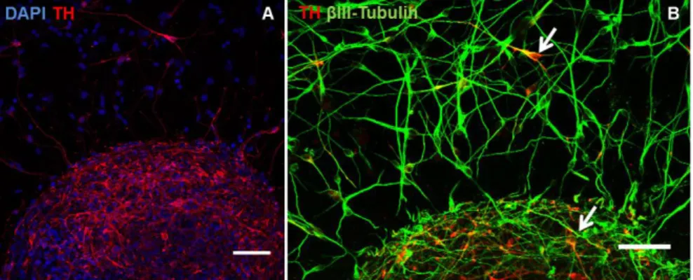
Given that one of the main objectives of this culture was the development of a human CNS 3D cellular model, with possible applications in the modeling of PD, the dopaminergic phenotype of differentiated neurospheres was also analyzed. At the end of the differentiation period (14D), a high proportion of cells within the neurospheres were TH+ (Fig. 7 - A). Fig. 7 - B demonstrates that these cells were co-expressing TH and βIII-Tubulin. TH (Tyrosine hydroxylase) is an enzyme of the dopamine synthesis pathway and is used as a marker of dopaminergic neurons (28), which suggests that a high number of neurons further Figure 6: Immunofluorescence microscopy characterization of hmNPC neurospheres from 7D (A) and 14D (B, C, D and E). Detection of βIII-Tubulin, nestin, MAPS and synaptophysin (nuclei were labeled with DAPI). E is a magnification of selected region in D. Arrows indicate examples of vesicular synaptophysin. Data are from one representative experiment of 3 independent experiments. (scale bars – A and B: 100 µm; C and D: 50 µm; E: 20µm).
Figure 7: Immunofluorescence microscopy characterization of hmNPC neurospheres from 14D. Detection of TH and βIII-Tubulin (nuclei were labeled with DAPI). Arrows indicate cells TH+/βIII-Tubulin+. Data are from one representative experiment of
differentiated into the dopaminergic lineage.
Gene expression results corroborate the previously described immunofluorescence observations. At the end of the differentiation period (14D) PCNA gene presented a reduction of 8-fold in the expression level (Fig. 8 – A), indicating that the rate of proliferation within the neurospheres was significantly reduced along differentiation. PCNA is a factor associated with DNA polymerase δ, induced during the interphase of the cell cycle to ensure the fidelity of the DNA replication [5]. Regarding dopaminergic differentiation, both nurr77-related receptor 1 (Nurr1) and TH presented a significant increase in gene expression (5 and 17-fold, respectively) (Fig. 8 – A). Nurr1 is a transcriptional factor with an important role in the terminal differentiation of mesencephalic dopaminergic neurons [46], namely in terms of TH expression and maintenance of mature dopaminergic neurons [47]. Therefore, the upregulation of these dopaminergic markers indicated that the 3D culture system allowed for differentiation into dopaminergic neurons.
Protein analysis results were also in agreement. A substantial increase in βIII-Tubulin and TH protein levels was observed during differentiation, the former to levels comparable to the 2D differentiation protocol (Fig. 8 – B). It was interesting to denote that Nestin and GFAP protein levels were higher when cells were cultured in 3D culture systems (in Fig. 8 – B; both proteins signal was not detectable in samples from 2D cultures, however, when the membrane was subjected to higher exposure times, the respective bands were visible – results not shown). Already at 7A, both nestin and GFAP showed an increased detection when compared to 2D proliferative cultures (Fig. 8 – B). This suggests that the 3D culture Figure 8: Gene expression and protein analysis of hmNPC cultured in SF culture system. (A) qRT-PCR analysis of hmNPC 2D and 3D cultures (fold changes in gene expression were normalized to proliferative hmNPCs expanded in 2D culture systems). Data are mean ± SEM of triplicates of at least 2 independent experiments. (B) Western blot analysis of hmNPC 2D and 3D cultures (α-Tubulin detection was used as loading control). Data are from one representative experiment of 3 independent experiments. (P0 – proliferative hmNPC expanded in 2D culture system, corresponding to SF inoculation; 7A – 7 days of aggregation; 7D and 14D – 7 and 14 days of differentiation; D – hmNPC differentiated in 2D culture system).
system establishes better conditions for the proliferation and maintenance of neural progenitor cells – radial glia – which are Nestin+/GFAP+. On the other hand, reports in literature have found that NPCs retain higher self-renewal and proliferation capacity in monolayer cultures [48] as opposed to a higher neurogenic potential in neurosphere cultures [40]. In fact, some degree of spontaneous differentiation has been described to take place within neurospheres, suggesting that the cell-cell interactions and paracrine factors released are sufficient trigger differentiation pathways [49].This could explain the few βIII-Tubulin+ cells that were detected in 3D culture by the end of the aggregation period (Fig. 6 – B). However, this is an inefficient process and differentiation still needs to be induced to yield higher numbers of differentiated cells. Moreover, along the course of differentiation, nestin protein levels decreased, whereas GFAP levels remained mostly constant (Fig. 8 – B). This could indicate a decrease of these radial glial progenitors in culture, which further differentiated into the astrocytic lineage (nestin-/GFAP+ cells) and neuronal lineage (nestin -/βIII-Tubulin+ cells).
Functionality assessment
The functionality of differentiated neuronal cells was assessed by a synaptic vesicle trafficking assay (section 2.8). Fig. 9 – A shows the FM 1-43 fluorescent signal of differentiated neurospheres after induction of endocytosis in the presence of the dye. At the end of data acquisition, the signal was substantially reduced (Fig. 9 – B). The initial fluorescence observed arised from endocytosed vesicles. After induction of exocytosis, labeled vesicles, fuse with the plasma membrane, and dye molecules rapidly diffuse into the surrounding membrane and departition into the extracellular solution, with consequent fluorescence loss. Thus, the reduction of fluorescence intensity over time (Fig. 9 – C) is a direct measure of exocytosis occurring within neurospheres.
Altogether, these results showed that following the differentiation protocol depicted in Fig. 3 using the shake flask culture system, a culture composed of highly differentiated neurospheres was obtained. At the end of the aggregation period, the culture was mostly Figure 9: Synaptic vesicle trafficking assay performed on hmNPC neurospheres at 14D. (A) Fluorescence microscopy image of FM 1-43 dye loaded after endocytosis stimulus. (B) Fluorescence microscopy image of FM 1-43 dye after 30 min of exocytosis stimulus. C: Fluorescence intensity analysis of FM 1-43 dye for the 30 minutes after induction of exocytosis. Data are from two independent experiments. (scale bars: 50 µm).
composed of viable, proliferative neurospheres, with minimal spontaneous differentiation observed. After differentiation, neurospheres were enriched in mature neurons (βIII-Tubulin+/MAPS+/synaptophysin+) which comprised an intricate network throughout the neurospheres and were capable of synaptic vesicle trafficking.
Also, specific dopaminergic differentiation was evident, both at mRNA and protein levels, by assessing the TH marker. However, the 2D differentiation protocol revealed higher levels of TH gene expression and protein than the 3D culture system. The mechanism of dopaminergic neurons development and differentiation is very complex and is regulated by various genes and factors, including Nurr1, Lmx1b-Pitx3, SHH, Engrailed 1, Engrailed 2, Wnt-1, Wnt-3, and Wnt-5 [50]. While all these factors interact with each other to a variable degree, Nurr1 primarily functions at late stages of development, contributing to the final differentiation of ventral mesencephalic late dopaminergic precursor cells (Nurr+) into mature dopaminergic neurons (TH+) [50]. Gene expression analysis of this marker showed higher levels of expression in 3D cultures when compared to 2D cultures at the end of differentiation. The level of Nurr1 expression is different in the different stages of development and, while remaining high in dopaminergic neurons through life, reaches the highest at earlier stages in development. These results suggested that in 3D cultures a less mature phenotype was attained and further improvement could be achieved by introduction of a maturation stage of the 3D differentiation protocol, which will be addressed in the following section.
Protein analysis results also suggested an increased differentiation into the astrocytic lineage. Nevertheless, the presence of mature astrocytes in culture needs to be confirmed through the specific astrocytic markers detection detection, such as GLT-1 [43]. Immunofluorescence microscopy and Western blot protocols for this marker are currently being implemented.
3.2. Maturation of 3D differentiated cultures of hmNPC
A 3D in vitro model of human CNS for modeling of PD must contain a strong dopaminergic phenotype since dopaminergic neurons are the cells specifically affected in the course of the disease. Results from the previous section indicated the need to extend the differentiation protocol, in order to obtain higher yields of dopaminergic neurons. However, further culture in differentiating conditions (DM) severely compromised structural integrity of the neurospheres and resulted in decreased viability of differentiated cells and marked decline in the expression of specific markers for dopaminergic phenotype (results not shown).
Several reports in the literature suggest that multipotent mesenchephalic progenitors go through four stages of development into dopaminergic neurons [51–53]. After commitment
with dopaminergic neuronal fate, there is an exit from the cell cycle. This is followed by initial expression of dopaminergic markers and a later maturation phase, where the establishment of connections with other neurons plays a very important role [51–53]. Recent publications report the generation and maintenance of dopaminergic neurons for at least 2 more weeks after removal of morphogenes [54], [55]. Based on these reports, the effect of the introduction of a maturation period in the culture was analyzed. After 7 or 14 days of differentiation, neurospheres were cultured in a medium without differentiation factors (Fusaric acid and Forskolin). cAMP levels were maintained during the maturation period since several reports suggest that it is crucial for the survival of several neuronal cells in peripheral and CNS [56].
The conditions tested are summarized in Fig. 3. The different maturation strategies were evaluated in terms of structural integrity and viability of the neurospheres and effect on hmNPC differentiation.
Viability assessment
During the maturation period, the culture presented a viability profile similar to the observed at the end of the differentiation period (Fig. 4), as monitored by the live/dead assay (Fig. 10). This suggests that the MM offers suitable conditions for maintaining differentiated hmNPCs in culture.
Differentiation assessment
Protein analysis revealed that an 18 day maturation period following the 14 days of differentiation allowed for the maintenance of βIII-Tubulin and GFAP protein levels (Fig. 11 – B), suggesting that these conditions were suitable for the preservation of differentiated neural cells in culture. In this assay was again evident the higher GFAP protein levels detected in 3D cultures. Regarding dopaminergic phenotype, gene expression results from Nurr1 and TH revealed an upregulation of both markers. Nurr1 expression increased up to 2-fold with each maturation period, significantly higher when maturation was started after a 14 days Figure 10: Fluorescence microscopy images depict results of live/dead assay (FDA - green; PI – red) performed in hmNPC 3D cultures differentiated in SF culture system during the maturation period. Data are from one representative experiment of 2 independent experiments. (7D and 14D – 7 and 14 days of differentiation; 10M and 18M – 10 and 18 days of maturation; scale bars: 100µm).