Outubro, 2013
Mónica Andreia Almeida Vieira
Mestre em QuímicaMonitoring antibiotics in the environment. Study of
Quinoxaline derivatives bioactivity
Dissertação para obtenção do Grau de Doutor em Química Sustentável
Orientador: João Paulo da Costa de Noronha, Professor Auxiliar, FCT-UNL
Co-orientadores:
Maria Cristina Prudêncio Pereira Soares, Professor Coordenador com Agregação, ESTSP-IPP
Monitoring antibiotics in the environment. Study of Quinoxaline derivatives bioactivity
Copyright © Mónica Andreia Almeida Vieira, Faculdade de Ciências e Tecnologia, Universidade Nova de Lisboa.
A Faculdade de Ciências e Tecnologia e a Universidade Nova de Lisboa têm o direito, perpétuo e sem limites geográficos, de arquivar e publicar esta dissertação através de exemplares impressos reproduzidos em papel ou de forma digital, ou por qualquer outro meio conhecido ou que venha a ser inventado, e de a divulgar através de repositórios científicos e de admitir a sua cópia e distribuição com objectivos
“One sometimes finds what one is not looking for.”
Sir Alexander Fleming
I
1
AGRADECIMENTOS
Uma tese de Doutoramento é uma pequena viagem na vida. Durante essa viagem, muitos são os que nos acompanham, os que desaparecem, os que nascem, os que permanecem…Neste espaço deixado ao agradecimento a todos aqueles que permitiram e apoiaram a realização deste trabalho, espero ser capaz de traduzir tudo aquilo que sinto…
Agradeço à Faculdade de Ciência e Tecnologia da Universidade Nova de Lisboa, pelo acolhimento e pela possibilidade de desenvolver e apresentar este trabalho. À Escola Superior de Tecnologia da Saúde do Porto do Instituto Politécnico do Porto, pelo apoio institucional e profissional e, sobretudo, pela disponibilização de equipamentos e material que possibilitaram o desenvolvimento deste trabalho. À Escola Superior de Biotecnologia da Universidade Católica Portuguesa, pela possibilidade de desenvolver parte deste trabalho ao seu abrigo. Agradeço à Fundação para a Ciência e Tecnologia pela atribuição da bolsa de doutoramento SFRH / BD / 48116 / 2008, que apoiou grande parte deste projecto.
Ao meu orientador, Prof. João Paulo, agradeço todo o empenho e dedicação. Obrigada pelo incentivo e por não me deixar desistir. E porque nem tudo foi trabalho, obrigada por partilhar comigo as suas obras, os seus interesses e a sua criatividade!
À Profª. Célia agradeço pela enorme disponibilidade e capacidade de traduzir a “Microbiologice” em “Quimice”. Muito obrigada por ter sempre um espaço para mim no seu laboratório e na sua agenda!
À Cristina… Por tudo!!! Pela amizade, que não tem preço! Pela camaradagem, pelo incentivo… Porque és para mim um exemplo a seguir, pela tua coragem, vontade de fazer sempre mais, alcançar todos os sonhos (mesmo aqueles que parecem
II impossíveis!), lutar pelo que acreditas e defender sempre os teus! O que aqui fica escrito para a posteridade não chega!!! Como costumas dizer: “Um grande bem hajas!!!”
Ao Rúben, o meu maninho de coração!!!! Sem ti, este trabalho teria sido impossível! Sabes que te agradeço por tudo, pelo teu apoio incondicional, pela confiança e pela tua enorme capacidade de nos fazeres andar para a frente!!
Ao Ricardo, o meu companheiro de uma já longa viagem académica! Obrigada pelo apoio e pela camaradagem, pelas horas que passamos a debater os projectos de doutoramento…e não só! Fomos caminhando juntos e aqui estamos!! Finally it’s done!!
Aos meus colegas docentes e não docentes da ESTSP, obrigada pelo apoio que me deram durante todo este tempo. À Joaninha Almeida, à Vanda, à Cris, à Ana Cláudia, ao “dótore” Peter Pan, adoro-vos!!! Aos meus alunos! Sem vocês seria difícil dar tanto valor ao que alcancei! Aos meus colegas da ESB, pela paciência e dedicação.
À minha família… Como agradecer?!?
Aos meus pais, que sempre acreditaram em mim e naquilo que quero alcançar! Obrigada por tudo, por estarem sempre ao meu lado e por me apoiarem em todas as decisões que tomei até hoje! A vossa menina conquista hoje mais um degrau em busca do céu…
Ao Jorge… Não tenho como te agradecer… Por todos estes anos ao teu lado, por tudo o que ultrapassámos, por tudo o que perdemos e por tudo o que conquistamos…OBRIGADA!!! Juntos, vamos crescendo e aprendendo que vida é feita de pequenas coisas que nos fazem sorrir todos os dias.
Ao Francisco, o meu maior projecto, o meu maior trabalho, o grande amor da minha vida!!! Obrigada por existires, filho, e por fazeres com que a vida seja todos os dias melhor!! Amo-te, filho!!
III Ao meu irmão Mário e à Tânia, ao meu pequeno Salvador, aos meus cunhadinhos lindos, Hélder, Renata, Sandra e Jorge, aos meus sobrinhos Inês e Dinis, obrigada por fazerem parte da minha vida. Aos meus sogros, obrigada por todo o apoio que sempre deram à família! À minha avó, que ainda me acompanha!
Ao meu “mais velho”, obrigada!! Pela tua amizade, pela tua companhia, por todos os momentos que partilhamos!! Fazes parte dos instantes de ócio deste projecto, o que considero uma participação significativa!
Aos meus amigos, por estarem ao meu lado nesta caminhada, por vezes penosa. Obrigada por terem estado lá quando mais precisei…
A todos aqueles que de alguma forma, mesmo que pequena, ajudaram na realização deste trabalho!
Muito obrigada a t
Muito obrigada a t
Muito obrigada a t
V
RESUMO
Os agentes antimicrobianos revolucionaram a medicina e promoveram um aumento da esperança média de vida das populações humanas, em todo o mundo. Este tipo de fármacos é utilizado não só em medicina humana como em veterinária, no tratamento e na prevenção de infecções, e em algumas regiões no mundo, como promotores de crescimento, garantindo uma maior e melhor produção animal. A utilização de antimicrobianos na produção animal faz com que resíduos destes fármacos contaminem o produto final e sejam, eventualmente, distribuídos na cadeia alimentar humana. Resíduos de agentes antimicrobianos, provenientes do consumo animal e humano, estão também presentes em esgotos, águas superficiais ou em lençóis de água. Ainda não se conhecem todas as consequências desta contaminação, mas há indícios de alterações na microbiota autóctone. O uso destes fármacos foi rapidamente seguido do aparecimento de resistência, o que levou à diminuição de eficácia e de compostos disponíveis. Portanto, a disseminação de antimicrobianos no ambiente também está associada ao aumento da resistência a este tipo de drogas.
O trabalho desenvolvido pretendeu estabelecer métodos de monitorização da presença de antimicrobianos em alimentos de origem animal, verificar se a presença de antimicrobianos no ambiente, designadamente em águas, em concentrações sub-inibitórias pode contribuir para a selecção de bactérias resistentes e caracterizar a actividade biológica de um conjunto de compostos, da família das quinoxalinas, como potenciais novos agentes antimicrobianos.
A fim de alcançar estes objectivos, foram utilizadas técnicas cromatográficas, para detecção e quantificação de agentes antimicrobianos, técnicas de microbiologia e biologia molecular para avaliação do comportamento de bactérias sob pressão selectiva. Foram, também, utilizadas várias estirpes de microrganismos eucariontes e procariontes, de forma a avaliar a actividade antimicrobiana de derivados N-óxido da quinoxalina. Recorreu-se, ainda, a culturas celulares, para avaliar a toxicidade destes potenciais novos antimicrobianos. Um novo método cromatográfico foi desenvolvido para a quantificação das formas reduzida e oxidada da glutationa, de forma a inferir o stress oxidativo celular provocado pela exposição aos compostos derivados da quinoxalina com comprovada actividade antimicrobiana.
VI Os resultados obtidos confirmam que as técnicas cromatográficas de HPLC-DAD são ferramentas potentes no controlo de qualidade alimentar. Indicam, ainda, que a presença de quantidades sub-inibitórias de agentes antimicrobianos no ambiente tem influência na dinâmica da população bacteriana de Escherichia coli sensível e resistente à ciprofloxacina.
Uma avaliação da actividade biológica de derivados de quinoxalina indicou os compostos estudados como potenciais novos agentes antimicrobianos, que demonstraram baixos efeitos tóxicos em linhas celulares e dano celular oxidativo de pequena extensão.
Palavras-chave: Antimicrobianos, Ambiente, Alimentos, Resistência a antimicrobianos, Quinoxalinas.
VII
ABSTRACT
Antimicrobial agents have revolutionized medicine and promoted an increase in average life expectancy of human populations worldwide. These drugs are used not only in human medicine but also in veterinary practice, in the treatment and prevention of infections, and in some regions in the world, as well as growth promoters, ensuring a greater and better animal production. The use of antimicrobial agents in animal production causes contamination of the final product with drug residues that are eventually distributed in human food chain. Residues of antimicrobial agents provenient from human and animal consumption are also present in sewage, surface water or ground water. It is still unknown all the consequences of this contamination, but there are indications of changes in indigenous microbiota. The use of these drugs was quickly followed by the emergence of resistance, which led to decreased efficacy and compounds available. Therefore, the spread of antimicrobial agents in the environment is also associated with increased resistance to such drugs.
The presented work intended to establish methods for monitoring the presence of antibiotics in animal foods, evaluate if the presence of antimicrobial agents in the environment at sub-inhibitory concentrations can contribute to the selection of resistant bacteria and characterize the biological activity of a number of compounds of the quinoxaline family as potential new antimicrobial agents.
In order to achieve these objectives, chromatographic techniques were used for detection and quantification of antimicrobial agents, methods of microbiology and molecular biology to evaluate the behavior of bacteria under selective pressure. Various strains of prokaryotes and eukaryotes microorganisms were also used to evaluate the antimicrobial activity of N-oxide derivatives of quinoxaline. We used, also, cell cultures to assess the potential toxicity of these new antibiotics. A new chromatographic method was developed to quantify the reduced and oxidized forms of glutathione, in order to infer the cellular oxidative stress induced by exposure to the quinoxaline derivative compounds with proven antimicrobial activity.
The results confirm that the chromatographic HPLC-DAD methods are powerful tools in monitoring food quality. They also indicate that the presence of sub-inhibitory amounts of ciprofloxacin in water may influence the dynamic of susceptible and resistant to ciprofloxacin Escherichia coli population.
VIII An assessment of the biological activity of quinoxaline derivatives indicated the compounds studied as potential new antimicrobial agents who have shown low toxicity in cell lines and oxidative cell damage in small extent.
Keywords: Antimicrobials, Environment, Food, Antimicrobial Resistance, Quinoxalines.
IX
CONTENTS INDEX
AGRADECIMENTOS ... I RESUMO ...V ABSTRACT ... VII CONTENTS INDEX ... IX FIGURES INDEX ... XVII TABLE INDEX ... XXI ABBREVIATION INDEX ... XXV1 GENERAL PLAN AND MAIN OBJECTIVES ... 4
MAIN OBJECTIVES ... 7 GENERAL FRAMEWORK... 8 2 INTRODUCTION ... 12 ANTIBIOTICS HISTORY ... 13 CLASSIFICATION OF ANTIBIOTICS ... 28 β-Lactams ...31 Glycopeptides ...46 Sulfonamides ...48 Aminoglycosides ...52 Tetracyclines ...58 Macrolides ...60
X Lincosamides ...64 Streptogramines ...65 Oxazolidinones ...67 Quinolones ...69 5-Nitroimidazoles ...73 Lipopeptides ...74
ANTIBIOTIC RESISTANCE:APUBLIC HEALTH CONCERNING ... 75
QUINOXALINE: THE LIGHT AT THE TUNNEL? ... 79
QUINOXALINE:ASTATE OF THE ART REVIEW ... 80
Abstract ...81 Introduction ...82 Biological Activity ...85 Antibacterial Activity ... 85 Antitubercular activity ... 93 Antiviral activity ... 94 Antifungal activity ... 96 Antiamoebic activity ... 97 Antiparasitic Activity ... 98 Antidiabetic activity ... 98 Anti-inflammatory activity ... 100 Anticancer activity ... 100 Antiglaucoma activity ... 102 Antiatherosclerotic activity ... 103
XI
Antidepressant Activity ... 104
Anti-glutamatergic activity ... 105
Industrial and Environmental applications of Quinoxalines ... 106
Corrosion Inhibition ... 106
Cu2+ detection - Colorimetric Sensor... 108
OLED - organic light-emitting diode ... 109
Conclusions... 110
3 MONITORING OF ANTIBIOTIC RESIDUES PRESENCE IN MILK ...114
OBJECTIVES ... 114
ABSTRACT... 114
INTRODUCTION ... 115
Antimicrobial agents and antibiotics ... 115
Aminoglycosides ... 116
β-Lactams ... 116
Chloramphenicol, florfenicol and thiamphenicol ... 117
Tetracyclines ... 117
Macrolides ... 117
Aminocyclitols ... 118
Fluoroquinolones... 118
Veterinary use of antibiotics ... 118
Antibiotics use in therapeutic and prophylaxis ... 119
Antibiotics use as growing promoters ... 121
XII
Antibiotics: water and environment problematic ... 123
Antibiotics detection and identification ... 125
MATERIAL AND METHODS ... 127
Standard Antimicrobial solutions ... 127
Samples collection ... 131
Procedure for sample treatment ... 132
Procedure for sample analysis and identification ... 132
Solid Phase Extraction (SPE) ... 134
High-Performance Liquid Chromatography – Diode Array Detector (HPLC-DAD) ... 135
RESULTS... 135
Calibration of a mixture of five antibiotics ... 136
Quantification of antibiotics in milk samples ... 137
DISCUSSION/CONCLUSION ... 138
4 SURVIVAL OF RESISTANT AND SUSCEPTIBLE BACTERIAL STRAINS IN THE PRESENCE OF SUB-INHIBITORY CONCENTRATION OF CIPROFLOXACIN ...142
OBJECTIVES ... 142
INTRODUCTION ... 142
MATERIALS AND METHODS ... 144
Bacterial strains ... 144
Exposure of bacteria to sub-inhibitory concentrations of ciprofloxacin ... 145
Confirmation of strain authenticity ... 146
XIII
Chromatographic method for ciprofloxacin quantification ... 148
Statistical methods ... 151
RESULTS... 151
Agitated microcosm assays ... 151
Multiwells assays ... 154
Bacterial stability ... 156
Resistance phenotypes ... 156
CIP concentration along time ... 157
DISCUSSION/CONCLUSION ... 159
5 ANTIMICROBIAL ACTIVITY OF QUINOXALINE DERIVATIVES ...164
OBJECTIVES ... 164
ABSTRACT... 165
INTRODUCTION ... 166
MATERIAL AND METHODS... 167
Quinoxaline N,N-dioxide and quinoxaline derivatives ... 167
Bacterial strains ... 169
Yeast strains ... 169
Microorganisms culture and zone inhibition ... 169
Minimum inhibitory assays ... 170
Cellular viability of bacteria ... 171
Cellular viability for eukaryotic model ... 172
XIV
Disk diffusion method ... 172
Minimum inhibitory concentration ... 173
Cellular viability and CFU variation for prokaryotic cells ... 177
Cellular viability and CFU variation for eukaryotic cells ... 178
CONCLUSIONS ... 180
ACKNOWLEDGEMENTS ... 182
6 CELL TOXICITY OF QUINOXALINE DERIVATIVES ...186
OBJECTIVES ... 186
INTRODUCTION ... 186
Antibiotic cellular effects of antibiotics ... 187
Quinolones ... 187
Quinoxaline derivatives ... 188
MATERIAL AND METHODS ... 189
Quinoxaline derivatives ... 189
Cell lines ... 190
Cell culture and in-vitro treatment ... 190
Toxicological assay ... 190
Statistical analysis ... 191
RESULTS AND DISCUSSION ... 191
CONCLUSIONS ... 195
7 OXIDATIVE CELLULAR DAMAGE OF QUINOXALINE DERIVATIVES ...200
XV
Abstract ... 201
The oxidative stress ... 202
Quinoxaline oxidative damadge ... 206
Materials and methods ... 208
Chromatographic system ... 208
Chromatographic methods ... 210
Liver extract samples ... 210
Glutathione kit assay ... 211
Blood Samples ... 212 Statistical analysis ... 213 Results ... 213 Discussion / Conclusion ... 218 8 CONCLUSIONS ...222 9 FUTURE PERSPECTIVES ...230
XVII
FIGURES INDEX
FIGURE 1.1: ANTIBIOTIC INTRODUCTION AND RESISTANCE OCCURRENCE (DATA FROM CDC, “ANTIBIOTIC
RESISTANCE THREATS IN THE UNITED STATES, THREATS REPORT 2013” [4]). ... 5
FIGURE 2.1: CHEMICAL REPRESENTATION OF 6-AMINOPENICILLANIC ACID (6-APA). ... 15
FIGURE 2.2: STRUCTURAL CORE REPRESENTATION FOR CEPHALOSPORIN (LEFT) AND CEPHAMYCIN (RIGHT). ... 16
FIGURE 2.3: REPRESENTATION OF THE CHEMICAL NUCLEUS OF CARBAPENEMS. ... 16
FIGURE 2.4: REPRESENTATION OF MONOBACTAM CORE STRUCTURE. ... 16
FIGURE 2.5: REPRESENTATION OF THE CHEMICAL STRUCTURE OF SULFONAMIDES. ... 17
FIGURE 2.6: TETRACYCLINE GENERAL STRUCTURE. ... 21
FIGURE 2.7: STRUCTURAL REPRESENTATION OF NALIDIXIC ACID. ... 22
FIGURE 2.8: STRUCTURAL REPRESENTATION OF QUINOLONE NUCLEUS - ADAPTED FROM [79]. ... 23
FIGURE 2.9: OXAZOLIDINONES PHARMACOPHORIC TEMPLATE (ADAPTED FROM [87]). ... 24
FIGURE 2.10: REPRESENTATION OF CHRONOLOGICAL APPROVAL OF ANTIBIOTICS CLASSIFIED BY CLASSES (ADAPTED FROM LACK OF DEVELOPMENT OF NEW ANTIMICROBIAL DRUGS: A POTENTIAL SERIOUS THREAT TO PUBLIC HEALTH [100], WITH LICENCE) ... 27
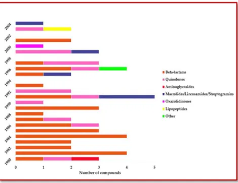
FIGURE 2.11: NUMBER OF NEW MOLECULES, DISTRIBUTED BY CLASS OF ANTIBIOTIC, OVER THE LAST DECADES (ADAPTED FROM POWERS, J. H. [98] ... 28
FIGURE 2.12: SCHEMATIC REPRESENTATION OF THE MECHANISM OF ACTION OF ANTIBIOTICS (ADAPTED FROM [3]). ... 30
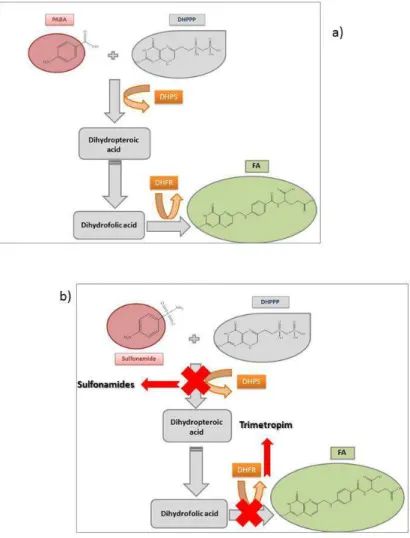
FIGURE 2.13: ANFOTERIC FORMS OF SULFONAMIDES. ... 48 FIGURE 2.14: SYNTHESIS OF FA: A) NORMAL PATHWAY FOR FA SYNTHESIS FROM PABA; B) SULFONAMIDES
ACTION ON FA SYNTHESIS BY DIHYDROPTEROATE SYNTHETASE (DHPS) INHIBITION AND
XVIII
OF COMPOUNDS (SULFONAMIDE + TRIMETROPIM) IS NORMALLY USED IN ORDER TO IMPROVE EFFICACY. ... 50 FIGURE 2.15: AMINOGLYCOSIDE ANTIBACTERIAL MECHANISM OF ACTION: A) GENERAL CHEMICAL
STRUCTURAL OF SOME AMINOGLYCOSIDE ANTIBIOTICS; B) 70S PROKARYOTIC RIBOSOME STRUCTURE (30S SUBUNIT IN BLUE AND 50S SUBUNIT IN BROWN); C) AMPLIFICATION OF THE BINDING SITE OF AMINOGLYCOSIDES, THAT (16S, IN BLUE), THAT DISTURBS PROTEIN SYNTHESIS. GENTAMICIN (YELLOW) AND NEOMYCIN (RED) ARE BONDED TO THE DECODING SITE. (ADAPTED FROM FELDMAN ET AL [135], WITH LICENSE). ... 53 FIGURE 2.16: QUINOXALINE COMPOUND: LEWIS STRUCTURE (LEFT) AND 3D STRUCTURE (RIGHT) ... 82 FIGURE 2.17: QUINOXALINE SYNTHESIS REPRESENTATION. ... 84 FIGURE 2.18: 8-CHLORO-1,4-SUBSTITUTED[1,2,4]TRIAZOLO[4,3-A] QUINOXALINE DERIVATIVES CORE
STRUCTURE. ... 86 FIGURE 2.19: 2,3-DIFURYL-4-QUINOXALINE-R-METILCARBOXAMIDE DERIVATIVES ... 87 FIGURE 2.20: 2,3-DIMETHYL-6-(DIMETHYLAMINOETHYL)-6H-INDOLO[2,3-B]-QUINOXALINE ... 95 FIGURE 2.21:
6-CHLORO-3,3-DIMETHYL-4-(ISOPROPENYLOXYCARBONYL)-3,4-DIHYDROQUINOXALIN-2(1H)-THIONE. ... 96 FIGURE 2.22: 1-(THIAZOLE[4,5-B]QUINOXALINE-2-YL)-3-PHENYL-2-PYRAZOLINES CORE. ... 97
FIGURE 2.23: LIGANDS L1H2 AND L2H2. FOR L1H2, R=CH3 AND FOR L2H2, R=C6H5. ... 99
FIGURE 2.24: (N-ARYLCARBAMOYL AND N-ARYLTHIOCARBAMOYL) HYDRAZINEQUINOXALINE-2(1H) COMPOUNDS CORE. ... 99 FIGURE 2.25: QUINOXALINE DERIVATIVES WITH ANTICANCER ACTIVITY. ... 102
FIGURE 2.26: ALPHAGAN®CHEMICAL STRUCTURE (5-BROMO-N-(2H-IMIDAZOL-2-YL)QUINOXALIN-6-AMINE)
... 103 FIGURE 2.27: 6-ARYLAMINO-2,3-BIS(PYRIDIN-2-YL)-7-CHOLOROQUINOXALINE-5,8-DIONES CORE. ... 104 FIGURE 2.28: 2,3-QUINOXALINEDIONE (QD). ... 107 FIGURE 2.29: INDENO-1-ONE[2,3-B]-QUINOXALINE (INQUI). ... 108 FIGURE 2.30: ACENAPHTHO[1,2-B] QUINOXALINE (AQ). ... 108
XIX
FIGURE 3.1: CALIBRATION: CHROMATOGRAMS FOR THE FOUR CONCENTRATION STANDARD TESTED. (AMOX: AMOXICILLIN; CIP: CIPROFLOXACIN; SULFO: SULFAMETHOXAZOLE; CLOR:
CHLORAMPHENICOL; CLOX: CLOXACILLIN) ... 133 FIGURE 3.2: CALIBRATION CURVES FOR A FIVE ANTIMICROBIAL AGENTS MIXTURE. (SULFO:
SULFAMETHOXAZOLE; CLOR: CHLORAMPHENICOL; CIP: CIPROFLOXACIN; CLOX: CLOXACILLIN; AMOX: AMOXACILLIN) ... 137 FIGURE 4.1: PROCEDURE 1 – MONITORING OF CIP SUSCEPTIBLE AND RESISTANT STRAINS BEHAVIOR
ALONG TIME... 145 FIGURE 4.2: PROCEDURE 2 – MONITORING OF A MIXTURE OF TWO STRAINS (SUSCEPTIBLE AND RESISTANT
TO CIP) BEHAVIOR ALONG TIME. ... 146 FIGURE 4.3: E. COLI S3R9 AND S3R22 BEHAVIOR ALONG TIME: TOP GRAPH – [CIP] = 2.00 MG/L. THE
SUSCEPTIBLE STRAIN DIES BEFORE 7 DAYS OF EXPOSURE; LOWER GRAPH – E. COLI S3R9 CFU
COUNTINGS FOR 4 WEEKS, UNDER A [CIP] = 0.05 MG/L. ... 153 FIGURE 4.4: LOG CFU/ML FOR MIXTURES OF E. COLI S3R9 AND S3R22, WITH (RIGHT) AND WITHOUT (LEFT)
CIP. ... 153 FIGURE 4.6: RAPD PROFILE FOR E.COLI CIP RESITANT STRAIN (H1FC22) AT [CIP] = 0.05 MG/L, AT: 1) 0 DAYS, 2)
56 DAYS, 3) FRESHLY UNFROZEN CELLS. ... 156 FIGURE 4.7: CALIBRATION CURVE FOR CIP QUANTIFICATION BY HPLC-DAD. ... 157 FIGURE 5.1: EXAMPLE OF VARIATION FOR GRAM NEGATIVE PROKARYOTIC STRAINS (E. COLI TEM-1) IN THE PRESENCE OF QUINOXALINE DERIVATIVE AT MIC DETERMINED. ... 178 FIGURE 5.2: VARIATION ALONG TIME FOR S. CEREVISIAE IN ABSENCE/PRESENCE OF THE COMPOUNDS
TESTED. ... 180 FIGURE 6.1: METABOLIC RATE RESULTS FOR SEVEN CELL LINES EXPOSED TO FIVE QUINOXALINE
DERIVATIVES WITH BIOLOGICAL ACTIVITY, MEASURED AFTER. A) EXPOSURE TO 2HF; B) EXPOSURE TO MQNX; C) EXPOSURE TO QNX; D) EXPOSURE TO 3M2MQNXC; D) EXPOSURE TO 2A3CQNX. (*: P < 0.05;
N = 3; RESULTS ARE EXPRESSED AS MEAN ± SD; C1: 10-3; C2 10-4; C3: 10-6 µG/L). 3T3-L1: MURINE
FIBROBLASTS, GF: GINGIVAL FIBROBLASTS, MSC: HUMAN MESENCHYMAL STEM CELLS, B16-F10: MURINE SKIN MELANOMA, HT29: HUMAN COLORECTAL ADENOCARCINOMA, BC3H1: MURINE BRAIN CELLS, MG-63: OSTEOSARCOMA CELL LINE. ... 193 FIGURE 7.1: SCHEMATIC REPRESENTATION OF THE RECYCLING PROCESS OF GLUTATHIONE. IN THE
PRESENCE OF ROS (REACTIVE OXYGEN SPECIES), GSH (REDUCED FORM OF GLUTATHIONE) IS OXIDIZED TO THE GSSG FORM. IN THE PRESENCE OF GLUTATHIONE REDUCTASE, AND USING NADPH AS AN ELECTRON DONOR, GSSG CAN BE REDUCED BACK TO GSH. ... 204
XX
FIGURE 7.2: ABSORPTION SPECTRUM FOR GSH (LEFT) AND GSSG (RIGHT) FORMS. ... 209 FIGURE 7.4: GSH AND GSSH CHROMATOGRAM: GSH FORM CORRESPONDS TO THE LEFT PEAK, WITH A TIME
OF RETENTION (TR) OF 3.96 MIN; GSSG CORRESPONDS TO THE RIGH PEAK, WITH A TR = 8.24 MIN. ... 213 FIGURE 7.5: CALIBRATION CURVES FOR GLUTATHIONE (OXIDIZED AND REDUCED FORMS) BY HPLC-DAD
METHOD. ... 214 FIGURE 7.6: CHROMATOGRAM FOR OXIDATIVE STRESS ANALYSIS, IN LIVER TISSUE, AFTER 24 HOURS
EXPOSED TO 3M2QNXC ... 216 FIGURE 7.3: CALIBRATION CURVE FOR GLUTATHIONE KIT ASSAY, AT 405 NM (SIGMA-ALDRICH, CO., USA
XXI
TABLE INDEX
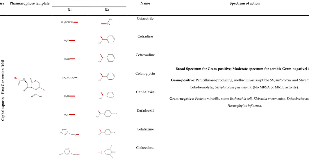
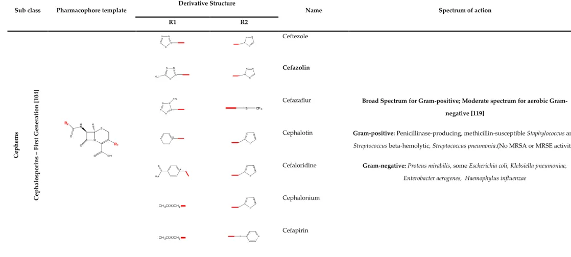
TABLE 2.1: Β-LACTAM CLASS OF ANTIBIOTICS – PENICILLIN: BIOLOGICAL PROPERTIES ... 33 TABLE 2.2: Β-LACTAM CLASS OF ANTIBIOTICS – CEPHEMS: BIOLOGICAL PROPERTIES (FIRST GENERATION
CEPHALOSPORINS) ... 34 TABLE 2.3: Β-LACTAM CLASS OF ANTIBIOTICS – CEPHEMS BIOLOGICAL PROPERTIES (SECOND AND THIRD
GENERATION CEPHALOSPORINS) ... 36 TABLE 2.4: Β-LACTAM CLASS OF ANTIBIOTICS – CEPHEMS BIOLOGICAL PROPERTIES (FOURTH GENERATION CEPHALOSPORINS) ... 40 TABLE 2.5: Β-LACTAM CLASS OF ANTIBIOTICS – CEPHEMS BIOLOGICAL (FIFTH GENERATION
CEPHALOSPORINS) ... 41 TABLE 2.6: Β-LACTAM CLASS OF ANTIBIOTICS - CEPHEMS BIOLOGICAL PROPERTIES (CEPHAMYCINS)... 42 TABLE 2.7: Β-LACTAM CLASS OF ANTIBIOTICS - CARBAPENEMS BIOLOGICAL PROPERTIES ... 43 TABLE 2.8: Β-LACTAM CLASS OF ANTIBIOTICS - MONOBACTAMS BIOLOGICAL PROPERTIES ... 44 TABLE 2.9: Β-LACTAM CLASS OF ANTIBIOTICS - Β-LACTAMASE INHIBITORS MICROBIOLOGICAL PROPERTIES ... 45 TABLE 2.10: CHEMICAL STRUCTURE AND BIOLOGICAL PROPERTIES FOR SOME GLYCOPEPTIDE ANTIBIOTICS ... 47 TABLE 2.11: CHEMICAL STRUCTURE AND BIOLOGICAL DATA FOR SOME SULFONAMIDES AND
P-AMINOBENZOIC ACID (STRUCTURES BUILT FROM SOFTWARE CAMBRIDGE CHEM3D PRO 12.0)... 51 TABLE 2.12: CHEMICAL STRUCTURE AND BIOLOGICAL PROPERTIES FOR AMINOGLYCOSIDE ANTIBIOTICS 54 TABLE 2.13: CHEMICAL STRUCTURE AND BIOLOGICAL PROPERTIES FOR TETRACYCLINE ANTIBIOTICS ... 59 TABLE 2.14: CHEMICAL STRUCTURE AND BIOLOGICAL PROPERTIES FOR MACROLIDE ANTIBIOTICS ... 61 TABLE 2.15: CHEMICAL STRUCTURE AND BIOLOGICAL PROPERTIES FOR LINCOSAMIDE ANTIBIOTICS ... 64 TABLE 2.16: CHEMICAL STRUCTURE AND BIOLOGICAL PROPERTIES FOR STREPTOGRAMINE ANTIBIOTICS . 66 TABLE 2.17: CHEMICAL STRUCTURE AND BIOLOGICAL PROPERTIES FOR OXAZOLIDINONE ANTIBIOTICS ... 68
XXII
TABLE 2.18: CHEMICAL STRUCTURE AND BIOLOGICAL PROPERTIES OF QUINOLONES CLASSIFIED
ACCORDING TO GENERATION – ADAPTED FROM [76] ... 71 TABLE 2.19: CHEMICAL STRUCTURES AND BIOLOGICAL PROPERTIES OF 5-NITROIMIDAZOLES ... 73 TABLE 2.20: CHEMICAL STRUCTURES AND BIOLOGICAL PROPERTIES OF SOME LIPOPEPTIDES ... 74 TABLE 2.21: QUINOXALINE PHYSICAL AND CHEMICAL PROPERTIES ... 84
TABLE 2.22: PUBLISHED EXPERIMENTAL DATA (PERCENTAGE OF BINDING, INTERCALATION, IC50 AND IG50)
OF QUINOXALINE DERIVATIVES AND THEIR SUBSTITUENTS ... 88 TABLE 3.1: ANTIMICROBIAL AGENTS TESTED FOR MULTIPLE DETECTION METHOD DEVELOPMENT. GREY
MARKED COMPOUNDS ARE THE UTILIZED IN THE CHROMATOGRAPHIC METHOD (GREY CELLS INDICATE THE ANTIMICROBIAL AGENTS SELECTED FOR THE DEVELOPED CHROMATOGRAPHIC METHOD). ... 128 TABLE 3.2: SAMPLE DISTRIBUTION OVER 4 GROUPS OF ANALYSIS ... 133 TABLE 3.3: LIMITS OF DETECTION AND QUANTIFICATION FOR THE FIVE ANTIMICROBIAL AGENTS STUDIED ... 137 TABLE 3.4: CIP QUANTIFICATION OF SPIKED MILK SAMPLES ... 138 TABLE 4.1: CHROMATOGRAPHIC METHOD FOR CIP DETERMINATION ... 150 TABLE 4.2: SURVIVAL OF E. COLI STRAINS S3R9 AND S3R22 IN WATER SPIKED WITH DIFFERENT
CONCENTRATIONS OF CIPROFLOXACIN, MONITORED WEEKLY ... 151 TABLE 4.3: Δ LOG CFU/ML (T = 21 – T = 0 DAYS) FOR MIXTURES OF E. COLI S3R9 AND CIP RESISTANT E. COLI
STRAINS AND Δ LOG CFU/ML FOR RESISTANT E. COLI STRAINS, IN THE PRESENCE AND ABSENCE OF CIP ... 155 TABLE 4.4: CIP QUANTIFICATION AT T = 0 AND T = 6 WEEKS ... 158 TABLE 5.1: QUINOXALINE N,N-DIOXIDE AND QUINOXALINE DERIVATIVES. ... 168 TABLE 5.2: ZONE DIAMETER INTERPRETATIVE STANDARDS (ENTEROBACTERIACCEAE) AND EQUIVALENT
MINIMAL INHIBITORY CONCENTRATION (MIC) ACCORDING TO CLSI DOCUMENT [393]. ... 174 TABLE 5.3: DILUTION FACTOR AND TIME INTERVALS FOR UFC/ML DETERMINATIONS... 174 TABLE 5.4:DISC DIFFUSION DIAMETERS FOR THE QUINOXALINE DERIVATIVES. ... 175
XXIII
TABLE 5.5: CLASSIFICATION OF EACH STRAIN AS SUSCEPTIBLE (S) OR RESISTANT (R) ACCORDING TO CLSI IN PRESENCE OF CEFOXITIN DISK (FOX) AND CIPROFLOXACIN DISK (CIP) BY DISK DIFFUSION METHOD [393]. ... 176 TABLE 5.6: MINIMUM INHIBITORY CONCENTRATION (µG/L) RESULTS FOR EACH GROUP BACTERIAL
STRAIN/COMPOUND BY MICRODILUTION METHOD, CLSI [393]. ... 176 TABLE 6.1: QUINOXALINE N,N-DIOXIDE AND QUINOXALINE DERIVATIVES THAT PRESENTED GRAM
NEGATIVE ANTIBACTERIAL ACTIVITY. ... 189 TABLE 6.2: SUMARY STATISTICAL ANALYSIS FOR METABOLIC RATE OF 7 CELL LINES EXPOSED TO
QUINOXALINE DERIVATIVES ... 194 TABLE 7.2: RESULTS FOR [GSH]/[GSSG] ANALYSIS IN LIVER TISSUE EXPOSED TO QUINOXALINE DERIVATIVE
COMPOUNDS WITH BIOLOGICAL ACTIVITY. ... 215 TABLE 7.1: DATA FOR THE KIT ASSAY CALIBRATION, ACCORDING TO THE PRODUCER’S GUIDELINES
XXV
ABBREVIATION INDEX
2A3CQNX 2-Amino-3-cyanooylquinoxaline-1,4-dioxide 2HF 2-Hydroxyphenazine-N,N-dioxide 2M3BenzoylQNX 2-Methyl-3-benzoylquinoxaline-1,4-dioxide 2M3BQNX 2-Methyl-3-benzyloylquinoxaline-1,4-dioxide 2MQNX 2-Methylquinoxaline-1,4-dioxide 3M2QNXC 3-Methyl-2-quinoxalinecarboxamide-1,4-dioxide 5-HT 5-Hydroxytryptamine6-APA 6-Aminopenicillanic acid
7-ACA 1-Aminocephalosporanic acid
AA Arachidonic acid
AIDS Acquired immunodeficiency syndrome
AML Amoxicillin
AMOX Amoxicillin
AMPA-R α-amino-3-hydroxy-5-methyl-4-isoxazole-propionic acid receptor
AMR Antimicrobial resistance
AQ Acenaphtho[1,2-b]quinoxaline
ATCC American Type Culture Collection
ATP Adenosine-5'- triphosphate
CDC Center for Disease Control and Prevention
CFU Colony-forming unit
CIP Ciprofloxacin
CISA Centro de Investigação em Saúde e Ambiente
CLOR Chloramphenicol
XXVI
CLSI Clinical and Laboratory Standards Institute
CV Coefficient of variation
Cys Cysteine
DAD Diode Array Detector
DHFR Dihydrofolate reductase
DHPP Dihydropteridine pyrophosphate
DHPS Dihydropteroate synthetase
D-MEM Dubelco-Modified Eagle's Medium
DMF Dimethylformamide
DMSO Dimethyl sulfoxide
DNA Desoxirribolnucleic acid
DOI Digital object identifier
DTNB Ditionitrobenzoic acid
EARSS European Antimicrobial Resistance Surveillance Sustem
ECD Electron Capture Detector
EDTA Ethylaminotetracetic acid
EEA Excitatory amino acid
ESAC European Sulveillance of Antimicrobial Consumption
EtOH Ethanol
FA Folic acid
FCT-MEC Fundação para a Ciência e Tecnologia - Ministério da Educação e Ciência
FDA Food and Drug Administration
FDNB 2,4-Dinitrofluorobenzene
FID Flame Ionization Detector
FL Fluorescent Detector
FOX Cefoxitin
FPD Flame Photometric Detector
G- Gram-negative
XXVII
GC Gas Chromatography
GI50 50% Growth inhibition
GPCR G-protein coupled receptor
GPx Glutathione peroxidase
GSH Reduced glutathione
GSSG Oxidized glutathione
GST Total glutathione
HIV Human immunodeficiency virus
HPLC High Performance Liquid Chromatography
HSV Herpes symplex vírus
IMA Indomethacin
INQUI Indeno-1-one[1,2-b]quinoxaline
IRD Infrared Detector
IUPAC International Union of Pure and Applied Chemistry
LC Liquid Chromatography
LD Limit of Detection
LEV Levofloxacin
LOX Lipoxygenase
LQ Limit of quantification
MAO Monoamine oxidase
MDR Multi-drug resistant
MeOH Methanol
MHPQ 3-methyl-2-(2-hydroxyphenyl)quinoxaline
MIC Minimum inhibitory concentration
MMtHPQ 3-methyl-2-(3-methoxy-4-hydroxyphenil)quinoxaline
MPA Mycophenolic acid
MPQ 3-methyl-2-phenylquinoxaline
MRL Maximum residue limit
XXVIII
MRSA Methicillin-resistant Staphylococcus aureus MRSE Methicillin-resistant Staphylococcus epidermis
MS Mass Spectrometer
NADH Nicotinamide adenine dinucleotide
NADPH Nicotinamide adenine dinucleotide phosphate
NGP Natural growing products
NOR Norfloxacin
NPD Nitrogen-Phosphorus Detector
NSAID Non-steroidal anti-inflammatory drug
OD Optical density
OGTT Oral glucose tolerance test
OLED Organic light-emitting diode
PABA p-Aminobenzoic acid
PBP Penicillin-Binding Protein
PCA Plate count agar
PHOLED Phosphorescent organic light-emitting diodes PHPQ 3-Phenyl-2-(2-hydroxyphenyl)quinoxaline
PPQ 2,3-Diphenylquinoxaline
PYCC Portuguese Yeast Culture Collection
QD Quinoxalinedione
QNX Quinoxaline -1,4-dioxide
R Resistant
RAoSMC Rat aortic smooth muscle cell
RAPD Random Amplified Polymorphic DNA
RNA Ribonucleic acid
ROS Reactive oxigen species
RP Reverse Phase
RT Reverse transcriptase
XXIX
SMC Smooth cell
SOD Superoxide dismutase
sp. Specie
SPE Solid phase extraction
spp. Species
SSA 5-Sulfosalicylic acid
SULFO Sulfomethoxazole
SXT Sulfamethoxazole/Trimetropim
TB Tuberculosis
tBOOH Tertiary-butylhydroperoxide
TCA Trichloroacetic acid
TCD Thermal Conductivity Detector
TCY Tetracyclin
TFA Trifluoroacetic acid
TIC Ticarcillin
TNB 2-Nitro-5-thiobenzoate
tRNA Transfer ribonucleic acid
TSA Tryptic soy agar
TSB Tryptic soy broth
USA United States of America
UTI Urinary tract infections
UV Ultra-violet
VRE Vancomycin-resistant Enterococcus
VRSA Vancomycin-resistant Staphylococcus aureus
WHO World Health Organization
XTT 2,3-bis-(2-methoxy-4-nitro-5-sulphophenyl)-2H-tetrazolium-5-carboxanilide YEPD Yeast extract peptone dextrose
2
Chapter 1: General plan and main objectives
1.1 Main Objectives 1.2 General Framework
4
1
GENERAL PLAN AND MAIN OBJECTIVES
The discovery of antibiotics, in the 1930s, revolutionized the medicine world. It became possible to treat serious lethal bacterial infections. Enthusiasm took over the medical community and the consequence was overprescribing.
Bacteria communities and cells can easily evolve and become adapted to changing environmental conditions, often through modification in the genome [1]. When exposed to antibiotics, bacteria find ways to avoid their deletery effects. Abusive and inappropriate use of antibiotics can, thus, result in bacterial resistance to antibiotics. The eradication of these resistant bacteria requires higher doses of medicine or stronger antibiotics. Because of antibiotic overuse, certain bacteria have become resistant to some of the most powerful antibiotics available today [2, 3].
In the past 40-50 years bacteria have become increasingly unsusceptible to the antibiotics in our medical arsenal. At the same time, scientific community and pharmaceutical industry have developed only a small number of new drugs to take the place of those that have become useless (Figure 1.1). As antibiotic effectiveness decreases, patients require longer, more toxic and expensive treatments. As a consequence, hospitals have themselves become spots of highly resistant pathogens, like methicillin-resistant Staphylococcus aureus (MRSA), increasing a paradoxal risk: hospitalization may actually kills instead of heal [4].
5
Figure 1.1: Antibiotic introduction and resistance occurrence (Data from CDC, “Antibiotic Resistance Threats in the United States, Threats Report 2013” [4]).
6 Antibiotics are not used only in human medicine. Animal production for consumption has taken gigantic proportions and animals are usually kept in large groups and the animal density is high. These are conditions for illness introduction and spread. Antibiotics then begun to be used for animal treatment, as well as for preventing infection appearance. Antibiotics are also used as growth promoters, since these properties have been identified and since the livestock industry deals with profit rates, and thereby achieves a faster and larger animal production. In the middle 1990s, multi-drug resistant strains of antibiotics were found in livestock and in food from animal origins. These resistant strains were mostly related to the antibiotics used as growth promoters. The use of these antibiotics in veterinary medicine and as growth promoters may result in trace residues of these substances in food from animal origin and have consequences in the human commensal flora [5, 6]. Nowadays, the use of antibiotics as growth promoters is forbidden in Europe [7], as “part of the Commission’s overall strategy to tackle the emergence of bacteria and other microbes resistant to antibiotics, due to their overexploitation or misuse”. In the USA, the use of antibiotics is still allowed, although there are appeals for the reduction and ban of these drugs [8].
Antimicrobialy active residues have been also detected in surface waters, soil and manure. The input of these microbial active substances is associated to municipal sewage, hospital effluents and agricultural and livestock activities [9-12].
The worldwide spread of resistance to antimicrobials has become "one of the world's most pressing public health problems." (Centers for Disease Control and Prevention - CDC). World Health Organization (WHO) established a set of recommendations, in 2001, in order to limit the emergence and spreading of antimicrobial resistance [13].
In this perspective, this research work intends to make a monitoring of the presence of antimicrobial residues in surface water and in food of animal origin (milk), by the development of a multiresidue antibiotic detection method, including the most
7 used and representative of different classes of antimicrobials. Because one of the most important consequences of the contamination with antimicrobial residues is the selection of antibiotics resistant bacteria, a model system with ciprofloxacin and Escherichia coli strains resistant and susceptible to that antimicrobial was used to assess such an effect. Knowledge of these effects is crucial for understanding the consequences of the use and disposal of antimicrobials.
One of the current problems related to resistance to antimicrobial agents is the lack of new substances that can replace those that have lost effectiveness. Thus, this study intends to evaluate the antimicrobial and antitumor activity, as well as toxicity of some quinoxaline derivatives. This family of compounds has been evaluated by several research groups in recent years. Additionally, there are data indicating them as a possible alternative to the fight against resistant bacterial strains, thus replacing other antimicrobials that are currently losing effectiveness.
1.1 MAIN OBJECTIVES
The main objectives proposed for this work were:
a) Develop chromatographic methods in order to evaluate antibiotic residues presence in several samples, as water and milk;
b) Compare the behavior of ciprofloxacin-resistant and susceptible strains of Escherichia coli in the presence of sub-inhibitory concentrations of that antimicrobial drug;
c) Evaluate antimicrobial activity and cellular toxicity of quinoxaline derivatives in human cells;
8 d) Determine the oxidative stress in liver extract caused by exposure to
quinoxaline derivatives solutions.
Specific objectives were traced in order to achieve the main goals for this work and are described along the text, in each chapter.
1.2 GENERAL FRAMEWORK
The present work is divided into 9 chapters. The present chapter (Chapter 1) presents the framework of the developed work and the main objectives. In Chapter 2, is presented a general introduction to the main topic of antibiotics, resistance to antibiotics and the public health concerning. A state-of-the-art review on quinoxaline derivatives biological activity is also presented. The content of this chapter is under review at the European Journal of Medical Chemistry (Manuscript Number: EJMECH-D-13-01270). Another paper was published in the development of the present work, addressing the resistance topic: Bacterial Resistance, Ricardo Ferraz, Cristina Prudêncio, Mónica Vieira, Ruben Fernandes, João Paulo Noronha, Zeljko Petrovski, Biochemistry & Pharmacology: Open Access, 1-7, 2012 (doi:10.4172/2167-0501.1000e138). Part of this article is textually exposed in section 2.3 of this document.
In Chapter 3 is presented the development of a new chromatographic method for antibiotic detection and its application on the analysis of surface water and food samples. The contents of this chapter was submitted to the international journal Talanta (Manuscript Number: TAL-D-13-02953) and is under review.
Chapter 4 reports the behavior of several bacterial strains under sub-inhibitory concentrations of ciprofloxacin in a time course assay. Susceptibility and phenotypic
9 profile was evaluated. The contents of this chapter are being submitted to an international peer review journal.
Chapter 5 reports the antimicrobial activity of quinoxaline derivatives tested. This chapter has been published at Microbiological Research (http://dx.doi.org/10.1016/j.micres.2013.06.015).
Chapter 6 presents the results obtained on the cellular toxicity in the human cells assays performed in order to evaluate quinoxaline derivatives as potential antimicrobial agents. The content of this chapter has been submitted to European Journal of Medicinal Chemistry (Manuscript Number: EJMECH-S-13-02430) and is under review.
Chapter 7 reports the oxidative damage results obtained for liver extract exposed to the quinoxaline derivatives. The content of this chapter has been submitted to Journal of Biotechnology (JBIOTEC-D-13-01223) and is under review.
Discussion of the results obtained and major conclusions are presented in Chapter 8. Future perspectives and current works are explained in Chapter 9.
10
Chapter 2: State of the Art
2. Introduction
2.1 Antibiotic History
2.2 Classification of Antibiotics
2.3 Antibiotic Resistance: A Public Health Concerning
2.4 Quinoxaline: the light at the tunnel?
12
2
INTRODUCTION
"From the beginning of human existence, their struggle for survival was established with other living beings, of all sizes. Some of these beings are so small that they were not seen nor touched. However, these microorganisms, in the battle for survival, inflicted heavy losses to the human species. There were pests, such as in Egypt, pests such as the bubonic plague and, more recently, epidemics such as influenza and cholera."
The natural products were the earliest therapeutic substances used by man, since there were observations that these substances controlled the symptoms of some diseases. Mercury, for example, had been used since the sixteenth century in the treatment of syphilis. Quinine and ipecacuanha root were brought to Europe in the seventeenth century and are effective against parasites of malaria and amoebic dysentery.
Man has used antimicrobial drugs for centuries without truly realizing its potential. It took several years to be able to identify and distinguish, at the microscope, bacteria, fungi and viruses. Antimicrobial drugs are the oldest therapy used to combat specific microorganisms such as bacteria and yeast. Nowadays, we look at the infections as minor diseases, but before the 1950s, cases of death at very young ages were very frequent due to infections such as tuberculosis, pneumonia, meningitis, diphtheria, pertussis, measles and influenza. With the discovery of antibiotics there was a revolution in the practice of medicine and life expectancy has increased significantly [14, 15].
The Modern Era of Antibiotics begins in the decades of 1930-1940, when man managed to control infections caused by streptococci and pneumococci. Some authors consider the beginning of this age with the onset of the clinical use of sulfonamides in 1936. Other authors consider that, officially, antibiotics were discovered in the 1920s,
13 specifically in September 1928, by the renowned Scottish chemist Sir Alexander Fleming, who observed the behavior of certain substances, such as the presence of the fungus Penicillium notatum, which restricted the normal growth of Staphylococcus aureus. Since then, antibiotics are used on a large scale, in fighting infections by microorganisms.
Some authors argue that the history of antibiotics begins with the use of alkaloids, starting in 1619, date that recall the earliest records of the first effective treatments for malaria and dysentery with quinine and root extracts of ipecacuanha, respectively [16, 17]. Several scientists, such as Joseph Lister [18], Louis Pasteur, Jules François Joubert [19], Paul Ehrlich [20], among others, identified the antimicrobial potential of different products.
Currently, there are several known classes of antibiotics. The first to be used were of natural origin, produced by bacteria, but at the present days, the vast majority of antimicrobial active ingredients used are of synthetic or semi-synthetic origin. Molds and soil organisms are the most used today for the production of antibiotics, but technological evolution enabled a better production and purification of these substances [21].
2.1 ANTIBIOTICS HISTORY
The term antibiosis, used to designate the antibiotic function, was first proposed by Vuillemin, in 1889, and described the antagonism between living beings in general. The term antibiotic was used, for the first time, by Selman Waksman, in 1941, to describe molecules produced by microorganisms that were antagonists of growth or living of other organisms around them [21, 22]. The common use of the term made it
14 applicable also to antimicrobial agents of synthetic origin, such as the quinolones and sulfonamides.
The discovery of natural origin substances with antibacterial effects occurred with the discovery of penicillin. Fleming, after a period of absence of the laboratory, observed inhibition of growth of a culture of staphylococci around a mold, which accidentally appeared in his plate [23]. The mold Penicillium notatum produces the substance inhibiting the growth of bacteria, later identified as penicillin [24]. The substance that Fleming was referring to is benzylpenicillin or Penicillin G. Howard Florey and Ernst Chain followed up the discovery of Fleming, and shared the Nobel Prize in Physiology or Medicine with him, in 1945. The main achievement was the successful isolation of penicillin and its extraction it with good yields [25]. Florey and Chain proved, further, its non-toxicity, using models of mice previously infected with streptococci. In 1959, the 6-aminopenicillanic acid (6-APA), shown in Figure 2.1, was extracted enzymatically by Batchelor [26]. The 6-APA was achieved through a simple mechanism and in a good yield. This product was the starting point for achieving semi-synthetic penicillins, which present improved spectrum of activity compared to penicillin G. The first semi synthetic penicillin produced was ampicillin, which has demonstrated antibacterial activity against Gram-negative microorganisms [27-29]. Penicillin and its derivatives are included in the β-lactam group of antibiotics that share a β-lactam ring as common feature. Cephems, carbapenems and monobactams are the other sub-classes that complete this group of antibiotics.
β-Lactams are, since their application, the more prescribed antibiotics in human and veterinary medicine [30]. The structure of this class of antibiotics is based on a β-lactam cyclic system attached to a thiazolidine ring, forming the 6-aminopenicillanic acid (6-APA). This structure is obtained by the condensation of a valine molecule and a cysteine molecule, originating the characteristic double system. This structural element is essential to β-lactams biological activity and, if the β-lactam ring is destroyed, by metabolic transformation or chemical alteration, the antibacterial activity is lost [31].
15
Figure 2.1: Chemical representation of 6-aminopenicillanic acid (6-APA).
β-lactams are divided in four major classes (penicilins, cephems, carbapenems and monobactams) and a β-lactamase inhibitors group (Table 2.1-Table 2.9).
The cephem group of β-lactams includes cephalosporins and cephamycins. Cephalosporins (Figure 2.2) are semi-synthetic antibiotics, derived from cephalosporin C, a natural compound, produced by Cephalosporium acremonium. Cephalosporin C was discovered in 1948, by Giuseppe Brotzu [32]. Brotzu noticed that Salmonella typhi, the bacterial strain that caused the typhoid fever, was inhibited by substances produced by C. acremonium. The cephalosporin active chemical nucleus is the 7-aminocephalosporanic acid (7-ACA), similar to 6-APA, as it possesses also a β-lactam ring. Cephamycins and cephalosporins are very similar structurally and are all classified as cephalosporins, by some authors [33, 34]. Cephamycins (Figure 2.2) are naturally produced by members of the Streptomyces genus and characterized by the 7-α-methoxyl chemical group. This sub-class of β-lactams were first reported in 1972, by Stapley and Miller, from the Merck Sharp and Dome Research Laboratories [35].
16
Cephalosporin chemical nucleus Cephamycin chemical nucleus
Figure 2.2: Structural core representation for cephalosporin (left) and cephamycin (right).
Carbapenems (Figure 2.3), another sub-class of β-lactam antibiotics, were developed from thienamycin, a natural product produced by Streptomyces cattleya, discovered in 1976 [36]. Thienamycin revealed chemical and biological instability. Therefore, scientists developed new compounds, more stable and profitable [37]. Structurally, carbapenems are also similar to penicillin, but they present a carbon atom, instead of a sulfur atom, in the position 1 of the β-lactamic structure
Figure 2.3: Representation of the chemical nucleus of carbapenems.
Monobactams are a sub-class of β-lactam antibiotics of synthetic origin. They may also be produced by actinomycetes, unlike cephalosporin and penicillin. These compounds present a chemical structure different from other β-lactams, since they are monocyclic (Figure 2.4) [38, 39]. The denomination of monobactams was employed by Sykes [40] and this class of compounds were discovered about 50 years after Fleming’s finding. Aztreonam was the first synthetic monobactam reported and the first, from a series of compounds, that actually reached medicine practice [41].
17 There is another functional sub-class of compounds, included in the β-lactam antibiotics, the β-lactamase inhibitors. Bacteria, after exposure to β-lactam antibiotics, have developed mechanisms of resistance to these drugs, through the action of β-lactamase enzymes that hydrolyze the active β-lactam ring, thus deactivating the antibacterial action. The lactamase inhibitors present higher affinity to the β-lactamase enzymes than the β-lactams, and bind irreversibly to them [42]. The clavulanic acid, sulbactam and tazobactam are the compounds that belong to this sub-class of β-lactams.
The β-lactam antibiotics are bactericidal, since they interfere in the cell wall synthesis, specifically by inactivation of enzymes involved in the synthesis of peptidoglycan, a fundamental biomolecule in cell wall constitution, leading to cell death due to osmotic pressure. The β-lactams target enzymes are the Penicillin-Binding Proteins (PBPs) that are located at the cytoplasmatic membrane [43].
In addition to β-lactam antibiotics, there are other classes of antibiotics with very different chemical structures that present different antibacterial activity.
In the 1930s, a series of discoveries, carried out in Germany and France, were called "the miracle of miracles," for the man provided an effective weapon to overcome bacterial infections: the sulfonamides or sulfa drugs [14]. These drugs were active only on some bacterial strains and had no effect on strains that caused lethal infections. The assembly referred to as sulfa drugs or sulphonamides have a common set of atoms that form the sulfonamide functional group (Figure 2.5).
S O O N R1 R2 R3
18 Sulfonamides are antimicrobial agents with considerable use in human and veterinary medicine. These antimicrobial agents were the first to be used to fight bacterial infections in human medicine [44].
In the 1940s, the sulfonamides were considered drugs of choice for the treatment of bacterial infections in humans. Currently, and in human medicine, the sulfonamides are used only in specific infections, such as urinary tract infections(UTI), but have also a wide application in veterinary medicine.
In 1908, Gelmo and collaborators have synthesized, for the first time, a sulfonamide, while searching for new azo dyes [45]. Following this work, Höerlin and colleagues found that dyes with sulfanyl group present in its composition had a higher affinity for silk and wool proteins [46], but also promoted some protection against septicemia in mice caused by hemolytic streptococci [47]. This discovery led Eisenberg, in 1913, to observe that chrysoidine showed bactericidal properties [47, 48], and, in 1919, Jacobs and Heidelberger [47] detected that some azo dyes, synthesized from hydrocupreine and diazotized sulfonamides , presented bactericidal action. In 1932, Domagk was awarded the Nobel Prize in Physiology or Medicine for his discovery of the therapeutic Prontosil® (p-aminobenzenesulfonamide, synthesized by Gelmo), which showed protective action, in rats and rabbits, against staphylococci and hemolytic sterptococci. Although not convinced that Prontosil®, that was so effective in animal models, could be effective in humans, Domagk administered a dose of it to his daughter, when she came down with an infection caused by streptococci. The recovery was complete, but this fact Domagk concealed until 1935, when the results become available for clinical trials in patients [49]. Following this discovery, several sulfonamides were synthesized in the 1930s, and many of these compounds revealed antibacterial activity against various strains of pneumococci and other sterptococci. In 1941, several sulfapyrimidines were presented, with higher antibacterial activity and
19 lower toxicity than the sulfonamides synthesized previously [50]. Presently, there are more than 5000 known compounds belonging to the class of sulfonamides, but only about 30 are used in human and veterinary medicine [51, 52]. Of these, we highlight 5, with main antibacterial application: sulfanilamide, sulfamethoxazole, sulfacetamide, sulfadiazine and sulfamerazine. From the central structure of sulfonamides (aniline group + sulfonium group) several compounds were synthesized and have pharmaceutical application as antibacterial agents. Some of the derivatives obtained may also act as diuretics and antidiabetogenic agents [50-52].
Sulfonamides exhibit activity against Gram-positive and Gram-negative bacteria as well as against certain fungi and protozoans, and therefore are classified as broad spectrum antibiotics [53].
Glycopeptides are complex heterocyclic molecules, constituted of a multi-peptide backbone with several sugars attached to it. These molecules are bulky and cannot penetrate the outer membrane of Gram-negative bacteria, reducing, thereby, their spectrum of action to Gram-positive bacteria [54]. Vancomycin, the first glycopeptide, was isolated in 1953, by Edmund Komfeld, from Amycolatopsis orientalis (formerly Nocardia orientalis), present in soil samples. It is a rare example of a haloorganic natural compound, with two chlorine atoms [55]. Bleomycin was discovered in 1966, by Hamao Umezawa [56], and is produced by Streptomyces verticillus. This antibiotic was found when Umezawa was looking for compounds with antitumor activity. Bleomycin is used as an anticancer agent, in Hodgkin’s lymphoma, squamous cell carcinoma and testicular cancer. It is also used in the treatment of plantar warts and in the chemical pleurodesis procedure [57]. Teicoplanin was discovered in the early 1990s and corresponds to a mixture of closely related compounds [58, 59]: five major components and four minor components. Telavancin, a semi-synthetic lipoglycopeptide, was approved in late 2000s [60].
20 Aminoglycoside is another class of antibiotics, and this name is related to its chemical structure. Aminoglycosides consist of two or more aminosugars linked to an aminocyclitol nucleus, an aminosubstituted cyclic polyalcohol. The different aminoglycosides are distinguished by the amino sugars in their constitution [61].
Aminoglycosides were introduced in 1944, with streptomycin, discovered by Selman Waksman [62]. Streptomycin is of natural origin, produced by Streptomyces griseus. Its discovery was an important goal, since it presented activity against Mycobacterium tuberculosis. Neomycin was the subsequently aminoglycoside, and was presented in 1949, extracted from Streptomyces fradiae, followed by kanamycin, that is produced by Streptomyces kanamyceticus [63, 64]. Amikacin, a derivative of kanamycin was introduced in 1972, and was the first semi-synthetic aminoglycoside [65]. In 1963, gentamicin was introduced, extracted from Micromonospora purpurea, an actinomycete, and netilmicin, a semi-synthetic derivative of sisomicin, produced by Micromonospora species, was presented in 1976 [66, 67]. The name of aminoglycosides follow the rule: substances originated from the Streptomyces genus present the suffix –mycin and those originated from the Micromonospora genus are named with the suffix –micin [68].
Another sugar related class of antibiotics is the macrolides. This class of antibiotics is characterized by the presence, in their structure, of a macrolide ring, a 14-, 15- or 16- macrocyclic lactone ring substituted by several sugars [69, 70]. Macrolide antibiotics were discovered in 1952, with the introduction of erythromycin, a natural product of Streptomyces erythreus. This compound is susceptible to acidic conditions, like those experienced in the stomach passage. With the purpose of adjust this behavior, scientists modified erythromycin structure. Semi-synthetic macrolides derivatives, like azithromycin and clarithromycin, were discovered by Croatian and Japanese drug companies, respectively, between late 1970s and early 1980s [54].
Tetracyclines were described in 1945 by Benjamin Duggar [71], on his research on Streptomyces species, although there are reports of tetracycline present in bones of
21 ancient Egyptians, from the fourth century [72]. Chlortetracycline was the first tetracycline described, extracted from Streptomyces aureofaciens. The following compounds were oxytetracycline and tetracycline. These antibiotics are used in human medicine for decades and, as they are broad spectrum and low cost antibiotics, are widely used in veterinary medicine, for treatment and production of animals, since they avoid infections in the livestock facilities, and as growth promoters [73]. The more common used tetracyclines are tetracycline itself, chlortetracycline, oxytetracycline and doxycycline [74]. The chemical structure of this class of antibiotics is derived from a hydronaphthacene nucleus containing four fused rings.
Figure 2.6: Tetracycline general structure.
Tetracycline antibiotics have similar chemical structures. They are strong chelating agents and biological membranes present a good permeability to these compounds. Due to their good lipossolubility, allied to low cost, residues of tetracyclines have been detected and reported in several food products and water [75].
Quinolones appeared in 1962 in the development of therapies for malaria. Quinolones are not natural substances of microbial origin, like penicillin, but have been synthesized, like the sulfonamides. The first substance of the class to which it was assigned antibacterial properties was nalidixic acid [76].
22
Figure 2.7: Structural representation of Nalidixic Acid.
The use of this substance was limited due to their narrow spectrum of activity, low maximum amounts of application and toxicity. In the 1980s, when resistance to antibacterial agents used, by strains of Shigella spp. and Escherichia coli, was detected, this class of antibacterial agents regained the attention of scientists, in the treatment of diarrhea and UT infections [76, 77]. It was the beginning of a period of synthesizing new substances, in order to replace the β-lactams and macrolides, looking for broader action spectra and less toxicity. The first quinolones developed from the nalidixic acid were pipemidic acid, cinoxacin and oxolinic acid. The skills acquired in the development of new drugs and the perception that simple changes in the side chains of the main structure caused significant changes in the action of these substances led to the development of more than 10,000 substances. However, only about 2% of these were subjected to the clinical trials and only some 20 substances have reached the market. Quinolones developed from the nalidixic acid exhibit activity against Gram-positive and Gram-negative bacteria, anaerobic bacteria and pathogenic mycobacteria [78]. Flumequine was the first fluoroquinolone substance, which includes a fluorine atom at the C-6 position, in its constitution, which demonstrated increasing applications of this class of antibacterials.
Quinolones (Figure 2.8) are structurally based on a 4-quinolone nucleus [79, 80], which is derived in four groups of compounds, called generations.
23
Figure 2.8: Structural representation of quinolone nucleus - adapted from [79].
Quinolones available for human and veterinary medicine use are divided, on the basis of their spectrum of activity, in four generations [79]. This classification is not unanimous among all authors. Flumequine is classified as a fluoroquinolone of 1st generation, together with nalidixic acid and its first derivatives, and is no longer used clinically. The 2nd generation quinolones, all fluoroquinolones with a fluorine atom at the C-6 position, have the advantage of greater effectiveness against Gram-negative bacteria. The most relevant quinolones of this generation are ciprofloxacin (CIP) and norfloxacin (NOR), as well as levofloxacin (LEV), which currently have clinical application. Are also part of this generation, sparfloxacin and grepafloxacin, which have improved activity against Gram-positive bacteria. The 3rd generation of quinolones, besides showing activity against Gram-positive organisms, yet has activity against anaerobes. This increase in the spectrum of action was obtained by introducing a methoxy group, at position C-8, and pyrrolidines or alkyl-substituted piperazines at position C-7. Trovafloxacin (or naphthyridone), although not presenting a methoxy substitution at C-8, belongs to this generation of quinolones and was the developed substance with wider spectrum recorded, but presented hepatotoxicity, so its clinical use was short lived [76]. Some authors consider that the quinolones with the methoxy group belong to the 4th generation [80, 81]. The 4th generation of quinolones, for some authors, is called des-fluoroquinolone generation [76], since encompasses substances free of fluorine at the C-6 position. The presence and location of fluorine atom in quinolones of 2nd and 3rd generations is associated with genotoxicity risks and side effects in the central nervous system. The introduction, in the central active molecule,
24 of substituents with antibacterial capacity increasingly improved, led to the removal of fluorine atom in position 6. Garenoxacine is one of the substances belonging to the 4th generation of quinolones [82], according to this classification, and contains no fluorine in its constitution.
Lincosamides, oxazolidinones and streptogramines are classes of antibiotics usually grouped together because they present similar mode of action and antibacterial spectra. They are active essentially against Gram-positive bacteria
Lincomycin, the first lincosamide antibiotic, was discovered in 1963, by Mason and co-workers [83], and is a natural product of Streptomyces lincolnensis. Lincomycin was supplanted by clindamycin, a semisynthetic derivative, due to its improved antibacterial activity [84]. Chemicaly, lincosamides are characterized by the presence of an alkyl 6-amino-6,8-dideoxy-1-thio-D-erythro-α-D-galacto-octopyranoside [83]. Lincosamides present antibacterial activity similar to macrolides [85] and resistance to erythromycin, a macrolide, affects lincosamides as well [54].
Oxazolidinones are a synthetic class of antibiotics. Linezolid was the first oxazolidinone approved for clinical use [86]. Some of the oxazolidinone antibiotics are the last generation of antibiotics used against Gram-positive pathogens and against multidrug-resistant bacteria, such as Methicilin-resistant Staphylococcus aureus (MRSA) [87]. The oxazolidinone essential chemical structure, or pharmacophoric template, is characterized by the structure in Figure 2.9.
25 Since oxazolidinones are a completely synthetic class of antibiotics, many investigation groups are developing new synthetic routes in order to obtain new effective antibiotics [86, 87], especially against multidrug-resistant bacteria.
Each member of the streptogramines family corresponds to two antibiotics, components A and B, produced as synergetic mixtures by various species of Streptomyces [54] and was first isolated from Streptomyces graminofaciens [88]. The two compounds in the streptogramin antibiotics are structurally different and present, alone, anti-bacterial activity, being bacteristatic. The combination of the two compounds has a bactericidal action improved relatively to the isolated components. The component A corresponds to a polyunsaturated peptide and component B to a hexadipeptide [89].
The imidazoles were introduced in the late 1950s and are a group of nitroheterocyclic compounds with antimicrobial activity that covers almost all microbial spectra, since they present activity against viruses, protozoa, helminthes and bacteria [54, 90].
The highly selective effect of these drugs is due to reduction of these drugs by nitroreductase enzymes resulting in the formation of highly reactive free radical species. Studies revealed mutagenic activity in bacteria and carcinogenic in rodents, although there is no proved carcinogenicity or mutagenicity in human beings [91].
Depending on the nature of substituents and the position of the nitro group, the nitroimidazole derivatives can show various pharmacological activities. The 5-nitroimidazoles are the class of imidazoles that present antibacterial activity. The best known 5-nitroimidazol is metronidazole [54], a white to pale-yellow powder, insoluble in water. Metronidazole is active against anaerobic bacteria and resistant strains are so uncommon that it is the elected treatment for infections of this kind of bacteria. Related 5-nitroimidazoles are dimetridazole, ipronidazole, ronidazole.
![Figure 1.1: Antibiotic introduction and resistance occurrence (Data from CDC, “Antibiotic Resistance Threats in the United States, Threats Report 2013” [4])](https://thumb-eu.123doks.com/thumbv2/123dok_br/15459153.1030341/38.892.225.621.134.915/antibiotic-introduction-resistance-occurrence-antibiotic-resistance-threats-threats.webp)
![Figure 2.10: Representation of chronological approval of antibiotics classified by classes (adapted from Lack of development of new antimicrobial drugs: a potential serious threat to public health [100], with licence)](https://thumb-eu.123doks.com/thumbv2/123dok_br/15459153.1030341/60.892.195.741.284.618/representation-chronological-approval-antibiotics-classified-development-antimicrobial-potential.webp)
![Figure 2.12: Schematic representation of the mechanism of action of antimicrobial agents (Adapted from [3])](https://thumb-eu.123doks.com/thumbv2/123dok_br/15459153.1030341/63.1263.225.1051.141.689/figure-schematic-representation-mechanism-action-antimicrobial-agents-adapted.webp)