U
NIVERSIDADE DE
L
ISBOA
F
ACULDADE DE
C
IÊNCIAS
D
EPARTAMENTO DE
B
IOLOGIA
V
EGETAL
Mycobacteria manipulation of host proteases and
inflammatory pathways during infection within
human macrophages and dendritic cells
Joana Pereira Marques
DISSERTAÇÃO
M
ESTRADO EM
M
ICROBIOLOGIA
A
PLICADA
U
NIVERSIDADE DE
L
ISBOA
F
ACULDADE DE
C
IÊNCIAS
D
EPARTAMENTO DE
B
IOLOGIA
V
EGETAL
Mycobacteria manipulation of host proteases and
inflammatory pathways during infection within
human macrophages and dendritic cells
Dissertação orientada pela Prof. Doutora Elsa Anes (FFUL)
e pela Prof. Doutora Margarida Gama Carvalho (FCUL)
Joana Pereira Marques
M
ESTRADO EM
M
ICROBIOLOGIA
A
PLICADA
Mycobacteria manipulation of host proteases and
inflammatory pathways during infection within
human macrophages and dendritic cells
Joana Pereira Marques
Master Thesis
2013
This thesis was fully performed at CPM-URIA of Faculty of Pharmacy of University of
Lisbon under the supervision of Prof. Dr. Elsa Anes.
Prof. Dr. Margarida Gama Carvalho was the internal designated supervisor in the
scope of the Master in Applied Microbiology of the Faculty of Sciences of the
University of Lisbon.
Table of Contents
Acknowledgements ... i
Communications in Scientific Meetings ... ii
Abbreviations ... iii
Abstract ... iv
Resumo ... v
1. Introduction ... 1
1.1. Tuberculosis ... 1 1.2. Mycobacteria ... 1 1.3. Pathogenesis of Tuberculosis ... 21.4. Interaction of mycobacteria with antigen presenting cells ... 3
1.4.1. Macrophages as the main reservoir for mycobacteria ... 3
1.4.2. Dendritic cells as important targets during mycobacteria infection ... 5
1.5. Mycobacterial manipulation of host cathepsins ... 7
1.6. Thesis Goals ... 9
2. Materials and Methods ... 10
2.1. Role of mycobacteria during macrophage activation and DC maturation ... 10
2.1.1. Bacterial cultures and growth conditions ... 10
2.1.2. Peripheral Blood Mononuclear Cells: Isolation Procedure ... 11
2.1.3. Human macrophage and dendritic cell differentiation ... 12
2.1.4. Treatment with IFN-γ and TNF-α ... 12
2.1.5. Mycobacteria infection of human macrophages and dendritic cells ... 12
2.1.6. Cell surface staining and flow cytometry ... 13
2.2. Role of host cathepsins during mycobacteria infection in human macrophages and dendritic cells ... 13
2.2.1. Mycobacterial manipulation of cathepsins in human macrophages and DCs... 13
2.2.1.2. Mycobacteria infection of human macrophages and dendritic cells ... 13
2.2.1.3. Western Blot ... 14
2.2.2. The effect of host cathepsins on mycobacteria intracellular survival ... 14
2.2.2.1. Bacterial cultures and growth conditions ... 14
2.2.2.2. THP-1 cell line and growth conditions ... 14
2.2.2.3. HEK 293T cell line and growth conditions ... 14
2.2.2.4. Stable knockdown of cathepsin genes in THP-1 cells using lentiviral vectors ... 15
2.2.2.5. Quantification of mycobacteria intracellular survival in silenced macrophages ... 16
2.2.2.6. Statistical analysis ... 17
2.3. Antibodies used to perform Flow Cytometry and Western Blot ... 17
3.
Results ... 18
3.1. Role of mycobacteria during macrophage activation and DC maturation ... 18
3.1.1. Characterization of resting macrophages and immature dendritic cells ... 18
3.1.2. Role of mycobacteria during macrophage activation ... 20
3.1.3. Role of mycobacteria during dendritic cell maturation ... 23
3.2. Role of cathepsins during mycobacteria infection of human macrophages and dendritic cells ... 27
3.2.1. Mycobacterial manipulation of host cathepsins in human macrophages and dendritic cells ... 27
3.2.2. Effect of cathepsins on M. tuberculosis intracellular survival within THP-1 macrophages ... 30
4. Discussion ... 33
i
Acknowledgements
The completion of this dissertation was only possible with the support of many to whom I wish to thank.
I would like to thank my supervisor, Prof. Elsa Anes, for giving me the great opportunity to work in her lab group and for the support and critical review of this thesis. I also thank Prof. Margarida Gama Carvalho, my internal supervisor, for her availability. I’m grateful to CPM-URIA of Faculty of Pharmacy, University of Lisbon, for allowing me to use their facilities to develop my work.
I want to thank to all my lab colleagues, David Pires, Nuno Carmo, Joana Bugalhão, Paulo Bettencourt, Pedro Timóteo e João Pombo, for their support during my first steps in the scientific research. A special acknowledge to David Pires for all the guidance and helpful advices during this thesis. Thank you, for sharing your wisdom and for allowing me to learn with your experience. I also thank Nuno Carmo for his help in the beginning of my work and for the opportunity to learn from his technical expertise, mainly in lentivirus production and cell transduction. Thank you so much Joana Bugalhão for your unconditional support and friendship during these two years. Thank you for all your patience and constantly motivating words and thank you for all the great moments that we spent together during this journey.
I also want to express my absolute gratitude to my friends and family, for being so supporting and understanding during this period of my life.
To “Biólogos e Companhia”: Carlota, Catarina S., Catarina P., Francisco, Joana, Rafael, Sofia and “ Afanados Team”, in particular to Cláudia and David, a sincere thank you for your unconditional friendship during the last years and for always being by my side.
A special acknowledgment to my parents and grandparents for always support my decisions and for being there for me, whenever I needed. Last but not least, to my sister for being my best friend, supporting me and giving me the courage to pursue my goals.
Finally, the support from the Portuguese Funding Agency, Fundação para a Ciência e Tecnologia (FCT), Projects PTDC/BIA-BCM/102123/2008;PTDC/SAU-MII/098024/2008 and PIC/82859/2007, is gratefully acknowledged.
ii
Communications in Scientific Meetings
- David Pires, Nuno Carmo, Joana Marques, Joana Bugalhão, Paulo Bettencourt and Elsa Anes (2012). To control or to be controlled during M. tuberculosis infection? Cathepsins and their inhibitors within host dendritic cells or macrophages. EMBO Conference, Tuberculosis 2012: Biology, Pathogenesis, Intervention strategies. Institute Pasteur, Paris, France. (Poster).
- Nuno Carmo, David Pires, Joana Bugalhão, Joana Marques, Paulo Bettencourt, Pedro Timóteo and Elsa Anes (2012). Host factors affecting Mycobacterium tuberculosis macrophage infection. EMBO Conference, Tuberculosis 2012: Biology, Pathogenesis, Intervention strategies. Institute Pasteur, Paris, France. (Poster).
iii
Abbreviations
APCs Antigen Presenting Cells BCG Bacillus Calmette-Guérin
BSA Bovine Serum Albumin
CD Cluster of Differentiation
CFU Colony Forming Units
CLIP Class II-associated invariant chain peptide
DC Dendritic Cells
GFP Green Fluorescent Protein
GM-CSF Granulocyte-Macrophage Colony-Stimulating factor iFBS Inactivated Fetal Bovine Serum
IFN-γ Interferon-gamma
IL-4 Interleukin-4
imDCs Immature Dendritic Cells mDCs Mature Dendritic Cells
HIV Human Immunodeficiency Virus
Ii Invariant chain
M0 Resting macrophages
M1 Classical activated macrophages M2 Alternative activated macrophages M-CSF Macrophage Colony-Stimulating Factor MFI Mean Fluorescence Intensity
MHC Major Histocompatibility Complex MOI Multiplicity of infection
MR Mannose Receptor
OADC Oleic acid, Albumin, Dextrose, Catalase PBMCs Peripheral Blood Mononuclear Cells
PBS Phosphate Buffer Saline
PMA Phorbol 12-myristate 13-acetate shRNA Short hairpin RNAs
iv
Abstract
Macrophages and dendritic cells (DCs) play an essential role during mycobacterial infection, acting as key effector cells in bacterial killing and antigen presentation. Cathepsins are host proteases involved in pathogen destruction and antigen presentation. This makes these proteases, as well as both cell types, perfect targets for mycobacterial manipulation.
In the first part of this thesis the role of mycobacteria during macrophage activation and DC maturation was addressed. By flow cytometry the surface expression of activation and maturation markers of mycobacteria infected human macrophages and DCs, was analyzed. It was demonstrated that: (1) M. tuberculosis H37Ra prevents the induction of the classical activated macrophage phenotype; (2) induces the maturation process of immature DCs; (3) the outcome of infection with M. tuberculosis H37Ra differs from that with M. smegmatis. These results indicate that M. tuberculosis infection has a differential role during macrophage activation and DC maturation and that M. tuberculosis H37Ra interferes with these processes differently of the non-pathogenic M. smegmatis.
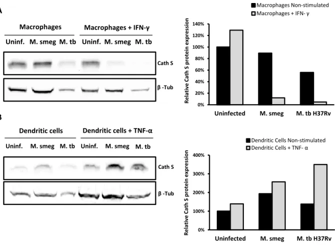
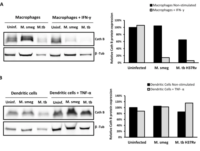
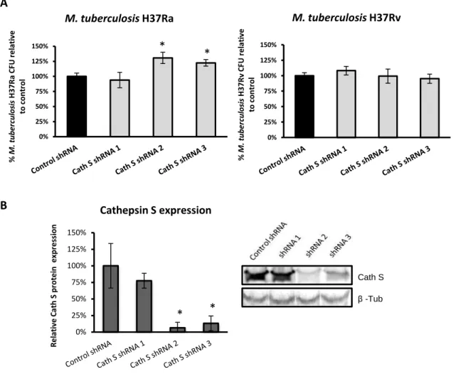
In the second part, we aimed to decipher the role of cathepsins during mycobateria infection of macrophages and DCs. Cathepsins protein levels were analyzed by western blot after infection with pathogenic or non-pathogenic mycobacteria. It was shown that cathepsin B and S levels during M. tuberculosis infection are distinct from those of host cells that internalized M. smegmatis and this is dependent on the host cell species tested. In addition, the role of these proteases in M. tuberculosis intracellular survival was evaluated by silencing cathepsins expression, using shRNA lentiviral vectors. It was observed, that cathepsin B and S knockdowns led to an increase in M. tuberculosis H37Ra intracellular survival. Altogether, these evidences point for a role of both cathepsins in the control of M. tuberculosis intracellular growth and demonstrate that M. tuberculosis modulates these cathepsins differently of the non-pathogenic M. smegmatis.
Keywords
Mycobacterium tuberculosis, Mycobacterium smegmatis, macrophages, dendritic cells, cathepsins, antigen presentation.
v
Resumo
A tuberculose representa um dos mais graves problemas de saúde pública, causando anualmente cerca de dois milhões de mortes em todo o mundo. Estima-se que cerca de um terço da população mundial se encontra infectada com Mycobacterium tuberculosis, o microrganismo patogénico causador desta doença infecciosa. A emergência de estirpes de M. tuberculosis multirresistentes e extensivamente resistentes à terapêutica antibiótica utilizada, aliado ao facto de a única vacina disponível possuir baixa eficácia contra a tuberculose pulmonar, vem realçar a necessidade urgente de se encontrar novos alvos terapêuticos e novas estratégias para controlar a doença.
O sucesso deste patogénio reside, principalmente, na sua capacidade de sobreviver e de se dividir no interior de células imunitárias. Após ser internalizado pelos macrófagos, M. tuberculosis inibe a fusão do fagossoma com o lisossoma, escapando assim à acção das enzimas proteolíticas (catepsinas) e ao ambiente acídico do fagolisossoma. Em paralelo, os macrófagos infectados induzem uma resposta pró-inflamatória, que promove o recrutamento de células imunitárias da corrente sanguínea para o local da infecção. Forma-se o granuloma, a estrutura característica da tuberculose, que se pensa ser capaz de conter a infecção micobacteriana. Aqui, algumas micobactérias podem permanecer num estado de latência durante um longo período de tempo. Apenas 5 a 10% da população infectada desenvolve a doença activa, o que na maioria dos casos se deve a uma imunodepressão do hospedeiro.
Nos pulmões, M. tuberculosis é internalizado por macrófagos alveolares residentes, mas também por células dendríticas recrutadas da corrente sanguínea. Estes dois tipos de células apresentadoras de antigénios desempenham papéis distintos na imunidade contra a tuberculose. Os macrófagos ao induzirem os seus mecanismos bactericidas são os principais responsáveis pela destruição das micobactérias. Por outro lado, as células dendríticas são especializadas na apresentação de antigénios micobacterianos às células T naïve, que se encontram nos nódulos linfáticos. De modo a conseguirem desempenhar estas funções, é necessário que ocorra um processo de activação dos macrófagos assim como um processo de maturação das células dendríticas. Estudos anteriores mostram que as micobactérias têm a capacidade de interferir com estes processos.
As catepsinas são proteases que estão envolvidas num grande número de processos celulares em macrófagos e células dendríticas, tais como: destruição dos patogénios, processamento e apresentação de antigénios, processamento de várias enzimas celulares do hospedeiro e também apoptose. O seu vasto número de funções, e em particular o seu papel na destruição dos patogénios e na apresentação de antigénios, faz destas proteases alvos perfeitos para manipulação por micobactérias durante a infecção de macrófagos e células dendríticas.
Na primeira parte desta tese, é explorado o papel das micobactérias durante a activação dos macrófagos e a maturação das células dendríticas. Foram infectados macrófagos e células dendríticas, diferenciados a partir de monócitos isolados do sangue periférico de dadores humanos
vi
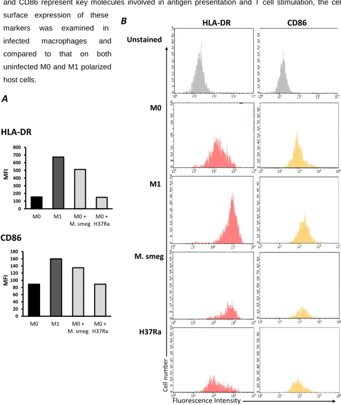
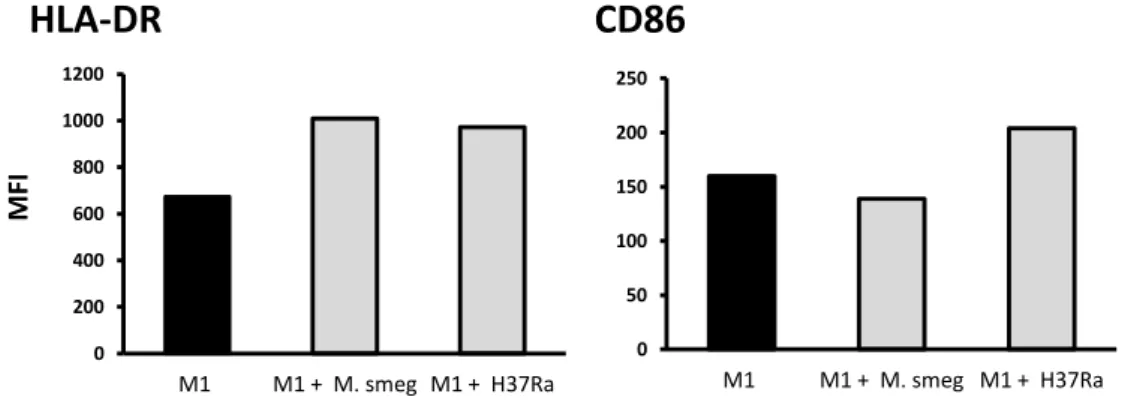
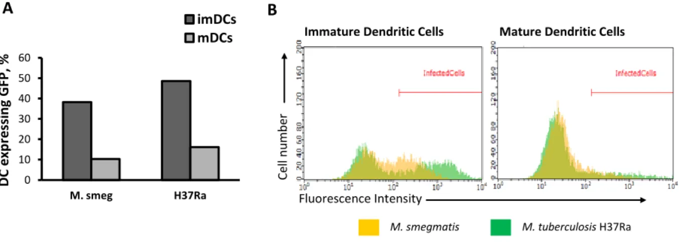
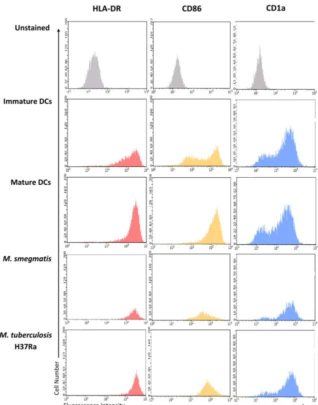
saudáveis, com micobactérias que expressavam a proteína verde fluorescente (GFP), nomeadamente a estirpe avirulenta M. tuberculosis H37Ra GFP e a espécie não patogénica M. smegmatis. Através de citometria de fluxo, foram seleccionadas apenas as células infectadas com micobactérias fluorescentes e o efeito da infecção foi analisado pela medição da expressão de marcadores superficiais de activação e maturação. Ao infectar macrófagos não activados (M0) com M. tuberculosis H37Ra, verificou-se que a infecção impede a indução da clássica resposta pró-inflamatória M1, ao nível das moléculas apresentadoras de antigénios. Pelo contrário, a infecção de macrófagos M0 com M. smegmatis tem um efeito estimulatório, resultando num fenótipo idêntico ao induzido pela estimulação destas células com interferão-gama. Tendo em conta que o número de células infectadas com M. tuberculosis H37Ra GFP e com M. smegmatis GFP é semelhante, podemos concluir que as diferenças observadas ao nível dos marcadores de activação resultam, efectivamente, de diferenças de virulência entre ambas as espécies. Por outro lado, a infecção de macrófagos previamente estimulados com interferão-gama (M1), com M. tuberculosis H37Ra, não inibiu a activação destas células, pelo menos ao nível das moléculas apresentadoras de antigénios. Isto sugere que, com uma baixa multiplicidade de infecção, a activação induzida pelo interferão-gama consegue superar os mecanismos inibidores induzidos pelo M. tuberculosis H37Ra, ao nível das moléculas apresentadoras de antigénios.Relativamente às células dendríticas, verificou-se que a infecção com M. tuberculosis H37Ra induz a maturação das células dendríticas imaturas, uma vez que se observou um aumento da expressão das moléculas apresentadoras de antigénios nas células infectadas. Foi também observado um aumento da expressão destes marcadores de maturação, após a infecção com M. smegmatis. No entanto, observou-se uma maior indução da maturação, pela estirpe avirulenta M. tuberculosis H37Ra, do que pela espécie saprófita M. smegmatis. As diferenças na virulência entre estas duas espécies poderão justificar o facto da infecção com M. tuberculosis H37Ra ser muito mais estimulatória do que com M. smegmatis. Estas evidências levam-nos a especular que a maturação das células dendríticas, observada após a infecção com M. tuberculosis H37Ra, poderá representar uma forma da micobactéria tirar partido das características migratórias das células dendríticas maturas e assim se disseminar pelo hospedeiro. Outra possível explicação seria que a indução da maturação pela infecção constitui um mecanismo de defesa do hospedeiro, na medida em que as células dendríticas infectadas representam fortes estimuladores das células T CD4+.
Concluindo, verifica-se que a infecção com a estirpe avirulenta M. tuberculosis H37Ra induz diferentes fenótipos, de acordo com o tipo de célula hospedeira infectada. Isto provavelmente está relacionado com as diferentes funções dos macrófagos e das células dendríticas na resposta imune contra a tuberculose. Em macrófagos humanos, esta estirpe avirulenta impede a expressão do fenótipo M1, ao nível das moléculas apresentadoras de antigénios, o que indica que provavelmente induz um fenótipo anti-inflamatório. Nas células dendríticas, a indução da maturação após a infecção com M. tuberculosis H37Ra pode reflectir um modo de disseminação utilizado pela micobactéria, ou um mecanismo de defesa do hospedeiro que lhe permita iniciar uma resposta imune celular específica. Para além disto, a infecção M. tuberculosis H37Ra induziu fenótipos distintos nas células imunes, comparativamente a M. smegmatis.
vii
Na segunda parte desta tese, foi abordado o papel das catepsinas durante a infecção micobacteriana de macrófagos e células dendríticas. Focámos o nosso estudo nas catepsinas S e B, as quais estão envolvidas na apresentação de antigénios via moléculas MHC classe II. A expressão destas catepsinas foi avaliada ao nível das proteínas, em macrófagos e células dendríticas infectados com a estirpe virulenta M. tuberculosis H37Rv ou com a espécie não patogénica M. smegmatis, de forma a analisar se a infecção por micobactérias modula a expressão destas proteases. Demonstrou-se que, em macrófagos M0, a infecção com M. tuberculosis H37Rv diminui a expressão das catepsinas S e B, enquanto que com M. smegmatis não provoca qualquer alteração nestas proteases.Por outro lado, a infecção micobacteriana afectou de forma diferencial a expressão das catepsinas S e B em células dendríticas e macrófagos humanos. Verificou-se que a infecção, tanto com M. tuberculosis H37Rv como com M. smegmatis, parece não interferir com os níveis de catepsina B nas células dendríticas. Relativamente à catepsina S, observou-se um aumento dos seus níveis após a infecção, tanto com M. tuberculosis H37Rv como com M. smegmatis. A infecção de células dendríticas imaturas com a estirpe virulenta resultou num aumento mínimo dos níveis de catepsina S, enquanto que a infecção de células dendríticas maturas resultou num grande aumento dos níveis desta protease. Apesar de não termos uma clara explicação para estes resultados, especulamos que, nas células dendríticas imaturas, M. tuberculosis H37Rv impede o aumento dos níveis de catepsina S, interferindo assim com apresentação de antigénios. Apesar de estas células após a infecção adquirirem um fenótipo de maturas, como anteriormente descrito, as moléculas apresentadoras de antigénios que estas expressam, encontram-se provavelmente num estado imaturo. Por outro lado, as células dendríticas maturas parecem ser capazes de superar este efeito inibitório e por isso se observa um grande aumento dos níveis desta protease.
Em paralelo, fomos também explorar o papel das catepsinas S e B na sobrevivência intracelular de M. tuberculosis em macrófagos. Para isso, monócitos humanos THP-1 foram silenciados para estas catepsinas, através do uso de vectores lentivirais a expressar short hairpin RNAs. Posteriormente, estas células foram diferenciadas em macrófagos e estes foram infectados com a estirpe virulenta M. tuberculosis H37Rv ou com a estirpe avirulenta M. tuberculosis H37Ra. Ao analisar os perfis de sobrevivência, verificou-se que o silenciamento da catepsina S aumenta significativamente a sobrevivência intracelular de M. tuberculosis H37Ra, enquanto que parece não interferir com a sobrevivência intracelular de M. tuberculosis H37Rv. Tendo em conta que a estirpe virulenta parece ter a capacidade de manipular a catepsina S em macrófagos humanos, é provável que o seu silenciamento mimetize esta modulação e, como tal, não se observem alterações na sua sobrevivência relativamente ao controlo sem silenciamento. A catepsina S parece ser relevante para o controlo do crescimento intracelular de M. tuberculosis H37Ra, mas enquanto a estirpe virulenta M. tuberculosis H37Rv parece desenvolver um mecanismo para evitar o efeito desta protease, a estirpe avirulenta M. tuberculosis H37Ra, parece não desenvolver o mesmo mecanismo de manipulação ou então desenvolve mas é menos efectivo. Foi também demonstrado que o silenciamento da catepsina B em macrófagos aumenta significativamente a sobrevivência intracelular de M. tuberculosis H37Ra e de M. tuberculosis H37Rv. Isto evidencia um papel desta protease no controlo da sobrevivência
viii
intracelular de ambas as estirpes em macrófagos.Concluindo, verificou-se que os níveis de catepsina S e B, durante a infecção com M. tuberculosis H37Rv, são diferentes dos observados durante a infecção com M. smegmatis. Para além disso, também se observou que os níveis de catepsina S e B diferem consoante a espécie das células imunitárias infectadas, o que poderá estar relacionado com diferentes cinéticas de acidificação e de actividade proteolítica existentes entre elas. Para além disto, os resultados mostram um papel de ambas as catepsinas no controlo do crescimento intracelular de M. tuberculosis H37Ra e M. tuberculosis H37Rv em macrófagos. Porém, a estirpe virulenta M. tuberculosis H37Rv parece ser capaz de subverter os efeitos bactericidas da catepsina S, ao diminuir os seus níveis proteicos. No entanto, a estirpe avirulenta M. tuberculosis H37Ra parece ser mais susceptível aos efeitos de ambas as proteases.
Palavras-chave
Mycobacterium tuberculosis, Mycobacterium smegmatis, macrófagos, células dendríticas, catepsinas, apresentação de antigénios.
1
1. Introduction
1.1.
Tuberculosis
Tuberculosis, one of the oldest recorded human diseases, is considered one of the leading causes of infectious disease mortality worldwide (Daniel, 2006; WHO, 2012). During the first half of the XIX century, this disease was the leading cause of death in Europe, as a result of the population explosion and the growth of large urban centers. The only treatment then, which consisted of fresh air, a good healthy diet and rest, was available in the so called sanatoriums (Harries et al., 2006; Smith, 2003). The first microbiological advances in this disease were made when the German physician Robert Koch first identified the tubercle bacillus, in 1882 (Barry III and Cheung, 2009). During the XX century, the incidence of tuberculosis was greatly reduced in the developed countries, due to the improvement of public health practices, the widespread use of the live attenuated M. bovis BCG (Bacillus Calmette-Guérin) vaccine, as well as the discovery of antibiotics, such as streptomycin, the first effective therapy for tuberculosis. By that time, many experts believed that tuberculosis had been nearly eradicated. However, in the mid-1980s the number of new cases increased with the deterioration of socio-economic conditions and the emergence of Human Immunodeficiency Virus (HIV), especially in southern and east Africa (Barry III and Cheung, 2009; Smith, 2003). This lead in 1993, to the World Health Organization to declare the disease as a global emergency (Harries et al., 2006).
Nowadays, tuberculosis still remains a major public-health problem with one-third of world’s population infected with Mycobacterium tuberculosis and 8.7 million new cases of tuberculosis reported, in 2011 (World Health Organization, 2012). Although tuberculosis still causes almost two million deaths annually worldwide, the disease particularly affects developing regions such as Africa, which has the highest proportion of tuberculosis cases co-infected with HIV (Corbett et al., 2003; Smith, 2003; World Health Organization, 2012). The currently available BCG vaccine, which provides protection against the disease in childhood, has proven to be ineffective against the most frequent outcome of tuberculosis, the lung infection in adults (Soualhine et al., 2007). This situation became even more alarming with the emergence of multidrug and extremely drug-resistant strains of M. tuberculosis worldwide (Nguyen and Pieters, 2009). Therefore, new strategies for the prevention and treatment of tuberculosis are urgently needed. A better understanding of the mechanisms of interaction between M. tuberculosis and the host immune system is crucial to define novel therapeutic targets and strategies to control the disease.
1.2.
Mycobacteria
The genus Mycobacterium comprises rod-shape bacteria characterized by a very complex lipid-rich cell envelope that differs substantially from the typical cell wall structure of gram-positive and also gram-negative bacteria. The cell envelope contains, in addition to the cell membrane and
2
peptidoglycan layers, a large hydrophobic layer composed by mycolic acids associated to a vast array of other lipids and glycolipids (Forrellad et al., 2013; Glickman et al., 2001; Russell, 2001). This unique envelope confers intrinsic resistance to antibiotics and dehydration being also responsible for the acid-fast staining property used to identify mycobacteria, in Zhiel-Neelsen acid-acid-fast stain (Forrellad et al. 2013; Scherr and Nguyen, 2009). The majority of the species that compose the genus are non-pathogenic environmental bacteria, such as Mycobacterium smegmatis, which presents similarities to soil bacteria from the genus Streptomyces (Cosma et al., 2003; Scherr and Nguyen, 2009). However, a few species are highly successful pathogens including Mycobacterium leprae, Mycobacterium ulcerans and those present in the Mycobacterium tuberculosis complex.The Mycobacterium tuberculosis complex consists in a group of genetically closely related species namely Mycobacterium tuberculosis, Mycobacterium africanum, Mycobacterium bovis, Mycobacterium microti, Mycobacterium canetti, Mycobacterium caprae and Mycobacterium pinnipedii (van Soolingen et al., 1997; Aranaz et al., 1999; Aranaz et al., 2003; Cosma et al., 2003; Cousins et al., 2003), which are the etiologic agents of tuberculosis in humans and other animals. Although, all the members of the complex are facultative intracellular pathogens that cause tuberculosis, they diverge in terms of their phenotypic properties, host tropisms and virulence (Ahmad, 2011). The most well known member is Mycobacterium tuberculosis, a slow-growing and obligate aerobe bacterium, which the only known natural host is the human species (Cosma et al., 2003; Pieters, 2001).
1.3.
Pathogenesis of Tuberculosis
Tuberculosis is usually a lung infection but may affect almost any organ of the body. This infection is caused by inhalation of aerosols containing the infectious bacilli, generated from the cough of an infected individual which has developed the active respiratory disease (Mortellaro et al. 2009). In the lung, M. tuberculosis is internalized by resident alveolar macrophages but also by dendritic cells and monocytes recruited from the bloodstream (Bhatt and Salgame, 2007). Once inside macrophages, M. tuberculosis subverts the major killing mechanisms employed by these host cells (Hestvik et al., 2005; Smith, 2003). This pathogen prevents fusion of phagosomes with lysosomes (Armstrong and Hart, 1971) and restricts its phagosome acidification to a mild pH (Sturgill-Koszycki et al., 1994), escaping from the degrading forces of proteolytic enzymes and from an acidic environment inside the phagolysosome. Therefore, the bacilli have the capacity to avoid immediate destruction, which allows them to establish a niche inside the macrophage (Houben et al., 2006).
Simultaneously, the M. tuberculosis infected macrophages induce a localized pro-inflammatory response, through the release of cytokines and chemokines, which promotes the recruitment of additional macrophages and other immune cells from the neighboring blood vessels to the infection site. These cells are the building blocks for the granuloma, the hallmark structure of tuberculosis (Dietrich and Doherty, 2009; Russell, 2007). The granuloma is constituted by an organized cluster of immune cells composed by infected macrophages in the center, surrounded by additional macrophages and lymphocytes, which delineate the periphery of the structure in association with a fibrous extracellular matrix. Dentritic cells, neutrophils and natural killer cells may also populate
3
this structure (Cosma et al., 2003; Russell, 2007). Although the precise function of the granuloma is not completely understood, it is believed that the formation of this structure contributes to the physical containment of the mycobacterial infection, limiting the pathogen dissemination to the rest of the lung tissue (Ahmad, 2011; Forrellad et al., 2012). However, the pathogen is not completely eradicated in some individuals since some M. tuberculosis bacilli may remain dormant for a long period of time, during which there are no disease symptoms and the individual does not transmit the infection to others. In this stage the infection is referred to as latent tuberculosis (Ahmad, 2011; Forrellad et al., 2012; Smith, 2003). Thus, only 5-10% of the infected population will develop the active disease in their lifetime (Mortellaro et al., 2009). When the immune system of a latently infected individual becomes weakened as a consequence of malnutrition, old age or HIV co-infection, the dormant bacilli are able to reactivate and replicate into the lung and other tissues (Ahmad, 2011). Under this situation, the granuloma center becomes necrotic, undergoes caseation, resulting in the destruction of the surrounding host tissue (Houben et al., 2006). This leads to the rupture of the granuloma walls and to the release of infectious bacilli into the airways (Dietrich et al., 2009; Russell, 2007).1.4.
Interaction of mycobacteria with antigen presenting cells
The interaction of mycobacteria with antigen presenting cells (APCs) is a major feature in the pathogenesis of tuberculosis. Both macrophages and dendritic cells play a central role in inducing the immune response against M. tuberculosis infection. Macrophages act as key effector cells in mycobacterial killing and dendritic cells in mycobacterial antigen presentation and consequent stimulation of naïve T cells. Therefore, both cell types are perfect targets for mycobacterial induced manipulation (Hope et al., 2004; Mortellaro et al., 2009).
1.4.1. Macrophages as the main reservoir for mycobacteria
Macrophages constitute the first line of cellular defense against microbial invasion and are known to be the main reservoir of infection by M. tuberculosis. Upon reaching the local of infection, the major role of macrophages is the rapid elimination of the invading microorganisms (Hestvik et al., 2005; Russell, 2001). A variety of cell surface receptors promote M. tuberculosis phagocytosis, these include among others, complement receptors, the mannose receptor (MR) and Fc receptors (Pieters, 2008). Following uptake, M. tuberculosis and other pathogens are retained within a phagocytic vacuole called the phagosome. If the normal phagosome maturation cycle occurs, a series of sequential fusion events with vesicles from the endocytic pathway occur, the phagosome fuses with lysosome and the pathogen encounters a hostile environment that includes acidic pH levels, reactive oxygen (ROI) and nitrogen intermediates (RNI) and lysosomal proteases, such as cathepsins. Together, these mechanisms will promote the pathogen destruction as well as the processing and presentation of antigens to T cells, in the context of major histocompatibility complex (MHC) molecules (Hope et al., 2004; Poirier and Av-Gay, 2012).
4
Contrasting with this scenario, pathogenic mycobacteria have developed strategies to avoid these killing mechanisms (Hestvik et al., 2005; Smith, 2003). Armstrong and Hart (1971) showed that after being phagocytosed, M. tuberculosis arrests normal maturation of its phagosome and thus prevents its fusion with pre-formed lysosomes, avoiding direct exposure of the bacilli to the degrading force of lysosomal hydrolases. Moreover, phagosomes containing M. tuberculosis do not undergo further acidification, presenting a mild pH of 6.2- 6.3, due to the exclusion of vacuolar proton-ATPase from the bacilli-containing phagosome membrane (Sturgill-Koszycki et al., 1994; Tailleux et al., 2003). These mechanisms not only allow the bacilli to evade proteolytic degradation but also contribute to keeping its antigens from being processed and loaded onto MHC class II molecules for antigen presentation (Baena and Porcelli, 2009). In addition to interfering with phagosome maturation, M. tuberculosis also manipulates intracellular trafficking in macrophages, retaining access to the recycling endosome system, in order to obtain the required nutrients for the bacilli intracellular growth (Clemens, 1996; Mortellaro et al., 2009).Macrophage activation and its consequences during M. tuberculosis infection
In response to pathogen invasion and inflammation mediators, resting macrophages (M0) undergo different programs of activation (polarization). Activated macrophages have been generally classified into two groups: classical (M1) and alternative (M2) activated macrophages (Benoit et al., 2008). M1 macrophages result from the stimulation with type-1 cytokines (Interferon gamma - IFN-γ) and microbial products. These cells are inflammatory and their microbicidal activity is enhanced (Lugo-Villarino et al., 2011). In contrast, M2 macrophages are poorly microbicidal and play a critical role in the resolution of inflammation by producing anti-inflammatory mediators. M2 activation program is driven by the stimulation of resting macrophages with type-2 cytokines, interleukin 4 (IL-4) and interleukin 13 (IL-13). In general, polarization of M1 macrophages is part of the common host immune response against bacterial infection (Andrade et al., 2012; Benoit et al., 2008).
In resting macrophages, pathogenic mycobacteria in order to survive and replicate, inhibit phagosome-lysosome fusion. However, IFN-γ activated macrophages (M1) are able to overcome the blockage of phagosome maturation from infected cells. Furthermore, IFN-γ activated macrophage response is characterized by the secretion of large amounts of pro-inflammatory cytokines and chemokines, high production of ROI and RNI, and enhanced phagocytosis. It also enhances the expression of MHC class II and costimulatory molecules such as CD80 and CD86 for antigen presentation to T cells (Herbst et al., 2011; Lugo-Villarino et al., 2011; Poirier and Av-Gay, 2012).
Antigen presentation pathway and their modulation by M. tuberculosis
Once activated by IFN-γ, macrophages have the capacity to present mycobacterial antigens to T cells. However, unlike dendritic cells which present mycobacterial antigens to naïve T cells in lymph nodes, macrophages present antigens to effector T cells at sites of infection (Baena and Porcelli, 2009).
5
Antigen presentation mediated by MHC molecules is a complex process that involves distinctive pathways. MHC class I molecules are expressed on all nucleated cells, presenting antigens to antigen-specific CD8+ T cells. This pathway is specialized to present endogenous antigens, which are processed in the cytosol via proteasome. In contrast, MHC class II molecules expression is restricted to the professional APCs, including macrophages, dendritic cells and B cells. This pathway allows the presentation of exogenous antigens, such as mycobacterial antigens, that are processed in the endosomal route and presented to CD4+ T cells (Crevel et al., 2002; Hsing and Rudensky, 2005). Moreover, there is another antigen presentation pathway involved. Nonclassical MHC-like molecules, such as CD1, present among others lipids, mycobacterial lipoproteins to CD1-restricted T cells, and are expressed on both macrophages and dendritic cells (Crevel et al., 2002). Besides this, dendritic cells and macrophages are also capable of performing cross-presentation. This pathway enables presentation of peptides derived from exogenous antigens on MHC class I molecules and consequent stimulation of CD8+ T-cells (Rock and Shen, 2005).CD4+ T cell activation is essential for control of mycobacterial infection, therefore interfering with MHC class II pathway for antigen presentation in macrophages, might be a strategy employed by the bacterium to avoid destruction and to persist in host immune cells (Chapman, 2006; Koul et al., 2004). Indeed, several mechanisms have been described by which M. tuberculosis can affect antigen presentation, including downregulation of the expression of MHC class II and costimulatory molecules, and sequestering mycobacterial antigens from molecules required for CD4+ T-cell activation (Koul et al., 2004; Noss et al., 2000; Russell, 2001). Components of mycobacterial cell wall, such as the 19-kDa lipoprotein, down-regulate surface expression of MHC class II molecules, reducing the antigen presentation capacity of the infected macrophages (Noss et al., 2001). Furthermore, mycobacterial species were found to interfere with macrophage IFN-γ signaling in order to inhibit the synthesis of MHC class II molecules (Noss et al., 2000).
1.4.2. Dendritic cells as important targets during mycobacteria infection
Dendritic cells (DCs) are the most potent antigen presenting cells of immune system, given their unique ability to capture the pathogen at the infection site and to migrate to secondary lymphoid organs in order to present pathogen-derived antigens to naïve T-cells. Hence, they are likely to have a major role in initiation of the adaptive immunity against tuberculosis (Banchereau et al., 2000; Herrmann and Lagrange, 2005; Savina, 2007).
Dendritic cells are monocytic bone marrow derived cells, present in most peripheral tissues such as the skin, intestine and the lungs, where they act as sentinel cells, monitoring the contact of the body surfaces with incoming pathogens (Demangel and Britton, 2000; Hope et al., 2004). In these sites, they are considered as immature dendritic cells. They express low to moderate levels of surface MHC class II and co-stimulatory molecules such as CD40, CD80, CD86, while having a high capability to phagocyte invading microorganisms. Therefore, immature dendritic cells are characterized by a high ability for antigen uptake, but low T cell stimulatory activity (Hope et al., 2004; Martino, 2008). In the lungs, immature dendritic cells underline alveolar spaces and upon mycobacterial encounter, these
6
cells effectively uptake the M. tuberculosis bacilli (Hanekom et al., 2003; Mortellaro et al., 2009). Upon interaction with pathogens, tissue injury or exposure to inflammatory mediators, these efficient antigen capturing cells undergo phenotypic and functional changes, a process termed maturation, that occur while they migrate into secondary lymphoid organs such as lymph nodes. During this maturation process, dendritic cells lose the capability to capture antigens, but acquire an increased ability to present antigens effectively, expressing high levels of antigen presenting molecules in their surface, such as MHC class II and CD1, and high levels of co-stimulatory molecules as CD40, CD80 and CD86 (Banchereau et al., 2000; Martino, 2008). In lymph nodes, mature dendritic cells present processed antigens in association with MHC class II molecules to naïve CD4+ T lymphocytes, inducing their activation and differentiation into effector T cells (Figure 1).Depending on the type of pathogen that is recognized by the dendritic cells and on the cytokine environment, CD4+ T cells differentiate into T helper 1 (Th1) cells which secret IFN-γ, inducing the killing of intracellular pathogens or T helper 2 (Th2) cells characterized by the production of interleukin 4 (IL-4), effective against extracellular pathogens (Koul et al., 2004). During M. tuberculosis infection, the Type 1 cytokines IL-12, IL-1 and IL-18 are secreted, polarizing the T cell response towards Th1 phenotype (Mortellaro et al., 2009). Thus, the effector Th1 cells, activated following antigen presentation by dendritic cells, migrate to the site of infection and produce cytokines such as IFN-γ in order to activate M. tuberculosis infected macrophages and consequently eliminate the bacilli (Bodnar et al., 2001; Poirier and Av-Gay, 2012).
Because dendritic cells play a crucial role in induction and regulation of a protective immunity against pathogens, modulation of dendritic cell generation and/or maturation may be a significant mechanism by which these infectious agents evade immune surveillance and disseminate within the host (Martino et al., 2004). Considerable in vitro evidence exists that dendritic cells can internalize pathogenic mycobacteria by phagocytosis (Henderson et al., 1997), although the outcome of this
Figure 1. Role of dendritic cells (DC) in the immune system. In peripheral tissues, immature DCs, act as
sentinel cells, monitoring these tissues against invading pathogens. Upon pathogen capture, immature DCs initiate their maturation and migration through the lymph into lymph nodes. Pathogen-derived antigens are presented by the mature DCs to naïve CD4+ T cells, inducing their activation and differentiation into effector T cells. This figure was adapted from Geijtenbeek et al. (2002).
7
interaction is still not completely understood. In particular, the ability of these pathogens to interfere with dendritic cell maturation remains controversial. Henderson et al. (1997) found that human monocyte-derived dendritic cells uptake M. tuberculosis efficiently, promoting the up-regulation of cell surface maturation markers in infected cells, including surface molecules involved in antigen presentation and interaction with T cells. This phenotype was consistent with activation of the dendritic cells, suggesting that infected dendritic cells produced cytokines that lead to maturation (Henderson et al., 1997). Furthermore, Thurnher and colleagues (1997) described the effects of BCG on cultured human blood dendritic cells. Infection with BCG resulted in the down-modulation of endocytosis and the up-regulation of surface maturation markers. The maturation of infected dendritic cells was at least in part induced by the secretion of tumor necrosis factor-alpha (TNF-α) by these cells in response to BCG infection. In contrast to these findings, suggesting that dendritic cell maturation is induced by mycobacteria infection, other workers have reported that M. tuberculosis inhibits maturation of human monocyte-derived dentritic cells in vitro. Infected dendritic cells presented a minimal and reversible upregulation of cellular surface maturation markers and were compromised in their ability to activate naïve T cells (Hanekom et al., 2003). Altogether, these indicated that infection with mycobacteria might have diverse phenotypic effects on immature dendritic cells, in vitro.1.5.
Mycobacterial manipulation of host cathepsins
Cathepsins are a larger family of proteases composed mostly by cysteine proteases, with the exception of cathepsins D and E which are aspartic proteases and cathepsin A and G which are serine proteases (Beers et al., 2003; Zavasnik-Bergant and Turk, 2006). Their classification is based on their structure and catalytic type (Reiser et al., 2010). In general, the cysteine cathepsins are stable in acidic cellular compartments as lysosomes and endosomes (Reiser et al., 2010). All cathepsins are synthesized as inactive proenzymes that require activaction by proteolytic cleavage (Zavasnik-Bergant and Turk, 2006). The major regulators of cathepsin activity are cystatins, endogenous protein inhibitors, which bind to the target enzyme, obstructing the active site and thereby preventing substrate hydrolysis (Hsing and Rudensky, 2005).
Cathepsins are involved in a number of important cellular processes in APCs such as pathogen destruction, processing of several host cell enzymes, antigen presentation and apoptosis. Considering antigen presentation, these proteases are implicated in the generation of mature MHC class II molecules and in antigen processing within endolysosomal compartments, where they degrade pathogens internalized by APCs into antigenic peptides presentable in MHC class II molecules (Baena and Porcelli, 2009; Conus and Simon, 2010; Zavasnik-Bergant and Turk, 2006).
Newly synthesized MHC class II αβ heterodimers assemble in the endoplasmic reticulum (ER) and bind to a chaperone, the invariant chain (Ii), before leaving ER towards endocytic compartments. The invariant chain not only function as a chaperone, assuring the proper folding and assembly of MHC class II molecules, but also directs trafficking of these molecules to the endosomal compartments, which contain the antigen peptides for loading onto MHC class II complex (Hsing and Rudensky, 2005; Riese et al.,1996). During the translocation through the endocytic pathway, the
8
invariant chain undergoes a stepwise proteolytic degradation (Figure 3) mediated by cathepsins that generate various cleavage intermediates, such as Iip22 and Iip10 intermediates. The initial cleavages can be executed by asparagine endopeptidase (AEP) although other proteases can perform this cleavage in its absence (Trombetta and Mellman, 2005). The processing of Ii culminates in the generation of the class II-associated invariant chain peptide (CLIP) (Beers et al., 2003; Nepal et al., 2006). The CLIP peptide remains in the MHC class II peptide binding groove and prevents premature peptide loading. A second chaperone, human leukocyte antigen (HLA)-DM, will then catalyze the removal of CLIP, in exchange for the antigen peptides. The resulting peptide-loaded MHC class II complexes are then transported to the cell surface and prepared for presentation to CD4+ T-cells in combination with co-stimulatory molecules (Chapman, 2006; Hsing and Rudensky, 2005; Torres et al., 2006).Macrophages and dendritic cells are equipped with a diverse group of cathepsins, most of which are cysteine proteases. Cathepsin B, L and S are the major cysteine proteases expressed in these cells and have all been suggested to participate in the processing of internalized antigens for CD4+T cell presentation and/or processing of invariant chain (Hsieh et al., 2002; Nepal et al., 2006). Cathepsin S is the principal protease involved in the late steps of li degradation in macrophages, dendritic cells and B cells, specifically in the conversion of Iip10 into CLIP, while in cortical thymic epithelial cells this process is mediated by cathepsin L (Nepal et al., 2006). Moreover, both cathepsin S and L have also been shown to be important in the generation of certain antigenic peptides (Hsing and Rudensky, 2005; Plüger et al., 2002), as well as cathepsin B (Matsunaga et al., 1993).
The infection with M. tuberculosis elicits a MHC class II-restricted CD4+T cell response that is essential to control the primary mycobacterial infection. Therefore, the integrity of the class II MHC
Figure 2. Invariant chain degradation events. The invariant chain (Ii chain) undergoes successive
cleavages, during the translocation of Ii chain-MHC class II complex through the endocytic pathway of antigen presenting cells. This proteolytic degradation is initiated by asparagine endopeptidase (AEP) or other unidentified proteases, originating the li chain intermediate p10. Further cleavage (involving cathepsin S or L, depending on the cell types) results in the class II-associated invariant chain peptide (CLIP). Subsequently, a second chaperone, human leukocyte antigen (HLA)-DM, catalyzes the removal of CLIP in exchange for the antigen peptides. This figure was adapted from Trombetta and Mellman ( 2005).
9
antigen presentation pathway is necessary to ensure that the bacilli are contained and that adaptive immunity is established. Since, cathepsins play an important role in MHC class II antigen processing and presentation, pathogenic mycobacteria modulate MHC class II pathway by interfering with these host proteases (Nepal et al., 2006). In this context, in vitro experiments showed that M. bovis BCG infection causes inhibition of IFN-γ induced cathepsin S expression, in infected human acute monocytic leukemia cell line (THP-1) cells (Sendide et al., 2005). This was associated with an intracellular accumulation of MHC class II molecules complexed with cathepsin S lip10 substrate and with the export of immature MHC class II molecules to the cell surface. This effect was reversed by the addition of neutralizing antibodies that suppress IL-10, restoring the expression of active cathepsin S and the export of mature MHC class II molecules to the surface of infected cells. Thus, M. bovis BCG inhibits cathepsin S expression through the induction of the inhibitory cytokine IL-10 (Sendide et al., 2005). In addition, the inhibition of expression or activity of other cathepsins, such as cathepsin L, in murine bone marrow-derived macrophages infected with either M. avium or M. tuberculosis, was also reported. In contrast, the cathepsin B and S activity has no obvious alteration upon mycobacterial infection. The inhibition of expression of this cathepsin may influence the types of T cell epitopes generated in antigen presenting cells (Nepal et al., 2006).1.6.
Thesis Goals
The major goal of this project is to characterize mycobacterial manipulation of host macrophage activation and dendritic cell maturation, in the context of host proteases such as lysosomal cathepsins. The study of these mycobacteria-host interactions may provide us the knowledge to develop new strategies for the prevention and treatment of tuberculosis.
In the first part of this thesis we intent to elucidate the role of mycobacteria during macrophage activation and dendritic cell maturation. For this purpose, human macrophages and DCs were infected with the avirulent M. tuberculosis H37Ra or the non-pathogenic M. smegmatis expressing green fluorescent protein (GFP) and their surface phenotype was analyzed using flow cytometry. We focused our analysis in the expression of surface activation or maturation markers, involved in antigen presentation and T cell stimulation. We also studied the influence that these mycobacteria species had on macrophage activation and DCs maturation, previously induced by IFN-γ and TNF-α, respectively.
The second part of this thesis, addresses the role of lysosomal cathepsins during mycobacteria infection of host macrophages and DCs. First, we evaluated whether mycobacteria infection modulates lysosomal cathepsins expression, by analyzing their protein levels in human macrophages and dendritic cells infected with the virulent M. tuberculosis H37Rv or the non-pathogenic M. smegmatis. Second, we investigated the importance of these cathepsins on M. tuberculosis intracellular survival within macrophages, by silencing cathepsins expression in THP-1 cells, using a shRNA lentiviral library, followed by infection with M. tuberculosis H37Rv or M. tuberculosis H37Ra strain and consequent quantification of intracellular survival through colony forming units counting.
10
2. Materials and Methods
2.1.
Role of mycobacteria during macrophage activation and DC maturation
2.1.1. Bacterial cultures and growth conditions
M. tuberculosis H37Ra (25177, ATCC) expressing green fluorescent protein (GFP) (M. tuberculosis H37Ra GFP) was generated by electroporation with pMN437 plasmid (Addgene plasmid # 32362), which harbors a gene that encodes GFP protein (mycgfp2+) and an hygromycin resistance gene for selection (a kind gift from Michael Niederweis). This green fluorescent protein (GFP)-expressing strain was cultivated in complete growth medium containing Middlebrook’s 7H9 broth (Difco) supplemented with 0.2% (v/v) glycerol (Sigma), 10% (v/v) OADC (BD- Becton, Dickinson and Company) and 0.05% (v/v) Tyloxapol (Sigma). The medium was also supplemented with 50μg/ml of hygromycin (Invitrogen) to maintain the selective pressure.
M. smegmatis mc2155 (700084, ATCC) harbouring a p19-(long-lived) EGFP plasmid (M.
smegmatis GFP) was grown in medium containing Middlebrook’s 7H9 broth (Difco) and Nutrient broth
(Difco), supplemented with 0.5% glucose, 0.05% (v/v) Tyloxapol (Sigma) but also with 50μg/ml of hygromycin (Invitrogen) in order to stabilize GFP expression (Anes et al., 2006). Both cultures were incubated at 37°C with 100 rpm agitation.
Prior to infection, in order to obtain a single cell suspension, all mycobacterial cultures were centrifuged at 3000 x g for 10 minutes, washed with PBS (Phosphate buffer saline) and centrifuged a second time in the same conditions. Then, the bacterial pellet was resuspended in the desired cell culture medium without antibiotics. To remove the clumps, the mycobacterial suspension was passed through a 21G needle and then sonicated for 5 minutes. Residual aggregates were eliminated by a low speed centrifugation (350 x g) for 1 minute. Single cell suspension was verified by light microscopy. Finally, optical density at 600 nm (OD600nm) was used to determine cellular density in the
mycobacterial suspension, assuming that OD600 nm = 0.1 is equivalent to 1x10 7
mycobacteria/ml.
Construction of Mycobacterium tuberculosis H37Ra GFP
o pMN437 plasmid purification – Transformation of E. coli with pMN437 plasmid
The chemically competent E. coli strain was placed on ice, in contact for 10 minutes with 100 ng of pMN437 DNA. The mixture was then incubated at 45 ºC for 45 seconds (heat-shock transformation), followed by another 10 minutes on ice. The bacteria cells were inoculated into SOC medium (Super Optical Broth; Sigma) and incubated at 37 ºC, under agitation for 1 hour. Following this, the cultures were plated in Luria-Bertani (LB) agar medium (Sigma) containing hygromycin and were incubated at 37 °C overnight (Hanahan and Harbor, 1983). As a confirmation of E. coli transformation with the pMN437 plasmid, colonies with GFP fluorescence were observed by fluorescence microscopy. The selected transformants were grown in LB broth with 50 µg/ml
11
hygromycin at 37 °C, under agitation, overnight. The pMN437 plasmid was purified using a PuroLink® Quick Plasmid Miniprep Kit (Invitrogen), according to the manufacturer’s instructions, and was quantified using NanoDrop ND 100 (ThermoCientific).o Electroporation of M. tuberculosis H37Ra with pMN437 plasmid
To prepare electrocompetent mycobacteria cells, M. tuberculosis H37Ra cultured as described above, was incubated on ice for 90 minutes and then centrifuged at 3000 x g for 10 minutes. Following this, mycobacteria cells were washed three times in ice-cold 10 % glycerol and before storage at -80 ºC, bacterial cells were also resuspended in ice-cold 10 % glycerol.
The pMN437 plasmid (2 µg DNA) was electroporated into electrocompetent M. tuberculosis H37Ra, applying one single pulse of 1.8 kV, 25 µF, with the pulse-controller resistance set at 200 Ω resistance, in 0.2 cm cuvettes. Electroporated mycobacteria were incubated in Middlebrook’s 7H9 broth (described above) at 37 ºC for 36 hours, to allow the expression of the antibiotic-resistant gene carried on the pMN437 plasmid. Subsequently, mycobacteria cells were plated on Middlebrook 7H10 agar medium (Difco) supplemented with 10 % (v/v) OADC (BD), 0.5 % (v/v) glycerol (Sigma) and 50 μg/ml of hygromycin, and incubated at 37 ºC. After 4 weeks, transformant colonies were isolated and then inoculated in the Middlebrook’s 7H9 broth (described above) with 50 μg/ml of hygromycin. Adapted from (Goude and Parish, 2008).
2.1.2. Peripheral Blood Mononuclear Cells: Isolation Procedure
Peripheral Blood Mononuclear Cells (PBMCs) were obtained from buffy coat preparations kindly provided by Instituto Português do Sangue. The mononuclear cells were isolated by density gradient centrifugation using Ficoll-PaqueTM Plus(GE Healthcare). Briefly, the buffy coat was diluted (1:1) in MACS Buffer, which was prepared by diluting MACS BSA (bovine serum albumin) Stock Solution 1:20 with autoMACSTM Rinsing Solution (both purchased from MACS, Miltenyi Biotec). Then, the diluted buffy coat was gently overlaid on Ficoll-PaqueTM Plus(GE Healthcare) and an 800 x g centrifugation for 20 minutes at room temperature was performed without brakes and with minimal acceleration. The resulting interface, which contains the PBMCs, was collected and washed two times with cold MACS Buffer, through a 450 x g centrifugation for 10 minutes at 4 ºC, followed by a second centrifugation at 350 x g for 5 minutes at 4º C. Subsequently, monocytes were positively selected from total PBMCs, using anti-CD14-labeled magnetic beads and MACS separation LS columns (MACS, Miltenyi Biotec), according to the manufacturer’s instructions. Depending on the following experiments, monocytes were plated at a desired density in the appropriate plates, in order to differentiate into macrophages and dendritic cells. Adapted from (Wang et al., 2010).
12
2.1.3. Human macrophage and dendritic cell differentiation
For macrophages differentiation, CD14 monocytes were resuspended in RPMI-1640 medium with GlutaMAXTM (Gibco) supplemented with 1 % HEPES (Gibco) at pH 7.4 and were allowed to attach to the plate for 2 hours at 37 ºC. Following this, cells were cultured with RPMI-1640 medium with GlutaMAXTM (Gibco) supplemented with 10 % (v/v) Heat-Inactivated Fetal Bovine Serum (iFBS) (Invitrogen), 1 % HEPES (Gibco) at pH 7.4, 1 % (v/v) sodium pyruvate (Gibco), 1 % (v/v) Penicillin-Streptomycin (Gibco), 0.1 % 2-Mercaptoethanol (Gibco) and 20 ng/ml M-CSF (macrophage-colony-stimulating factor) (Immunotools), for 7 days at 37 ºC in 5 % CO2 atmosphere. On day 4, fresh
medium with a full dose of M-CSF was added to the culture.
For dendritic cells differentiation, monocytes were resuspended in RPMI-1640 medium with GlutaMAXTM (Gibco) containing 10 % (v/v) iFBS (Invitrogen), 1 % HEPES (Gibco) at pH 7.4, 1 % (v/v) sodium pyruvate (Gibco), 1 % (v/v) Penicillin-Streptomycin (Gibco), 0.1 % 2-Mercaptoethanol (Gibco), 20 ng/ml IL-4 (Immunotools) and 10 ng/ml GM-CSF (granulocyte-macrophage colony-stimulating factor) (Immunotools), for 7 days at 37 ºC in 5 % CO2atmosphere. On day 2 and 4, new medium with
a full dose of IL-4 and GM-CSF was added to the culture.
To analyze cell surface markers expression of human monocyte-derived macrophages and dendritic cells, monocytes were plated in 12-well plates, at a density of 1x10⁶ cells per well.
2.1.4. Treatment with IFN-γ and TNF-α
When required, human monocyte-derived macrophages were stimulated with human IFN-γ (Immunotools) at 100 UI/ml and human monocytes-derived dendritic cells were stimulated with TNF-α (Immunotools) at 50 ng/ml, overnight, and prior to infection.
2.1.5. Mycobacteria infection of human macrophages and dendritic cells
For analysis of cell surface markers expression of mycobacteria infected human monocyte-derived macrophages and dendritic cells, monocytes were plated in 24-well plates, at a density of 5x10⁵ cells per well and following 7 days of differentiation, human macrophages and dendritic cells were stimulated as described above or were left unstimulated. Then, these cells were infected with single-cell suspensions of M. smegmatis GFP or M. tuberculosis H37Ra GFP (prepared as described above), at a multiplicity of infection (MOI) of 1 and were incubated with the macrophage or dendritic cell differentiation medium without antibiotics (infection medium), for 3 hours at 37 °C, to allow the uptake of mycobacteria. After 3 hours infection, in order to remove non-internalized extracellular mycobacteria, cells were washed with infection medium and cultivated for an additional 24 hours in infection medium supplemented with 10 μg/ml of gentamicin (Gibco). One day after infection, cells were analyzed by flow cytometry. Uninfected human macrophages and dendritic cells, non-stimulated and stimulated were used as controls.
13
2.1.6. Cell surface staining and flow cytometry
Uninfected or infected human macrophages and dendritic cells were washed with PBS, harvested by incubation with 5 mM EDTA (Ethylenediaminetetraacetic acid) (Gibco) in PBS for 10 minutes, followed by fixation with 4 % paraformaldehyde. Subsequently, cells were washed with MACS buffer (MACS, Miltenyi Biotec) and incubated for 30 minutes at room temperature with the appropriate fluorochrome-conjugated antibodies (Table 1). Cells were washed twice before cell surface markers expression was assessed, using Guava easyCyteTM HT flow cytometer (Millipore). Data analysis was performed using Guava InCyte software (Millipore) and results were expressed as the Mean Fluorescence Intensity (MFI) of each surface marker, percentage of positive cells for each surface marker or as the percentage of GFP-positive cells. A total of 5,000 events per sample were analyzed. Only viable cells were considered in the analysis.
2.2.
Role of host cathepsins during mycobacteria infection in human
macrophages and dendritic cells
2.2.1. Mycobacterial manipulation of cathepsins in human macrophages and DCs
2.2.1.1. Bacterial cultures and growth conditions
M. smegmatis was grown as described above but without hygromycin addition. M. tuberculosis H37Rv (25618, ATCC), was cultivated in complete growth medium containing Middlebrook’s 7H9 broth (Difco) supplemented with 0.2 % (v/v) glycerol (Sigma), 10 % (v/v) OADC (BD- Becton, Dickinson and Company) and 0.05 % (v/v) Tyloxapol (Sigma) and were incubated at 37 ºC with 100 r.p.m agitation. Before any experiment, the individualization of mycobacterial culture was performed as previously described.
2.2.1.2. Mycobacteria infection of human macrophages and dendritic cells
CD14 monocytes isolated from the peripheral blood (as described above) were seeded at a density of 2x10⁶ cells per well, in a 6-well plate and differentiated into human macrophages and dendritic cells with the appropriate cytokines, for 7 days, as described above. The human monocyte-derived macrophages and dendritic cells (non-stimulated and stimulated) were infected with single-cell suspensions of M. smegmatis or M. tuberculosis H37Rv at a MOI of 1 for 3 hours. Following 3 hours infection, cells were washed and cultivated for an additional day, in infection medium supplemented with 10 μg/ml of gentamicin to destroy extracellular bacteria. To analyze the cathepsins expression, the infected cells were lysed and the proteins collected 24 hours post-infection.
14
2.2.1.3. Western Blot
The adherent cells were scraped with Laemmli buffer 2x (Sigma) diluted in PBS (1:2) and the cells in suspension were centrifuged and ressuspended with the same buffer. The resulting cell lysates were denatured at 95 ºC for 5 minutes. The same volume of protein extracts were loaded on a 12 % SDS-PAGE gel (Sodium Dodecyl Sulfate PolyAcrylamide Gel Electrophoresis) and the protein separation was performed by electrophoresis at 200 V for 1 hour. Then proteins were transferred to a nitrocellulose membrane (0.45 µm pore size; Bio-Rad) at 30 V, 4 ºC, overnight, in a wet transfer system, Mini Trans-Blot® module (Bio-Rad). The membrane was blocked for 1 hour in Blocking Buffer, prepared with 5 % Bovine Serum Albumin (BSA; Merck) in TBS-T (Tris-buffered saline 1x, 0.1 % Tween20), and then incubated for 2 hours at room temperature with the appropriate primary antibodies (Table 2(a)) diluted in TBS-T with 1 % BSA (Merck). After washing the nitrocellulose membrane with TBS-T, incubation with the appropriate horseradish peroxidase-conjugated secondary antibody (Table 2(b)) diluted in TBS-T with 5 % BSA (Merck) was performed, for 1 hour at room temperature. The membrane was washed again, before enhanced chemiluminescence detection. Cathepsin bands were visualized using ECL Prime Western Blotting Detection Reagents (Amersham). Quantification of blot bands was performed using ImageJ 1.45 software (National Institutes of Health, USA). Anti-tubulin antibody was used as a normalization control.
2.2.2. The effect of host cathepsins on mycobacteria intracellular survival
2.2.2.1. Bacterial cultures and growth conditions
M. tuberculosis H37Ra and M. tuberculosis H37Rv were cultivated as described above but without hygromycin addition. Before any experiment, the individualization of mycobacterial culture was performed as previously described.
2.2.2.2. THP-1 cell line and growth conditions
Human acute monocytic leukemia cell line, THP-1 (TIB-202™, ATCC), was grown in RPMI-1640 medium with GlutaMAXTM (Gibco) supplemented with 10 % (v/v) iFBS (Invitrogen), 1 % (v/v) L-glutamine (Gibco), 1 % (v/v) HEPES (Gibco) at pH 7.4, 1 % (v/v) sodium pyruvate (Gibco), 1 % (v/v) MEM-non essential amino acids (Gibco) and 1 % (v/v) Penicillin-Streptomycin (Gibco), and incubated at 37 °C in 5 % CO2 atmosphere.
2.2.2.3. HEK 293T cell line and growth conditions
Human embryonic kidney 293T cell line, HEK 293T (CRL-11268™, ATCC), was maintained in Dulbecco’s Modified Eagle Medium (DMEM) (Gibco) supplemented with 10 % (v/v) iFBS (Invitrogen) and 1 % (v/v) Penicillin-Streptomycin (Gibco) and incubated at 37 °C in 5 % CO2 atmosphere.