MESTRADO INTEGRADO EM MEDICINA
Cerebrovascular Diseases and Cancer
Sofia de Castro Neves Padilha
M
INSTITUTO DE CIÊNCIAS BIOMÉDICAS ABEL SALAZAR, UNIVERSIDADE DO PORTO
MESTRADO INTEGRADO EM MEDICINA
CEREBROVASCULAR DISEASES AND CANCER
Junho, 2019
Estudante: Sofia de Castro Neves Padilha
Número de estudante: 201304859
Endereço de correio eletrónico: [email protected]
Orientador: Luís Filipe Oliveira da Maia
Professor Auxiliar Convidado do ICBAS
Assistente Hospitalar de Neurologia no CHUP
Co-Orientador: Rui Jorge Rocha Felgueiras
Assistente Hospitalar de Neurologia no CHUP
i
ACKNOWLEDGEMENTS
I would first like to thank my thesis advisor, Dr. Luís Maia, for the support and availability during my thesis development, for all the suggestions and advices to take this work to a higher level.
I would also like to thank my co-advisor, Dr. Rui Felgueiras, for his guidance in the collection of data and for his valuable comments on this thesis.
My sincere thanks also go to Professor Rui Magalhães for his patience, his assistance and for always being available even in busier days.
To Dr. Diogo Pereira and to the medical students that also made this thesis possible, for their persistence and hard-work.
And finally, a special thanks to my family, my boyfriend and my friends, that endured the best and the worst moments, always giving motivation and support until this thesis was complete.
ii
RESUMO
Introdução: O cancro é uma doença complexa que pode provocar eventos cerebrovasculares.
Foram já propostos vários mecanismos para explicar os acidentes vasculares cerebrais (AVC) de causa neoplásica, em particular a ocorrência de um estado de hipercoagulabilidade que favorece a trombose e a isquemia. É, portanto, plausível que mecanismos semelhantes possam provocar acidentes isquémicos transitórios (AITs). Nós pretendemos determinar a incidência de cancro após ataques neurológicos transitórios (ANTs) e avaliar se os AITs ou os ANTs não-isquémicos estariam associados com o desenvolvimento de cancro.
Métodos: Fizemos um estudo retrospetivo de todos os doentes com ANT incluídos no registo
prospetivo ACIN 2, que foram admitidos no Hospital de Santo António (HSA), Porto, Portugal. Os participantes do estudo foram classificados em ANT não-isquémico ou AIT e caraterizados de acordo com dados sociodemográficos e fatores de risco vascular na altura do evento. Recorrendo ao processo clínico eletrónico e a entrevistas telefónicas, fizemos uma pesquisa sistemática pelos seguintes desfechos num período de 7 anos após o evento índice: óbito e causa de óbito, diagnóstico de cancro, localização e estadiamento, AVC, AIT e ANT.
Resultados: Entre 2009 e 2011, 594 doentes foram diagnosticados com o primeiro ANT. Na coorte
final (510 doentes), 32% tiveram um evento índice AIT; 60.6% eram do sexo feminino e a idade média era de 63 anos. Foi encontrado cancro em 15.3%, e desses, 56.1% destes ocorreu após o evento índice. A incidência global de cancro (exceto pele) foi de 1064.4/100000/ano e de 1665.2/100000/ano nos doentes com AITs. O cancro foi mais prevalente nos doentes com AIT comparado com ANT não-isquémico (13.5% vs. 6.9%, p=0.016). O tipo mais frequente de cancro foi o gastrointestinal (28.3%). Foram encontradas metástases em 40.9% dos doentes com cancro diagnosticado após o AIT/ANT, mais frequentemente no grupo de AIT (p=0.028). A sobrevivência livre de cancro foi menor nos doentes com AIT do que ANT (log rank teste com p=0.010). Ainda assim, isto não foi confirmado na análise multivariada. AVC e AIT foram desfechos mais frequentes em doentes com AIT (25.8% vs. 3.7% para AVC; 13.5% vs.1.4% para AIT; p<0.001). A mortalidade também foi mais elevada no grupo com AIT (22.7% vs. 9.2%, p<0.001).
Discussão: A taxa de incidência global de cancro e no grupo de doentes com AIT foi superior à taxa
de incidência da população na região norte de Portugal. Ainda assim, a ocorrência de um AIT não foi um preditor independente de cancro. O baixo número de ocorrências de cancro pode ter limitado as nossas conclusões.
iii
ABSTRACT
Introduction: Cancer is a complex disease that can lead to cerebrovascular events. Several
mechanisms have been proposed to explain previously documented cancer-related strokes, in particular the occurrence of a hypercoagulable state that favours thrombosis and ischemia. Considering this, it is plausible that similar mechanisms could lead to transient ischemic attacks (TIAs). We aimed to determine the incidence of cancer after transient neurological attacks (TNAs) and evaluate if either TIAs or non-ischemic TNAs were associated with cancer.
Methods: We did a retrospective study of all the patients with TNA included in the ACIN 2
prospective registry, that were admitted to Hospital de Santo António (HSA), Porto, Portugal. Study participants were classified in non-ischemic TNA or TIA, characterized according to demographic data and vascular risk factors at the time of the event. Using e-medical records and telephone interviews, we did a systematic search for the following outcomes for a period of up to 7 years after the index event: death and cause of death, cancer diagnosis, location and staging, stroke, TIA and TNA.
Results: Between 2009 and 2011, 594 patients were diagnosed with the first TNA. In the final cohort
(510 patients), 32% of patients had a TIA index event; 60.6% were female and mean age was 63 years. Cancer was found in 15.3% and 56.1% of them occurred after the index event. Global cancer incidence (excluding skin cancer) was 1064.4/100000/year and 1665.2/100000/year in TIA group. Cancer was more prevalent in TIA patients compared to non-ischemic TNA (13.5% vs. 6.9%, p=0.016). The most common type of cancer was gastrointestinal (28.3%). Metastasis were found in 40.9% of patients with cancer diagnosed after the TIA/TNA, most frequently in TIA group (p=0.028). Cancer-free survival time was shorter in TIA patients than non-ischemic TNA (log rank test with p=0.010), still this was not confirmed in a multivariate analysis. Stroke and TIA were more common outcomes in TIA patients (25.8% vs. 3.7% for stroke; 13.5% vs.1.4% for TIA; p<0.001). Mortality was also higher in TIA group (22.7% vs. 9.2%, p<0.001).
Discussion: The global incidence rate of cancer and in TIA group was superior to the incidence rate
of the population of the north region of Portugal. Still, occurrence of a TIA was not an independent predictor for cancer. The low number of cancer occurrences may have limited our conclusions.
iv
LIST OF ABBREVIATIONS
TIA – Transient ischemic attack TNA – Transient neurological attack
v
TABLE OF CONTENTS
INTRODUCTION... 1 METHODS ... 3 RESULTS ... 6 DISCUSSION ... 9 APPENDICES ... 13 REFERENCES ... 26vi
LIST OF TABLES
TABLE I – Study population baseline characterization and outcomes TABLE II – Cancer Incidence rates in the study population
TABLE III – Univariate and multivariate Cox regression analysis of variables associated with increased cancer occurrence
TABLE IV – Univariate and multivariate Cox regression analysis of variables associated with increased stroke occurrence
TABLE V – Univariate and multivariate Cox regression analysis of variables associated with increased TIA occurrence
TABLE VI – Univariate and multivariate Cox regression analysis of variables associated with increased mortality
vii
LIST OF FIGURES
FIGURE 1 – ACIN 2 study population selection diagram FIGURE 2 – Kaplan-Meier curve for cancer-free survival FIGURE 3 – Kaplan-Meier curve for metastasis-free survival
FIGURE 4 – Kaplan-Meier curve for gastrointestinal cancer-free survival FIGURE 5 – Kaplan-Meier curve for stroke-free survival
FIGURE 6 – Kaplan-Meier curve for TIA-free survival FIGURE 7 – Kaplan-Meier curve for overall survival
1
INTRODUCTION
Cancer is a complex disease that manifests in the most diverse ways and various systems. One of the systems that is frequently affected by cancer is the nervous system, which can suffer a series of neurologic complications due to neoplastic disease. Neurologic complications of cancer have been divided in direct and indirect complications. Direct complications can implicate either central nervous system or peripheral nervous system, through brain, leptomeningeal, spine and peripheral nervous system metastasis or by the tumour itself (in the case of a nervous system neoplasm), which produce direct effects over nervous tissues and structures, such as compression, infiltration or oedema[1]. Indirect effects of cancer over the nervous system include paraneoplastic neurologic
disorders, cerebrovascular complications, infections and treatment effects[1]. Some of the most
studied indirect effects of cancer in the nervous system are paraneoplastic syndromes, that produce neurological symptoms through autoimmune mechanisms[1-3]. These mechanisms are
explained by the tumour production of an antigen usually expressed in the nervous system that leads to an anti-tumour response by the immune system that creates the neurologic symptoms[2, 3]. Effects of cancer treatment result either for toxicity due to chemotherapy agents or from physical
damage induced by radiation, and infections of the central nervous system are subsequent to the immunocompromised state of the cancer patient[1, 3]. Cerebrovascular complications, like ischemic
stroke, are another indirect effect of cancer over the nervous system and are due to a hypercoagulable state induced by neoplastic diseases[1]. And, although haemorrhagic stroke is also
a possible effect of cancer, this is less frequent and usually occurs in patients with bleeding tumours or metastasis[1]. There are other ways by which a neoplasm can lead to ischemic stroke without
involving coagulopathy, mainly through some of the effects mentioned above, such as cancer-therapy effects, infections and direct tumour effects, such as invasion or compression of blood vessels or embolization[1, 4, 5].
In 1865, Trousseau noted that in the presence of cancer there was “a special condition of the blood predisposed to spontaneous coagulation”[6] and, since that time, many authors have reported this
association between cancer and hypercoagulable states. Although they are still not completely understood, potential mechanisms implicated in the prothrombotic effects of cancer, such as cancer-related stroke, are divided into two categories: the immune response to the neoplasm, which involves the production of acute phase reactants, abnormal protein metabolism and haemodynamic compromise, and the direct effects caused by the tumour cell[7]. The tumour cell is
able to secrete prothrombotic and fibrinolytic substances, as well as cytokines that promote an inflammatory procoagulant state[5, 7]. In addition, the tumour-cell has the ability to interact with
2 mechanisms can result either in localized thrombosis or in a series of coagulation disorders, such as disseminated intravascular coagulopathy and non-bacterial thrombotic endocarditis[4, 8].
In about 50% of cancer patients, and up to 95% of those with metastasis, there is abnormal haemostasis[8], which may reflect the importance of cancer-related pro-coagulant mechanisms in
the development of ischemic stroke. And although the most frequent causes of stroke in cancer patients are the same as in the population – risk factors such as hypertension, diabetes mellitus, dyslipidaemia, tobacco use – cancer itself increases the risk of having a stroke[9], which indicates a
causal relationship between these two entities.
Therefore, it is plausible that similar mechanisms to those involved in stroke in cancer patients could lead to transient ischemic attacks (TIAs). TIAs are defined by the American Heart Association/American Stroke Association as brief episodes of neurological dysfunction resulting from focal cerebral ischemia not associated with permanent cerebral infarction[10] and are often
described as focal subtype of transient neurological attacks (TNAs)[11].
TNAs are attacks with neurological symptoms that last less than 24 hours, either focal, non-focal or both[11]. TNAs can also be classified in ischemic TNAs, known as transient ischemic attacks (TIAs),
and non-ischemic TNAs. Some examples of non-ischemic TNAs are migraine with aura, focal seizures, hypoglycaemic encephalopathy, hyperventilation syndromes, transient global amnesia, among other rare syndromes[12]. It is unclear, however, if non-ischemic TNAs might have some
relation with cancer, since this subject hasn’t been sufficiently studied so far. Considering that the hypothesis of cancer-related TIA is based on its ischemic/prothrombotic underlying mechanism, it seems unlikely that the same mechanisms would be involved in a cancer-related non-ischemic TNA. However, as mentioned before, other mechanisms can be implicated in the development of neurological symptoms in cancer patients, such as paraneoplastic syndromes, that could be the origin of these non-ischemic TNAs. Additionally, many TNAs remain without a clear aetiology, sometimes difficult to distinguish from TIAs[13], and therefore, these could eventually be associated
with cancer.
Having this in mind, we will determine the incidence of cancer in patients after the first TNA and evaluate if TIAs or non-ischemic TNAs are associated with cancer. If this is the case, our study may lead to include the search for occult malignancies in the investigation protocol of patients with TIAs particularly in those with undetermined aetiology.
3
METHODS
We aimed to determine the incidence of cancer in the following years after a TIA or a non-ischemic TNA in (ACIN 2) study participants. ACIN 2 is a prospective community-based registry of all neurological attacks (stroke, TIA and non-ischemic focal TNA) in the north of Portugal (including urban and rural areas) during the years and 2009-2011 (ACIN2)[14].
Study Participants
In the present study we identified all the patients with TNA included in the ACIN 2 prospective registry. Such patients were evaluated by two study neurologists (Rui Felgueiras and Manuel Correia) to classify them into non-ischemic TNA or TIA. TIA was defined as a clinical syndrome characterized by acute loss of focal cerebral or monocular (amaurosis fugax) function lasting 24 hours and thought to be attributable to inadequate cerebral or ocular blood supply as a result of arterial thrombosis or embolism associated with arterial, cardiac, or haematological disease[15]. TNA
included all transitory abnormal symptoms or signs that presumably were the result of a focal structural or functional non-ischemic cerebral alteration, or in rare exceptions also global, with an acute onset and duration of less than 24h, including symptoms or signs affecting vision, speech, sensitive, motor system (including strength, coordination and balance), cognition or vertigo. Patients with vertigo with defined peripheral aetiology were not included in our study. The study was approved by local ethical committee and all the patients signed an informed consent to participate in the ACIN2 study.
Study Design
We did a retrospective study of all the patients with TNA included in the ACIN 2 prospective registry, that were admitted to the emergency department of Hospital de Santo António (HSA), Porto, Portugal. Study participants were classified in non-ischemic TNA or TIA, characterized according to demographic data, vascular risk factors at the time of the event: sex, age and vascular risk factors for stroke, such as arterial hypertension, diabetes mellitus, dyslipidemia, tobacco use, peripheral artery disease, ischemic heart disease, previous history of thrombotic events and atrial fibrillation. For that we considered previous diagnosis recorded in medical registries or, in the case of hypertension, diabetes or dyslipidemia, the use of the respective drug. Tobacco use considered both current and past smokers. Previous history of thrombotic events included superficial and deep venous thrombosis, pulmonary thromboembolism and previous stroke.
4 Using, e-medical records and a structured telephone interviews, we did a systematic search for the following outcomes for a period of up to 7 years (2560 days) after the index event: death and cause of death, cancer diagnosis, location and staging, stroke, TIA and non-ischemic TNA. All this data was collected by medical students and a neurology resident. Concerning the cerebrovascular event, which was the inclusion criteria in the ACIN study, we obtained the following parameters: date of hospital admission, definite diagnosis after adequate investigation (TIA or non-ischemic TNA) and the aetiology of the event, using the dichotomy “Known aetiology/Unknown aetiology”. We also collected information about relevant outcomes, namely death, stroke, TIA or non-ischemic TNA following the index event and related information. Occurrence of cancer was considered based on the patient’s physician previous diagnosis. We also collected the following parameters: date of diagnosis, primary location of cancer and cancer staging. Primary location of cancer was divided in 6 categories: gastrointestinal (which also included oral cavity cancer), genitourinary (which comprised bladder and urothelium, prostate, testicles, utherus, cervix, ovaries cancer), respiratory (including lung and respiratory epithelium cancer), skin, breast and others (thyroid, haematologic, central nervous system cancer, etc.). Cancer staging was divided in 3 categories: metastatic, non-metastatic or unknown (in cases where the staging wasn’t available or wasn’t conducted) and was based in consultations registries, as well as in results of complementary diagnostics.
Data collection started in December 2019 and ended in April 2019.
Statistical Analysis
We compared TIA and non-ischemic TNA patients according to their demographic characteristics, risk factors and possible outcomes (stroke, TIA, non-ischemic TNA, death and, our main outcome, cancer), using descriptive statistics and hypothesis tests. For comparing continuous variables, we performed independent samples t-test. For categorical variables, we conducted chi-square analysis. We also separated our groups into “Known aetiology” or “Unknown aetiology” and performed the same tests in these subgroups, to evaluate if there was a significant difference between these categories.
Regarding cancer outcome, the descriptive statistics and tests mentioned above were conducted in order to evaluate differences between the total of cancer patients, patients who had a previous diagnosis of cancer and patients with a cancer diagnosis after the main cerebrovascular event (TIA or non-ischemic TNA as criteria of inclusion in the ACIN study). We calculated 95% confidence intervals for cancer incidence using a Poisson distribution. Differences between cancer types and staging was assessed. Kaplan-Meier statistics and log-rank test were used to compare survival.
5 Univariate and multivariate cox regression analysis was conducted to assess the independent contribution of each parameter in cancer patients versus patients without cancer.
Kaplan-Meier and cox regression analysis were also performed for other endpoints, namely: cancer with metastasis, gastrointestinal cancer, stroke, TIA, TNA and death.
All statistical analysis was performed using IBM SPSS Statistics 25 software. A p value<0.05 was considered statistically significant.
6
RESULTS
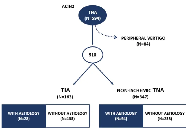
Between 2009 and 2011, 594 patients had their first transient focal neurological symptoms and were included in the ACIN 2 registry. We excluded 84 patients (14.1%) that had vertigo of known peripheral aetiology. In the final cohort, 163 patients had a TIA (32.0%), and 347 patients had a non-ischemic TNA diagnosis (68.0%). In Figure 1 we depict the patients included in the study and the reasons for exclusion. Table I shows a comparison between these two groups.
Patients had a mean age of 63 18 years, which was lower in the non-ischemic TNA group (59 19) compared to the TIA group (71 14). Most patients were female (60.6%), 81 within TIA group (49.7%) and 228 within non-ischemic TNA group (65.7%).
The most common risk factors were hypertension (51.4%), dyslipidemia (34.5%), diabetes mellitus (18.2%) and tobacco use (14.7%). Hypertension was more frequent in the TIA group (68.7% vs. 43.2%) than in the non-ischemic TNA group (p<0.001). Dyslipidemia was found in 52.1% of TIA patients, whereas in TNA patients it accounted for only 26.2% (p<0.001). Tobacco use was also more common in TIA patients (20.2% vs. 12.1%) than non-ischemic TNA (p=0.015). The prevalence of other vascular risk factors (diabetes mellitus, peripheral artery disease, previous acute myocardial infarction, coronary artery disease, previous history of thrombotic events and previous diagnosis of atrial fibrillation) was not different in both groups.
The diagnosis of cancer was found in 78 patients (15.3%), during follow-up of 7 years. Forty-six patients (56.1% of cancer patients) were diagnosed after the index neurological event (TIA or non-ischemic TNA). Cancer wasn’t significantly different between TIA and non-non-ischemic TNA groups (p=0.062). However, when considered only diagnosis of cancer posterior to the neurological event (9% of all patients), cancer prevalence was higher in TIA patients compared to non-ischemic TNA patients (13.5% vs. 6.9%, p=0.016), as shown in Table I. Global cancer incidence (excluding skin cancer) was 1064.4/100000/year (CI 95% 753.2-1461.1), accounting for 1665.2/100000/year in TIA patients and 782.2/100000/year in TNA patients, as shown in Table II. Mean age by the date of cancer diagnosis was 73.812.1 years and wasn’t significantly different in both groups.
The most common cancer diagnosed after the neurological event was originated from gastrointestinal system (28.3%), followed by genitourinary system (21.7%), skin (17.4%, none malignant melanoma), respiratory tract (13.0%), breast (6.5%) and others (13.0%). Gastrointestinal, genitourinary and respiratory tract cancers were more frequent among TIA patients (36.4% vs. 20.8%, 22.7% vs. 20.8% and 18.2% vs. 8.3%, respectively), whereas skin, breast and other type of cancers were more common in non-ischemic TNA patients (20.8% vs. 13.6%, 12.5% vs. 0% and 16.7% vs. 9.1%, respectively). There was one case of central nervous system cancer in the TIA group.
7 Regarding cancer staging, metastasis were found in 15 patients, 9 patients in TIA group and 6 in non-ischemic TNA group. From the 22 patients with cancer diagnosed posteriorly to the TIA, 40.9% had metastasis, which was more frequent than in cancer patients of the non-ischemic TNA group (p=0.028), that had a metastasis in 25% of the patients. From all patients, only one TIA patient had brain metastasis.
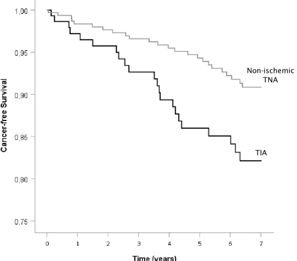
By the end of follow-up, estimated cumulative proportion of cancer-free patients in the TIA group was 82.1%, against 90.9% in the TNA group. Cancer-free survival time was shorter in TIA patients than TNA patients, with a Kaplan-Meier survival plot demonstrating different survival distribution (log rank test with p=0.010), as shown in Figure 2.
Increasing age, male sex, hypertension and current or past smoking habits were associated with cancer in univariate analysis (HR=1.04, p<0.001; HR=2.37, p=0.004; HR=1.96, p=0.033; HR=2.02, p=0.036; respectively), as shown in Table III. Other variables (diabetes mellitus, dyslipidemia, peripheral artery disease, ischemic heart disease, previous thrombotic events and previous atrial fibrillation) didn’t demonstrate any statistically significant difference. In multivariate analysis, only age was an independent factor (p<0.001), but male sex and tobacco use showed tendency towards significance (p=0.094 and p=0.090, respectively). Other variables did not reach statistical significance in multivariate analysis. We found that there was a higher risk of cancer in TIA patients, only in univariate analysis (HR=2.11, p=0.011 in univariate analysis vs. HR=1.36, p=0.329 in multivariate analysis).
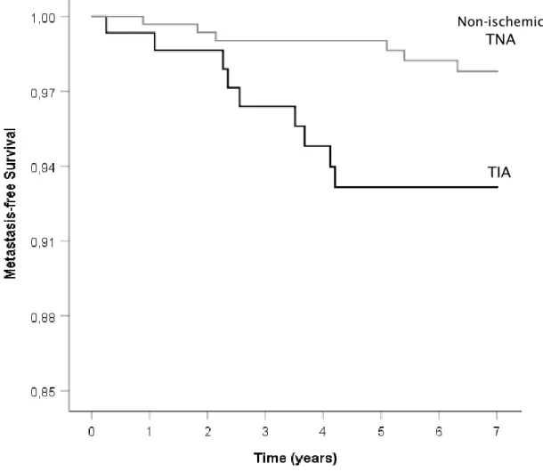
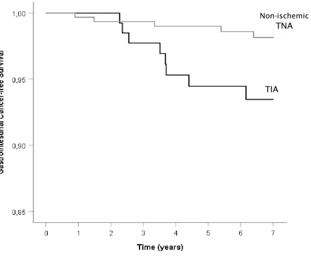
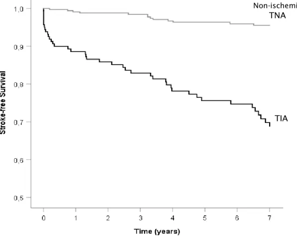
Kaplan-Meier analysis was also used to compare metastasis-free survival and gastrointestinal cancer-free survival. It showed that both metastasis-free survival and gastrointestinal cancer-free survival time was lower in the TIA group (log rank test with p=0.012 and p=0.014, respectively) (Figures 3 and 4). A cox regression analysis was not performed given the small number of patients. Concerning neurological outcomes, stroke and TIA occurred more frequently after a TIA as index event, with stroke occurring in 25.8% of TIA patients against 3.7% in non-ischemic TNA group (p<0.001) and TIA occurring in 13.5% TIA patients against 1.4% in non-ischemic TNA group (p<0.001). Of all strokes, 3 were haemorrhagic (5.5% of all strokes). There wasn’t statistically significant difference in the incidence of non-ischemic TNA after the index event between both groups. To find if there was an independent relationship between neurological outcomes and our index event, a Kaplan-Meier analysis was performed, showing smaller survival time before stroke or TIA in TIA patients than TNA patients (log rank test with p<0.001 in both cases) (Figures 5 and 6). By the end of follow-up, estimated cumulative proportion of surviving patients for stroke endpoint in the TIA group was 68.8%, against 95.5% in the non-ischemic TNA group and 84.6% against 98.4% for TIA endpoint. When repeated for a non-ischemic TNA endpoint, this analysis didn’t demonstrate significant difference in survival distribution in both groups (log rank test with p=0.085). Univariate
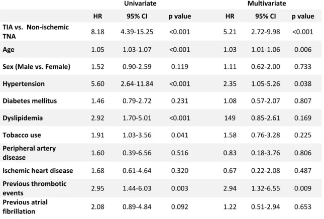
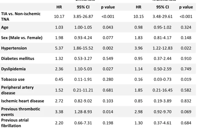
8 analysis and multivariate analysis confirmed TIA as an independent factor associated to stroke and TIA outcomes (HR=8.18, p<0.001 in univariate analysis and HR=5.21, p<0.001 in multivariate analysis) (Table IV and V). In univariate analysis, other variables showed an independent relationship with stroke: age (HR=1.05, p<0.001), hypertension (HR=5.60, p<0.001), dyslipidemia (HR=2.92, p<0.001), tobacco use (HR=1.91, p=0.041) and previous thrombotic events (HR=2.95, p=0.003). Previous atrial fibrillation showed tendency towards significance (HR=2.08, p=0.092). In multivariate analysis, some of these variables did not reach significance, except for age (HR=1.03, p=0.006), hypertension (HR=2.35, p=0.038) and previous thrombotic events (HR=2.94, p=0.009). Regarding the relationship between TIA outcome and variables other than the index event, age (HR=1.03, p=0.043), hypertension (HR=5.37, p=0.002), dyslipidemia (HR=2.36, p=0.027) and previous thrombotic events (HR=3.38, p=0.014) were demonstrated to be independent predictors for this outcome in univariate analysis. In multivariate analysis, only hypertension (HR=3.96, p=0.022) was confirmed as an independent factor for TIA. Other variables didn’t show statistically significant difference for TIA events.
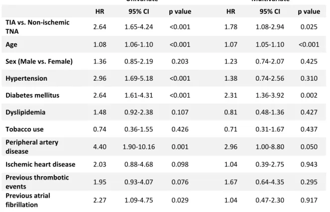
Mortality was higher in TIA group (p<0.001), with a total of 13.5% of deaths, 22.7% in TIA patients and 9.2% in non-ischemic TNA patients. The most common cause for death was infection (26.1%), followed by cancer (13%), cardiovascular events (4.3%), cerebrovascular events (4.3%) and other (5.8%). In 46.4% of patients we were not able to find the cause of death. Death by infection, cancer and cerebrovascular events was higher in TIA patients, unlike death by cardiovascular events and other causes, which were higher in non-ischemic TNA patients, as shown in Table I. Death of unknown cause was also more common in non-ischemic TNA patients. Kaplan-Meier analysis showed that survival time was lower in the TIA group (log rank test with p<0.001) (Figure 7). Considering these results, a cox regression analysis was performed, demonstrating TIA is an independent factor associated mortality both in univariate and multivariate analysis (HR=2.64, p=<0.001 in univariate analysis and HR=1.78, p=0.025 in multivariate analysis), as shown in Table VI. Regarding other variables, only age and diabetes maintained as independent predictors for death in multivariate analysis.
9
DISCUSSION
We found that the global incidence rate of cancer (except for skin non-melanoma cancer) in our group of patients (1064.4/100000/year) was superior to the population incidence rate which was 502.1/100000/year in the year 2012 (Registo Oncológico Regional do Norte[16]) Strikingly, TIA
incidence rate (1665.2/100000 /year) was also superior to global incidence rate for the population of the north region of Portugal. In comparison, the non-ischemic TNA group incidence rate appears to be similar to the population. This is in line with our initial hypothesis, supporting a link between ischemic events and cancer. Consistently, we also found that, cancer occurring after the index event, was more frequent in TIA patients than TNA patients. To the best of our knowledge this is the first time such observation is documented, although several investigators considered the same goal regarding stroke and cancer[17-23]. The similarity of the pathomechanisms underlying stroke and
TIA, may justify this observation. Having said that, to complete our assessment, we showed that there was different cancer free-survival between TIA and non-ischemic TNAs. However, the association between TIA and cancer was not observed in the cox regression analysis. In fact, only age of the patient was confirmed as an independent risk factor for cancer development when comparing the 2 groups, which is also consistent with previous knowledge[24]. Still the non-ischemic
TNA group may not be the ideal control since it may enclose TIA patients wrongly diagnosed or non-ischemic TNAs may have also some relation to cancer through a different mechanism.
The most common cancers diagnosed after the neurological event were gastrointestinal tumours, as it has been described about the association of stroke and cancer[17, 20]. Genitourinary system was
also one of the most common type of tumour, which could be due to its diversity in possible primary locations (bladder, prostate, uterus, ovaries, cervix, testicles). The high incidence of skin cancer is probably explained by its high incidence in the population[25], since none of the identified skin
cancers was malignant melanoma. Some of the vascular risk factors mentioned above, particularly tobacco use, are also risk factors for several types of cancer[26-29], which could explain a higher
incidence of gastrointestinal, genitourinary and respiratory cancers in TIA patients, without necessarily being a direct relationship between TIA and each of these types of cancer. Besides, a few of those risk factors are also risk factors for breast cancer[30], which we found was more
common in TNA patients. Still these observations are limited by the low number of events.
Statistical significance was also found regarding the difference of metastasis incidence in TIA and non-ischemic TNA groups. Since only one patient had brain metastasis, we can assume that direct compression or infiltration of brain structures is unlikely to be the mechanism that lead to the index event. However, other mechanisms, such as metastasis emboli or vascular compression, have been
10 proposed to explain cerebral ischemia in cancer patients[1, 5], which supports our hypothesis of an
ischemic background in cancer patients’ neurologic complications.
In our study, we found that mean age was higher in TIA patients, by a ten years difference, and that, while female patients were more common in non-ischemic TNAs, male sex was the most frequent gender in TIAs. This information is consistent with previous studies[13, 31, 32], that confirm higher
prevalence of TIA in men and older patients. As for non-ischemic TNA, the association with age and sex is less certain, which is explained its diverse aetiology, characterized by different demographic prevalence, but in general comprising a group of younger patients[33-36].
Regarding risk factors, we found that hypertension, dyslipidemia and tobacco use were significantly different between these two groups, with a higher prevalence in TIA, as was expected. TIA has been consistently associated with cardiovascular risk factors in general[13, 37, 38], in resemblance to stroke,
due to its vasculo-thrombotic mechanisms[39], therefore, it’s predictable that these variables are
associated with TIA events, rather than non-ischemic TNAs. Although other vascular risk factors (diabetes mellitus, peripheral artery disease, previous acute myocardial infarction, coronary artery disease, previous history of thrombotic events and previous diagnosis of atrial fibrillation) weren’t significantly different between both groups, several of these factors have been previously associated with TIA, despite not always being found a significant association with the cerebrovascular event[31, 37]. Systolic blood pressure, total cholesterol and angina pectoris have also
been described as independent risk factors for focal TNA[11], which could be an explanation for its
high prevalence in TNA patients.
Concerning neurological outcomes, it has been previously described that there is a great risk of stroke after TIA[40, 41], but recurrent TIA is also a frequent outcome[42, 43]. That information is
consistent with our findings, translated in a statistically significant difference between TIA and non-ischemic TNA patients’ outcomes. The excessive difference in survival time estimates between TIA and non-ischemic TNA group regarding stroke or TIA endpoints, conveys exactly the same message, which is again demonstrated in univariate and multivariate analysis. It’s interesting to see that, although TIA shares some risk factors with stroke and recurrent TIA, this event is for itself an independent predictor of stroke and recurrent TIA. Besides TIA, other variables were found to be independent predictors for these outcomes, namely hypertension (in both TIA and stroke outcomes), age and previous thrombotic events (solely considering stroke events), which is again consistent with risk factors previously described in literature[13, 37].
Given that both cancer and stroke are more frequent outcomes in TIA patients, it would be expectable that mortality would be higher in this group. As such, not only mortality by these causes was superior in TIA patients, but also death by infection and global mortality accompanied this distribution. The fact that mortality by cardiovascular events, unlike the previously mentioned
11 causes, was higher in non-ischemic TNA patients is possibly due to the fact that all heart diseases in total account for more mortality than cerebrovascular disease in general population of Portugal[44]. Death by cancer in total was high, as expected, not only because it is one of the main
causes of death in Portugal[44], but also because the incidence of cancer was found to be superior
in TIA than in general population. However, death by cerebrovascular disease in all patients was low, considering it’s high incidence in Portuguese population[44]. This might be explained by the
great amount of cases in which the cause of death wasn’t known (about 46.6% of death cases). Considering these facts, it was also expectable that survival time would be superior in non-ischemic TNA patients and that TIA would be an independent predictor for death, as we determined. The retrospective design is the main limitation in our study: recording in electronic patient hospital records is not done systematic across all patients; the reliability of information given by patients during telephone interview; patient absenteeism. All these arguments might have undervalued the number of patients diagnosed with cancer and other possible outcomes. In addition, our initial purpose also included a comparison between two subgroups of known and unknown aetiology to see if there was a significant difference in outcomes between these categories. However, we found that, in our group of patients, the distribution of Known and Unknown aetiology patients didn’t match previous descriptions in literature. In a study about aetiology, duration and prognosis of TIAs in Germany, the frequency of TIAs of unknow aetiology was 43.2%, not very different from stroke of unknown aetiology (29.5%)[45], which is similar to what has been previously described by the
American Heart Association/American Stroke Association[46]. These values are very different from
what we found in our TIA patients, in which the frequency of unknown aetiology was 82.8%. This was due to the fact that aetiology was determined only based on the first assessment of the patient at the emergency department. For this reason, we decided not to compare these groups. Still, this issue needs to be addressed and future studies should clarify if there is a difference in the incidence of cancer between TIAs of different aetiologies and TIAs of unknown aetiology, as a way to shorten the eligible group of TIA patients for occult cancer screening.
12
CONCLUSION
Cancer incidence is higher in our group of patients compared to the general population of the same geographical origin. This is particularly striking for the TIA group that presents a higher incidence of cancer, namely GI tract and metastasized cancers.
Our study suggests that there might be a link between cancer and TIA. However, our results don’t allow us to state with certainty that occult cancer is the cause for the index cerebrovascular event, since multivariate analysis didn’t confirm the difference between TIA and non-ischemic TNA group found in previous tests. Future studies could deepen this discussion and help to clarify if there is in fact a causal relationship between cancer and TIAs.
If this cause-effect relationship is proven, healthcare professionals should consider screening for occult malignancies in patients with TIAs, particularly those with unknown aetiology, as well as in stroke patients, since an early diagnosis of cancer has a great impact on patients’ survival [47].
13
APPENDICES
APPENDIX A – TABLE I
Table I – Study population baseline characterization and outcomes
TIA (N=163) Non-ischemic TNA (N=347)
Total (N=510) p value
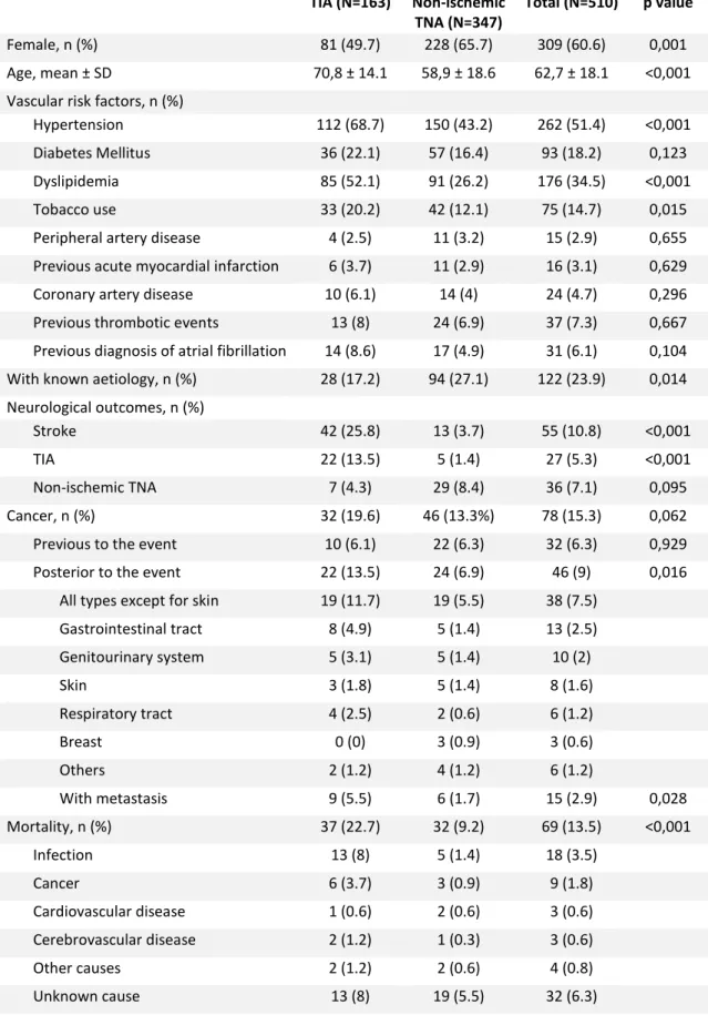
Female, n (%) 81 (49.7) 228 (65.7) 309 (60.6) 0,001
Age, mean ± SD 70,8 ± 14.1 58,9 ± 18.6 62,7 ± 18.1 <0,001
Vascular risk factors, n (%)
Hypertension 112 (68.7) 150 (43.2) 262 (51.4) <0,001
Diabetes Mellitus 36 (22.1) 57 (16.4) 93 (18.2) 0,123
Dyslipidemia 85 (52.1) 91 (26.2) 176 (34.5) <0,001
Tobacco use 33 (20.2) 42 (12.1) 75 (14.7) 0,015
Peripheral artery disease 4 (2.5) 11 (3.2) 15 (2.9) 0,655
Previous acute myocardial infarction 6 (3.7) 11 (2.9) 16 (3.1) 0,629
Coronary artery disease 10 (6.1) 14 (4) 24 (4.7) 0,296
Previous thrombotic events 13 (8) 24 (6.9) 37 (7.3) 0,667
Previous diagnosis of atrial fibrillation 14 (8.6) 17 (4.9) 31 (6.1) 0,104
With known aetiology, n (%) 28 (17.2) 94 (27.1) 122 (23.9) 0,014
Neurological outcomes, n (%)
Stroke 42 (25.8) 13 (3.7) 55 (10.8) <0,001
TIA 22 (13.5) 5 (1.4) 27 (5.3) <0,001
Non-ischemic TNA 7 (4.3) 29 (8.4) 36 (7.1) 0,095
Cancer, n (%) 32 (19.6) 46 (13.3%) 78 (15.3) 0,062
Previous to the event 10 (6.1) 22 (6.3) 32 (6.3) 0,929
Posterior to the event 22 (13.5) 24 (6.9) 46 (9) 0,016
All types except for skin 19 (11.7) 19 (5.5) 38 (7.5)
Gastrointestinal tract 8 (4.9) 5 (1.4) 13 (2.5) Genitourinary system 5 (3.1) 5 (1.4) 10 (2) Skin 3 (1.8) 5 (1.4) 8 (1.6) Respiratory tract 4 (2.5) 2 (0.6) 6 (1.2) Breast 0 (0) 3 (0.9) 3 (0.6) Others 2 (1.2) 4 (1.2) 6 (1.2) With metastasis 9 (5.5) 6 (1.7) 15 (2.9) 0,028 Mortality, n (%) 37 (22.7) 32 (9.2) 69 (13.5) <0,001 Infection 13 (8) 5 (1.4) 18 (3.5) Cancer 6 (3.7) 3 (0.9) 9 (1.8) Cardiovascular disease 1 (0.6) 2 (0.6) 3 (0.6) Cerebrovascular disease 2 (1.2) 1 (0.3) 3 (0.6) Other causes 2 (1.2) 2 (0.6) 4 (0.8) Unknown cause 13 (8) 19 (5.5) 32 (6.3)
14 APPENDIX B – TABLE II
Table II – Cancer Incidence rates in the study population
Incidence rate /100000 per year 95% CI /100000 per year Total 1064.4 753.2 - 1461.1 TIA patients 1665.2 1002.6 - 2600.4
15 APPENDIX C – TABLE III
Table III – Univariate and multivariate Cox regression analysis of variables associated with increased cancer occurrence Univariate Multivariate HR 95% CI p value HR 95% CI p value TIA vs. Non-ischemic TNA 2.11 1.19-3.77 0.011 1.36 0.74-2.49 0.329 Age 1.04 1.02-1.06 <0.001 1.04 0.91-3.23 0.094
Sex (Male vs. Female) 2.37 1.31-4.25 0.004 1.72 1.02-1.06 <0.001
Hypertension 1.96 1.06-3.63 0.033 1.26 0.63-2.55 0.517 Diabetes mellitus 1.46 0.74-2.87 0.278 1.12 0.54-2.30 0.764 Dyslipidemia 1.35 0.75-2.42 0.323 0.82 0.43-1.56 0.547 Tobacco use 2.02 1.05-3.90 0.036 1.91 0.90-1.03 0.090 Peripheral artery disease 1.00 0.14-7.24 0.998 0.59 0.07-4.70 0.620 Ischemic heart disease 1.52 0.47-4.89 0.485 1.07 0.30-3.81 0.913
Previous thrombotic
events 1.30 0.47-3.63 0.613 1.22 0.40-3.69 0.728 Previous atrial
16 APPENDIX D – TABLE IV
Table IV – Univariate and multivariate Cox regression analysis of variables associated with increased stroke occurrence Univariate Multivariate HR 95% CI p value HR 95% CI p value TIA vs. Non-ischemic TNA 8.18 4.39-15.25 <0.001 5.21 2.72-9.98 <0.001 Age 1.05 1.03-1.07 <0.001 1.03 1.01-1.06 0.006
Sex (Male vs. Female) 1.52 0.90-2.59 0.119 1.11 0.62-2.00 0.733
Hypertension 5.60 2.64-11.84 <0.001 2.35 1.05-5.26 0.038 Diabetes mellitus 1.46 0.79-2.72 0.231 1.08 0.57-2.07 0.807 Dyslipidemia 2.92 1.70-5.01 <0.001 149 0.85-2.61 0.169 Tobacco use 1.91 1.03-3.56 0.041 1.58 0.76-3.28 0.225 Peripheral artery disease 1.60 0.39-6.56 0.516 0.83 0.18-3.76 0.806 Ischemic heart disease 1.68 0.61-4.64 0.320 0.67 0.22-2.08 0.487
Previous thrombotic
events 2.95 1.44-6.03 0.003 2.94 1.32-6.55 0.009 Previous atrial
17 APPENDIX E – TABLE V
Table V – Univariate and multivariate Cox regression analysis of variables associated with increased TIA occurrence Univariate Multivariate HR 95% CI p value HR 95% CI p value TIA vs. Non-ischemic TNA 10.17 3.85-26.87 <0.001 10.15 3.48-29.61 <0.001 Age 1.03 1.00-1.05 0.043 0.98 0.95-1.02 0.324
Sex (Male vs. Female) 1.98 0.93-4.24 0.077 1.83 0.81-4.17 0.148
Hypertension 5.37 1.86-15.52 0.002 3.96 1.22-12.83 0.022 Diabetes mellitus 1.32 0.53-3.27 0.549 0.95 0.37-2.44 0.910 Dyslipidemia 2.36 1.10-5.03 0.027 1.14 0.50-2.59 0.749 Tobacco use 0.45 0.11-1.91 0.280 0.16 0.03-0.73 0.019 Peripheral artery disease 1.52 0.21-11.21 0.681 1.85 0.21-16.45 0.582 Ischemic heart disease 2.72 0.82-9.02 0.103 0.85 0.19-3.89 0.832
Previous thrombotic
events 3.38 1.28-8.93 0.014 2.98 0.92-9.70 0.069 Previous atrial
18 APPENDIX F – TABLE VI
Table VI – Univariate and multivariate Cox regression analysis of variables associated with increased mortality Univariate Multivariate HR 95% CI p value HR 95% CI p value TIA vs. Non-ischemic TNA 2.64 1.65-4.24 <0.001 1.78 1.08-2.94 0.025 Age 1.08 1.06-1.10 <0.001 1.07 1.05-1.10 <0.001
Sex (Male vs. Female) 1.36 0.85-2.19 0.203 1.23 0.74-2.07 0.425
Hypertension 2.96 1.69-5.18 <0.001 1.38 0.74-2.56 0.310 Diabetes mellitus 2.64 1.61-4.31 <0.001 2.31 1.36-3.92 0.002 Dyslipidemia 1.48 0.92-2.38 0.107 0.81 0.48-1.36 0.427 Tobacco use 0.74 0.36-1.55 0.426 0.71 0.31-1.67 0.437 Peripheral artery disease 4.40 1.90-10.16 0.001 2.96 1.00-8.80 0.050 Ischemic heart disease 2.03 0.88-4.68 0.098 1.04 0.39-2.75 0.943
Previous thrombotic
events 1.95 0.93-4.07 0.076 1.67 0.64-4.35 0.295 Previous atrial
19 APPENDIX G – FIGURE 1
20 APPENDIX H – FIGURE 2
Figure 2 – Kaplan-Meier curve for cancer-free survival
TIA
Non-ischemic TNA
21 APPENDIX I – FIGURE 3
Figure 3 – Kaplan-Meier curve for metastasis-free survival
Non-ischemic
TNA
22 APPENDIX J – FIGURE 4
Figure 4 – Kaplan-Meier curve for gastrointestinal cancer-free survival
Non-ischemic
TNA
23 APPENDIX K – FIGURE 5
Figure 5 – Kaplan-Meier curve for stroke-free survival
Non-ischemic
TNA
24 APPENDIX L – FIGURE 6
Figure 6 – Kaplan-Meier curve for TIA-free survival
Non-ischemic
TNA
25 APPENDIX M – FIGURE 7
Figure 7 – Kaplan-Meier curve for overall survival
Non-ischemic
TNA
26
REFERENCES
1. Giglio, P. and M.R. Gilbert, Neurologic complications of cancer and its treatment. Current oncology reports, 2010. 12(1): p. 50-59.
2. Darnell, R.B. and J.B. Posner, Paraneoplastic Syndromes Affecting the Nervous System. Seminars in Oncology, 2006. 33(3): p. 270-298.
3. Dalmau, J., H.S. Gultekin, and J.B. Posner, Paraneoplastic Neurologic Syndromes:
Pathogenesis and Physiopathology. Brain Pathology, 1999. 9(2): p. 275-284.
4. Dardiotis, E., et al., Cancer-associated stroke: Pathophysiology, detection and management
(Review). International journal of oncology, 2019. 54(3): p. 779-796.
5. Dearborn, J.L., V.C. Urrutia, and S.R. Zeiler, Stroke and Cancer- A Complicated Relationship. Journal of neurology & translational neuroscience, 2014. 2(1): p. 1039-1039.
6. Trousseau, A., Lectures on clinical medicine, delivered at the Hotel-Dieu, Paris. 1865. 7. Caine, G.J., et al., The hypercoagulable state of malignancy: pathogenesis and current
debate. Neoplasia, 2002. 4(6): p. 465-73.
8. Yeh, P.-S. and H.-J. Lin, Cerebrovascular Complications in Patients with Malignancy: Report
of Three Cases and Review of the Literature. Vol. 13. 2004. 34-8.
9. Bang, O.Y., et al., Ischemic stroke and cancer: stroke severely impacts cancer patients, while
cancer increases the number of strokes. Journal of clinical neurology (Seoul, Korea), 2011.
7(2): p. 53-59.
10. Easton, J.D., et al., Definition and Evaluation of Transient Ischemic Attack. Stroke, 2009. 40(6): p. 2276-2293.
11. Bos, M.J., et al., Incidence and Prognosis of Transient Neurological Attacks. JAMA, 2007. 298(24): p. 2877-2885.
12. Abbatemarco, J.R. and A.D. Rae-Grant, Transient neurologic syndromes: A diagnostic
approach. Cleve Clin J Med, 2018. 85(2): p. 155-163.
13. Fonseca, A.C. and P. Canhão, Diagnostic difficulties in the classification of transient
neurological attacks. European Journal of Neurology, 2011. 18(4): p. 644-648.
14. Correia, M., et al., Changes in stroke incidence, outcome, and associated factors in Porto
between 1998 and 2011. International Journal of Stroke, 2017. 12(2): p. 169-179.
15. Correia, M., et al., Transient ischemic attacks in rural and urban northern Portugal:
incidence and short-term prognosis. Stroke, 2006. 37(1): p. 50-5.
16. RORENO. Registo Oncológico Regional do Norte 2012. Instituto Português de Oncologia do Porto, e.P., 2019.
17. Carrilho Romeiro, A., A. Valadas, and J. Marques, Acute Ischemic Stroke on Cancer Patients,
a Distinct Etiology? A Case-Control Study. Acta Med Port, 2015. 28(5): p. 613-8.
18. Navi, B.B., et al., Association between incident cancer and subsequent stroke. Annals of neurology, 2015. 77(2): p. 291-300.
19. Taccone, F.S., S.M. Jeangette, and S.A. Blecic, First-Ever Stroke as Initial Presentation of
Systemic Cancer. Journal of Stroke and Cerebrovascular Diseases, 2008. 17(4): p. 169-174.
20. Kim Seon, G., et al., Ischemic Stroke in Cancer Patients With and Without Conventional
Mechanisms. Stroke, 2010. 41(4): p. 798-801.
21. Álvarez-Pérez, F.J., et al., Frequency and Mechanism of Ischemic Stroke Associated with
Malignancy: A Retrospective Series. European Neurology, 2012. 68(4): p. 209-213.
22. Quintas, S., et al., Predictors of unknown cancer in patients with ischemic stroke. Vol. 137. 2018.
23. Cocho, D., et al., Predictors of Occult Cancer in Acute Ischemic Stroke Patients. Journal of Stroke and Cerebrovascular Diseases, 2015. 24(6): p. 1324-1328.
24. Koene, R.J., et al., Shared Risk Factors in Cardiovascular Disease and Cancer. Circulation, 2016. 133(11): p. 1104-1114.
27 25. Bray, F., et al., Global cancer statistics 2018: GLOBOCAN estimates of incidence and
mortality worldwide for 36 cancers in 185 countries. CA: A Cancer Journal for Clinicians,
2018. 68(6): p. 394-424.
26. Johnson, C.M., et al., Meta-analyses of colorectal cancer risk factors. Cancer Causes & Control, 2013. 24(6): p. 1207-1222.
27. Pienta, K.J. and P.S. Esper, Risk Factors for Prostate Cancer. Annals of Internal Medicine, 1993. 118(10): p. 793-803.
28. Burger, M., et al., Epidemiology and Risk Factors of Urothelial Bladder Cancer. European Urology, 2013. 63(2): p. 234-241.
29. de Groot, P. and R.F. Munden, Lung Cancer Epidemiology, Risk Factors, and Prevention. Radiologic Clinics, 2012. 50(5): p. 863-876.
30. ROJAS, K. and A. STUCKEY, Breast Cancer Epidemiology and Risk Factors. Clinical Obstetrics and Gynecology, 2016. 59(4): p. 651-672.
31. Bejot, Y., et al., Trends in the Incidence of Transient Ischemic Attacks, Premorbid Risk Factors
and the Use of Preventive Treatments in the Population of Dijon, France from 1985 to 2004.
Cerebrovascular Diseases, 2007. 23(2-3): p. 126-131.
32. Bots Michiel, L., et al., Transient Neurological Attacks in the General Population. Stroke, 1997. 28(4): p. 768-773.
33. Manzoni, G.C. and P. Torelli, Epidemiology of migraine. The Journal of Headache and Pain, 2003. 4(Suppl 1): p. s18-s22.
34. Dennis, M. and C. Warlow, Migraine aura without headache: transient ischaemic attack or
not? Journal of Neurology, Neurosurgery & Psychiatry, 1992. 55(6): p. 437-440.
35. Carlson, C., et al., Sex differences in seizure types and symptoms. Epilepsy & behavior : E&B, 2014. 41: p. 103-108.
36. Banerjee, P.N., D. Filippi, and W. Allen Hauser, The descriptive epidemiology of epilepsy-a
review. Epilepsy research, 2009. 85(1): p. 31-45.
37. Zvan, B., B. Pecnik, and T. Pogacnik, Transient ischemic attacks, risk factors, and precerebral
color doppler angiosonography. Journal of Stroke and Cerebrovascular Diseases, 1994. 4(3):
p. 161-165.
38. Heyden, S., et al., Cardiovascular mortality in transient ischemic attacks. Stroke, 1980. 11(3): p. 252-255.
39. W. Johansson, B., Cerebral Artery Spasm - A Possible Mechanism of TIA? Vol. 58 Suppl 2. 1986. 157-60.
40. Coutts, S.B., et al., Recurrent Events in Transient Ischemic Attack and Minor Stroke. Stroke, 2008. 39(9): p. 2461-2466.
41. Amarenco, P., et al., One-Year Risk of Stroke after Transient Ischemic Attack or Minor
Stroke. New England Journal of Medicine, 2016. 374(16): p. 1533-1542.
42. Walz, E.T., T. Brink, and A. Slivka, Pattern and frequency of recurrent transient ischemic
attacks. Journal of Stroke and Cerebrovascular Diseases, 1997. 6(3): p. 121-124.
43. Claiborne Johnston, S., et al., A comparison of risk factors for recurrent TIA and stroke in
patients diagnosed with TIA. Neurology, 2003. 60(2): p. 280.
44. Estatísticas da Saúde 2014: Mortalidade Geral. Lisboa, I.N.d.E., I.P.; 2016.
45. Weimar, C., et al., Etiology, Duration, and Prognosis of Transient Ischemic Attacks: An
Analysis From the German Stroke Data Bank. JAMA Neurology, 2002. 59(10): p. 1584-1588.
46. Association, A.H.A.A.S., Understanding Diagnosis and Treatment of Cryptogenic Stroke: A
Healthcare Professional Guide. 2015.
47. Hawkes, N., Cancer survival data emphasise importance of early diagnosis. BMJ, 2019. 364: p. l408.