s
REGULATION OF THE PORE-FORMING TRANSLOCON OF THE
TYPE III SECRETION SYSTEM BY THE EspC SERINE
PROTEASE IN ENTEOPATHOGENIC ESCHERICHIA COLI
by
Leticia Tavares Gomes
REGULATION OF THE PORE-FORMING TRANSLOCON OF THE TYPE III SECRETION SYSTEM BY THE EspC SERINE PROTEASE IN
ENTEROPATHOGENIC ESCHERICHIA COLI
Thesis presented to Escola Superior de Biotecnologia of the Universidade Católica Portuguesa to fulfill the requirements of Master of Science degree in Microbiology
by
Leticia Tavares Gomes
Place: Centre of Interdisciplinary Research in Biology – Collège de France, Paris
Supervision: Guy TRAN VAN NHIEU
Aos meus pais,
por tudo o que fizeram e construiram,
por continuarem a fazer tudo pelos filhos.
Resumo
As bactérias do grupo Escherichia coli enteropatogénica (EPEC) são uma das maiores causas de diarreia fatal em crianças com menos de cinco ano nos países em desenvolvimento. A virulência de EPEC depende da presença do sistema de secreção do tipo III (SST3). O SST3 é constituído por um corpo basal situado entre a membrana externa e interna da bactéria, prolongado por uma agulha que é projetada a partir da superfície da bactéria. No caso de EPEC, a agulha do SST3 é prolongada por um filamento formado pela proteína hidrofílica, EspA. Depois de contactar com a célula hospedeira, o sistema ativa a secreção de duas proteínas hidrofóbicas, EspD e EspB. Estas proteínas inserem-se na membrana plasmática da célula hospedeira para formar o transclocão. O translocão é uma estrutura chave para formar o poro na membrana da célula hospedeira pelo qual os efetores são inseridos na célula.
O laboratório onde se realizou este trabalho estuda o papel da proteína EspC, que pertence à família SPATE (serine protease autotransporter of Enterobacteriaceae), na regulação e funcionamento do translocão. EspC é secretada pelo sistema de secreção do tipo V, sendo independente do SST3. Foi demonstrado que EspC tem um papel importante no controlo da formação do poro membranar e da virulência mediada pelo SST3. EspC tem preferencialmente como alvo os complexos de EspA-EspD, envolvidos na formação do poro membranar, presumivelmente após o destacamento do translocão do filamento da agulha.
Este trabalho de estágio utilizou procedimentos de purificação para isolar o complexo EspA-EspD sensível à proteólise de EspC com o objetivo de realizar a caracterização estrutural deste complexo por Microscopia Eletrónica. Implementamos várias fases de purificação distintas para aumentar a pureza das amostras contendo os complexos desejados. Conseguimos isolar complexos homogéneos contendo EspA que mostram um estrutura em forma de anel com um diâmetro externo de 10 nm e um interno de 4 nm.
Abstract
Enteropathogenic E. coli (EPEC) is a major cause of fatal diarrhea in childs under 5 years in developing countries. EPEC virulence is dependent on the presence of type III secretion system (T3SS). T3SSs are used by many pathogenic Gram-negative bacteria to inject bacterial effectors into host cells. T3SSs consist of a basal body embedded in the bacterial outer and inner membrane, prolonged by a needle protruding from the bacterial surface. In the case of EPEC, the T3SS needle is extended by a filament formed by hydrophilic EspA protomers. Upon cell contact, the system triggers the secretion of two hydrophobic proteins EspD and EspB, that insert in the host cell plasma membrane to form the translocon. The translocon is critical for the injection of type 3 effectors, presumably by forming a pore into host cell plasma membranes through which the effectors are channeled.
The host lab studies the role of EspC, a serine protease autotransporter of Enterobacteriaceae (SPATE) family, on the regulation of the translocon function. EspC is a protein secreted by EPEC by a type V secretion system independently of the T3SS, although the translocation into epithelial cells requires active type III secretion. EspC has been shown to play a role in controlling pore formation and cytotoxicity mediated by the T3SS. EspC was shown to preferentially target an EspA-EspD complex, involved in the T3SS-dependent pore formation, supposedly following detachment of the translocon from the T3SS needle filament. This internship work took advantage of fractionation procedures worked out to isolate the EspA-EspD complex sensitive to EspC proteolysis, with the aim to perform a structural characterization of this complex by electron microscopy analysis. In this study, we increased the yield of EspA-EspD complex purification, that for the first time allowed the detection of EspB together with EspA-EspD complexes. We implemented additional purification steps to increase the purity of protein complexes. We were able to isolate homogenous EspA-containing complexes showing a ring-link structure with an external diameter of 10 nm of and a 4 nm-inner diameter.
ACKNOWLEDGMENTS
I would like to thank all the persons that make this experience unforgettable. Paris is always a good idea.
First, I would like to thanks Guy Tran Van Nhieu, for the opportunity to work at Collège de France, in his team, and for all the knowledge that he passed to me. Thank you for all the time that you spend correcting this master thesis.
Thanks to all the team: César, for all the help in the lab, all the discussions and fun that we had together; Mariette for the support in all situations, thank you for be the one that always know what to say and how to help; Javier, thank you for all your tips and tricks in the lab, for all the availability to help; Stephane, Daniel and Nicole, thank you for all the help and the good time that we spend together!
All the good friends that I’ve made at Collège. A special thanks to Richard that since the beginning was here to hear all the fears and expectations that I had. Marion and Stephanie, you already know… our triplet is the best.
A special word to the ones that walked by my side in Coimbra – “Saudade desta cidade, levo comigo P’rá vida”. Obrigada à Universidade de Coimbra, à Associação Académica de Coimbra e às pessoas que por lá passaram comigo.
Obrigada a quem sempre me incentivou a correr atrás dos meus objetivos, ainda que algumas decisões não tenham sido fáceis. Obrigada às de sempre, por tudo que somos juntas.
Obrigada ao Porto que me acolheu. À Escola Superior de Biotecnologia, especialmente aos professores com que me cruzei, alguns dos melhores da minha vida de estudante. Aos colegas, especial à Dandára por ter sido a minha metade em todos os trabalhos, no dia a dia do Porto e no skype.
Obrigada às minhas tias e madrinha, pelo suporte que foram para mim em Paris. Especialmente à tia São, que me acolheu com tudo o que tinha.
À Barbara e ao Nelson, por me terem acompanhado sempre!
Aos meus pais. Faltam-me as palavras para agradecer tudo aquilo que me permitiram viver. Obrigada por me terem deixado caminhar sozinha, por me terem visto cair, mas por estarem lá para me levantarem. Desculpem pelos momentos menos bons… Mas a força que sempre me guiou, foi o nunca vos querer desiludir. Sei que vêm esta minha vitória como vossa, e ela é toda dedicada a vocês. São o melhor de mim, a minha inspiração de vida!
C
ONTENTSR
ESUMO 3A
BSTRACT 4A
CKNOWLEDGMENT 5A
BBREVIATIONS 8I
NTRODUCTION 9 I. Escherichia coli 9II. Enteropathogenic E. coli 10
III. EPEC Pathogenesis 11
A. Adherence 12
B. Signal transduction and intimate attachment 13
IV. Locus of enterocyte effacement (LEE) 13
A. LEE regulation 14
V. Type III Secretion System (T3SS) 16
A. General Structure 16
B. Biogenesis of the T3SS 18
C. The basal body and Needle Complex 20
D. Control of the needle length 22
E. Tip complex and translocon 22
VII. Regulation of Pore formation by a bacterial serine protease 28
VIII. The Serine Protease EspC 29
A. SPATES – General overview of EspC 29
B. Cytotoxicity of EspC 30
C. EspC regulates pore formation and cytotoxicity mediated by the T3SS 31
R
ATIONALE 32M
ATERIAL ANDM
ETHODSI. Analysis of secreted proteins 33
II. EspC Purification 34
III. EspA/D Purification 34
IV. Electron Microscopy 36
R
ESULTS 37I. Testing the secretion of proteins of interest 37
II. EspC Purification 39
III. EspA-EspD Purification 40
A. Testing the levels of protein production 41
B. EspA/D Purification 42
D
ISCUSSION 48A
BBREVIATIONS E. coli – Escherichia coliEPEC – Enteropathogenic Escherichia coli SST3 – Sitema de Secreção Tipo III T3SS – Type III Secretion System
SPATE – Serine Protease Autotransporters of Enterobacteriaceae AE – Attachment and Effacing
IECs – Intestinal Epithelial Cells LEE – Locus of enterocyte effacement Tir – Translocated Intimin Receptor BFP – Bundle-forming pili
Ler – LEE-encoded regulator
H-NS – Histone-like nucleoid-structuring protein Hfq – RNA binding protein
T3SA – Type III Secretion Apparatus LPS – Lipopolysaccharide
EspA – Hydrophilic protein EspD – Major hydrophobic protein EspB – Minor hydrophobic protein EM – Electron Microscopy NC – Needle Complex TC – Tip Complex RBC – Red blood Cell
NMR – Nuclear Magnetic Resonance DOC – Sodium deoxycholate FAK – Focal adhesion kinase TCA – Trichloroacetic acid ON – Overnight
FPLC – Flow Pressure Liquid Chromatography L – Load
FT – Flow Through
I
NTRODUCTIONI. Escherichia coli
Theodor Eschrich was the first to report the isolation of a microorganism from infant stool which he named Bacterium coli commune in 1885 (reprinted in English: Eschrich and Bettelheim, 1988). Only in 1954, the name Escherichia coli was fully adopted (Cowan, 1954). In early studies, it was considered as a harmless commensal of the gastrointestinal tract in warm-blooded animals. Because some strains were found not to invade cells or release diffusible toxins, doubts about their pathogenic potential were raised in the 1960s and 1970s. The isolation of pathogenic bacteria inducing diarrhea in human volunteers provided the decisive evidence for E. coli strains as true human pathogens (Levine et al., 1978).
The diseases caused by pathogenic E. coli strains affect the gastrointestinal tract, as well as extraintestinal sites such as the urinary tract, the bloodstream, and central nervous system (Croxen and Finlay, 2010). Pathogenic E. coli strains have been classified in two main groups: diarrhoeagenic E. coli or extraintestinal E. coli. Enteropathogenic E. coli (EPEC), enterohaemorrhagic E. coli (EHEC), enterotoxigenic E. coli (ETEC), enteroinvasive E. coli (EIEC), enteroaggregative E. coli (EAEC) and diffusely adherent E. coli (DAEC) correspond to the six well studied pathovars that are diarrhoeagenic. The extraintestinal group contains two pathovars: the uropathogenic E. coli (UPEC) and neonatal meningitis E. coli (NMEC).
The genome size of E. coli strains can differ by a million base pairs between commensal and pathogenic variants – comparative genomics studies between 186 E. coli genomes have shown a "core genome" of about 1,700 homolog gene clusters, and a flexible gene pool of about 16,400 gene clusters (Kaas et al., 2012). The pathogenicity of E. coli is regulated by the flexible gene pool trough the gain or loss of genetic material (Croxen et al., 2013). Horizontal gene transfer is an important way to disseminate traits in recipient organisms and is crucial to promote the fitness and survival of the pathogen during infection. Pathogenicity islands (PAIs) are clusters of virulence genes found on plasmids or integrated into the chromosome, that are usually bordered by mobile genetic elements, PAIs are not found in non-pathogenic bacteria.
II. Enteropathogenic E. coli
EPEC was the first diarrheagenic E. coli to be identified, being responsible for a series of outbreaks of infantile diarrhea in the 1940s and 1950s. E2348/69 strain has been used worldwide as a prototype strain to study EPEC and the complete sequence of the strain was published in 2009 (Igushi et al., 2009). EPEC is a non-invasive microorganism that does not produce enterotoxins. It was with studies on the E2348/69 strain that the firsts insights in the pathogenic strategy of EPEC began to emerge. With the discovery of LEE (Locus of Enterocyte Effacement), EPEC was classified in a group of bacteria known as attaching and effacing (A/E) pathogens. AE pathogens classification is based on their ability to form distinctive lesions on the surfaces of intestinal epithelial cells (IECs). The EPEC group is subdivided in atypical and typical EPEC based on the presence or absence of the E. coli adherence factor plasmid, respectively (pEAF) (Kaper et al., 2004). While humans are the only known reservoir for typical EPEC, atypical strains have been isolated from human and animal sources like dogs, rabbits, monkeys and sheep (Moura et al., 2009).
The enteropathogenic E. coli is a major cause of fatal diarrhea in childs under 5 years in developing countries. The prevalence of EPEC infection varies from the studies and the data showed until today, depend on the population of study, and age distribution or the methods used for detection or diagnosis. Also, the geographic region or the socioeconomic class may influence the epidemiology of EPEC. Based on molecular methods and identification of intimin gene, for example, EPEC was found responsible for 5 to 10% of pediatric diarrheal episodes in developing countries, while if the determination is made based on the HEp-2 adherence pattern or serotyping, the estimated prevalence is higher, going from 10 to 20% (Ochoa et al., 2008).
EPEC causes diarrhea often accompanied with fever, vomiting and dehydration in children under 5 years. EPEC infections often lead to acute diarrhea, but persistent cases have also been reported to last more than 2 weeks (Nataro and Kaper, 1998). In comparison to infection with other diarrheal pathogens such as adenovirus, rotavirus, Campylobacter and
Salmonella, infection with EPEC is more likely to lead to development of persistent diarrhea
III. EPEC Pathogenesis
AE lesions are a hallmark of EPEC pathogenesis, characterized by effacement of brush border microvilli at the site of bacterial attachment. The disruption of the intestinal brush border is accompanied by the formation of actin pedestals that extend from the surface of the epithelium into the lumen. The pedestal-like structures are produced following secretion of a bacterial receptor protein (Tir) through the Type III secretion System (T3SS). The T3SS is absolutely required for EPEC pathogenesis.
A three-stage model of EPEC pathogenesis was first described by Donnenberg and Kaper, including localized adherence to host cells, signal transduction and intimate attachment (Donnenberg and Kaper, 1992). During intimate attachment, a series of bacterial effectors proteins are injected into host cells, where they interfere with actin dynamics and others host cellular processes (Dean and Kenny, 2009). The three-stage model is represented in Figure 1.
Figure 1. Attaching and Effacing lesions induced by EPEC. (a) Electron micrograph of EPEC expressing type IV fimbria
known as bundle-forming pili (BFP); (b) Electron micrograph of an EPEC bacterium engaged in attaching and effacing activity with a host intestinal epithelial cell. Note the loss of microvilli and the formation of a cuplike pedestal to which the bacterium is intimately attached; (c) A model of EPEC pathogenesis. (Donnenberg et al., 2001).
A bacterial aggregate, connected by bundles of BFP fibers, is shown near an intestinal epithelial cell (Fig. 1a). As infection proceeds, the bacteria detach from the pilus fibers, disaggregate, and attach to the host cell through a surface appendage that contains EspA (Fig. 1b). It is believed that Tir, EspB, EspD and EspF (see table 1.) travel through this appendage to the host cell. EspF is not required for attaching and effacing activity but plays a role in disruption of intestinal barrier function and host cell death. EspB, EspD and Tir are required for attaching and effacing activity (Fig. 1c).
Table 1. Summary of virulence factors in EPEC O127:H6 adapted from K.J. Spears et al. 2006
Virulence factor Encoding region Function
Adhesins
Type 1 fimbriae fim operon Adherence to α-D-mannose-containing glycoproteins Type IV pilus (Bfp) 14 genes in the
EAF plasmid
Initial adherence specificity to phosphatidylethanolamine
OmpA ompA Mediates HeLa cell adherence
Escherichia coli factor for
adherence (Efa)
(O157) lifA Regulation of T3SS
Flagellin fliC Motility abd adherence
LEE-encoded
Intimin eae (LEE5) Intimate attachment adhesion molecule
Translocated intimin tir (LEE5) Intimate attachment; AE lesion formation; actin polymerization
Receptor (Tir)
Mitochondrion-associated protein (Map)
map (LEE5) Disruption of TER; mitochondrial membrane potential; filopodium formation
EspF espF (LEE4) Disruption of TER; mitochondrial membrane potential; cell death
EspH espH (LEE3) Disruption of cytoskeleton Non-LEE-encoded
Secreted serine protease espC Epithelial/tight junction disruption, mucinase, interference with complement cascade
A. Adherence
The localized adherence phenotype of EPEC is the result of a surface appendage known as the bundle-forming pilus (BFP), which is encoded by the EAF plasmid. BFP is responsible for the formation of bacterial clusters, because of its ability to form reversible aggregates into rope-like bundles. If any of the genes required for the formation of BFP is inactivated by mutation, the bacteria fails to form aggregates and is not able to display localized adhesion (Donnenberg et al., 2000).
A/E lesions are caused by the ability of bacteria to intimately attach to the host cell membrane, destroying the microvilli and inducing the formation of pedestal-like structures enriched in cytoskeletal proteins (Fig. 1b). The LEE is a 35 kb genetic locus responsible for this event. It has been shown that the LEE from EPEC strain E2348/69 cloned into a non-pathogenic E. coli strain is sufficient to confer attaching and effacing activity (Frankel et al., 1998).
B. Signal transduction and intimate attachment
Formation of the A/E lesions occurs by subversion of actin dynamics within host cell by EPEC through the injection of type III effectors. It is mediated by the interaction between intimin, a bacterial surface protein and the bacterial translocated intimin receptor, Tir. All the strains capable to induce the A/E lesions have the eae gene, which encodes intimin.
IV. Locus of enterocyte effacement (LEE)
The genes encoding T3SS components and related proteins including regulators, chaperones, and some effectors, are clustered in a 35-kb chromosomal region termed the locus of enterocyte effacement (LEE). The LEE contains 41 genes organized in five major operons, designated LEE1, LEE2, LEE3, LEE4 and LEE5, and several smaller transcriptional units (Mellies et al. 2007). The LEE1, LEE2, and LEE3 operons encode the type III apparatus including a basal body that spans the inner and outer membranes, prolonged by a needle composed of EscF. The LEE4 operon encodes the EspA protein that polymerizes to form the filament that prolongs the T3SS needle; EspB and EspD form the translocon in the host cell membrane; EspF is injected into the host cell and targeted to the mitochondria, where it plays a role in the cell death pathway. The LEE5 operon encodes the Tir and intimin proteins, which are necessary for intimate attachment to the host epithelium; and CesT, a chaperone for Tir (Mellies et al. 2007).
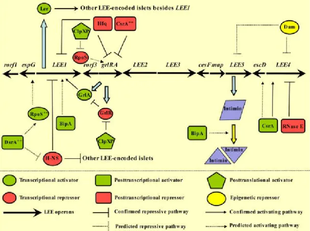
A. LEE regulation
The regulation of virulence genes expression in EPEC in not completely understood. The
LEE1-located Ler (LEE-encoded regulator) plays a central role in the regulation of the AE
The expression of LEE genes is thermo-regulated: AE lesions do not form if the bacteria are incubated at 28°C prior to infecting host cells at 37°C. H-NS (histone-like nucleoid-structuring) is responsible for repressing LEE1 at 27°C by binding to LEE1, LEE2 and LEE3. At 37°C H-NS are not able to bind to these loci, therefore protein secretion via T3SS in EPEC occurs maximally at 37°C (Mellies et al., 2007). Under inducible conditions, H-NS mediated repression of LEE1 is relieved and the transcription of ler is induced (Bhatt et al., 2011). Ler primarily activates transcription from the other LEE operons that allows the synthesis and assembly of the T3SS. Ler is 15kDa protein with a strong control over the LEE genes. It has been shown that a ler mutant of the prototypical EPEC strain E2348/69 severely decreased its ability to form AE lesions in epithelial cells in culture (Elliott et al., 2000).
Figure 2. Transcriptional and extratranscriptional control of LEE. (From Bhatt et al., 2011)
When reaching host body temperature, Ler increases the transcription of the LEE2, LEE3,
LEE4 and LEE5 operons. Among others, Ler also increases the expression of EspC protein.
EspC will further discussed in the chapter VIII (EspC serine protease). During infection of cultured cells, Ler-induced expression is necessary only during the early stages of the EPEC infection process (Leverton et al., 2005). By real-time PCR the Leverton demonstrated that transcription of the EPEC LEE3, LEE4 and LEE5 operons increased over 3 hours
post-infection, while the expression of LEE1, carrying ler, decreased during the same period in a Ler-dependent manner (Leverton et al., 2005). This shows that during infection, other factors regulate Ler function leading to repression of LEE1. Mutation of ler in EPEC also affects the pattern of EPEC adherence (Elliott et al., 2000). Wild-type EPEC normally shows localized adherence to cells, with the formation of microcolonies. Mutation of ler in EPEC is rather associated with the appearance of a diffuse adherence phenotype. Interestingly, electron microscopy studies have shown that ler mutants present changes in fimbrial expression accompanied by the alterations in adherence.
Moreover, Ler is also regulated by post-translational factors, such as Hfq. Hfq is a conserved RNA-binding protein that regulates diverse cellular processes through post-transcriptional control of gene expression, functioning as a chaperone of sRNAs. Hfq has been shown to have a critical impact on bacterial pathogenicity in a wide array of bacterial species (Shakhnovich et al., 2009). It is known that in EHEC hfq mutants increase Ler production leading to an increase in the quantities of T3SS effectors into the supernatant even though an hfq mutant shows compromised growth.
The ability to form AE lesions, intimate attachment to the host cell and formation of “pedestals”-like structures are characteristics shared with Enterohaemorrhagic Escherichia
coli (EHEC) that also expresses a T3SS and shows genetic similarities with EPEC. (Mellies et al., 2007). The T3SS is used by Gram-negative bacteria to translocate effector proteins into
the cytosol of eukaryotic host cells while, in the case of EPEC, remaining extracellular (Frankel et al., 1998).
V. Type III Secretion System (T3SS)
Michiels et al. observed that the Yersinia “Yop” proteins were translocated into host cells without the Sec-dependent cleavage of their N-terminal sequence, leading to the discovery of a new secretion system, named Type III Secretion System (Michiels et al., 1991).
The Type III Secretion System (T3SS) is involved in a wide spectrum of interactions, from mutualism to pathogenesis, between Gram-negative bacteria and various eukaryotes, including plants, fungi, protozoa and mammals (Egan et al., 2014). T3SSs are highly homologous to flagella, which drive cell motility, but are specialized in the delivery of bacterial effectors in to eukaryotic cells (Abby and Rocha, 2012).
Bacteria may express one or several T3SSs dedicated to the injection of specific subsets of effectors and show different infectious strategies. For example, Salmonella expresses two T3SSs, one to translocate proteins required for cell invasion during the early stage of infection and a second for proteins that are needed for intracellular survival and replication inside the phagosome (Shea et al., 1996). Shigella, on the other hand, only expresses one T3SS to invade intestinal epithelial cells and to replicate freely in the cytosol of the infected cells (Clerc et al., 1986).
Because they play a critical role in the virulence of human and plant pathogens (E. coli,
Shigella, Salmonella, Yersinia, Vibrio, Pseudomonas, Chlamydia), T3SSs represent an
attractive target for the discovery or design of novel anti-infective agents and vaccine approaches.
A. General Structure
T3SSs are highly complex nanomachines consisting of the type III secretion apparatus (T3SA) and type III effectors and chaperones. T3SA contain between 20 to 30 components, with at least 9 of them conserved among plants- and animal-pathogenic bacteria (Bogdanove
et al., 1996). Despite the high degree of homology of some T3SA components, the field is in
need of a general nomenclature, such as the one used for flagellar components from different species to facilitate the cross-species comparisons. Table 2 adapted from Notti et al., 2016, illustrate the diversity of names of used for T3SA orthologs and a recently proposed nomenclature.
Table 2. Unified nomenclature for the homologous core components of the T3SS, adapted from Notti RQ, 2016
Universal
Nomenclature Salmonella Shigella EPEC
Yersinia spp.
Pseudomonas aeruginosa
SctC InvG MxiD EscC YscC PscC
SctD PrgH MxiG EscD YscD PscD
Basal Body
SctJ PrgK MxiJ EscJ YscJ PscJ
Inner Rod SctI PrgJ MxiI EscI YscI PscI
Needle
Filament SctF PrgI MxiH EscF YscF PscF
Needle Length Regulator
SctP InvJ Spa32 EscP YscP PscP
SctV InvA MxiA EscV LcrD PcrD
SctR SpaP Spa24 EscR YscR PscR
SctS SpaQ Spa9 EscS YscS PscS
SctT SpaR Spa29 EscT YscT PscT
Inner Membrane Machinery
SctU SpaS Spa40 EscU YscU PscU
SipB IpaB EspD YopB PopB
SipC IpaC EspB YopD PopD
Needle Tip and
Translocon SipD IpaD EspA LcrV PcrV
Coiled Coil
Linker SctO InvI Spa13 EscA YscO PscO
ATPase SctN InvC Spa47 EscN YscN PscN
SctQ SpaO Spa33 SepQ YscQ PscQ
SctK OrgA MxiK YscK
Sorting Platform
SctL OrgB MxiN EscL YscL PscL
Export
Regulator SctW InvE MxiC SepL/SepD YopN PopN
The function of this nanostructure is to deliver bacterial effectors into the cytosol of eukaryotic target cells. While there may be minor differences between species, T3SAs share a general structure and functional properties.
T3SAs consist of a basal body spanning the two bacterial membranes and the peptidoglycan, prolonged by a hollow needle protruding from the bacterial surface. In some instances, the T3SA needle may be connected to a filament (animal pathogens) or to a long pilus (plant pathogens) (Cornelis, 2010). The length of the needle is tightly regulated (Cornelis, 2006), presumably to allow the precise exposure of the so-called "tip complex" at the bacterial LPS surface layer (Mota et al., 2005). The tip complex is composed of two proteins, one hydrophobic and one hydrophilic. The tip complex is involved in the negative regulation of the T3SS and plays an important role in the recognition of the host cell. It is proposed to act as a scaffold for the assembly of the minor hydrophobic protein. The minor
hydrophobic protein is secreted upon cell contact and together with the major hydrophobic protein, is required to allow translocation of proteins from the bacterium to the host cell cytoplasm. Because the hydrophilic- as well as translocon proteins are required for type III effector injection into host cells, these are also called "translocator" components.
In the past years the application of structural and biochemical approaches to the study of the T3SS has provided numerous insights into the assembly and function of the system. We will present a general overview of T3SSs based on the Salmonella, Yersinia and Shigella
systems that are the best characterized.
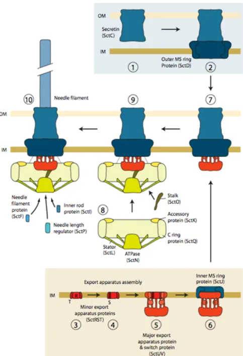
B. Biogenesis of the T3SS
The first studies on the biogenesis of T3SA have been performed in Salmonella (Diepold and Wagner, 2014). Today it is accepted that the injectisome assembly starts with the Sec-dependent formation of a stable ring in the outer membrane, the secretin composed of SctC, followed by the formation of a set of rings associated with the inner membrane, the MS ring composed of SctD and SctJ (Figure 3, panel 1-6). One of the studies that support this proposal shows that the secretin and MS rings associate in a process independent of type 3 secretion, since it can occur in the absence of the T3SS ATPase (Kubori et al., 2000). In support for an initial role in T3SA assembly Schraidt et al. showed that in the absence of secretin fewer complexes were observed, indicating a role for the secretin in the complex stability (Schraidt
et al., 2010). In accordance with those findings, in the case of EPEC it was found that all three
basal proteins (EscCDJ) were required for assembly of the T3SA (Ogino et al., 2006).
Assembly of the inner and the outer membrane rings (Figure 3, panel 7) is presumed to allow the recruitment of the cytoplasmic components (Figure 3, panel 8 and 9). The cytosolic components (Figure 3, panel 8) assemble at the proximal side of the MS rings, as observed for the flagellum (Diepold et al., 2010). The type III ATPase and the C ring require the presence of each other for their recruitment. Both of them interact with accessory proteins (YscK and YscL in the case of Yersinia) to assemble at the proximal side of the basal body (Diepold et
al., 2012). The observation that the C ring, stalk and accessory proteins co-purify with the
cytosolic domain of the switch protein SctU suggests that the switch protein provides the docking site (Botteaux et al., 2010).
The next step of the T3SS assembly is the formation of the needle filament (Figure 3, panel 10). The assembly of SctUVJ with SctC and SctD allows the recruitment of the cytoplasmic components, leading to a functional T3SS. At this step the T3SS secretes the
early substrates, among others the needle filament protein that needs the co-secretion of the inner rod and the needle length regulator (Chapter V, D).
Figure 3. Model of injectisome assembly. The secretin ring in the OM assembles independently and triggers the assembly of
the outer MS ring (1, 2). In the inner membrane, MS ring assembly nucleates the minor export apparatus proteins and progresses via the recruitment of the switch protein and major export apparatus protein (3–5). Both the secretin – outer MS ring protein complex and the export apparatus complex can associate with the inner MS ring protein (6, 7). Integration of the two assemblies into one complex allows subsequent recruitment of the cytoplasmic components (7–9). This leads to a functional type III secretion system, firstly secreting early substrates, amongst others the components of the inner rod and needle filament proteins (10). From Diepold and Wagner 2014.
The secretion of the needle subunit is assisted by a chaperone, which prevents premature filament formation in the bacterial cytosol (Sal-Man et al., 2013). This leads to needle elongation that occurs by subunit polymerization at the distal end, through partial refolding of the subunit protomers from alpha-helix into beta-strand conformation (Poyraz et
al., 2010). The length of the needle is controlled by a mechanism involving the "molecular
ruler" protein SctP and the switch protein SctU.
Several studies have demonstrated that effector protein translocation starts within seconds after host cell contact and that the translocation rates of effector proteins can vary between effectors (Enninga, 2005; Rosenshine, 2009). The different rates suggest that a hierarchy of effectors' translocation is required for the efficient manipulation of host cellular pathways. Furthermore, the temporal regulation of effector protein translocation might prevent interference of effector proteins with antagonistic activities, as shown for SopE, that induces actin polymerization, and SptP that disrupts the actin cytoskeleton in the case of Salmonella
spp.
C. The basal body and Needle Complex
The basal body is composed of the secretin in the outer membrane connected by a hinge region to the MS rings associated with the inner membrane (Schraidt et al., 2011). EM reconstructions of the Salmonella Typhimurium injectisome have shown that the basal body is formed by InvG, PrgH and PrgK (SctC, SctD and SctJ in the universal nomenclature) (Marlovits et al., 2004). In Salmonella, quantitative amino acid analysis revealed that the components of the base InvG:PrgH:PrgK are present in 1:1:1 molar ratio, suggesting that the three proteins are structurally linked by a shared rotational symmetry. The symmetry of inner membrane MS rings' structure appears to be conserved among T3SSs (e.g.: Shigella, Hodgkinson et al., 2009), while the stoichiometry of the secretin, InvG (SctC) may vary between species (e.g.: Yersinia, Kudryashev et al., 2013).
Within the lumen of the basal body, SctI forms a cylindrical “inner rod” structure that supports the extracellular needle formed of SctF. The needle complex protruding from the extracellular side of the T3SS basal body is formed by a helical assembly of SctF, with an outer diameter of 8 nm and an inner diameter of 2 - 3 nm (Radics et al., 2013). In the case of EPEC, a filament formed of polymerized EspA prolongates the needle structure composed of EscF (SctF). This filamentous extension of the hydrophilic protein from the needle is unique
to EPEC family (Knutton et al., 1998). The EspA filament has an internal diameter of 2.5 nm, similar to that of the EscF needle (Cheung et al., 2015). This suggests that the function of the EspA filament is to extend the T3SS transport conduit to reach the target cell membrane through the intestinal glycocalyx (Mueller et al., 2008).
Figure 4. Overview of the injectisome and its components. Left and middle panels show surface representations of 3D
reconstructions of NCs based on cryo-electron microscopic data. Right panel shows a drawing of the T3S holo-complex indicating all its components. From Diepold and Wagner, 2013.
The EPEC EscF is a 8-kDa protein (Wilson et al., 2001), showing homology to the major components of T3SS needle structures of Salmonella (PrgI, 24% identity), Shigella (MxiH, 25% identity), and Yersinia enterocolitica (YscF, 20% identity) (Ogino et al., 2006). The EspA forms a hollow filament with helical symmetry with 5.6 subunits/turn for a 1 start helix and an inner channel permitting translocation of proteins (Daniell et al., 2003). The formation of the EspA filament is dependent on T3S and EscN (the T3SS ATPase), EscC (the outer membrane secretin) and EscF. The polymerization of EspA filaments is mediated by coiled–coil interaction between subunit polypeptides (Delahay et al., 1999) in the same way as flagella are assembled from flagellin (Hyman and Trachtenberg, 1991).
The flagella and injectisomes have considerable organizational similarity, pointing to common and specialized functions of the different T3SS parts. Phylogenetic studies suggested that both systems share a common ancestor (Gophna et al., 2003). In particular, the basal structures are very similar. The needle structure of the T3SS corresponds to the flagellar hook
(Daniell, 2001). The EspA filament has approximately half the diameter of flagellum (120 vs 230 Å) and is required for effector injection as opposed to bacterial motility.
D. Control of the needle length
This regulated length of the needles has been explained in Yersinia by the "molecular ruler" model. In this model, the type III substrate SctP is presumed to "measure" the length of the needle by being physically stretched by the extending needle. In agreement with this model, there is a correlation between the number of the residues in a central domain of SctP and the needle filament length. Upon maximal SctP extension, a signal is transmitted leading to a stall of secretion of the needle component and recognition switch towards tip complex components (Notti and Stebbins, 2016). Alternatively, in the case of Salmonella, Marlovits et
al., suggested that SctP regulates the needle length through control of the inner rod assembly.
This alternate model is based on the observation that mutants lacking SctP show a decrease density in the inner rod-supporting region, generating long needles (Marlovits et al., 2006). Thus, the same team proposed that SctI and SctF are simultaneously secreted and that the completion of the inner rod assembly terminates needle growth in a SctP-dependent manner. Consistent with this model, the overexpression of SctF or SctI leads to longer and shorter needles, respectively (Galan et al., 2014).
E. Tip complex and translocon
Many of the properties of the tip complex and the translocon are not well understood, in particular in the case of EPEC. The outstanding questions are the following: how is the translocon inserted in the membrane? Is this associated with conformational changes in the needle and translocon components? How is the link between the needle and the translocon components regulated? How is the translocon pore formed? What regulates pore formation? We will review below studies pertinent to these questions in systems that have been better characterized.
After the assembly of the needle tip complex, the T3SS is inactive. Although it remains inactive, it does not secrete effector proteins efficiently. This “standby mode” requires the presence of a “plug” protein complex including a “gate-keeper” protein (SctW), which is thought to be located at the cytosolic interface of the injectisome; as well as the tip complex components, consisting of a hydrophilic and the major hydrophobic proteins.
Translocation of effector proteins into the eukaryotic cell cytosol requires the T3SS translocon, inserted into the host plasma membrane upon cell contact. After the host cell contact the minor hydrophobic protein of the translocon (EspB in EPEC) is secreted (Figure 5).
The translocon consists of two hydrophobic proteins, a major and minor one: in the case of EPEC, these hydrophobic proteins are EspD, the major component and EspB, the minor component (Mueller and Cornelis, 2008).
Figure 5. Scheme showing the transition from the tip complex to translocon insertion upon cell contact. Modified from a
figure kindly provided by Eduardo Soto from «Departamento de Genética Molecular, Instituto de Fisiología Celular Universidad Nacional Autónoma de México».
The major and minor hydrophobic components form a hetero-oligomer inserted and forming a pore in the host cell plasma membrane.
Among the different species, the tip complex and translocon components do not share extensive homology, but are believed to perform the same function. The hydrophilic
component acts as a link between the major hydrophobic protein and the T3SS needle. The needle interacts with the hydrophilic component at least in part, through its elongate coiled-coil motif observed in all T3SS hydrophilic tip proteins described to date.
In the case of Shigella and Salmonella, some studies showed that the hydrophilic protein (IpaD/SipD respectively) possesses an amino-terminal auto-chaperoning sub-domain (Notti et al, 2016). This hydrophilic protein's domain may prevent the premature oligomerization of the major hydrophobic component. However, this domain has not been identified in Yersinia or Pseudomonas tip complex components, suggesting that it may not represent a general feature of T3SSs.
In Shigella during in vitro growth, the T3SS is inactive. The composition of the tip complex has been studied under various conditions and using bacterial mutants. Picking published the first detailed characterization of the tip complex (Picking WL et al. 2005). After the construction of several ipa mutants, different tests were performed in RBC (red blood cells) membranes. Lysis of RBCs provides a read-out of the T3SS-dependent pore formation into host cell membrane and permits to measure the pore size through osmoprotection studies. The results revealed that the pore size, presumably corresponding to the translocon inner-diameter, corresponds to 2.5 to 3 nm.
Mutation in the N-terminal (aminoacid 1 to 20) of IpaD leads to a de-regulated secretion of IpaB and IpaC. These results are in accordance with the hypothesis that the tip complex serves as a T3SA plug, preventing type III secretion at basal state. Consistently, ipaB and ipaD mutants also show constitutive type III secretion. Findings of the group of William Picking suggest that at the basal state, the tip complex is composed of a homopentamer composed of five copies of the hydrophilic component IpaD. Those findings are consistent with Muller et al., who showed that the TC of Yersinia is also formed by a homopentamer of the ortholog LcrV (Muller et al., 2005). However, the TC's composition may vary depending on the growth conditions (Picking, 2016). When Shigella was grown to medium supplemented with 2.5 mM deoxycholate (DOC) – a concentration that simulated the physiological range observed in the human intestine – IpaB was detected on the surface of the bacteria. Immuno-labeling of the bacterial surface indicates that IpaD can be observed without IpaB, but not the opposite. How DOC leads to IpaB recruitment to the needle is not clear allowing the speculation that it is based on conformational changes induced by DOC (Stensrud et al., 2008). Using molecular docking simulation on the IpaD crystal structure and fluorescence energy transfer, it was predicted that DOC binds IpaD in a cleft formed by the
central coiled-coiled and the N-terminal domain of IpaD, presumed to regulate IpaB recruitment. During their travel in the intestinal lumen, bacteria encounter bile salts, including DOC, that are present at high concentrations in the host duodenum. As a result of the interaction of IpaD with bile salts, conformational changes in IpaD stimulate the recruitment of IpaB at the needle tip (Picking et al., 2008).
Contrarily to what is expected based on Shigella results, bile salts repress Salmonella T3SS and invasiveness (Wang et al., 2010). In Salmonella, bile salts repress the transcription of T3SS genes and reduce the secretion of T3SS effector (Prouty and Gunn, 2000). NMR studies indicate that bile salts interacted with nearly the same set of SipD residues. However, the largest chemical shift perturbations occurred at a distance from the predicted bile salt binding site in IpaD. These differences may explain the opposite effects of bile salts on T3SS activity observed in Shigella and Salmonella.
There is a controversy about the Shigella TC's composition. Ariel Blocker and colleagues found that the TC is a heteropentamer consisting of four molecules of IpaD and one molecule of IpaB (Blocker et al., 2008). More recent EM studies have corroborated this model (Cheung et al., 2015). In this model, the four IpaD copies are proposed to assemble first. Following secretion, the first copy of IpaD would assemble onto the needle via interaction with an MxiH subunit located below. The second, third and fourth copies of IpaD would then assemble through similar interactions with MxiH subunits as well as with a neighboring flanking IpaD copy. Because of the helical nature of the MxiH needle, the fifth site formed by the IpaD copies would differ from the others and be lined on both sites by IpaD. They assumed then that the unique nature of this site would favor the insertion of IpaB. There are several reasons that explain the lack of precise characterization of the EPEC Tip Complex:
1) As opposed to Shigella, the induction of the expression of genes involved in T3SS assembly requires bacterial growth in a eukaryotic cell culture medium. Growth in this cell medium also triggers the secretion of translocator components and type III effectors in the bacterial supernatant;
2) The EPEC hydrophilic component polymerizes to form a filament, introducing another levels of complexity in the regulation. For example, the EspA filaments show different lengths and it is not clear what regulates EspA polymerization and switch to the translocon component secretion;
3) While the expression of EspA and EspD, together with that of other LEE components, is up-regulated during growth in cell medium compared to regular growth in classical bacterial culture medium, there is an additional up-regulation of EspA-D expression upon cell contact, indicative of other levels of control. For all these reasons, it has not been possible to "freeze" the system in a configuration where the tip complex caps the T3SS needle, prior to host cell contact.
VI. AB Toxin-like model
The fact that the coiled-coil domains of hydrophobic translocon components show analogy with pore forming toxins (Barta et al., 2012) supports the idea that both have common origins and oligomerization mechanisms. In the case of Yersinia, a model different from the canonical T3SS model has been proposed (Edgren et al., 2012). This model resembles the classical AB5 toxin delivery mechanism and is based on the following observations. Akopyan and colleagues performed immunoelectron microscopy of HeLa cells infected with Yersinia pseudotuberculosis to analyse the spatial localization of effectors upon host-cell interaction (Akopyan et al., 2010). They observed that the effector protein YopE was distributed at the bacterium-target cell interface and was not associated with distinct foci or macromolecular structures, as expected for T3SS canonical translocation. In addition, the bacterial outer membrane was in close contact with the host cell plasma membrane, in the absence of bridging structures. Under growth conditions at low Ca2+ concentrations leading to
Yop protein expression, YopE was primarily found on the surface of the bacteria and not in the cytoplasm as predicted in the typical T3SS model (one step model). Moreover, the surface localized YopE did not colocalize with T3SAs. Both YopH and YopD were also mainly detected on the surface of Y. pseudotuberculosis. The next step was to investigate if the surface localized Yops were translocated to the HeLa cells being able to induce cytotoxicity. YopH is a phosphotyrosine phosphatase (PTPase) responsible for the inhibition of the immediate-early Ca2+ response during infection (Andersson et al., 1999). When YopH purified in vitro was added in the extracellular medium of cells infected by a yopH mutant, the same blockage of Ca2+ response was observed, but not in non-infected cells or cells infected with a T3SS-deficient strain. These findings suggest that the surface-localized YopH is translocated in cells in a T3SS-dependent manner.
Figure 6. Progression of injectisome assembly and activation. In the early stage the assembled basal body recognizes early
substrates (green) for secretion. Those substrates are required for the needle assembly. Once the needle assembly is completed, YscU (orange) undergoes autocleavage, which triggers a substrate specificity switch (middle stage). YopN associates with the basal body to allow middle substrates (red) to be secreted. Those middle substrates are required to form the tip complex and translocation pores. Upon cell contact, YopN is released from the basal body and secreted triggering the transition for the late stage. In the one-step model, the pore complex assembles at the tipoff the needle creating a continuous channel, through the late substrates (yellow) are injected. In the two-steps model, late substrates are secreted into the extracellular space and interact with pore proteins (YopB/D) allowing the translocation into the host cell. Adapted from Dewoody et al., 2013.
A two-steps model is proposed whereby YopH, secreted in the extracellular medium hijacks translocon components by binding to YopB associated with host cell membranes (Akopyan et al., 2011). Endocytosis of the YopB-YopH complex then accounts for YopH injection into cells. This model is reminiscent of a binary AB-toxin-like mechanism, where the major hydrophobic translocon component plays the role of the toxin B component that mediates the translocation of the catalytic A-moiety across the host-cell plasma membrane.
While this model may represent an alternate pathway for type III effector injection, these studies also raise important questions about the actual evidence for type III effectors translocation through a pore formed by the translocon in the host cell membrane.
The hydrophilic protein does not integrate the membrane, but is necessary for pore formation. Does this hydrophilic protein act as an assembly platform for proper oligomerization of the translocon components or is it mostly involved in the connection between the T3SS needle and the translocon during injection?
In the canonical view, type III effector proteins cross a sealed conduct that does not allow exchanges with the extracellular medium, to reach the host cell cytosol. In support of this view, several Cryo-EM studies show a short spacing between the bacterial surface and the host membranes at T3SS contact. In particular, a recent study showed that multiple
Chlamydia T3SS-host membrane contact with an average spacing of 33 nm (Nans et al.,
2015).
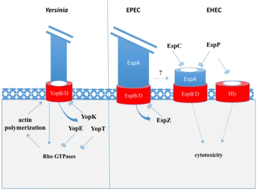
VII. Regulation of Pore formation by a bacterial serine protease
As described before, the T3SS-mediated injection of bacterial effector through eukaryotic cell plasma membranes requires the insertion in host cell membranes of the translocon connected to the T3SS needle by the hydrophilic protein (Fig. 7). Insertion and regulation of the translocon into the host cell membrane is a complex process. Sheahan and Isberg have identified host cell factors required for Yersinia T3SS-associated pore activity and showed that numerous cytoskeletal and membrane trafficking proteins are involved (Sheahan and Isberg, 2015).
Briefly, upon cell contact the Yersinia YopB/D translocon components activate Rho GTPases leading to the polymerization of actin and T3SS-dependent pore formation (Bliska et al., 2013). Injected T3 effectors, such as YopK, YopE and YopT (Figure 7), downregulate T3-pore formation and effector translocation. YopK directly acts on the T3 translocon while YopE and YopT inhibit Rho GTPases (Dewoody et al., 2013). In EPEC, EspZ acts in the same way as YopK, by interacting with the translocon components EspD and preventing the translocation of T3SS effector into infected cells (Berger et al., 2012).
Recently the host lab has reported a novel mechanism of controlling T3SS-dependent pore formation by EPEC and EHEC (Guignot et al., 2015). EspC is a bacterial toxin secreted by a type V (autotransporter) secretion mode. EspC belongs to the serine protease autotransporter of Enterobacteriaceae (SPATE) family and has been involved in EPEC virulence through its proteolytic activity on host proteins, as will be developed further. Interestingly, Guignot et al showed that EspC degrades the EspA and EspD components, following contact with epithelial cells, thus down-regulating T3SS-dependent pore formation and cytotoxicity. EspC does not prevent type III effector injection or actin pedestal formation, suggesting that it may recognize a specific conformation of EspA/D. It is speculated that EspC targets a complex of EspA-EspD, presumably in association with the translocon inserted in the host cell membrane. Interestingly, EspP (the EspC orthologue in EHEC) which shows the same proteolytic activity towards EspA and EspD, has been involved in the proteolytic degradation of the E. coli hemolysin Hly, a pore forming cytolysin, leading to the inactivation of the pore-forming activity (Brockmeyer et al., 2011). These findings point to a role for these SPATEs in the controls of bacterial-induced pore in the host cell plasma membranes, and the control of host cell death and inflammatory processes.
VIII. The Serine Protease EspC
A. SPATES – General overview of EspC
SPATEs are widely spread among Enterobacteriaceae. SPATEs have a common structure, being composed of three different parts: a N-terminal domain composed of a cleavable peptide signal, a central passenger domain that corresponds to the mature protein with a conserved serine protease motif (GDSGSG) and a C-terminal autotransporter domain (Figure. 8). They cleave proteins implicated in diverse functions, such as fodrin, pepsin A and
hemoglobin (Dautin, 2010). Autotransporters are exported in the bacterial periplasm through the Sec pathway. They promote their secretion by using their autotransporter domain that forms a beta-barrel in the outer membrane, through which the passenger domain translocates. Following cleavage, the mature form the SPATE is released in the extracellular medium (Henderson et al., 2004).
Figure 8. Autotransporter Proteins, common domain organization of AT proteins and Type V Secretion System. EspC as a
SPATE is composed of 3 parts: signal peptide in the N-terminal; autotransporter (ß) domain in the C-terminal that allows the secretion of the mature protein in the extracellular milieu; passenger domain that corresponds to the mature proteins and linker domain. Adapted from (Navarro-Garcia et al., 2010).
EspC is the first protein secreted by EPEC during cellular infection. Even though EspC is encoded by non-LEE locus, it is regulated by Ler (Chapter V). The secretion of EspC in the extracellular milieu is independent of T3SS. However, it has been showed that while EspC secretion is T3SS-independent, EspC is translocated into the epithelial cells in a process requiring active type III secretion (Vidal and Navarro-Garcia, 2008). Indeed, EspC is detected in the cytoplasmic fraction of cells infected with wild-type EPEC, but not in an escN mutant deficient for the T3SS ATPase, despite the fact that the escN mutant secretes EspC (Vidal and Navarro-Garcia, 2008). This process is highly specific for EspC since another SPATE (Pic) is not internalized in the same conditions (Vidal and Navarro-Garcia, 2006). Interestingly, the expression of EspC is coupled to that of the T3SS suggesting a coordinated function of these virulence determinants (Guignot et al., 2015). However, how exactly EspC “hijacks” the type III secretion system to gain access to the eukaryotic cell cytoplasm is not known.
B. Cytotoxicity of EspC
EspC induces the cleavage of several eukaryotic proteins, such as the coagulation factor type V, pepsin and hemoglobin (Dautin, 2010). EspC also induces the cleavage of fodrin, a protein associated with the actin cytoskeleton and two other focal adhesion proteins,
paxillin and focal adhesion kinase (FAK), following its translocation into host cells, presumably leading to cytotoxicity of epithelial cells (Navarro-Garcia et al., 2004). The intracellular effects of EspC are sequential, first leading to fodrin degradation, followed by paxillin degradation, FAK dephosphorylation and FAK degradation, associated with cell rounding, cell detachment and cell death. Those events are not found in an espC isogenic mutant, even though this mutant causes AE lesions and is proficient for the injection of type III effectors (Navarro-Garcia et al., 2014).
C. EspC regulates pore formation and cytotoxicity mediated by the T3SS
Analysis of the bacterial secretion profile was the first hint of an activity of EspC on EPEC translocator components. When bacteria are grown in vitro in cell culture medium, EspC decreases the amounts of secreted EspA and EspD, while the levels of EspB are not affected (Guignot et al., 2015). The EspC-mediated degradation are linked to its proteolytic activity, since it is inhibited by treatment with the serine protease inhibitor PMSF, and not observed with EspC-S256I, a catalytically inactive variant. Similar effects are observed when using purified proteins. These experiments also pointed to a predominant proteolytic activity of EspC on a fraction containing EspA and EspD fraction, compared to a fraction containing EspA filaments. Immunofluorescence microscopy indicated that the EspA-EspD fraction was associated with punctiform structures, indicative of specific complexes. Consistently, EspC also regulates the levels of EspA and EspD during the early stages of cell infection by EPEC.
EspC was shown to negatively regulate the formation of T3SS-dependent pores in the plasma membrane of cells during bacterial infection, in fluorescent dye loading experiments. Increased pore formation in cells infected with the ∆espC mutant also increases bacterial-induced cell death, compared to WT EPEC.
R
ATIONALEThis Master internship builds from the hypothesis that EspC targets a specific translocon conformer that may disconnect from the T3SS needle to form a pore in the host cell membrane.
It aimed at taking advantage of the fractionation procedures worked out to isolate the EspA-EspD complex sensitive to EspC proteolysis to perform a structural characterization of this complex by electron microscopy analysis. Further, we will attempt to gain insights into the interaction between the protease and EspA-EspD by performing the structural characterization of EspC-EspA-EspD complex.
M
ATERIAL ANDM
ETHODS I. Analysis of secreted proteinsAll the strains used in this study are listed in the table 3.
Strains were grown in Luria-Bertani broth (LB) for 16 hours in a 37 °C shaking incubator at 200 rpm. To induce the expression of LEE genes and activate type III secretion, EPEC LB pre-cultures were diluted LB 1:200 in DMEM containing 1 g/L glucose (Gibco) and incubated for 5h at 37 °C in a 10% CO2 incubator. The equivalent of 10 ml of bacterial
culture were centrifuged at 8000 g for 10 min at 4°C.
Table 3. Bacterial strains and plasmids list.
Strain Characteristics Reference
WT E2348/69 EPEC wild-type strain 2348/69 (Levine et al., 1985) ΔespC espC isogenic mutant of WT E2348/69
(espC::kan)
(Guignot and Tran Van Nhieu, 2015)
ΔespA espA isogenic mutant of WT E2348/69 (Knutton et al., 1998) ΔespD espD isogenic mutant of WT E2348/69 (Garmendia et al., 2004) ΔescN escN isogenic mutant of WT E2348/69
(escN::kan)
(Garmendia et al., 2004)
pJLM174 espC cloned in pBAD30 (Mellies et al., 2001)
pJLM174-S256I espC-S256I cloned in pBAD30 (Guignot and Tran Van Nhieu, 2015)
The supernatants were filter-sterilized using a 0.22 µm-pore-size filter (Millex) and transferred to 15 ml-Falcon tubes. Proteins were subjected to precipitation using trichloroacetic acid (TCA) at a 5% final concentration for 1 hour on ice. Samples were centrifuged for 5 min at 8000 g at 4°C in a (Eppendorf, 5810 R) centrifuge. Pellets were washed with 500 µL of acetone pre-cooled at -20 °C to remove salts. Samples were immediately centrifuged for 5 min at 8000 g (Eppendorf, 5424). The wash with acetone was repeated 2 times. Pellets were resuspend in 50 µL of Laemmli loading buffer (0,0625M Tris-HCl pH 6.8; 2% SDS; 10% Glycerol; 0.005% Bromophenol Blue; 5% βME) and boiled for 10 minutes. 25 µL of each sample were analyzed by SDS-PAGE using a 12.5% polyacrylamide gel. Samples were allowed to migrate for 1h30 at a constant amperage of 25 mA and analyzed by Coomassie blue staining, by incubating for 30 min in 10% acetic acid, 50% ethanol and
40% H2O containing 0.25% Coomassie Blue, followed by incubation successive washes in
distaining buffer (6.25% acetic acid; 5% ethanol).
II. EspC Purification
To purify EspC, the strain DH5-α (pJLM174) over night (ON) LB pre-culture was diluted at 1:200 in 1 L LB medium supplemented with arabinose (0.2%) and ampicillin (100 µg/ml) at 37°C with shaking at 200 rpm for 16 h. All subsequent steps were carried at 4°C unless otherwise stated. Cultures were centrifuged at 6000 g for 15 min in a Beckman (J-26 XP) centrifuge. Clarified supernatants were filtered through a 0.22-µm-pore-size filter (Stericup, SCGPU05RE, Millipore). Proteins from filter-sterilized supernatant were precipitated by the addition of ammonium sulfate at 40% final concentration. Precipitation was performed by batch addition of the appropriate amounts of ammonium sulfate over a two hours-period, to ensure protein precipitation. Batches were added to the chilled supernatant with constant stirring, until complete crystal dissolution. Samples were centrifuged at 6000 g for 10 min. The supernatants were discarded and the pellets were resuspended in 25 mM Tris-HCl (pH 7.4), 25 mM NaCl and 1 mM β-mercaptoethanol (βME) in a volume equal to 1:50 of the supernatant. Samples were dialyzed 3 times against 100 volumes of the same buffer, using a 3.5 kD cut-off dialysis membrane (Spectra/Por, 132720). The dialyzed samples were filtered through a 0.22 µm-pore-sized filter to remove potential aggregates. Samples were subjected to FPLC (Flow Pressure Liquid Chromatography) using an anion exchange column (Mono Q, GE Healthcare) and a 20 ml 25 mM – 1000 mM NaCl linear gradient, collecting 1 ml fraction. The fractions were analyzed by SDS-PAGE and Coomassie staining, as described in
Analysis of Protein Samples section. III. EspA/D Purification
EspA and EspD were purified from the supernatant of ΔespC strain using modifications from published procedures (Guignot et al., 2015). The optimal growth conditions were determined in a set of pilot experiments (see Results section, (III)). Bacterial pre-cultures performed in LB were used to inoculate (dilution 1:20) DMEM cultures containing 0.1M HEPES at 37°C with 200 rpm shaking for 5 or 6 hours in a 5 liters-Erlenmeyer flask for 1 liter culture. All subsequent steps were carried at 4°C unless otherwise stated. Samples were centrifuged at 6000 g for 15 min to remove the bacteria in a Beckman (J-26 XP) centrifuge. Supernatants were filtered through 0.22-µm-pore-size filters. Proteins from the sterilized supernatant were precipitated using 40% ammonium sulfate, with constant stirring at 4°C as
described for the EspC purification protocol. Samples were centrifuged at 6000 g for 10 min at 4°C. The supernatants were discarded and pellets were resuspend in 50 mM Tris-HCl (pH 7.4), 50 mM NaCl and 1 mM β-mercaptoethanol (βME) in a volume equal to 1:50 of the supernatant. Samples were dialyzed 3 times for 1 hour against 100 volumes of the same buffer using a 3,5 kD-cut off dialysis membrane. The supernatants were filtered through 0.22-µm-pore-size filters and proteins where separated by FPLC using an anion exchange column (Mono Q, GE Healthcare) using a 20 ml-linear gradient of 50mM – 600mM NaCl, collecting 1 ml fraction. The fractions were analyzed by SDS-PAGE and Coomassie staining, as described in Analysis of Protein Samples section.
The fractions containing EspA and EspD, were pooled and dialyzed against 20 mM HEPES and 20 mM NaCl. The GraFix protocol was applied to the pooled fractions (Stark, 2010). The buffers were prepared based on the table 4 and filtered through a 0.22-µm-pore-size filter (Stericup, SCGPU05RE, Millipore). Using a specialized gradient mixer to form a continuous gradient a 4.4-ml centrifuge tube was filled with 2.1 ml of each buffer. 200 µl of the samples was load in the top of the gradient. The ultracentrifugation was performed at 50,000 RPM for 16h (Beckmann SW55-Ti rotor). The fractionation was performed using a capillary to pump the gradient out from the bottom to the top, taking fractions of 200 µl. The fractions were analyzed by SDS-PAGE (5% polyacrylamide gel) and Coomassie staining, as described in Analysis of Protein Samples section.
Table 4. Buffers composition to the gradient of GraFix procedure.
Buffer A (top) Buf (bottom)
50 mM HEPES pH: 7.5 50 mM NaCl 10% Glycerol - 50 mM HEPES pH: 7.5 50 mM NaCl 30% Glycerol 0.15% Glutaraldehyde
200 µl of the pooled fraction from anion exchange column (Mono Q) were subjected to size exclusion chromatography (Superdex 200, 25 ml column, GE Healthcare) using the same buffer as the dialysis buffer, collecting 0.5 ml-fractions. Absorption (A280 nm) peaks were
IV. Electron Microscopy
EspA and EspD containing samples were analyzed by transmission electron microscopy following negative staining. Before the staining of the samples, grids were placed in paper in a carbon coated machine, until the vacuum reaches a level of 10-5 Torr. 4 µl of each sample were applied for one minute to the glow discharged carbon coated grids (CF400, EM, USA). Samples are processed for contrasting with 2% Uranyl acetate in water. The images were recorded on Tecnai Spirit (FEI, USA) under 120kv with an Eagle (FEI, USA) 4k x 4k camera using the TIA software. The observations were done using TEM Bright Field SA 49000X.