i
ARCHMAT
(ERASMUS MUNDUS MASTER IN ARCHaeological MATerials Science
)
Mestrado 2 Ciclo
Exploring Trace Elemental Analysis of human remains from
San Pablo Medieval site using ICP-MS
Deepshikha Sharma
M36177
Dr. Jose Miguel Carretero Díaz Supervisor, Universidad de Burgos Dra. Rebeca García González
Co-Supervisor, Universidad de Burgos Prof. Nick Schiavon
Co-supervisor, Universidade de Evora
ii
ARCHMAT
(ERASMUS MUNDUS MASTER IN ARCHaeological MATerials Science
)
Mestrado 2 Ciclo
Exploring Trace Elemental Analysis of human remains from
San Pablo Medieval site using ICP-MS
Deepshikha Sharma
M36177
Dr. Jose Miguel Carretero Díaz Supervisor, Universidad de Burgos Dra. Rebeca García González
Co-Supervisor, Universidad de Burgos Prof. Nick Schiavon
Co-supervisor, Universidade de Evora
iii
Explorando Trace Elemental Análise de restos humanos do local medieval de San Pablo usando ICP-MS
Resumo
A abordagem analítica do uso de Trace Elements (TEs) pode ser usada não só para entender o ambiente vivo e a dieta, mas até a diagênese pós mortem de qualquer resíduo humano usando a variabilidade nas composições elementares de dentes e ossos. Oito indivíduos foram amostrados do convento de San Pablo em Burgos usando seus dois tipos de tecido ósseo (ossos cortical e trabecular) do fêmur e esmalte dentário para cada caso. Três dessas amostras pertenciam à nave da igreja (século 16th -19th) enquanto o resto era encontrado no pátio do
claustro pertencente ao século XIV-XVI. Essas amostras foram processadas na Espectrometria de Massa Plasmática Acoplada Induzamente (ICP-MS) para analisar as concentrações de Ca e P, bem como os TEs não essenciais e bio-essenciais, a fim de poder estabelecer a integridade das amostras e para descobrir quais TEs podem ser úteis para fazer inferências sobre a dieta antiga e a absorção diagenética e em que medida. Os dados de TE foram tratados usando diferentes ferramentas estatísticas e testes para encontrar possíveis diferenças de gênero ou mesmo diferenças intra-locais na dieta e também foram corroborados com informações coletadas a partir da análise de microwear. Verificou-se que a dieta dos indivíduos era de tipo misto com componentes vegetais e de carne, enquanto a presença de alimentos marinhos não pôde ser confirmada. Isto foi de acordo com os resultados de microwear para algumas das amostras. Usando as razões de Ba e Sr por Ca, verificou-se que os ossos corticais deram os resultados mais confiáveis para inferências sobre dieta, excluindo o uso de níveis de Mn e Fe que foram altamente afetados pela absorção diagenetica nos tecidos ósseos. Além disso, as amostras enterradas na nave da igreja podem estar consumindo mais proteínas de carne do que as enterradas no claustro, o que pode indicar uma diferença em seu status social ou uma mudança na dieta ao longo do tempo. Como esperado, para a maioria dos elementos, como Pb, Mn, Fe, Cu, os tecidos trabeculares foram os mais afetados pela absorção diagenetica, ademais, na em sua maioria superfície interna do eixo do fêmur.
Palavras-chave: Trace Elements, ICP-MS, San Pablo, palaeodiet, diagénese, microwear, estatísticas
Abstract
The analytical approach of using Trace Elements (TEs) can be used not only to understand the living environement and diet but even post-mortem diagenesis of any human remains using the variability in the elemental compositions of both teeth and bones. Eight individuals were sampled from the convent of San Pablo in Burgos using their two types of bone tissues (cortical and trabecular bones) from the femur and tooth enamel for each case. Three of these samples belonged to the church nave (16th -19th C) while the rest were found from the cloister courtyard belonging to 14th -16th Century. These samples were processed in solution mode Inductively Coupled Plasma Mass Spectrometry (ICP-MS) to analyse concentrations of Ca and P as well as both non-essential and bio-essential TEs in order to be able to establish the integrity of the samples and to find out which TEs can be helpful in making inferences on ancient diet and diagenetic uptake and to which extent. The TE data was treated using different statistical tools and tests to find possible gender differences or even intra-site differences in the diet and was also corroborated with information gathered from microwear analysis. It was found that the diet of the individuals was of a mixed type with both vegetal and meat components while presence of marine food could not be confirmed. This was in accordance with the microwear results for some of the samples. Using the Ba and Sr ratios to Ca, it was found that cortical bones gave the most reliable results for inferences on diet excluding the use of Mn and Fe levels which were both highly affected by diagenetic uptake in the bone tissues. Additionally, the samples buried in church nave might be consuming more meat proteins than those buried in the cloister which might indicate a difference in their social status or a change in the diet through time. As expected, for most of the elements such as Pb, Mn, Fe, Cu, trabecular tissues were the most affected by diagenetic uptake moreover mosly at the inner surface of the femur shaft.
v
Acknowledgments
First of all I wish to be grateful to the Universidad de Burgos and especially my supervisor and co-supervisors for guiding me throughout the length of this research. A special mention of the Parque Scientifico Technologico (PCT) lab and my labmates is necessary who worked with me as hard as possible all the way until the end. Without such assistance, this work might not have been produced. There was never a time when they were not available with their guidance and support. I cannot thank enough my friends, classmates and relatives for giving me the motivation to keep going even when the going seemed tough at times. I am deeply grateful to my friend and classmate, Rebecca MacRoberts for her timely support and guidance and also to Pedro Barrulas for taking time from his busy schedule to help with the thesis. And lastly but not the least, the ArchMat program and everybody associated with it, for giving us such pathbreaking opportunities in the course of two years. Surely, it has been a life changing experience for all of us involved.
vi
Contents
List of Tables and Figures ... viii
1 Introduction ... 1
2 Historical and Archaeological context ... 3
2.1 Structure and architectural insights ... 8
2.2 The archaeological background ... 9
2.2.1 Evaluation of the results from the archaeological intervention (2002-2003) ... 11
2.2.2 Burials and material remains found ... 12
3 Scientific Background ... 16
3.1 Elements in the human body ... 16
3.2 The major elements ... 17
3.3 The trace elements ... 17
3.3.1 Essential elements ... 17
3.3.2 Non-Essential elements ... 18
3.4 The Structure of Bones and Teeth ... 19
3.4.1 Teeth ... 20
3.4.2 Bones ... 22
3.5 Trace Elements in Bones and Teeth ... 22
3.6 Main factors for TE uptake in bone and teeth ... 24
3.6.1 Diet ... 24
3.6.2 Diagenesis ... 25
3.7 Trace Element Detection and Quantification using ICP-MS ... 27
4 Objectives ... 29
5 Materials and Methods ... 31
5.1 Bone sampling ... 31
5.2 Bone Sample preparation ... 32
5.3 Tooth Sampling ... 33
5.4 Tooth sample preparation ... 35
5.5 Sample preparation for ICP-MS ... 36
5.6 Semiquantitative and quantitative analysis using ICP-MS ... 36
5.7 Comparison with previous literature ... 38
5.8 Statistical analysis ... 39
vii
5.8.2 Two-tailed t-test assuming unequal variances ... 39
5.8.3 Scatter plots ... 40
5.8.4 One way Analysis of Variance (ANOVA) and Tukey’s Honestly Significant Difference (HSD) ... 40
5.8.5 K-means cluster Analysis... 40
5.9 Microwear analysis for diet reconstruction ... 40
6 Results and Discussion ... 42
6.1 State of preservation and other observations ... 42
6.2 Comparison with previous literature values ... 43
6.3 Regression analysis ... 46
6.3.1 Correlations between elemental concentrations ... 46
6.3.2 Correlation between Ba/Ca and Sr/Ca ratios ... 52
6.4 T-test for gender differences and differences between the burial areas ... 55
6.4.1 Gender differences ... 55
6.4.2 Differences between church and cloister samples ... 57
6.5 Scatter plots ... 59
6.6 One-way ANOVA and Tukey’s HSD ... 64
6.7 Cluster analysis ... 66
6.8 Discussions on diet ... 69
6.8.1 Comparison with microwear analysis results ... 78
7 Conclusion ... 80 7.1 FutureDirections ... 82 Bibliography ... 85 Appendix I ... 98 Appendix II ... 101 Appendix III ... 103 Appendix IV………. 106
viii
List of Tables and Figures
1. Tables
Table 1: Development of permanent dentition (Simon & Stevenson, 1975). ... 21
Table 2: Details of the samples analysed in this study. ... 31
Table 3: Details of bone samples collected. ... 34
Table 4: Details of tooth enamel samples collected. ... 35
Table 5: Digestion program used for the samples. ... 37
Table 6: The NIST SRMs and recovery percentages for all the elements analysed. ... 38
Table 7: Summary of Ca and P measurements for all the three kinds of tissues. ... 43
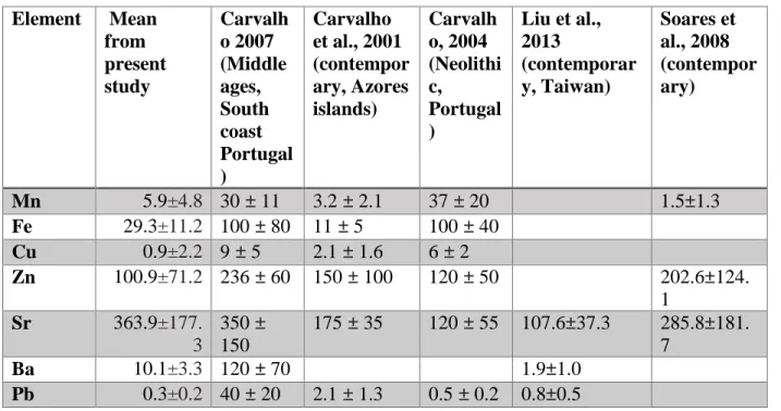
Table 8: Comparison of concentrations (ppm) found in current study with previous studies for tooth enamel. ... 43
Table 9: Additional elemental comparison with previously published values for tooth enamel (Appendix II). ... 101
Table 10: Comparison of literature values of various elements with the current study for compact bones (ppm). ... 44
Table 11: Table showing t-test results for Sr in teeth enamel in males and females (Appendix III). ... 103
Table 12: Table showing mean (ppm) for each trace element for Females (F) and males (M) in tooth enamel and cortical bone levels. ... 57
Table 13: Table showing t-test results for Pb in teeth enamel among samples from church nave and cloister courtyard (Appendix III). ... 103
Table 14: Results for t-test between cloister courtyard and church nave samples from tooth enamel, cortical and trabecular bones. ... 59
Table 15: Example of ANOVA for Mn. F value > Fcritical which leads to the rejection of null hypothesis that all the means are equal. p = 0.05 (Appendix III). ... 103
Table 16: ANOVA for Zn showing the difference between the means is not significant. F<Fcritical, P>0.05 (Appendix III). ... 104
Table 17: Example of Tukey’s HSD result for strontium ... 64
Table 18: Tukey’s HSD results at confidence interval of 95%age ... 104
Table 19: Results for significant difference among elemental concentration between different tissues. ... 65
Table 20: Cluster analysis based on tooth enamel. ... 66
Table 21: Cluster analysis based on compact bones. ... 67
Table 22: Cluster analysis based on Sr and Zn levels. ... 68
Table 23: Trace Elemental results (ppm) for tooth enamel samples (Appendix II). ... 101
Table 24: Trace Elemental results (ppm) for trabecular bone samples (Appendix II). ... 101
ix
Table 26:Variance in the results for concentration of each element (Appendix II).………102
Table 27: log Ba/Sr, log Ba/Ca, log Sr/Ca, Zn/Ca values for cortical bones (Appendix II). ... 102
2. Figures Figure 1: Geological map of Burgos along with the legend (info.igme.es) ... 3
Figure 2: The view of the San Pablo monastery from Isidro Gill (Casillas & Alvarez, 2005) ... 4
Figure 3: Engraving of the city of Burgos, of century XIX, with the view of the convent from the east, by Guesden (Casillas & Alvarez, 2005) ... 5
Figure 4: Map of the 2002-2003 archaeological excavation which shows the total area (22,000 m2) (Memoirs of archaeological intervention 2002-2003) ... 10
Figure 5: Map showing the location of burials (Memoir of archaeological intervention 2002-2003) ... 13
Figure 6: The burials and ossuaries found from the Convent of San Pablo (Casillas & Alvarez, 2005) .... 13
Figure 7 (a, b): Comparison of literature values of various elements with the current study for tooth enamel (ppm) ... 45
Figure 8 (a, b): Correlation between Ca and P concentrations (ppm) in spongy bones and compact bones ... 47
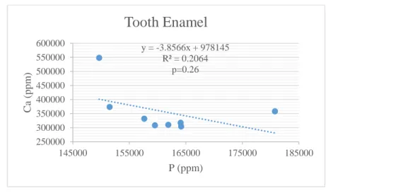
Figure 9: No correlation between Ca and P concentrations (ppm) in tooth enamel………47
Figure 10 (a, b): Inverse correlation of Ca with Cu and Ba in trabecular bones ... 48
Figure 11 (a, b): Inverse correlation of Ca and P with Fe in trabecular bones ... 48
Figure 12 (a, b): Inverse correlation of P with Cu and Ba in trabecular bones ... 48
Figure 13: Inverse correlation of phosphorus levels with manganese ... 49
Figure 14 (a, b): Correlation of iron with manganese in trabecular and compact bones ... 49
Figure 15: Iron does not display any correlation with manganese in tooth enamel ... 50
Figure 16 (a, b): Mn correlation with Cu and Pb in trabecular bones ... 50
Figure 17 (a, b): Ba correlation with Cu and Mn in trabecular bones... 51
Figure 18 (a, b): Ba correlation with Cu and Sr in the compact bone tissues ... 51
Figure 19 (a, b): Cu correlation with Fe in trabecular bones and tooth enamel ... 52
Figure 20 (a, b): Pb correlation with Fe and Zn correlation with Sr in trabecular bones ... 52
Figure 21: Correlation between log Sr/Ca (ppm/mg/g) and log Ba/Ca (ppm/mg/g) for cortical bones ... 54
Figure 22: Correlation between log Sr/Ca (ppm/mg/g) and log Ba/Ca (ppm/mg/g) for trabecular bones .. 54
Figure 23: Correlation between log Sr/Ca (ppm/mg/g) and log Ba/Ca (ppm/mg/g) for tooth enamel ... 54
Figure 24: Pb levels in different tissues in different samples ... 60
Figure 25: Fe levels in different tissues in different samples ... 61
x
Figure 27: Cu levels in different tissues in different samples ... 62
Figure 28: Zn levels in different tissues in different samples ... 62
Figure 29: Sr levels in different tissues in different samples ... 63
Figure 30: Ba levels in different tissues in different samples ... 63
Figure 31: Clusters based on Sr and Zn levels (ppm) in cortical bones ... 69
Figure 32: Mean log (Ba/Sr) values from published data for water and terrestrial samples (Burton & Price, 1990) ... 71
Figure 33: Log Ba/Sr values for all the samples ... 71
Figure 34: Correlation between Cu and log Ba/Sr values ... 72
Figure 35: Trophic level differentiation based on log Ba/Ca (ppm/ppm) and log Sr/Ca (ppm/ppm) values (Peek & Clementz, 2012) ... 73
Figure 36: Sr/Ca and Ba/Ca ratios for natural samples (Peek & Clementz, 2012) ... 74
Figure 37: log Ba/Ca (ppm/ppm) and log Sr/Ca (ppm/ppm) for the compact bones of the samples ... 75
Figure 38: Zn/Ca (ppm/mg/g) values for compact bones ... 77
Figure 39: Plot of NH/NT index vs NV/NT index for four dietary groups and the three specimen. Black lines represent the 95%age equiprobability ellipses of each dietary group and crosses are the centroid of these ellipse (Zuriñe Sanchez Puente, personal communication) ... 78
Figure 40: Discriminant function analysis of four dietary groups based on microwear measurements taken on the buccal surface of the three specimen. Black lines represent the 95%age equiprobability ellipses of each dietary group and crosses are the centroid of each ellipse (Zuriñe Sanchez Puente, personal communication) ... 79
Figure 41: Occlusal and buccal views of a) SP 7533b, b) SP 7525 (Appendix I) ... 98
Figure 42: Occlusal and buccal views of a) SP 7544, b) SP 7568 (Appendix I) ... 98
Figure 43: Occlusal and buccal views of a) SP 7535 b) SP 7581 (Appendix I) ... 98
Figure 44: Occlusal and buccal views of a) SP 7575 b) SP 7579 (Appendix I) ... 98
Figure 45: Sample pictures for bones after sample extraction a) SP 7533b, b) SP 7525, c) SP 7568, d) SP 7535, e) SP 7544, f) SP 7579, g) SP 7581, h) SP 7575 (Appendix I) ... 99
Figure 46: The femur bones after sample extraction from sample a) SP 7579, b) SP 7535, c) SP 7525, d) SP 7544, e) SP 7533b, f) SP 7581, g) SP 7568, h) SP 7575 (Appendix I) ... 100
1
Introduction
The convent of San Pablo de Burgos was one of the first and most important monasteries that the Dominicans founded in Castile, Spain. Many artists took part throughout its construction across the centuries, who made the cathedral famous and some of them later even asked to be buried in one of the chapels of the church. It was located in the city of Burgos founded by Santo Domingo of Guzman in the year 1218 (Conde et al., 1982) according to tradition but most of the historians place its foundation in the year 1224 (Serrano, 1997). It is unanimously agreed upon that the Dominican friars were already established in Burgos between 1219 and 1222.
The historic city of Burgos was the capital of Crown of Castile at one point of time and could be considered as one of the most important centres of medieval Spain. In that time, the convent was an important landmark of the urban Burgales landscape. The history of the convent is in a way entwined with the history of this city. If its history, very close to that of the city, was substancial in historical times until the 18th century, its ruin in the 19th century was also absolute. The oldest site of the convent was located outside the city which was later shifted to its last location. Today, after having gone through turbulent times such as Spanish War of Independence and Spanish confiscation in the nineteenth century, the convent ceases to exist not only physically but also from the citizens’ memory who do not even remember the existence of the convent. The monastery undoubtedly has a lot of archaeological, historical and artistic information about the thriving middle ages of Burgos city and the surrounding areas which have been continuously inhabited since more than 800,000 years ago. The life of the San Pablo convent had always been in close relation with the life of the city and needs to be studied and thus recued from oblivion in order to return it to the popular culture and memory of the city.
Being one of the most influential religious institutions in Burgos city during the 13th to 18th centuries, the convent was preferred by the contemporary inhabitants as the burial place regardless of their economic or social status. Unsurprisingly, numerous burials were recorded in the site during the excavation in 2002-2003. The convent was developed into a prison, barracks and even hospital during the second half of 19th century. By 1870, the ministry of war ordered
2
the still standing ruins of the convent and the church to be demolished for building barracks. Consequently, the convent completely disappeared and the barracks continued to be used for one more century until they were destructed in 1973. By 2010, the museum of Human Evolution was developed on the same site but not before a thorough archaeological investigation during 2001-2004.
The historical, archaeological and anthropological studies on the remains from San Pablo have already provided a lot of information about the medieval communities and their lifestyle in the contemporary times. Nonetheless, there have not been any previous archaeometric investigations into the remains from San Pablo whose history could be recorded better by understanding various aspects of the lost site. The main research goals of this study are to explore the diet of some of the individuals buried in the cloister courtyard and the church nave of the convent in order to find differences based on genders, socio-economic status or chronological period of these burials using trace elemental analysis and statistical tools such as t-tests, regression analysis, ANOVA, cluster analysis and a few others. Apart from this, it is also a focus of this study to be able to make preliminary explorations into developing a methodology to notice diagenetic changes in different TEs using different skeletal elements from the same specimen which might be helpful in cases like this where the original archaeological context and the soil from the site is not available for further examinations. It will be interesting to note which TEs and which skeletal tissues (tooth enamel, cortical bone or trabecular bone) provide more reliable data for inferences on diet and diagenetic uptake.
3
2
Historical and Archaeological Context
Burgos is located in the Castile and Leon province of Spain which has been the most important region and even an important centre of Spain in the past. The city is surrounded by Miocene formations consisting of limestones along with marls while the actual city was fed by seven rivers in the past. Thus, it is situated on river terraces most important of which is the Arlanzon River. These are therefore quaternary fluvial sediments made up of gravel, sand and clay. The monastery most probably was situated on terraces created by Arlanzon, Urbel, Ubierna and Vena rivers (Figure 1).
4
The convent of San Pablo was the point of reference in the urban Burgales landscape and was a Dominican establishment. Although it was difficult to know exactly where the primary location was and also the exact date of its foundation. The excavations have established the location of its last standing claustro at the coordinates of 42.339073°, -3.697135° (Figure 2).
Figure 2: The view of the San Pablo monastery from Isidro Gill (Casillas & Alvarez, 2005)
We knew in 1218, that when the Santo Domingo of Guzman visited Spain on the 22 of December of 1216, honorio 3rd approved the order of preachers in Burgos (Casillas, 2003). It has also been determined that the first Dominicans settled in the city of Burgos around the 1220. They settled in the extramural of the city in the famed neighborhood of La Vega, close to the churches of Saint Cosme and Saint Damian in the south of the city (Casillas, 2003). Right from its foundation, the convent reveled in the patronage of the kings of Castile until the rule of Catholic Monarchs.
The Bull of Vitute Conspicuos established the Dominican friars as independent from the diocesan
friars and tried to eliminate the ambiguities regarding the burials in the temples and monasteries, giving full rights to the Dominican friars to decide about the burials. Nevertheless, the cathedral of Burgos held a long argument with them refusing the burials of clergy and nobility members inside the convent of San Pablo (Casillas, 2002). The clash started when they denied the burial of Juan Tomé in the convent (Casillas, 2003).
This resulted in a lawsuit filed by the Dominican friars at Rome drawing until 1302 when at last the convent and the Burgos cathedral made a settlement in which the Dominicans were to move
5
from their convent to a new building which is the site of the Museum of Human Evolution in Burgos today (Casillas, 2002). By the beginning of the 14th Century, the Dominicans had moved to their new monastery and it took them about 15 months to transfer their possessions and the bodies buried in the old building to the new convent (Serna, 1945). The real estate and monetary assets of many convents in this area including San Pablo increased after the 1470s due to the joining of many new individuals in the convents which led to their expansion and increase in the influence (Ocampo, 2009). The main benefactors who helped the building of this convent were the kings Don Alfonso el Sabio and Don Sancho the IV, his son, who gave a place to build the new convent.
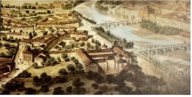
The fields that the community had occupied were close to 27000 sq metres. In the street of San Pablo there were different buildings that, after the confiscation of 1835, were sold like separate buildings. The convent that bordered these fields, had a series of properties attached to it like an orchard or an estate of recreation that was in La Quinta. And it also had a separate entrance called the noble door that was in a flank of the façade of the church and a second door for the service at the back adjoining with the San Lukas street (Figure 3) (Casillas, 2003).
Figure 3: Engraving of the city of Burgos, of century XIX, with the view of the convent from the east, by Guesden (Casillas & Alvarez, 2005)
6
The 13th century is the period in which the order acquired its moment of expansion. This was an order that soon earned the appreciation of the city, thanks to its preaching and cultural advice. They were preferred in matters of devotions and especially in matters related to death, and were solicited as business advisors and contractors, as witnesses and as testamentary agents. At the end of the 12th century, and at the beginning of the following, it was the time period when the mendicant
orders in Castile had a privileged position and when there were marked relations between the order and the Castilian oligarchies. This is reflected by the beginning of sponsorship of the chaplaincies, where they began to be buried (Casillas, 2003).
The 14th century is marked by the entrance of the Black Death in Spain (1346) which struck Castile hard. As a result of this, the power of the convents began to wane and the relaxation of the clergy was encouraged. With the intention of recovering the gaps in the order and the past and the plague, they began to deliver habits with great ease during this relaxation time which is known as the "claustra" (Casillas, 2003). Even with these internal weaknesses, the patronage and the burying of the dead continued to increase.
The convent maintained the continuum, but with little real support. So it could carry out some constructive activities among which the most innovative is the one that highlights the patronage of D. Leonor Henriquez, the granddaughter of Alfonso XI, who had a bulky sepulchre built in the center known as the tomb of the "beata" (Casillas, 2003). The 15th century was a good time for the convent under Bishop D. Pablo de Santa Maria, of Jewish origin, who had converted at maturity and was the bishop of Cartagena. He took possession to govern the diocese of Burgos in 1425.
His works were outstanding and promoted the improvement of the customs and important works in the diocese. He showed his distinguished patronage to the convent of St. Paul by choosing the Great Chapel for his burial as well as for those of his most notorious descendants. Among the works he performed as patrons were the finishing of the main chapel, covering of the vaults of the church, widening of the chapel of the chapter and other works in the cloister. This is the time when the convent acquired the form that it maintained, with small modifications, until its disappearance. The convent was not finished until September 31, 1430, having begun work 130 years earlier and marking the arrival date of the Dominicans around 1220 (Casillas, 2003).
The period between the end of the 15th century and the first half of the 16th century was the best time for the convent, which became an important study center for the formation of members of the
7
Order and for the education of the most notable people in Burgos. On the subject of death, the Dominicans were the favorites after the Franciscans and the convent was for many Burgaleses, the place chosen for their eternal rest. So the 16th century is marked by the greatest burial movement in the convent (Casillas, 2003).
The best time during the century was when the mercantile bourgeoisie appeared. A bourgeoisie whose development contributed to the best time of the city, which affirmed and consolidated its influence in the kingdom and materialized the creation of private chapels within the convent of San Pablo. As far as construction was concerned, by the end of the previous century the four wings of the cloister had already been closed, and other offices such as the library, dormitories, refectory, nursing, hostelry and novitiate were expanded.
By the second half of the 16th century the plague returned to the city. This was the main cause of Burgos's loss of demographic, economic and court power resulting in the fall of the Convent of San Pablo, which sent its best men to the convents of Valladolid or Salamanca. The decadence is also accused in terms of patronage having declined to its lowest intellectual level. The works that were carried out at this moment were no longer inside the convent edifice but in the premises of the convent with too much repetition and with an apparent lack of necessity, for example, structures like the prestiberio, the bookshop, the stairs or the room of "De Profundis" were modified several times in the same century (Casillas, 2003).
In the second half of the seventeenth century the last works of importance in the convent were carried out. Different chapels and facade of the building were modified, building a sumptuous belfry under the orders of Friar Jose de Torres, in addition to the rebuilding of the cloisters and of their ornamentation with pictures (Casillas, 2003).
But without a doubt, the booming years for the convent of San Pablo ended in 1807 when the Napoleonic troops arrived in Burgos, at which time the city had to establish barracks, schools, and private houses and evidently also used the convents for these military purposes. The troops occupied the entire ground floor, except for the inn and the kitchen, an occupation that lasted until 1808 when, after the Battle of Gamonal having defeated the army from Extremadura, the troops entered the city under the consent of General Lasalle, setting it on fire and plundering it for days (Casillas, 2003). The building suffered by ruination, the church was dismantled and was without altars.
8
During the period of military occupation the convent was destined to diverse uses, like lodging of troops, jail for the prisoners, military hospital and even warehouse. Several friars were executed and the rest of them left the convent. Their property was plundered and destroyed, some were saved, transferring them to numerous parish churches, such as San Lesmes, San Gil or the Cathedral, as well as to several towns in the province.
After the departure of the weak troops of the city, some of the friars returned to the convent in 1813, the Friar Tomas de la Iglesia being the head. They rehabilitated the rooms and according to the chronicles of that time, it was remarkable how quickly the convent returned to its day to day functioning (Casillas, 2003).
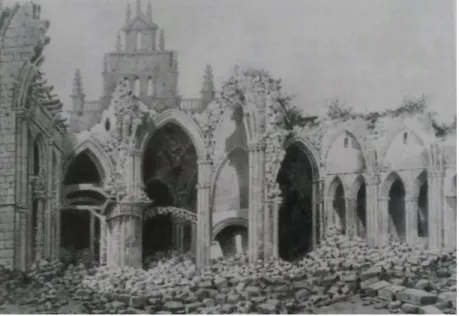
In 1827, thanks to the Friar Manuel Martínez, the works took great impulse but the joy lasted for a very short time, since in December of 1835 the friars were expelled from their monastic dependencies. After the expulsion, the objects of worship and the estates began to be sold. On the other hand, the buildings, the church and the convent were occupied by the army, for the lodging of troops, hospital or ammunition store. Thus it deteriorated, until the army itself decided to destroy it in order to build the barracks that were inaugurated on June 19, 1883 and remained standing until 1975 (Casillas, 2003). Few monasteries achieved such a restoration and a few have known such a disastrous end after the exclaustration of 1835. The engravings from the ruins kept in the City Hall show us that it was a sumptuous building and the church especially was an architectural marvel.
2.1 Structure and architectural insights
It can be mentioned that there is no proper Dominican architecture. In the beginning, the order of the Dominicans felt a great disinterest in building their edifices. But things changed from the XIII century when the order of the mendicants proposed a new typology that had a great acceptance in Spain, a same aesthetic taste that the Franciscans as well as the Dominicans shared. This type of buildings were raised close to the local tradition, seeking to maintain the functionality. Buildings of that time were being gradually modified to adapt to the fashion of the moment.
In the first attempt, the friars looked for a space where they could officiate the Eucharist, and later extended the wish to have a great choir, which influenced the design and a polygonal form was developed. This was united with a body formed by one or three naves and generally a roof of wood.
9
The ornamentation at first was very scarce but to cover the pedagogical needs the Cistercian rigidity was abandoned and they began to open up to the religious arts. Until the end of the 14th century it was not possible to even mention the entrance of images in the Dominican temples, although in Burgos in the second half of the 14th century, the cloister was decorated pictorially (Casillas, 2003).
An artistic austerity existed in the primary years which has nothing to do with what happened in the future. The changes in altarpieces, great artistic works and numerous paintings covering the walls of the monastic stays account for the works from great artists of the time.
The construction stages for the convent can be divided into three specific periods (Casillas, 2003).
a) A first period that covered the 14th century and reached the beginning of the 15th century, where the basic plan was built.
b) A second phase, in the first half of the 16th century, in which the convent was widened and decorated in the plateresque style.
c) A third Baroque period, which was going to be maintained until the beginning of the 19th century.
The large church was attached to the convent on the north side with a structural plan of a Latin cross and nave, besides having a greater chapel. The convent of the two floors was organized around a large cloister, the lower part with a more public character and the upper part dedicated to the needs of the community. Each wing of the cloister was also dedicated to a function, the north wing being for the passage, the east wing was the noble zone with the chapter room, the wing of medidodia was destined to the life of the community and the west wing was dedicated to the study (Casillas & Alvarez, 2005). The materials used were those being used commonly in the city of Burgos such as Hontoria’s stone (a pure limestone), brick, wood, plaster and stucco.
2.2 The archaeological background
The first action carried out on the remains of the convent of San Pablo (Burgos) was carried out in 2001 (February-April), under the orders of the archaeologist Jose Luis Ibarra, by the company Wyngaerde. This intervention had the objective to verify the existence of material remains of the
10
Dominican convent, to be able to define in detail the construction phases of the convent. In addition to the archaeologists mentioned, the team included researchers José Miguel Carretero Díaz, researcher Rolf M. Quamm, and paleontologist Yahya Bensaid who conducted a study on the animals consumed in the monastery, to provide data on daily life and diet of the friars (Casillas & Alvarez, 2005).
Prior to the archaeological intervention, a survey was carried out, in which it was possible to verify the existence of walls, ceramic remains and human remains, as well as a pavement sample, which could be recovered and is currently represented in one of the buildings that make up the complex of the human evolution museum.
After the pre-excavation survey, the archaeological action was allowed, which was carried out delimiting the enclosure in different zones.The first one was called zone B, located geographically in the northwest area of the plot with dimensions of approximately 8,100 m². The surveys on the other zone (Zone C) are about 16,500 m², and were subdivided into two other zones (C1 and C2) (Casillas & Alvarez, 2005) (Figure 4).
Figure 4: Map of the 2002-2003 archaeological excavation which shows the total area (22,000 m2) (Memoirs of archaeological intervention 2002-2003)
11
The archaeological work was decided to be started in the area of the cloister, which presented better results and remains than the remaining of the church, which was estimated beforehand thanks to the pre-excavation survey. These burials were documented through "Burial Files" in which the layout of the skeleton, the burial form, the treasure accompanying it and the stratigraphic relationships were recorded (Casillas & Alvarez, 2005).
In the church the plan of the convent was defined by obtaining the outline of the head chapel and of other chapels. In addition to this, possible exhumation of 242 burials was found in the nave and in the lateral chapels. The excavation was completed by another company "Aratikos Arqueólogos", which under the direction of Angel Palomino and Javier Abarquero, carried out the investigation between June and September 2004 (Casillas & Alvarez, 2005).
2.2.1 Evaluation of the results from the archaeological intervention (2002-2003)
The studies carried out during the archaeological intervention (2002-2003) were decisive to know the constructive sequence of the old convent of San Pablo. Referring to the history of the convent it was possible to establish how the arrival of the Dominican friars to the city of Burgos had the date of 1220-1222, but it was not until 1302 when the construction of the convent began. However, burials are recorded prior to the commencement of the construction of the convent, since along with the individuals buried there are coins of Alfonso X (1252-1284) and Sancho IV (1284-1295) (Casillas & Alvarez, 2005).
The construction of the church began in the early years of the 14th century and was completed thanks to the impulse of D. Pablo de Santa Maria in 1430. It was a Gothic church with three naves with several chapels located between the buttresses. This chronology has been confirmed by the archaeological intervention. After the church began the construction of the most essential units, among which the capitular room initially built with a short height, stands out.
The cloister was realized in several phases being primarily of low height. In the year 1380 the documents report the news of the construction of a new cloister which was completed at the end of 14th century. The cloister was used as a place of burial. Since the end of the 13th century some
of these burials were adapted to the wall, so it has been possible to confirm the existence of tomb altars (Casillas & Alvarez, 2005).
12
In the 16th century new works and reforms were carried out in the premises of the convent. The cloister was remodeled along with the chapter room in which a second mortar floor was found with eleven burial pits. The construction of thirty tombs of cooked bricks for the members of the monastic community between the 17th and 18th centuries in the chapter room is verified.
An important work was produced in the church called the chapel of the eleven thousand virgins. It was demarcated as a funerary chapel in 1563 and given to the Maluenda family who built a crypt that was in use until the 18th century. In the 19th century this chapel was filled with children's
burials, along with a large registry of architectural and decorative remains.
In the 16th century, the naves were multiplied in the Church's edifice for the family and individual graves, where burials were performed in lime-filled coffins and successive reutilizations took place with the generation of ossuary until at least 1782 (Casillas & Alvarez, 2005). With the creation of municipal cemeteries in the 18th and 19th centuries, burials were discontinued inside the convent building.
2.2.2 Burials and material remains found
The burials found in the excavation season of 2002-2003 were located in three areas: the first is a necropolis in the courtyard of the cloister that dates from the Medieval period, a second area with graves in the Chapter Room where the monks would have been buried belonging to the monastery and a society of greater social class and a third burial zone located inside the church, more or less near the high altar according to social class (Figure 5).
There have been found 428 burials and an indeterminate number of ossuaries (Figure 6). The intervention of 2004 numbered another hundred individuals and many more ossuaries which were in different chapels.
13
Figure 5: Map showing the location of burials (Memoir of archaeological intervention 2002-2003)
14 Cloister
The burials of the cloister belong to the 14th to 16th centuries, and were found in earthen graves, making a set of 118 burials of which 54 correspond to children (Casillas & Alvarez, 2005). The adult individuals buried there, appear to be oriented according to the Christian ritual that has been practiced since the Middle Ages, according to which the head lies in the west and is facing east, so that in the resurrection they stand facing god. In addition to the burials there was a varied treasure, composed of coins and copper pins. The presence of pins indicates that the bodies were buried with a shroud. Coins along with burials, due to pagan reminiscences, could indicate two things. The first that they took something valuable to the other world and a second theory could refer to the payment of the boatman. Infantile individuals did not present the same discourse as adults, and this may be because they were not yet baptized and therefore were not Christian bodies (Casillas & Alvarez, 2005).
Several levels of burials have been found, especially in the northwest area, in which up to three superimposed burial phases have appeared. In most of these cases of overlap, one of the layers corresponds to ossuary (Excavation Report, 2002-2003 (cited in Casillas & Alvarez, 2005)). The state of conservation of these skeletons is diverse, being the best conserved, adult skeletons and those located in lower levels. These burials are assigned to the stratigraphic unit (U.E) 1-250 (Excavation Report, 2002-2003 (cited in Casillas & Alvarez, 2005)).
A study of 16 individuals from this strata indicated that life in medieval times was appalling, the poverty of the diet is evident in the study of teeth and malnutrition is reflected in the growth pattern of children, well below the current one demonstrated through previous studies. The poorest population was buried in the courtyard of the cloister, leaving the church for noble characters (Casillas & Alvarez, 2005). Five of these remains will be examined in the study at hand.
Church
A second burial site is the church, where burials appear in both the central nave and the side chapels (located in the southern corridor and are total nine in number). The chronology of these burials is from the 16th to the 19th centuries. All the burials here were arranged from West to East. The state
of conservation of these skeletal remains is not optimal, possibly due to a higher acidity of the substrate or due to the remodeling in the 19th century (Casillas & Alvarez, 2005)
15
In the 2002-2003 campaign, 224 west-east skeletons were documented (Casillas & Alvarez, 2005). There is an area of overlapping tombs at the feet of the church up to four levels. The side chapels on the other hand have a single level and a treasure rich in objects of personal adornment and coins, associated with burials. All the burials of the central nave belong to the U.E. 1-350, except for some examples of individual tomb presenting another U.E. (Excavation Report, 2002-2003 (cited in Casillas & Alvarez, 2005)). Three of the individuals buried in the central nave will be examined in this work.
Chapter Room
The capitular room is the third place of burial. It was part of the political-administrative space of the Dominican friars but it also had a sepulchral function. During the archaeological intervention carried out in the chapter hall, which was located to the right of the east part of the cloister, 26 tombs were found with their corresponding architectural structures. They were formed by varied materials: brick, stone and various moldings. (Excavation Report, 2002-2003 (cited in Casillas & Alvarez, 2005))
These burials belong to successive historical periods of the 14th and 19th centuries. An early period refers to all the burials associated with the architectural structures of the tombs. A second phase, anterior in antiquity to the first, corresponds with a second burial. A third phase, with a superior antiquity with respect to the previous ones, corresponds with a level of ossuary and under this another level of ossuary of a superior antiquity was found (Excavation Report, 2002-2003 (cited in Casillas & Alvarez, 2005)).
The intervention in the capitular room concluded with a total of 56 burials of which one belongs to a subadult individual and in most of them the head was located to the east which is the opposite to that in the cloister (Excavation Report, 2002-2003 (cited in Casillas & Alvarez, 2005)). The recovered materials are diverse such as bone buttons, remains of leather sandals, rosary beads, scapulas, coins, everything related to the rudimentary and the shroud of the friars. As a general conclusion, the excavation campaign (2002-2003) has managed to provide data on the construction of the old convent of San Pablo and confirms the historical data proposed with archaeological evidence.
16
3
Scientific Background
There is a vast variety in the culture and diversity among humans in both space and time. Archaeology helps to study all the various past cultures and with the advent of archaeometry it has become easier not just to understand the material remains of past humans but also their way of life, origin, mobility, health status, and diet and so on. Physical anthropology now is being assisted by the chemical and physicochemical analysis of the skeletal remains along with the conventional anatomic/anthropological study. This methodology can enhance our knowledge in various ways and can help to develop quite similar reconstructions about all the aforementioned questions and many more (Szostek et al., 2003).
Trace element (TE) analysis is a versatile analytical approach for archaeometry which can be utilized to provide basis for the reconstruction of the food economy, living habits, environment, and dietary habits of the ancient populations throughout the history of humans (Boscher-Barre & Trocellier, 1993; Molleson et al., 1988; Reiche et al., 1999; Brenn et al., 1999; Elliott & Grime, 1993).The processes of incorporation of trace elements in the bioapatite of the skeletal system are active right from the beginning of the life of the individual directly from its environment till after the death burial period, termed as diagenesis (Reynard & Balter, 2014).
Hence not just post-mortem diagenesis but even living habits of any remains can be inferred from the variability in the elemental compositions of both teeth and bones (Seiler et al., 1994). Thus it might be concluded that archaeological investigations related to ancient populations and their living habits and environment can be carried on with the help of such elemental markers by profiling the elements in ancient human remains (Carvalho et al., 2000).
3.1 Elements in the human body
The elements present in the human body have been divided roughly into three (or two in some cases) categories namely:
a) Major elements
The six elements namely oxygen, carbon, hydrogen, nitrogen, calcium and phosphorus which make up around 99%age of the human body are considered as major elements.
17
b) Minor elements
Potassium, sulfur, sodium, chlorine, and magnesium make up the most of the remaining composition of the body and are termed as minor elements.
c) Trace Elements
Elements such as iron, zinc, silicon, strontium, bromine, lead, copper, manganese, barium and many more make up less than 1%age of the body composition and are known as trace elements. Some of them are essential or have a favorable effect on the body, while some seem to be toxic or do not have any known function (https://sciencenotes.org/).
Some authors have classified major and minor elements together as major elements and the rest as trace elements or minor elements (Underwood, 1959).
3.2 The major elements Phosphorus and Calcium
Phosphorus is an indispensable part of the apatite structure of both teeth and bones. The analysis of phosphorus present in the remains can be used as a measure of the extent of degradation and diagenesis acting on them. Ca/P ratio is usually measured for such inferences. Calcium makes up the largest percentage in bone and teeth mineral which is up to 38%age for bones and is more or less constant for all cases except when found in archaeological context with fully preserved collagen it can range between 26%age-38%age (Burton, 2008). Thus it is usually measured in order to assess the quality of the sample in most cases (Allmäe et al., 2012).
3.3 The trace elements
The TEs assimilated in the apatite of any living being can be broadly divided into two following categories.
3.3.1 Essential elements
Essential elements are those which are required by the human body for regulating various functions and thus play a very important role in our metabolism and other biochemical pathways. Their concentration has to be controlled in such a way as to fall exactly between the thresholds of toxicity and deficiency i.e. neither too much nor too less. Consequently, by measuring the concentration of such elements in the archaeological remains, it is possible to figure out any possible deficiencies
18
or toxicities in the past societies (Patterson et al., 1987; Rasmussen et al., 2008). Essential elements can help to determine the diet, metabolic activity and so on for the individual since they are important for the bodily functions in one or the other form (Reynard & Balter, 2014). They are also actively involved in the activities of the enzymes and proteins which are essential for bone growth (Yamaguchi et al., 1986).
Three important examples of such bioessential elements are zinc, iron and copper. All of these three play a vital part in the metabolism in human body. Their concentration in the body is governed to be lie between the toxic and deficient levels since they are so crucial for many bodily functions. Zinc is a component of more than 300 metalloproteins which function as enzymes or have other important structural properties (Cousins, 1985). Iron is well known to be present in haemoglobin which is involved in the transfer of oxygen and transport of electrons within the body. Copper is highly associated with iron in metabolic processes and pathways and facilitates the transfer of electrons through biochemical reactions. Manganese is another essential element whose high concentration in the human remains can give evidence of high ratios of plant foodstuff in the diet of the individual but it is not always the case (Allmäe et al., 2012).
3.3.2 Non-Essential elements
Non-essential elements are termed so because they do not yet have any recognized function in the human body but due to their similar properties to bioessential elements, they tend to replicate the behaviour of such elements. These are the elements which get incorporated in the apatite replacing the essential element. Hence in general, they are not part of any metabolic pathways and their biological behaviour is understood better by calculating their ratio in reference to that of the bioessential element that they mimic (Reynard & Balter, 2014). Their analysis can be helpful to rebuild the trophic chains by comparing their concentration with that of the essential element. They usually due to their similar size and chemical properties to an essential element, passively become part of biochemical processes and pathways in the biological organisms. For example alkali earth elements like Ba, Sr, Mg and others which might get accumulated in large amounts during the lifetime of an organism while others like rare Earth elements (REE) such as Hf, Th, U which get stored in the remains post-mortem and can give information regarding the tracing of the diagenetic processes and even dating of the remains (Reynard & Balter, 2014).
19
Strontium and barium are two important non-essential TEs which are associated with Ca and their ratios with Ca have now been used for over sixty years (Comar et al., 1957; Wasserman et al., 1957). The body undergoes the process of purification in which a healthy adult mammal reduces the ratio of Sr/Ca and Ba/Ca during the various metabolic pathways. This lowering of nonessential elements in the mammals leads to reduced ratios of Ba/Ca and Sr/Ca in tooth, body and bones as compared to that in the food when one moves up the trophic pyramid. Lead is another non-essential TE which, instead of being a vital part of nutrition for the body, is a rather harmful and toxic element for biological organisms. Lead is taken up by the body from the surroundings. The lungs or digestive tract facilitate the intake of lead in an individual (Bronner, 2008) which keeps getting built up in the bones throughout the life of the individual if and whenever the body is subjected to any source of lead pollution.
Apart from these two categories, there might also be some non-toxic elements which might get absorbed in the gastro-intestinal tract if they are needed by the body. Hence the composition of such elements is developed as a result of the balance between their intake in the diet and their metabolic requirement. Enameloblasts and osteoblasts are two more very particular types of cells which govern the precipitation of apatite crystals and in turn have a huge effect on the concentration of trace elements (Reynard & Balter, 2014).
From the point of view of palaeoecological and paleontological studies it is imperative to be able to differentiate the bio-essential elements from the non-essential elements since their behaviour and the processes are different in the body.
3.4 The Structure of Bones and Teeth
Teeth and bones are minerals consisting of a hydroxyapatite and protein matrix along with a calciumphosphate which is inorganic in nature. Interestingly enough, they both are helpful in monitoring the doses of various elements to which any human has been exposed to (Carvalho et al., 2004). Bones and teeth generally are composed of an inorganic matrix, an organic matrix and different cells. The creation of mineral tissues in both of them is being governed by these cells. Teeth and bones are both quite dynamic structures in their own but bones much more so than teeth. (McKee et al., 2005).
20
Bones and teeth are very similar in their compositions but with some vital differences owing to their distinct functions. The bones are composed of some amount of non-collagenous proteins, and collagen which is the main fibrous protein that makes up its organic matrix. It is the main protein that provides flexibility to tissues like tendons and ligaments but the actual rigidity required to bear greater loads comes from the inorganic mineral part which is present alongside the collagen matrix in the bones and teeth. Tooth dentin and cementum are also composed of collagen as the primary organic component but in the enamel there is no collagen present. The major mineral present in the enamel is similar to the hydroxyapatite mineral found in the inorganic matrix of bones. The nanocrystals present in bone apatite have a larger number of possibilities of substitution as a result of the various types of vacancies in the molar ratio of Ca/P which is not the case for the enamel apatite and thus is more approximate to the actual stoichiometric hydroxyapatite ratio of 1.67 (Boskey, 2007).
3.4.1 Teeth
Teeth are also composed of different tissues just like bones and can be differentiated at organ level. Teeth comprise of inorganic, organic and water fractions. Their inorganic stage consists of the unit cell (Ca,X)₁₀(P,C)₆(O,OH)₂₆. The microcations do not generally construct complex ion species and prefer coordination with oxygen instead (Liu et al., 2014). These cations have a small ionic radius and high charge/radius ratio and can substitute each other. The X in this chemical formula signifies an assortment of potential replacements for Ca, such as Sr, Ba and Pb (McConnell, 1973). The exact chemical formula for enamel is (OH)₂Ca₆[(P₅.₈C₀.₂)O₂₄](Ca₃.₁Mg₀.₁C₀.₅) which estimates the Ca/P ratio at about 2.02 (Gruner et al., 1937).
Tooth is basically made of four tissues namely enamel, dentine, cementum and dental pulp. The crown is made-up of these vital tissues and is covered in enamel at the top of the nape of the tooth which extends until the gumline. This enamel is very hard thanks to its almost completely mineral composition without the presence of any or very little protein. The crown of the tooth is visible once it get erupted (Liu et al., 2013). The dentin is the most important constituent of a tooth which is situated right under the enamel. This dentin then is separated from the surrounding jawbone by a composite material made of dentin and bone which is called cementum which along with the jawbone is connected with the tooth by a periodontal ligament surrounded by a membrane (Boskey, 2007).
21
The tooth is able to carry out its functions with the help of a regular supply of blood vessels and nerves into the pulp cavity inside the dentin. After mineralization, in early life, the enamel remains closed and will no more carry out notable physiological exchange of elements (Liu et al., 2013).
Human beings generally have twenty deciduous teeth in the primary stage of their life and thirty-two permanent teeth when they reach adult hood. Each type has its own time frame for calcification, growth and eruption (Table 1) (Hillson, 1996). Such differential growth of teeth in humans can therefore provide the possibility of combining multiple teeth of the same individual to create a composite time series using TEs. This combination allows the construction of longer continuous records of seasonal variations in paleo-environments or diets during the years in which the teeth mineralized (de Winter et al., 2016). Teeth are fundamentally classified into four groups viz. incisors, canines, premolars and molars.
Teeth were reported in 1930s to contain a variety of minor or trace elements (Dreal, 1936; Lowater & Murray, 1937) and thus conserve great data through a life span varying with environmental exposures (Liu et al., 2014).
Table 1: Development of permanent dentition (Simon & Stevenson, 1975). Designation Calcification begins Eruption Central incisor 3-4 months 7-8 years
Lateral incisor 10-12 months 8-9 years
Cuspid 4-5 months 11-12 years
First bicuspid 18-21 months 10-11 years
Second bicuspid 24-30 months 10-12 years
First molar birth 6-7 years
Second molar 30-36 months 12-13 years
22
3.4.2 Bones
Bone is a major part of the skeletal system. It is a living, dynamic structure which provides a supportive and defensive foundation for the body. It provides a repository of calcium and phosphate and functions in metabolism also. Core of the bone consists of marrow, which serves as a repository of nutrients and creates several forms of blood cells. The artificial segment is made of crystals which build up hexagonal plates that are arranged in a regular way on and parallel to the axis of the collagen fibers. Bones are made up of customarily hydroxyapatite, but they also consist of carbonate, citrate and lesser amounts of sodium, magnesium, potassium, chloride, fluoride, and a number of other elements (Tandon et al., 1997).
Bone tissue is divided into two sections: compact (cortical) bone and trabecular (spongy and porous, cancellous) bone based on the hardness, porosity and the content of soft tissue existent in them. Though not every bone can be determinedly categorized as compact or trabecular as some types are intermediate in porosity and challenging to classify. The compact bone is the rigid dense part enclosing the outer walls of all bones and is conjoint in the streak of the long bones. The trabecular bone is a pliable formation seen at the core of flat bones and at the edge of long bones. It is extremely filigree being soft and comprising mainly of bone marrow (Arnold et al., 1966).
Bone is an exemplar of a biological specimen that presents many challenges to acquire a sample for chemical scrutiny. Thus, it’s not surprising that dependable chemical composition data, especially for minor and trace elements, are few.
3.5 Trace Elements in Bones and Teeth
The concentration of different elements in the apatite mineral is linked with various factors such as their intake from water, food, metabolic pathways, respiration, and exposure to the environmental factors and also formation of some definite tissues while the individual is in the period of in utero development (Dolphin et al., 2005). There are certain physiochemical (external) parameters such as pH, salinity, temperature, soil composition etc. that govern the uptake of trace elements. This systematic occurrence is predictable (Darrah, 2009). TEs differ from one person to another and therefore can also be used for forensic objectives (Perrone et al., 2014). The metabolic reactions in the body do not have a strong influence on elements such as Sr and Ba which are thus more useful to derive palaeodietary inferences because of their ability to be directly correlated to
23
food habits of each individual. These elements are also incorporated in the body when they replace Ca in the hydroxyapatite crystal of the bone mineral and hence are also prone to diagenetic alterations as well (Pankowska et al., 2016).
Bones are one of the most dynamic structures in the human body and one of the hardest as well. They are dynamic since they keep getting remodelled periodically all throughout the life of the individual. This turnover of bone or its continuous replacement is called remodelling in which cycles of simultaneous formation of new bone and resorption of existing bones keep occurring. This process does not stop even when the growth of an individual might stop and thus leads to storing of trace elements even in adult skeletons (Swanston et al., 2012). Owing to the equilibrium between these two processes of resorption and formation, every year all through the life of any individual, about 10%age new bones are being formed in case of mature bones. The turnover rates for both of them are quite distinct, i.e. 4.3%age of the total mass of the Ca exchanged per year belongs to the compact tissue while 32%age of it can be attributed to the spongy tissues. What makes them really useful in archaeology is the fact that the bone structure survives death and thus can be encountered in fossil (Abbott et al., 1996) as well as archaeological records (Mulhern & Van Gerven, 1997).
Due to the process of turnover or remodelling of bones owing to cellular activities, there is a variation in the composition of bone depending on the environmental factors, health and the age of the tissues and the age of an individual. Even within the bone different localities within the trabeculae and osteons can have difference in their crystal size, chemical composition and in the mineral composition depending on the age of the tissues (Boskey, 2007).
Teeth also exhibit this variability in constitution and structure among the different components. For the mature tooth enamel there is no process of removal and re-deposition, in short there is no remodelling procedure for enamel (Boskey, 2007). Moreover, the organic part of its matrix is already eroded and additionally enamel is not made of a collagen matrix (Margolis et al., 2006). Thus the chemicals which can be used for repairing the damaged enamel by remineralization or some bacteria that release acids which can cause dental caries and cavities as a result of dissolution, are the only ways by which the composition of tooth enamel can be changed (Verdelis et al., 2007). There are many environmentally originated elements which get incorporated in the mineralised structures of the human body during its lifetime (Carvalho et al., 2000).
24
The amount of elements incorporated in the tooth enamel is much less than that in the bones (Eggins et al., 2003). This happens because hard tissues have different properties when it comes to absorption of minerals. While the enamel is being mineralized, entry of Ca is enhanced to the detriment of the Sr and Ba level and therefore they both are not present in enough quantities (Balter, 2004). Teeth can preserve ontogenetic data and are quite tough and impervious thanks to their highly crystalline structure. They are able to record the chronological development in the life of an individual in the form of element distribution at various degrees of mineralization. This variation in the distribution is caused by factors such as health, illness, stress and diet of an individual (Dolphin et al., 2005).
The research has delved deeper into micro and macro-elements and their investigation in skeletal remains in the last thirty years. This newly developed area of research has opened up new avenues of studies in the field of anthropology which offers various possibilities of research as has already been said such as studying diet, pathology and diseases (Glen´-Haduch et al., 1997), the social status and also the physiology of the ancient communities (Schutkowski, 1994; Schutkowski et al., 1999).
3.6 Main factors for TE uptake in bone and teeth
3.6.1 Diet
The food habits of past societies have long been a subject of enquiry. In case of populations belonging to historical times, information about the food can easily be found from historical records and remains. This information can be also complemented with archaeozoological and archaeobotanical remains and human evidences found from archaeological contexts.
Diet reconstructions have also been made possible by the analyses of chemical elements in the bones and teeth of past humans. Somewhere in the second half of the 20th century, the first attempt at trying to analyse the elements in human bones in order to understand and recreate the food habits of the ancient communities was undertaken by Gilbert (Gilbert, 1975) in 1975 and by Brown in 1973 (Brown, 1973). They consequently have been considered the forerunners of such studies and led to a rigorous wave of such researches in the 1980s. Since then, there have been many studies upon the dietary habits of past societies. Usually the study of essential elements plays an important
25
part in diet reconstructions. For example the presence of elements such as zinc (Zn) or copper (Cu) usually points towards the ingestion of meat in the diet (Buikstra et al., 1989).
Of late, trace element analysis has shifted its focus from palaeodiet reconstruction towards the understanding of diagenetic processes and their impact (King et al., 2011; Maurer et al., 2011) after settling on the fact that bone chemistry is quite complicated. The promise of a research field developed on the basis of the relation between trace elements in human apatite structures and particular trace element concentrations has now faded after the initial boost (Brown, 1974). Nevertheless, barium and strontium are still being used for diet reconstruction by scientists (Kamphaus, 2013).
3.6.2 Diagenesis
The composition of trace elements in teeth and bones is considered to be a good indicator of the level of the exposure of that individual to the elements present in the past environment and diet. But the complexities arise when these elements not only get incorporated in the body while alive but can also assimilate post-mortem from the archaeological burial environment of the remains (Swanston et al., 2012). Post-depositional chemical alteration or diagenesis in archaic human bone is the major problem in the use of trace element examination for dietary reconstruction. Due to its regular remodelling and recreation, bones are viewed as suitable dose monitors for some of the trace elements. But on the other hand, they are quite vulnerable also to diagenetic alterations even after the death of the individual owing to the inner channels in its porous structure and an open morphological structure. This post-mortem assimilation of trace elements differs from element to element. Elements like copper, iron, lead and manganese are very likely to get incorporated in the bones due to the surrounding enriched soil of the burial ground (Carvalho et al., 2004).
Diagenesis has been discussed a lot in the present study as well as in various other literatures. The main obstacle in the interpretation of trace elemental data in archaeological remains, especially human remains, is that the elements of interest such as Fe, Cu, Mn, Zn, Sr, Ba and Pb and others are subjected to post mortem changes due to diagenetic processes during the burial period. They are related with the climate and geology of any site and can vary within the same site between different burials. Thus it is imperative to thoroughly assess each site independently of the other (Dudas et al., 2016).