UNIVERSIDADE DE LISBOA
FACULDADE DE CIÊNCIAS
DEPARTAMENTO DE BIOLOGIA ANIMAL
The role of microRNAs in Haematopoiesis and Immunity
of Drosophila melanogaster
Mestrado em Biologia Evolutiva e do Desenvolvimento
Joana Gomes Campos de Carvalho
Dissertação orientada por:Élio Sucena
(Faculdade de Ciências da Universidade de Lisboa e Instituto Gulbenkian de Ciência)
Acknowledgements
Reaching the end of my first step in the big world of academic research was no easy task and being surrounded by amazing people throughout the process is highly advised and helps you to be amazing too. To everyone who contributed to this achievement, thank you.
I want to thank everyone from the groups of Élio Sucena, Patrícia Beldade and Christen Mirth from the EvoDevo community at Instituto Gulbenkian de Ciência. They created the best possible working environment and source of healthy motivation and constructive criticism one could ask for.
I thank Élio Sucena for trusting me since the beginning and giving me the creative freedom others can only dream of. Thank you for being such an inspiration and keeping me in the right track.
I want to thank Julien Marcetteau, the most talented and kind friend, who was essential for the success of this project and without whom it would not exist.
I also thank Tânia and Nuno, for the important experimental guidelines throughout the project, and the remaining of my friends who directly or indirectly contributed to the important comical reliefs of the story.
Finally, I want to thank my sisters and mother, who were strong enough to endure in the best and worst moments of my progress, and my all-time companion who gave me such valuable creative input and important life lessons without ever leaving my side.
Resumo em Português
A resposta imune de Drosophila melanogaster envolve múltiplas estratégias de defesa que são relativamente conservadas entre animais. De forma geral, esta resposta resulta da interacção entre uma componente humoral, também designada por imunidade sistémica, e uma componente celular, que implica a diferenciação de vários tipos de células especializadas.
A imunidade sistémica consiste na produção de péptidos antimicrobianos (PAMs) pelas células do corpo adiposo e de componentes das reacções de melanização e de stress oxidativo que actuam no sentido de neutralizar patogéneos. Os PAMs acumulam-se na hemolinfa e a sua produção ocorre principalmente através das vias de sinalização Toll e Immune defficiency (Imd), que são activadas após o reconhecimento de certos componentes da parede celular de bactérias gram-positivas ou fungos e bactérias gram-negativas, respectivamente. Além disso, existem outras vias de sinalização que tembém podem participar na resposta humoral. A via JNK (JUN N-terminal kinase) é activada por componentes da via Imd e tem um papel importante na indução de apoptose e na remodelação do citoesqueleto e adesão das células. Além disso, está envolvida na resistência ao stress oxidativo. A via JAK-STAT, por sua vez, está relacionada com a resposta à infecção viral e com a produção de citocinas e factores de resposta ao stress. Embora a existência de diferentes cascatas de sinalização confira especificidade à resposta imune, certos tipos de infecção podem levar a uma activação sinergística das mesmas vias. As lesões sépticas (picada com uma agulha embebida em solução bacteriana), por exemplo, activam tanto a via Toll como a via Imd, levando à produção de PAMs como a Diptericina e a Drosomicina.
A componente celular da resposta imune de D. melanogaster tem como intervenientes principais os hemócitos (células do sangue), que são descritos como funcionalmente análogos à linhagem mielóide dos vertebrados. Em homeostase, 90% dos hemócitos são plasmatócitos, que têm capacidade fagocítica e contribuem para a activação da produção de PAMs, e 10% são células cristal, que participam nas reacções de melanização e na regeneração de feridas. Além disso, mediante a ocorrência de uma ferida ou de endoparasitismo, há diferenciação de lamelócitos sob o controlo da via JAK-STAT, que são importantes para a encapsulação de microorganismos de maiores dimensões. Os efectores da componente celular da resposta imune são gerados e mantidos através do processo de desenvolvimento designado de hematopoiese, que ocorre ao longo do ciclo de vida em diferentes compartimentos. Nos estadios larvares, onde este processo é mais activo, a glândula linfática é o principal orgão hematopoiético em que células progenitoras dão origem a plasmatócitos e células cristal. Fora deste órgão, existem hemócitos em circulação e em agregados sésseis na epiderme dorsal ao longo dos segmentos corporais. Nestes tecidos existe também proliferação e diferenciação de hemócitos, que se encontram em equilíbrio dinâmico com o compartimento circulatório. Embora a exacta contribuição de cada uma das componentes da resposta imune ainda não esteja bem estabelecida, a sua acção combinada pensa-se que seja importante para a neutralização e eliminação de microorganismos invasores.
Os microRNAs (miRNAs), classificados como sequências curtas e não codificantes que actuam através do silenciamento da expressão génica, têm vindo a emergir como reguladores de muitos programas de desenvolvimento animal e de muitas respostas biológicas, incluindo a resposta imune. Enquanto nos vertebrados o repertório de funções descritas para os miRNAs inclui tanto a regulação da diferenciação e proliferação celular durante a hematopoiese, como a produção de anticorpos e citocinas, nos invertebrados, e particularmente em D. melanogaster, a informação relativa à função dos miRNAs torna-se relativamente escassa.
imune de D. melanogaster, através de screens sistemáticos que englobam a maioria dos microRNAs presentes no genoma deste organismo modelo. Incidindo tanto no processo de hematopoiese larvar, que dá origem aos efectores da componente celular, como na resposta imune sistémica dos adultos, cuja função influencia a resistência à infecção bacteriana, os nossos resultados indicam que vários miRNAs têm papéis regulatórios importantes nestas duas vertentes da resposta imune.
Os resultados obtidos através de infecção sistémica com Pseudomonas entomophila e
Pseudomonas putida, duas bactérias do tipo gram-negativo, mostram que as delecções dos mir-317 e
mir-137 aumentam a susceptibilidade à infecção bacteriana, causando taxas de mortalidade elevadas. Por outro lado, a delecção do cluster mir-100/let-7/mir-125 revela uma redução da mortalidade no contexto de infecção, provavelmente relacionada com uma maior resistência ou tolerância à infecção com bactérias gram-negativas. Apesar de apresentarem dinâmicas opostas entre as duas bactérias testadas, também as delecções do mir-31b e do mir-2b-1 apresentam diferenças significativas na susceptibilidade à infecção bacteriana. De acordo com as previsões bioinformáticas, os alvos destas sequências incluem vários componentes da resposta imune sistémica, nomeadamente proteínas das vias Imd e Toll, assim como componentes importantes para a regulação da resposta de stress oxidativo. Além disso, estes miRNAs têm como alvos proteínas importantes para a fisiologia geral do organismo, que também podem impactar a sobrevivência após infecção bacteriana.
Por sua vez, os miRNAs também desempenham funções importantes na hematopoiese em D.
melanogaster, particularmente no que toca à adesão e diferenciação de vários tipos de hemócitos. Os
nossos resultados indicam que as delecções do mir-969 e do mir-124 causam aumento e diminuição da densidade de plasmatócitos nos agregados sésseis, respetivamente. Isto tem consequências para os números de células cristal, que dependem do correcto estabelecimento da densidade de plasmatócitos, já que estas se transdiferenciam a partir dos mesmos. Estas duas sequências de miRNAs têm como alvos genes importantes para a adesão e migração celular, processos estes que podem influenciar a densidade de plasmatócitos nos órgãos hematopoiéticos de D. melanogaster. A delecção do mir-1 em heterozigotia, por sua vez, revelou um decréscimo significativo na densidade de células cristal, indicando que este miRNA tem um papel importante na regulação da transdiferenciação deste tipo celular, processo este que é mediado pela sinalização Notch. A previsão bioinformática dos alvos desta sequência revela que o mir-1 inclui vários componentes da via de sinalização Notch, reforçando a ideia de que poderá regular o processo de diferenciação de células cristal. Finalmente, a delecção do mir-980 causa uma redução acentuada nos números de plasmatócitos e células cristal, levando a uma disrupção da estrutura e integridade dos tecidos hematopoiéticos larvares. Isto deve-se a uma diferenciação ectópica de lamelócitos, que entram em circulação e levam à formação de tumores melanóticos, como resultado da encapsulação exacerbada dos própios tecidos. A análise in silico dos alvos do mir-980 revelou que o mRNA de domeless, que codifica para o receptor da via JAK-STAT, interage fortemente com o mir-980. Uma vez disrompido o mecanismo de silenciamento deste gene, por via da delecção do mir-980, a sobreexpressão de Domeless poderá levar à activação da via JAK-STAT e consequentemente à diferenciação exacerbada de lamelócitos. Resultados preliminares revelam que a remoção do mir-980 causa uma mudança no padrão de activação da via JAK-STAT, possivelmente em tecidos como o corpo adiposo ou os músculos somáticos e uma pequena percentagem de hemócitos.
No futuro, a análise detalhada da origem dos vários fenótipos observados permitirá a caracterização e compreensão do impacto deste mecanismo de regulação na resposta imune de D.
melanogaster. Particularmente, será importante analisar o tempo e local de expressão dos miRNAs
candidatos e os seus alvos, assim como a nessecidade e a suficiência de cada uma destas sequências para regulação dos vários mecanismos que contribuem para a resposta imune de D. melanogaster.
Palavras-chave
Hematopoiese; Imunidade; microRNAs; D. melanogaster
English Summary
The immune response of Drosophila melanogaster involves multiple innate defences that are conserved in higher organisms and have been largely characterized. Consisting of an interaction between a humoral and a cellular component, it is essential for the neutralization and elimination of invading microorganisms. While the humoral defences refer mainly to the production of antimicrobial peptides (AMPs), cellular defences include different haemocyte-mediated responses like phagocytosis, melanization and encapsulation. MicroRNAs, short non-coding sequences that silence gene expression, are emerging as regulators of several developmental programs and biological responses, including the immune response. Furthermore, they have been implicated in processes like antibody switching and lineage determination in mammalian haematopoiesis or regulation of AMP levels in D.
melanogaster. Here we proposed to dissect the role of these sequences in the immune system of D. melanogaster by performing a systematic loss-of-function screen that covers the majority of
microRNAs present in its genome. Focusing on both the larval haematopoiesis and the adult systemic response, we have revealed a set of candidate microRNAs that are important for the regulation of resistance to bacterial infection and haemocyte differentiation. Our results indicate 317 and mir-137 loss-of-functions dramatically increase susceptibility to bacterial infection by Pseudomonas
entomophila and Pseudomonas putida, leading to high mortality rates. On the other hand, the
mir-100/let-7/mir-125 cluster knock-out shows a significant decrease in mortality, related to higher resistance or tolerance to gram-negative bacterial infection. Furthermore, we have shown that mir-124 and mir-969 impact plasmatocyte adhesion, influencing the density of cells in the sessile haemocyte clusters of larvae. Deletion of mir-1 is important for the regulation of the differentiation of crystal cells, which are mainly involved in melanization and wound healing. Finally, we have shown that mir-980 loss-of-function causes ectopic lamellocyte differentiation, a cryptic cell type that is crucial for the encapsulation mechanism and normally triggered upon parasitoid wasp infection. In silico predictions have revealed that these candidates can directly target immune-related genes and modulate their expression levels. Further investigation will allow us to provide a better understanding of the impact of these microRNAs on their corresponding targets and the immune response pathways that include them.
Key-Words
Table of Contents
Acknowledgements
I
Resumo em Português
II
English Summary
IV
1. Introduction
1
1.1 The immune response of Drosophila melanogaster 1
1.2 microRNAs: Biogenesis and Function 4
1.3 microRNAs and the immune response 6
2. Materials and Methods
7
2.1 Fly Stocks and Maintenance 7
2.2 Systemic Immunity Screen and Bacterial Infection 8
2.3 Haematopoiesis Screen, Larval Imaging and Cell Counting 8
2.4 Lymph Gland Dissection and Haemolymph Extraction 9
2.5 Statistical Analysis 9
2.6 Retrieval of computationally predicted targets of miRNAs 9
3. Results
10
3.1 microRNAs have a role in systemic immunity of Drosophila melanogaster 10 3.2 The role of microRNAs in the haematopoiesis of Drosophila melanogaster 14 3.3 mir-980 controls lamellocyte differentiation in the sessile haemocyte clusters 16
4. Discussion
18
4.1 Experimental design 18
4.2 The role of microRNAs in the systemic immunity of D. melanogaster 19 4.3 microRNAs and the cellular defences of D. melanogaster 21
1. Introduction
The interactions between multicellular organisms and pathogens have long shaped the evolution of the immune system. Innate immunity, which consists in a first-line defense against pathogenic challenges, is likely to be common to all metazoans1. Insects, for instance, rely solemnly
on innate defences to mount a complex and multifaceted immune response2. Drosophila melanogaster,
widely used as a model to study innate immunity3, breed, lay eggs and develop in decomposing media,
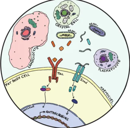
being constantly exposed to a wide variety of pathogens. Once such pathogens cross the epithelial barriers and enter the body cavity (haemocoel) both cellular and humoral defences are mobilized (Figure 1.1). While the digestive tract is the main contributor to haemocoel microbial load, septic injury and endoparasitism can also induce pathogenicity and shape immune response evolution3.
1.1 The immune response of Drosophila melanogaster
The immune response of D. melanogaster consists of an interaction between a humoral component known as the systemic immune response and a cellular component, the latter involving the differentiation of several specialized cell types2(Figure 1.1). The systemic immune response consists
of a coordinated activation of proteolytic cascades and production of melanization and oxidative stress components that neutralize invading pathogens2. The hallmark of this process is the systemic
antimicrobial response, the challenge induced synthesis and secretion of antimicrobial peptides (AMPs) by the fat body4. AMPs accumulate in the circulating haemolymph and have a distinct yet
overlapping specificity, targeting fungi, gram-positive and gram-negative bacteria5. The production of
AMPs involves two main pathways: Toll and Immune deficiency (Imd), both intracellular cascades that are activated when conserved components of the microbial cell wall are detected by and bound to recognition proteins2.
Gram-positive bacteria and fungi are recognized by both peptidoglycan-recognition proteins (PGRPs) and gram-negative binding proteins (GNBPs), preferentially activating the Toll pathway. Activation of the Toll pathway occurs after cleavage of the recognition complex, when the cytokine Spätzle binds to the extracellular region of the Toll receptor6. Such binding triggers conformational
changes in the receptor that activate the intracellular signalling cascade. Once Toll signalling is induced, a pre-signalling complex is assembled, consisting of MyD88, Tube and Pelle, that leads to phosphorylation and degradation of Cactus. The result is the graded release and nuclear translocation of Dif (Dorsal-related immunity factor), a nuclear factor κB (NF-κB), which induces the expression of AMP genes such as Drosomycin4. Gram-negative bacteria recognition is achieved through the sensing
of specific forms of peptidoglycan by PGRPs, which will mainly activate the Imd pathway4,5
. After
peptidoglycan components of the microbial cell wall are recognized by transmembrane PGRPs, the IMD adaptor mediates the action of the intracellular domains of PGRPs functioning as a signalling platform. One of its targets is the NF-κB-like transcription factor Relish4, which is cleaved by at least
three proteins: Imd, the adaptor Fadd (FAS-associated death domain) and the caspase Dredd (death-related ced-3/Nedd2- like protein)7. After cleavage, the Rel domain translocates to the nucleus and, as
the Toll pathway effector Dif, binds to κB binding sites on the promoter of target genes2which include
the AMP Diptericin, a common read-out of Imd pathway activation4.
In addition to initiating Relish signalling, immune stimulation of the Imd pathway also activates the JUN N-terminal kinase (JNK) pathway. The branch point between the two signalling cascades is thought to occur upstream of the cleavage of Relish, mediated by TAK18. The JNK
pathway is known to control the expression of a cluster of immune-dependent genes that include proteins involved in cytoskeleton remodeling, cell adhesion and apoptosis induction2,9. Importantly,
JNK signalling is involved in resistance to oxidative stress and may briefly control the expression of some AMP genes10.
Finally, another pathway implicated in the humoral response of D. melanogaster is the JAK-STAT signalling pathway. This includes the response against viral infection and the production of immune-related cytokines and stress response factors11. In fact, JAK/STAT is activated in response to
secreted Unpaired cytokines (Upd, Upd2, Upd3). Extracellular binding to the receptor Domeless induces a conformational change that triggers the intracellular phosphorylation of the janus kinase signal transductor (JAK), Hopscotch. This reaction allows the creation of docking sites for the phosphorylation of members of the STAT family of transcription factors, which in turn can translocate to the nucleus and activate the transcription of several target genes11.
The existence of different pathways and recognition molecules confers specificity to the immune response of Drosophila. However, some challenges can lead to the synergistic activation of several pathways. Septic injuries (pricking with a bacteria-soaked needle), for instance, strongly activate the Toll pathway and expression of Drosomycin, while transiently inducing Diptericin production through the Imd pathway. Furthermore, production of Drosomycin, the best described anti-fungal peptide, depends on the type of immune response that is triggered, being produced by both pathways in the case of the systemic response or just by one of them (Toll) in the case of the local one2,13.
Figure 1.1 – The humoral and cellular components of the immune response of Drosophila melanogaster. Humoral
defences include the activation in the fat body cells of Toll and Imd, intracellular signalling cascades that are responsible for the production of antimicrobial peptides (AMPs). Cellular defences include phagocytosis and melanization which are guaranteed, respectively, by plasmatocytes and crystal cells. Upon injury or parasitoid wasp infection lamellocytes are differentiated, mainly participating in the encapsulation reaction.
The immune response of D. melanogaster further includes a cellular component that acts in parallel with the humoral defences towards a more generalized capacity to efficiently cope with infection. The main effectors here are the haemocytes (blood cells), usually described as functionally analogous to the vertebrate myeloid lineage14. Based on their structural and functional features,
Plasmatocytes display phagocytic activity, mainly taking up apoptosis debris, particularly throughout development. Additionally, they are important for the phagocytosis of bacteria and contribute to the activation of AMP production15. Crystal cells are characterized by large prophenoloxidase (PPO)
inclusions and are involved in melanization and wound healing, by rupturing and releasing PPO into the haemolymph under the control of the JNK pathway16. Lamellocytes are large cells generated only
in response to specific immune challenges and are essential for the encapsulation of microorganisms too large to be engulfed exclusively by plasmatocytes2,17.
These effectors of the cellular immune response are generated and maintained through the developmental process of haematopoiesis. In Drosophila, it is established to occur during two developmental stages, embryonic and larval, in spatially separated compartments2. The first wave of
haematopoiesis takes place during embryogenesis, when cells of the head mesoderm of early embryos give rise to plasmatocytes and crystal cells that persist throughout larval development and into adulthood18. The second wave of haematopoiesis occurs in the larval stages of development and is
characterized by the expansion of the existing embryonic haemocytes and by differentiation within the larval haematopoietic organ, the lymph gland (LG)19. Different compartments contribute to the
maintenance and expansion of these cellular populations20. The LG is a specialized organ located in
the anterior end of the dorsal vessel that differentiates and disintegrates upon pupation or in response to infection by parasitoid wasps18. This organ contains prohaemocytes (haematopoietic progenitors) in
the medullary zone, kept in a pluripotent stage by the activation of the JAK-STAT pathway21.
Throughout normal larval development, prohaemocytes migrate towards the cortical zone, where they enter distinct cellular lineages and sequentially express different genetic markers. The specification of plasmatocytes requires the transcription factors Glial cells missing (Gcm) and Gcm2 and crystal cell fate determination is dependent on the cell-autonomous expression of the transcription factor Lozenge (Lz), in a Notch-dependent manner21,22. Depending on the maturation stage, plasmatocytes can be
identified by the expression of genes such as hemolectin and the phagocytosis receptor Eater, and crystal cells by the presence of Lozenge (Lz) or Prophenoloxidase A1 (ProPOA1)15,22.
In homeostasis, plasmatocytes constitute around 90% of the haemocyte population, while crystal cells account for the remaining 10%15. Such levels are maintained throughout the
haematopoietic process not only by the LG but by two other compartments. One of them includes the pool of circulating haemocytes that move freely in the haemolymph and take part in the surveillance of the larval body against microbes15,23. The other compartment comprises the sessile haemocyte clusters
under the dorsal subepidermal layer of the larva, which was recently determined to also be a true hematopoietic tissue15,20. This tissue, composed of sessile haemocyte pockets, contributes greatly to
the storage, multiplication and differentiation of blood cells, and is likely to be in dynamic equilibrium with the circulating compartment, with which there is constant exchange of cells in both directions16,24.
Moreover, these sessile clusters are fundamental to the increase of the haemocyte pool and their structure is necessary to achieve a correct proportion between the different types of cells in the larva20.
However, contrary to what is established to occur in the lymph gland, where crystal cells differentiate from prohaemocytes, in the sessile clusters they transdifferentiate de novo from mature plasmatocytes20. Both of these compartments consist of groups of plasmatocytes and crystal cells that
function as a way of easily mobilizing cells towards, for instance, an effective immune response15.
After pupariation, haematopoietic tissues disintegrate and only part of the haemocytes created during larval stages remains in the adult15. It was recently proposed that a third stage of haematopoiesis
occurs during adulthood, guaranteed by four haematopoietic blood cell clusters in the dorsal side of the abdomen. These haemocytes were shown to contribute to phagocytosis of bacteria and blood cell specification25.
The normal proportions of haemocytes can be disrupted in several circumstances, particularly when lamellocytes differentiate. Sterile wounding and parasitoid wasp eggs laid in Drosophila larvae
trigger lamellocyte differentiation in the LG and sessile haemocyte clusters15,26,27. In the LG, wasp
parasitism induces disruption of JAK-STAT signalling through the action of a domeless antagonist, lat, in the medullary zone, allowing the differentiation of prohaemocytes28. These LG-derived lamellocytes
appear at a relatively late stage of the response to parasitoid wasp infection, suggesting that the sessile compartment contributes greatly to the initial increase in lamellocyte numbers. A recent study reported that circulating haemocytes, via the production of the cytokines Upd2 and Upd3, activate JAK/STAT signalling in the somatic muscles and that such process is required for both lamellocyte differentiation and efficient encapsulation of parasitoid wasp eggs29. It is further established that lamellocytes
transdifferentiate from plasmatocytes, which go through distinct intermediate stages of differentiation and loose phagocytic activity until they reach a terminally differentiated lamellocyte fate30. The
ectopic differentiation of lamellocytes, as it is also described in some JAK/STAT and Toll pathway mutants, can further lead to the formation of melanotic tumors due to encapsulation of self-tissue31.
Despite the described role of haemocytes in phagocytosis, wound healing and encapsulation, the contribution of cellular defense mechanisms to the systemic immune response is still unclear. It has been previously reported that, during the larval immune response to orally administered pathogens, plasmatocytes contribute to an efficient activation of AMP production by the fat body cells32. In
contrast, in adult flies after septic injury blood cells are not required to mount normal Toll and Imd pathway activation32. However, plasmatocyte removal has been correlated with increased
susceptibility to specific bacterial infection in adult flies32,33. Regardless, this vast array of mechanisms
culminates in a highly orchestrated immune response against invading organisms in Drosophila, where both the humoral and cellular components play important roles in the neutralization and elimination of pathogens, respectively.
1.2 microRNAs: Biogenesis and Function
Since the discovery of lin-4 and let-7 in Caenorhabditis elegans a large number of microRNAs (miRNAs) have been described in a wide variety of organisms. These discoveries led to the conclusion that this class of noncoding RNA molecules represented a new layer of regulation of gene expression. miRNAs are currently characterized as short non-coding sequences that function through silencing of gene expression, by base-pairing with the 3’ untranslated regions (3’UTRs) of target messenger RNAs (mRNAs)34. Each one of these sequences targets hundreds of mRNAs,
resulting in a broad effect on the mRNA and protein content of cells and making miRNAs one of the main emerging regulators of a wide variety of biological processes24,35.
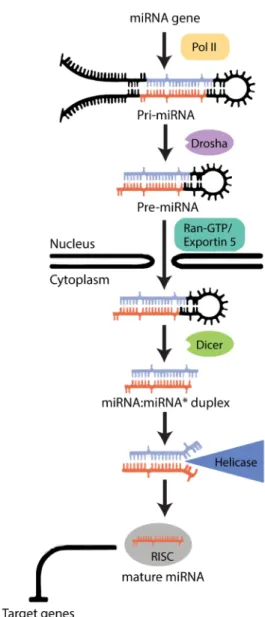
miRNAs are produced according to a multi-step process occurring both inside the nucleus and the cytoplasm of cells34(Figure 1.2). These sequences can either be found in (1) regions of the genome
that are quite distant from the annotated targets, implying that they constitute a different transcriptional unit, or (2) be included inside intronic regulatory regions, providing a mechanism to maintain a coordinated expression between proteins and their respective miRNAs36. Some miRNAs can be found
organized in clusters, constituted of groups of sequences which are evolutionarily related to each other and often coexpressed37.
What is known from most animal models is that the majority of miRNA genes are transcribed by RNA polymerase II (Pol II)36. The transcription of a miRNA gene generates a stemloop containing
the primary miRNA (pri-miRNA)38, which is processed within the nucleus by a multiprotein complex
called the Microprocessor, of which the core components are the RNase III enzyme Drosha and the double-stranded RNA-binding domain (dsRBD) protein DGCR8/Pasha. This complex cleaves the pri-miRNA stem loop, producing an hairpin precursor pri-miRNA (pre-pri-miRNA) that is around 70 nucleotides long34. Next, after translocation to the cytoplasm, the pre-miRNA is cleaved again by Dicer to produce
implicated in the production of interference RNAs (RNAi) and then recognized as important for miRNA maturation36.
Following the cleavage steps, the main RNA silencing pathways (siRNA and miRNA) converge and become biochemically indistinguishable36. The Argonaute proteins (Ago)
are recruited and, together with Dicer, form a trimeric complex that initiates the assembly of the RNA-induced silencing complex (RISC)34. Once
a single stranded miRNA is incorporated into RISC, the complex is guided to mRNA targets based on base-pairing interactions. Base-pairing with the 5’ end of miRNAs, especially to the so-called 2–7 nucleotide ‘seed’, is crucial for efficient targeting. In cases of perfect or near-perfect complementarity to the miRNA, target mRNAs are cleaved and degraded; imperfect miRNA-mRNA hybrids lead to translational repression36. The repression mechanism is far
more common and it is achieved by a destabilization of the mRNA sequence. The post-transcriptional step at which silencing occurs is still unclear, but either elongation of the transcript or initiation of translation are the most likely targets34.
Most biological processes are governed by highly regulated activation and suppression of specific gene programs that are dependent on the activity of miRNAs35. These sequences are
involved in animal development, in cell proliferation and differentiation, in apoptosis and in many other physiological and pathological processes34. Furthermore, due to their overlapping
functions and “buffering effect” on gene expression variation, they are reported to be crucial for the robustness of developmental and
physiological processes39. In Drosophila, one of the most important miRNAs, bantam, was isolated by
gain-of-function assays that revealed its capacity to promote tissue growth and inhibit apoptosis40.
miR-14 is also involved in this process, as well as in the regulation of fat metabolism in a
dose-dependent manner41.
1.3 microRNAs and the immune response
The regulation of the development and function of the immune system lies among the many processes in which miRNAs have been implicated in vertebrates. This includes the regulation of several components of the vertebrate immune response24. Indeed, it is known that miRNAs are
differentially regulated in haematopoietic cell lineages, as well as in a vast array of features of host-pathogen interactions42. Individual miRNAs have been implicated in Haematopoietic Stem Cells
Figure 1.2 – The biogenesis of microRNAs in
Drosophila melanogaster. After a miRNA is transcribed,
the generation of a mature miRNA includes both nuclear and cytoplasmic maturation, culminating in the repression of the translation of several target genes (Adapted from Bartel et al (2004)).
(HSCs) biology, contributing to their homeostasis and lineage commitment (e.g. mir-196b, mir-221 or mir-222)43. miRNAs also have a role in the development and function of vertebrate immune cells,
modulating key steps of cellular lineage determination42. This includes cells of the mammalian innate
immune system, like granulocytes or monocytes from the myeloid lineage (e.g. 21, 196, mir-181a, mir-223 or mir-150)43and the production of Toll-Like Receptors (TLRs) with a major role also
in vertebrates in the recognition of invading bacteria, viruses and other pathogens24. Furthermore,
miRNAs have been reported to be important for the regulation of several adaptive immunity components, such as development of B and T cells, antibody class switching and cytokine production (e.g. mir-17-92 cluster, mir-155 or mir-150)42. Importantly, altered expression levels of miRNAs have
been implicated in various haematopoietic disorders, such as leukaemias, lymphomas and even cancers with immune cell origin42.
However, the repertoire of described immune-related miRNAs and their impact on invertebrate immunity is limited and remains largely unexplored. Commonly observed in various host–pathogen systems is a differential abundance of host miRNAs following an infection, which varies over time and according to the type of pathogen involved44. Several miRNAs have been
implicated in the anti-viral immune response. In this case, abundance of several host miRNAs can be altered after infection in Bombyx mori or Aedes mosquitoes, where it has been speculated that these sequences could target viral genes, dampening replication rates44. This modulatory effect of pathogens
on the host miRNA profile is also reported during bacterial infection. This includes, for instance, the effect of Wolbachia infection in the miRNA profile of Aedes aegypti, where aae-miR-2940 and -309a are reported to have an increase in expression after infection. Also, infection by the Gram-negative bacterium Serratia marcescens in Apis mellifera leads to the downregulation of miRNAs such as ame-bantam, -mir-12, -34, -184, -278, -375, -989 and -1175, and the upregulation of let-7, ame-mir-2, -13a, and -92a44. The available information has been so far obtained from microarray experiments and
therefore still lacks vital functional analysis and validation. One of the very few miRNAs whose role in insect immunity has been experimentally established is mir-8, which negatively regulates AMPs such as Drosomycin and Diptericin in D. melanogaster45. The miRNA let-7 has also been
hypothesized to regulate Diptericin expression in Drosophila S2 cells through the action of the hormone Ecdysone. By repressing Diptericin translation in an ecdysone-dependent manner, let-7 may set a threshold for this AMP and possibly avoid over-stimulation of the innate immune response46. But
the complete function of these sequences in the immune system of invertebrates has yet to be fully understood. Taking into account that immune mechanisms are relatively well conserved among animals, further characterization of miRNA-mediated gene regulation during haematopoiesis should shed light on the homeostatic control of haematopoietic cell renewal and differentiation, while characterization of the humoral response should provide insights on several important features of host-pathogen interactions.
In order to dissect the role of miRNAs in the regulation of immune-related mechanisms in
Drosophila melanogaster, we propose to address two main questions that focus on each of the arms of
the immune system: the humoral and the cellular defences. It is known that the success of haemocyte function depends on their numbers and proportions. Hence, any defects in haematopoiesis, the developmental process that regulates haemocyte proliferation and differentiation, will compromise the availability of these cells throughout development. Our questions include (1) understanding the impact of miRNAs in the control of larval haematopoiesis, the most active and well characterized stage of this developmental process, and (2) dissecting the role of miRNAs in the regulation of the humoral response, mainly involved on the recognition of invading organisms and the triggering of the systemic antimicrobial response.
2. Materials and Methods
2.1 Fly Stocks and Maintenance
To identify miRNAs that modulate larval haematopoiesis and the adult systemic immune response we performed a systematic screen using a library of knock-out lines generated by ends-out homologous recombination by Cohen et al, 2014, covering the vast majority of the miRNAs present in the genome of D. melanogaster. This library encompasses 75 loci encoding a set of 130 single miRNA precursors, and 20 miRNA clusters encoding a total of 58 different miRNA precursors47. Both single
and multiple knock-out of abundant and evolutionarily conserved sequences were used, in which each deletion inactivates single miRNAs or the 8-nucleotide seed of a given cluster of miRNAs respectively. Due to availability constraints, our study focused on a panel of 83 out of the 95 lines of the original library (Supplementary Data – Table 1).
This panel of Drosophila melanogaster microRNA knock-out lines was kindly provided by Claudio Alonso Lab, University of Sussex, UK (Supplementary Data - Table1). The controls y1,w*and
w1118 together with all microRNA deletion lines were simultaneously crossed with the balancer chromosomes y1, w*, N1/FM7c, P{w[+mC]=GAL4-twi.G}108.4, P{UAS-2xEGFP}AX, w1118;
In(2LR)Gla, wgGla-1/ CyO, P{w[+mC]=GAL4-twi.G}2.2, P{UAS-2xEGFP}AH2.2 and w*; ry506Dr1/
TM3, P{Dfd-GMR-nvYFP}3, Sb1. Due to higher genetic background disparities, lines including miRNAs located on the chromosome X were crossed with the control y1,w*for an extra generation (Supplementary Data – Figure1). Due to differences in lethality between the different microRNA deletion lines (Supplementary Data – Table1), the screening process was performed in two modalities: (1) homozygous lethal mutations were tested in heterozygosity over a balancer or over a wild type chromosome; (2) the remaining lines were tested as homozygous. Depending on the modality, lines were compared to equally balanced or non-balanced controls. All fly stocks were maintained in standard fly food at 18ºC. Crosses and experiments were performed at 25°C, ~60% humidity and 12:12hr light:dark cycles.
2.2 Systemic Immunity Screen and Bacterial Infection
Pseudomonas entomophila and Pseudomonas putida were used for systemic infection in the
primary and secondary systemic immunity screens respectively. Following Martins et al (2014), bacterial stocks were kept in glycerol at -80 ºC, streaked in fresh Petri dishes and incubated at 30 ºC. Before use single colonies were picked and let to grow overnight at 30 ºC in Lysogeny Broth medium (LB). Cultures were centrifuged and adjusted to the desired optical density (OD, 1=5x10⁸cells/mL) (P.
entomophila, OD~0,01; P. putida, OD~10). For each of the lines tested and their respective controls,
adult flies (4–6 days after eclosion) were pricked in the thoracic region with a tungsten needle soaked in bacterial solution. Sterile LB medium was used for technical controls on control lines (X, 2 and 3). Each of the biological replicates included administration of bacterial treatments to 30 to 50 males, after which individuals were separated in random groups of 10 on small tubes with standard fly food and kept at 25ºC,~60% humidity and 12:12hr light-darkness cycle. Survival was monitored every day for 6 days in the case of P. entomophila and 7 days for P. putida. Individuals that did not recover from anaesthesia after 2h or were alive at the end of the experiment were counted as censored observations.
2.3 Haematopoiesis Screen, Larval Imaging and Cell Counting
Crystal cells and plasmatocytes were labelled with specific live genetic fluorescent markers on all mutant lines to perform the haematopoiesis screen and analyze several features of differentiation
and proliferation of larval haemocytes. Depending on the location of the microRNA deletion, the markers were distributed across the genome in three different combinations: w*; eater-DsRed;
BcGFP, or LzGal4, EaterGFP; +/+; UAS-Tomato or LzGal4, UAS-mCD8GFP; eater-DsRed; +/+.
Note that Eater marks plasmatocytes whereas Lozenge (Lz) and Black Cells (Bc) are crystal cell markers. The genetic crosses required to create lines carrying the cell reporters as well as each of the microRNA knock-outs were also carried out on the existing controls for each of the chromosomes analyzed. The sessile haemocyte compartment, particularly on the most posterior segment of the larval body, was used as a proxy for the remaining segments as previously validated48. All experiments were
performed in males, due to high variance associated to the haemocyte pool of females.
Ten late third instar (L3) male larvae were imaged for each of the screened lines. After staging and male selection, larvae were placed in a microcentrifuge tube with standard fly food at room temperature (RT) for at least one hour before imaging. Prior to acquisition, the selected larvae were washed twice in cold 1xPBS, dried on paper and placed on a cold metal block. Individual larvae were secured under a microscope slide and imaged dorsally on a Zeiss STereo LUMAR stereoscope equipped with a Hamamatsu Orca-ER CCD camera and a GFP fluorescence filterset. Together with a whole-body picture, a high-magnification caption was taken of the posterior-most haemocyte cluster of the larval body for quantification purposes. Cells were counted on Fiji49to assess proportions and
density. Plasmatocyte numbers and cluster area were determined automatically using the ITCN plugin50and crystal cells were counted manually using the Cell Counter plugin51.
2.4 Lymph Gland Dissection and Haemolymph Extraction
Detailed description of the phenotype of the miR-980 knock-out line required further crossing with: (1) +/+; eaterGal4, UAS-YFP, MSNF9-mCherry; BcCFP for labelling lamellocytes (MSNF9-mCherry) and the remaining types of haemocytes; (2) w1118; 10XStat92E-GFP for reporting activation of the JAK-STAT pathway. For lymph gland characterization, late L3 male larvae resulting from these crosses were collected and dissected according to Evans et al (2014). Samples were fixed for 30 minutes in 3.7% ρ-formaldehyde:PBS at RT, washed three times with 1xPBS for 5 minutes and mounted on a microscope slide with Vectashield (Vector). Confocal Z-series stacks were acquired on a Leica SP5 confocal using 20x and 40x air objectives. The circulating haemocyte compartment was characterized according to Leitão&Sucena (2015). Six L3 male larvae were disrupted on the ventral cuticle to minimize inclusion of cells from the dorsal clusters. Haemolymph from the dissected larvae was collected and placed on a 12-well slide for 20 minutes. Following fixation with 3.7% ρ-formaldehyde:PBS for 20 minutes, samples were washed three times with 1xPBS and sealed in Vectashield. Images were acquired on a Leica DMRA2 upright microscope using the 20x air objective.
2.5 Statistical Analysis
Statistical analysis and graphics were done on R Studio, version 3.2.2, and GraphPad Prism Software, version 6. Survival fractions for each time point were estimated using the Kaplan-Meyer method and analyzed in two statistical approaches: (1) Comparison of average survival curves using the Logrank (Mantel-Cox) test; (2) Comparison of hazard ratio regression coefficients using a Cox’s proportional hazards mixed effect model. The first approach allows comparison of survival curve dynamics over time, taking into consideration each time point. The Logrank comparison in particular gives equal weight to all time points. The second approach provides a global estimate of the risk of death after infection (hazard ratio), while allowing the exploration of the effects of several explanatory variables on survival. Due to the detection of a significant difference between biological replicates (date effect), hazard ratios were analyzed with the mutant lines defined as a fixed factor and date of the experiment and replicate vials were defined as random factors. Hazard ratios of independent trials for
each mutant line were also taken in consideration. Data obtained from the haematopoiesis survey was checked for normal distribution using a Saphiro-Wilk test. Samples were compared with their controls using t-tests when normally distributed and a Wilcoxon–Mann–Whitney test when the distribution departed from normal. Multiple comparisons from both datasets were corrected using the Bonferroni or the Holm-Bonferroni tests.
2.6 Retrieval of computationally predicted miRNA targets
microRNA predicted target retrieval was done using an online database based on several algorithms designed for this purpose (http://m3rna.cnb.csic.es/)52. These algorithms consider
properties like complementarity and structure of the miRNA-mRNA interaction and attribute each target gene different scores based on such features. There are two combinatorial approaches of several available predictive algorithms in order to make predictions more efficient: (1) Weighted Scoring by Precision (WSP), based on summing up the weighted scores for different databases and (2) Logistic Regression combined Scoring (LRS), which applies logistic regression to find the combined score of the algorithms taking in consideration the probability of experimental validation52. LRS was
preferentially used to assess the high score targets for each of the obtained candidate microRNAs and only the first 200 high-LRS score target genes for each miRNA were considered.
3. Results
3.1 microRNAs have a role in systemic immunity of Drosophila melanogaster
To assess if microRNAs play a role in the systemic immune response of Drosophila, we performed a primary screen on eighty-three different microRNA (miR) deletion lines by monitoring survival of adult flies after infection with Pseudomonas entomophila.
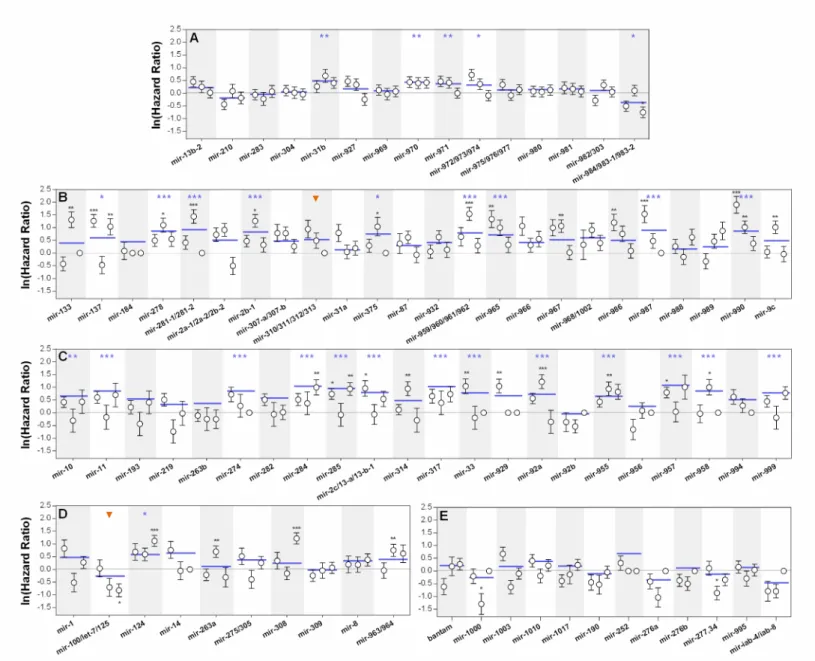
In general, results from the statistical comparisons of both survival curves and hazard ratios between controls and treatments are in agreement with each other (Supplementary Data – Table 2). However, some miRNA deletions report significant differences in the hazard ratios without producing detectable differences in the comparison of survival curves or vice-versa. Furthermore, analysis of the hazard ratios of independent biological replicates revealed that detectable differences are not always consistent among trials (Figure 3.1). Based on these distinct statistical outcomes the lines tested were stratified into four categories:
Rank 1 includes 14 lines which show consistent differences in both statistical approaches
when compared to their controls (Supplementary Data – Table 2). Hence, mutations included in this category significantly impact the risk of death after infection, generating very distinct survival dynamics over time. For instance, mir-990 deletion causes a striking decrease in survival after the first day of infection that is corroborated by a high hazard ratio coefficient, consistent across all biological replicates. Other lines, such as mir-137 and mir-285 deletions, report the same outcome, but independent replicates reveal disparate tendencies (Figure 3.1). s
Rank 2 represents situations where significant differences in the survival curves between
treatment and controls were reported, but hazard ratio comparison failed to corroborate these findings. For example, the accentuated increase in survival after infection of mir-100/let-7/mir-125 deletion line that is detected in the survival curve comparison, fails to be represented by a significant negative hazard ratio (Figure 3.1 – D), which is only mildly detected in one of the biological replicates.
Rank 3 refers to situations where hazard ratios were significantly different between treatments
and controls, but overall survival curves were not distinct enough to validate their disparity.
Figure 3.1 – Survival to bacterial infection. Natural logarithm of hazard
ratios between survival of flies with knocked-out microRNA genes and their controls upon infection with P.
entomophila. (A) microRNA knock-out
lines located on the X chromosome. (B)
Homozygous viable microRNA knock-out lines located on chromosome II and (C) III. (D) Homozygous lethal
microRNA knock-out lines located on the chromosome II and (E) III.
Significant deviations from the control line (x=0) represent significantly different hazard ratios (p<0,05). Positive and negative coefficients represent decreased and increased survival, respectively. White circles represent independent biological replicates of each line and blue lines refer to the hazard ratio obtained from the statistical model that accounts for a random date effect (See materials and methods). Vertical bars correspond to the 95% confidence intervals of the estimated hazard ratios. Data points that overlap with the basal line and lack vertical bars represent missing data. The orange arrowheads represent the two lines belonging to Rank 2, which reported significant differences exclusively for the average survival curve comparisons. *p<0,05; **p<0,01 ; ***p<0,001.
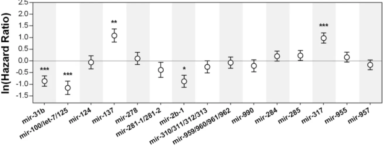
This ranking system was used to select the lines used in the subsequent phases of the screening process. We performed our secondary screen on fifteen candidates, including lines to which Rank 1 was attributed based on three biological replicates (13 lines) and two extra lines from Rank 2 that showed interesting differences in the overall survival curves dynamics. This screen consisted of a
Pseudomonas putida systemic infection. Figure 3.2 represents the normalized hazard ratios obtained
for the candidate microRNA deletion lines upon infection with P. putida. Out of the 15 lines considered, only five reported significant differences in survival after infection (p<0,05). The same differences were consistent between the comparison of survival curves and of hazard ratios (Supplementary Data – Table 3 and Figure 3). These include mir-31b, mir-2b-1 and the 100/let-7/125 cluster, which show a significant increase in survival when compared to controls, and mir-137 and mir-317 that had a significant decrease in survival after bacterial infection. Importantly, due to time constraints this dataset refers to only one trial. Further replication is necessary to firmly establish a role for these candidate microRNAs in the immunity of Drosophila melanogaster against
Pseudomonas systemic infection.
Figure 3.2 – Survival to bacterial infection of candidate microRNA knock-out lines. Natural logarithm of hazard ratios
between survival of flies with knocked-out microRNA genes and their controls upon infection with Pseudomonas putida. The basal line refers to the controls of each sample, thus significant deviations from zero represent significant differences in survival (p<0,05). Positive and negative hazard ratio coefficients represent decreased and increased survival, respectively. Vertical bars correspond to the 95% confidence intervals of the estimated hazard ratios. *p<0,05; **p<0,01; ***p<0,001.
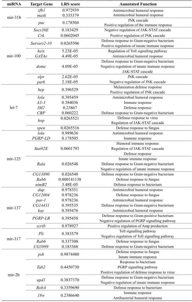
Throughout our trials, the observed phenotype of candidate microRNA deletions was found to be relatively consistent in some lines, while others showed reverse tendencies. This is the case of the mir-31b and mir-2b-1 deletions, which reported opposite survival tendencies between screens performed with different pathogenic bacteria from the genus Pseudomonas. The remaining lines that revealed significant differences were consistently susceptible or resistant/tolerant to bacterial infection – mir-100/let-7/mir-125 cluster, mir-137 and mir-317. Based on our observations, we then asked which genes could be targeted for post-transcriptional silencing by these miRNAs. Among the extensive list of target genes found to be complementary to our candidate miRNAs, we found annotated ontology categories associated with regulation of the general antimicrobial immune response, particularly of the Imd, Toll, JNK and JAK-STAT pathways (Table 3.1). Furthermore, some of the lists included genes that have annotated roles in both systemic and cellular immunity, particularly in haematopoiesis (Supplementary Data – Table 4).
Table 3.1 – Retrieved immune-related target genes for candidate miRNAs obtained from the systemic immunity screens
miRNA Target Gene LRS score Annotated Function
mir-31b
zfh1 0.972939 Antimicrobial humoral response Antimicrobial humoral response mask 0.335379
puc 0.178504 Positive regulation of the immune responseJNK cascade
mir-100
Socs36E 0.183429 Negative regulation of JAK-STAT cascade Crk 0.0602045 Positive regulation of JNK cascade Su(var)2-10 0.0265506 Defense response to Gram-negative bacteriumPositive regulation of innate immune response
heix 5.23E-05 Regulation of Toll signalling pathway
GATAe 4.49E-05 Antimicrobial humoral response
dome 4.09E-05 Negative regulation of innate immune responseDefense response to Gram-negative bacterium JAK-STAT cascade
slpr 2.62E-05 JNK cascade
park 2.18E-05 Negative regulation of JNK cascade
let-7
hep 0.396529 Positive regulation of JNK cascadeMelanization defense response
lola 0.395459 Antimicrobial humoral response
A3-3 0.384036 Immune response
IM2 0.23867 Defense response
CBP 0.060222 Defense response to Gram-negative bacterium hop 0.0265521 Regulation of JAK-STAT cascadeDefense response to virus spen 0.0265516 Defense response to fungus
mir-125
lola 0.989636 Antimicrobial humoral response
PGRP-LD 0.178505 Immune response
Stat92E 0.0601793 Regulation of JAK-STAT cascadeHumoral immune response Defense response
Rala 0.026548 Defense response to Gram-negative bacteriumInnate immune response Negative regulation of innate immune response CG13890 0.026548 Defense response to Gram-negative bacterium
Rab6 0.000141138 Defense response to fungus
nimB2 3.48E-05 Defense response to bacterium
mir-137
dup 0.978351 Antimicrobial humoral response
CG12004 0.978236 Defense response to fungus
par-1 0.978236 Antimicrobial humoral response
CG14411 0.395535 Defense response to Gram-negative bacterium kay 0.395476 Antimicrobial humoral response PGRP-LB 0.395458 Negative regulation of PGRP signalling pathwayDefense response to Gram-positive bacterium
scrib 0.978927 Positive regulation of Amp production
mir-317 Pli 0.383579
Toll signalling pathway
Negative regulation of Toll signalling pathway Rab6 0.337308 Defense response to fungus
CG5899 0.183368 Defense response to Gram-negative bacterium
mir-2b
psh 0.9874480 Defense response to fungusInnate immune response
Tab2 0.4450730 PGRP signalling pathwayResponse to bacterium
Positive regulation of defense response to virus upd1 0.3837370 Negative regulation of innate immune responseDefense response to Gram-negative bacterium
BobA 0.3359690 Defense response to bacterium
3.2 The role of microRNAs in the haematopoiesis of Drosophila melanogaster
The combined action of both humoral and cellular arms during the immune response of D.
melanogaster, together with the indication that miRNAs can have several haematopoiesis related
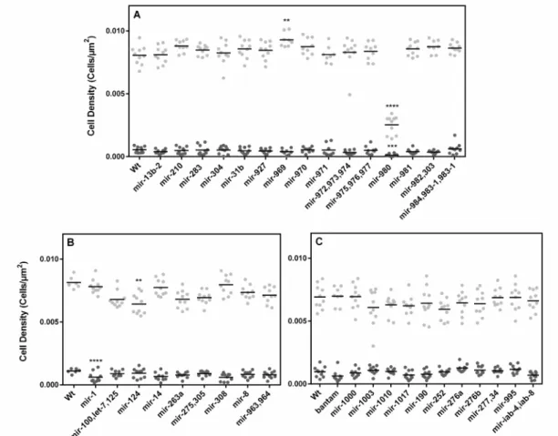
targets, led us to ask if miRNAs could play a role in haematopoiesis and cellular immunity. To tackle this, we performed a screen on the same panel of microRNA knock-out lines used above for haematopoietic defects. Using the most posterior sessile haemocyte cluster of the larval body as a proxy for total sessile haemocytes48, we quantified the following haematopoietic features of larvae:
plasmatocyte and crystal cell absolute numbers and sessile cluster size. These two measurements were used to determine the densities of each cell type in the sessile haematopoietic compartment (cells/µm2). Figure 3.3 refers to larval haemocyte densities at the posterior-most cluster of the several
microRNA deletion lines analyzed. Due to time constraints and crossing scheme limitations, only deletions located on the X chromosome (Figure 3.3 – A) and homozygous lethal from second and third chromosomes were included in our analysis (Figure 3.3 – B and C).
Figure 3.3 – Plasmatocyte and crystal cell density. Quantification of plasmatocyte ( ) and crystal cell ( ) densities in the
most posterior sessile haemocyte cluster of L3 male larvae (Number of cells/Cluster Area, Cells/μm2). Black horizontal lines
refer to the mean of each dataset. (A) Plasmatocyte and crystal cell density of microRNA knock-out lines located in the X
chromosome and of homozygous lethal microRNA knock-out lines located in the (B) chromosome II and (C) III. *p<0,05;
**p<0,01 ; ***p<0,001; ****p<0,0001.
Out of a total of 36 lines analyzed for this trait, four showed significant changes in either plasmatocyte and/or crystal cell densities (p<0,05): mir-969, mir-980, mir-1 and mir-124.
mir-969 hemizygous deletion shows a small yet significant increase in plasmatocyte density (Figure 3.3 – A) as a result of having reduced clusters that maintain average plasmatocyte numbers (Supplementary Data – Figure 4 and 5).
mir-980 hemizygous knock-out produced a striking decrease in the densities of both cell types (Figure 3.3 – A). Despite being associated with a reduction in cluster size (Supplementary Data – Figure 4), this is not significant, indicating that the detected differences in density are in fact a result of a marked decrease in cell numbers (Supplementary Data – Figure 5 and 6).
Additionally, mir-1 heterozygote knock-out shows a considerable decline in crystal cell density (Figure 3.3 – B) likely due to the observed non-significant decrease in crystal cell numbers (Supplementary Data – Figure 6).
Finally, mir-124 heterozygous deletion is associated with a reduction in plasmatocyte density (Figure 3.3 – B), again associated with a non-statistically detectable reduction in plasmatocyte numbers (Supplementary Data – Figure 5).
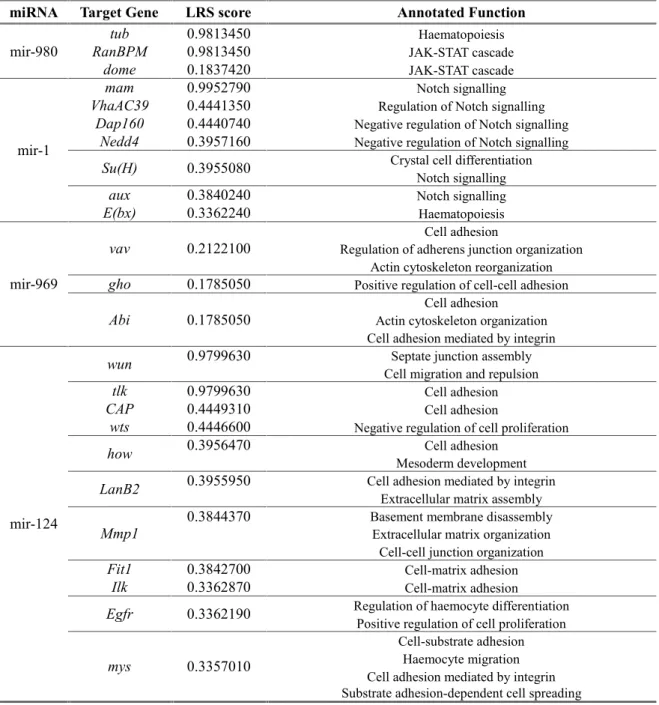
Collectively, our observations point towards the idea that microRNAs are important regulators of haematopoiesis. Among the predicted target genes of each of the miRNA candidates obtained from this screen, and reinforcing our observations, we found genes with annotated roles in haematopoiesis, JAK-STAT and Notch signalling and crystal cell differentiation, as well as cell adhesion and extracellular matrix assembly (Table 3.2). Further characterization of the remaining lines of the microRNA knock-out panel could reveal other important players in the dynamics of proliferation and differentiation in haematopoiesis that ultimately can influence other developmental processes and biological responses.
Table 3.2 – Retrieved immune-related target genes for candidate miRNAs obtained from the haematopoiesis screen.
miRNA Target Gene LRS score Annotated Function
mir-980 RanBPMtub 0.98134500.9813450 JAK-STAT cascadeHaematopoiesis
dome 0.1837420 JAK-STAT cascade
mir-1
mam 0.9952790 Notch signalling
VhaAC39 0.4441350 Regulation of Notch signalling
Dap160 0.4440740 Negative regulation of Notch signalling Nedd4 0.3957160 Negative regulation of Notch signalling
Su(H) 0.3955080 Crystal cell differentiationNotch signalling
aux 0.3840240 Notch signalling
E(bx) 0.3362240 Haematopoiesis
mir-969
vav 0.2122100 Regulation of adherens junction organizationCell adhesion Actin cytoskeleton reorganization gho 0.1785050 Positive regulation of cell-cell adhesion Abi 0.1785050 Actin cytoskeleton organizationCell adhesion
Cell adhesion mediated by integrin
mir-124
wun 0.9799630 Cell migration and repulsionSeptate junction assembly
tlk 0.9799630 Cell adhesion
CAP 0.4449310 Cell adhesion
wts 0.4446600 Negative regulation of cell proliferation how 0.3956470 Mesoderm developmentCell adhesion LanB2 0.3955950 Cell adhesion mediated by integrinExtracellular matrix assembly Mmp1 0.3844370 Basement membrane disassemblyExtracellular matrix organization
Cell-cell junction organization
Fit1 0.3842700 Cell-matrix adhesion
Ilk 0.3362870 Cell-matrix adhesion
Egfr 0.3362190 Regulation of haemocyte differentiationPositive regulation of cell proliferation
mys 0.3357010
Cell-substrate adhesion Haemocyte migration Cell adhesion mediated by integrin Substrate adhesion-dependent cell spreading
Table 3.2 (continued)
miRNA Target Gene LRS score Annotated Function
mir-124
parvin 0.2386660 Cell adhesion
girdin 0.2123550 Epithelial cell-cell adhesion
scb 0.2122480 Calcium-dependent cell-matrix adhesionCell-cell adhesion
mbt 0.2122350 Regulation of cell-cell adhesion mediated by cadherin
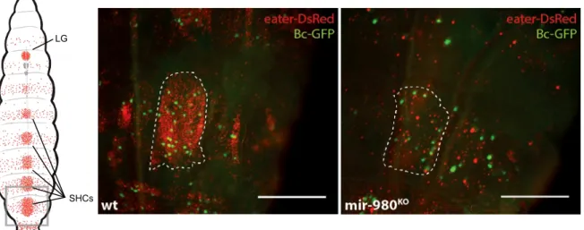
3.3 mir-980 controls lamellocyte differentiation in the sessile haemocyte clusters
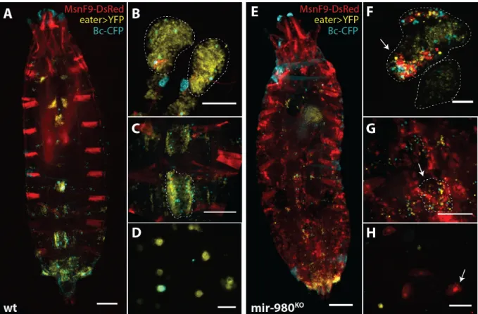
The deletion of mir-980 produced the most striking phenotype obtained from the primary screen for haematopoiesis defects. As shown in Figure 3.4, both crystal cells and plasmatocytes show a marked decrease in density in the sessile haemocyte clusters of mir-980 hemizygous male larvae. This outcome is not correlated with changes in cluster area, but rather with a strong decrease in the absolute numbers of both cell types (Supplementary Data – Figure 4 to 6). Additionally, preliminary observations of lymph gland structure did not reveal similar cellular features, pointing to the idea that such changes could be exclusive of the sessile haemocyte compartment. Additionally, we detected the presence of melanized nodules in the haemocoel of late L3 larvae and pupae. However, such structures were not common to all individuals, being found in around 10% of the population. Furthermore, no fluorescence was detected in dissected nodules for both crystal cell and plasmatocyte specific markers (data not shown).
Figure 3.4 – mir-980 knock-out decreases plasmatocyte and crystal cell densities. (Left) Schematic dorsal view of a third
instar larva where plasmatocytes and crystal cells are marked in red (eater-DsRed) and green (Bc-GFP), respectively. Highlighted is the most posterior cluster, used as a proxy to infer haemocyte densities (LG, Lymph Gland; SHCs, Sessile Haemocyte Clusters). (Center and Right) Magnified dorsal view of the most posterior haemocyte cluster of mir-980 larvae and the respective control. (scale=250µm)
The presence of melanized nodules in the larval haemocoel is common in previously described mutant phenotypes and is associated in some circumstances with ectopic lamellocyte differentiation53.
Furthermore, both lamellocytes and crystal cells transdifferentiate from plasmatocytes and depend on the number of the latter cell type15,20. We therefore asked if the accentuated decrease in plasmatocyte
numbers was caused by a change of cellular fate into lamellocytes. As shown in Figure 3.5 using the lamellocyte specific reporter for the gene misshapen MSNF9-DsRed, mir-980 deletion triggers ectopic lamellocyte differentiation and these cells are found circulating in the haemolymph in high numbers. Combined with a reduction in the numbers of plasmatocytes and crystal cells, we confirmed the severe disruption of the integrity of the sessile haemocyte compartment (Figure 3.5 – E and G) and the presence of melanotic nodules in the context of a mir-980 deletion. Furthermore, lamellocyte differentiation was found to occur in the lymph glands (Figure 3.5 – F). However, the fact that this
LG
organ is still intact in the mutant background, contrary to what was expected, suggests that the main contribution to the circulating lamellocyte population comes from the sessile haemocyte compartment only. Staged analysis of the phenotype also revealed that this exacerbated differentiation event begins after larvae reach the mid L3 stage and persists after pupariation, being found also in adults (data not shown). Collectively, our observations suggest that mir-980 plays an important role in inhibiting lamellocyte differentiation in homeostasis.
Figure 3.5 – mir-980 has a role in the regulation of lamellocyte differentiation. Comparison of the different haemocyte
compartments of third instar male larvae of control line (A-D) and mir-980 knock-out (E-H). Plasmatocytes are marked with
eater>YFP, crystal cells with Bc-CFP and lamellocytes with MsnF9-DsRed. (A, E) Dorsal view of the larval body
(scale=250µm) together with (B, F) dissected lymph glands (scale=100µm), (C, G) magnified sessile haemocyte clusters
(scale=250µm) and (D, H) circulating haemocytes (scale=50µm).
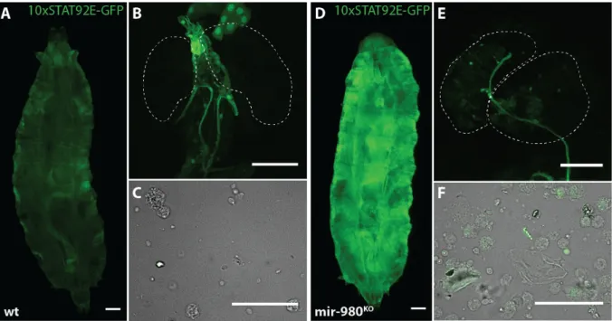
We next asked which gene products could be targeted for silencing by mir-980, ultimately contributing to the regulation of lamellocyte transdifferentiation. Among the high LRS score targets, we found that mir-980 has predicted complementarity to domeless, the transmembrane receptor of the JAK-STAT pathway28(Table 3.2). Activation of the JAK-STAT pathway has been previously reported
to be associated with ectopic lamellocyte differentiation and melanotic nodule formation11. Based on
the idea that deletion of mir-980 would lead to an increase in dome translation, we assessed the JAK-STAT activation status. Preliminary results show that mir-980 deletion causes a change in the activation pattern of Stat92E (Figure 3.6), the nuclear effector of the JAK-STAT pathway11. This is
particularly evident in the whole-body expression pattern (Figure 3.6 – D). Whether the change in expression occurs in the epidermis, fat body or any other widespread tissue still remains to be determined. Furthermore, the haematopoietic compartments failed to report strong changes in GFP expression. However, a markedly reduced number of haemocytes shows cytoplasmic GFP expression in the mutant context, both in the lymph glands and in the circulating haemocyte compartments (Figure 3.6).
Figure 3.6 – mir-980 knock-out impacts STAT92E expression pattern. Comparison of the STAT92E activation pattern in
third instar male larvae of control line (A-C) and mir-980 knock-out (D-F). (A, D) Dorsal view of the larval body
(scale=250µm) together with (B, E) dissected lymph glands (scale=100µm) and (C, F) circulating haemocytes (scale=50µm).
4. Discussion
In this project, we set out to unravel the roles of miRNAs in the immune response of D.
melanogaster assessing if (1) miRNAs play a role in the humoral defences, particularly in the
regulation of susceptibility to bacterial infection and (2) if miRNAs are important for the regulation of cellular defences, mainly in regards with the development of haemocytes.
4.1 Experimental design
Our results from a primary screen of miRNA knock-out lines challenged with P. entomophila systemic infection indicate that several miRNA genes are necessary for the regulation of susceptibility to bacterial infection and for the function of general immune defense mechanisms (Figure 3.1). P.
entomophila is a gram-negative bacterium that has the ability to trigger both local and systemic
immune responses after ingestion or systemic administration54,55. Furthermore, systemic infection with
P. entomophila is correlated with high virulence at low doses when compared to the oral infection
route, since the first bypasses the epithelial barrier defences inherent to the latter. Therefore, systemic infection leads to high mortality rates even with bacterial concentrations at the lower edge of detection. Steep mortality curves, where mortality reaches a plateau above 85% in the first three days, have low resolution. Hence, small effect mutations which may lead to higher mortality might go unnoticed and create false negatives. Decreasing the 24-hour sampling interval or using “slow-killing” pathogens in the primary screen might have had a positive effect in this loss of resolution. Nonetheless, we chose P. entomophila for our initial approach because, contrary to other bacteria, it is a natural pathogen of D. melanogaster and is able to trigger Imd activation after systemic infection2.
Moreover, due to viability constraints, some miRNAs were tested in heterozygosity, which might contribute to the presence of false negatives among our datasets. mir-8, for instance, has been previously implicated in the regulation of AMP levels in homeostasis45. However, due to high pupal
lethality, it was only tested in heterozygosity and we failed to detect differences in adult survival after bacterial infection (Figure 3.1). Importantly, the detection of a statistically significant date effect