1. Department of Physical Medicine and Rehabilitation, Bursa Sevket Yilmaz Training and Research Hospital, Bursa, Turkey 2. Department of Physical Medicine and Rehabilitation, Ankara Training and Research Hospital, Ankara, Turkey
3. Department of Radiology, Ankara Kecioren Training and Research Hospital, Ankara, Turkey
tenosynovitis score (p=0.003).
Conclusion: This study showed that RA has significant
negative impact on hand function and dexterity and the parameters used in the evaluation of hand function are mainly associated with tenosynovitis scores. Since tenosynovitis is a common pathology in RA, MRI can be used as a supportive method in early diagnosis of tenosynovitis and may be useful in identification of pa-tients requiring aggressive treatment.
Keywords: Hand; MRI; Rheumatoid arthritis; Tenosy
-novitis; Dexterity.
IntroductIon
Rheumatoid arthritis (RA) is a chronic, systemic, au-toimmune disease of unknown etiology leading to pro-gressive joint destruction and difficulties in performing daily living activities1. It is the most common inflam-matory arthritis, affecting ~ % 1 of the population2. RA affects especially synovial joints and tendons, so pri-marily is an inflammatory synovitis rather than arthri-tis. Inflammation of the synovial membrane leads to formation of highly cellular inflammatory pannus tis-sue. Pannus grows over and infiltrates cartilage, ten-dons and ligaments, which result in erosion of carti-lage and subchondral bone, distruption of ligamentous insertions and impaired tendon glide. These factors combine to cause pain, stiffness and deformities seen in RA3.
Hand deformity and loss of joint function are com-mon in patients with RA and it is estimated that the hands and wrists are affected in 80% to 90% of the pa-tients with RA4. Hand involvement is one of the major determinant of disease outcome affecting the ability to perform activities of daily living and other functional activities5,6. Up to 30% of patients have radiographic
Effect of rheumatoid arthritis on strength, dexterity,
coordination and functional status of the hand:
relationship with magnetic resonance imaging findings
Erol AM1, Ceceli E2, Uysal Ramadan S3, Borman P2
ACTA REUMATOL PORT. 2016;41:328-337
AbstrAct
Objective: To evaluate the effect of rheumatoid
arthri-tis (RA) on strength, dexterity, coordination and func-tional status of the hand and to determine the relation with magnetic resonance imaging (MRI) findings.
Materials and Methods: Thirty-eight patients with RA
and thirty-three controls were included in the study. There were five drop-outs in RA group. Pain was as-sessed by visual analog scale. Painful and swollen joints of the dominant hand were recorded. Hand deformities of the patients were noted. Hand grip strength and pinch strength of the dominant hand were evaluated. Hand disability was assessed by Duruoz hand index (DHI) and the Purdue pegboard test was used for as-sessment of coordination and dexterity. MRI of the dominant wrist and hand was performed in RA group. MRI scans were evaluated for synovitis, tenosynovitis, bone erosion and bone edema.
Results: Demographic characteristics were similar
be-tween groups. While DHI scores were significantly higher (p=0.000), Purdue pegboard test scores were significantly lower in RA group in comparison to con-trol group (p=0.000). Bone edema and synovitis scores were significantly higher in patients with longer disea -se duration (p=0.025, p=0.006 respectively). There were significant negative correlation between grip strength, pinch strength subgroups and tenosynovitis scores (p=0.001, p=0.001). When the Purdue pegboard scores were lower, tenosynovitis scores were signifi-cantly higher (p=0.019, p=0.013, p=0.043). There was a significant positive correlation between DHI score and
evidence of disease at the time of diagnosis, and over 60% have radiographic joint changes within 2 years of diagnosis. So accurate measurement of hand functions using objective and easy methods are important in RA patients5,7. An important aim of treatment in RA is to control disease activity, prevent joint deformities, pre-serve function, and thus maintain or improve quality of life4.
Hand and wrist involvement is of great importance in RA8. Diagnosing RA during its early stage is crucial, given the implications for therapeutic management9. Within the first year after symptom onset, joint syno -vial inflammation progresses to erosion of cartilage and bone in up to 47% of patients10. Structural joint da -mage has been traditionally evaluated by radiological images. However, only the late signs of preceding disea se activity can be visualised by radiography11. Magnetic resonance imaging (MRI) is an imaging method that can exceed many limitations of conven-tional radiography. MRI can detect the presence of inflammation in the synovium, tenosynovium, and pro -bably the periarticular bone earlier in RA10,11. Given these advantages, MRI has a major potential as an out-come measure in RA clinical trials and investigations. The aim of this study is to evaluate the effect of RA on pinch and grip strengths, range of motion (ROM), hand dexterity, coordination, and performing daily ac-tivities and to determine the relation of MRI findings with these parameters.
MAterIAls And Methods
Thirty-eight consecutive patients with RA, according to the 1987 revised American College of Rheumatolo-gy (ACR) criteria12, visiting the outpatient department of Rheumatology were enrolled in the study. The con-trol group comprised 33 age- and sex-matched sub-jects. The exclusion criteria were the presence of o ther hand and wrist diseases such as entrapment neuropa-thy, tendinitis, history of major hand trauma or surgery of the hand, or of neurologic diseases causing seque-lae in the hand. Those with a psychiatric disorder were also not included in the study. The control group was constituted from the non-rheumatoid patients who atten ded the outpatient clinic due to low back pain or knee pain, having no clinical symptoms referable to hand joints. There were five drop-outs in the RA group. One patient due to body weight over the safety res -triction of MRI table, two patients due to
claustropho-bia and two patients due to joint contractures causing difficulty in positioning in MRI machine, could not undergo MR imaging. The study was completed with 33 patients.
All participants’ demographic variables including age, gender, weight, height, dominant hand, occupa-tion, hand overuse history and comorbid diseases were recorded. Hand overuse was defined as hobbies and jobs required repetitive and frequent usage of hands. All patients in the RA group were receiving disease--modifying treatments during the study. Duration of disease and morning stiffness were also recorded. Accor ding to rheumatoid factor (RF) levels, patients were classified as RF positive and RF negative.
Presence of hand pain was evaluated on a 0-10 vi-sual analog scale (VAS). Painful and swollen joints of the dominant hand were recorded. Deformities of the wrist and fingers were defined. ROM was measured in degrees with a standard finger goniometer. For the wrist, four movements were assessed: extension, fle -xion, radial and ulnar deviation. Mobility of each fin-ger was assessed by measuring flexion and extension movements at the metacarpophalangeal (MCP), proxi -mal interphalangeal (PIP) and distal interphalangeal (DIP) joints. Total ROM was calculated for each fin-ger3.
Hand grip strength was measured on the dominant hand using Jamar hydraulic hand dynamometer. The subject’s arm was positioned according to the Ameri-can Society of Hand Therapist’s recommendations. The procedure was repeated three times. The average rea -ding was recorded in kilograms. Pinch strength was measured with Jamar hydraulic pinch gauge which as-sesses tip to tip pinch between the thumb and index finger, lateral pinch where the thumb is clasped against the radial side of the index finger (strongest pinch grip) and three jaw chuck where the pulp of the thumb is pinched against the pulps of the index and middle fin-gers. As for the power grip, the test was repeated three times and the average reading was recorded in kilo-grams13.
Purdue Pegboard test was used for evaluating fine coordination and dexterity of the hand. Four subtests comprise the test; right hand (RH), left hand (LH), both hands (BH) and assembly. Each stage of the test was administered three times14. Hand disability was assessed by Duruöz hand index (DHI). DHI is an 18--item questionnaire concerning daily living activities, each question being scored from 0 (performed wi thout difficulty) to 5 (impossible to do). Disability was
torum communis, extensor indicis; (V) extensor digiti minimi; (VI) extensor carpi ulnaris and three flexor ten-don groups: (1) the flexor carpi ulnaris tenten-don; (2) the flexor digitorum superficialis and profundus; (3) the flexor carpi radialis were scored. Grading was as fol-lows: grade 0 indicated no tendon sheath enhance-ment; grade 1, tendon sheath enhancement without tendon sheath thickening and grade 2, tendon sheath enhancement with tendon sheath thickening. Total tenosynovitis score was calculated (maximum possible score = 18) 9,10.
The ethics committee of hospital approved the study and all participants were given a written informed con-sent.
stAtIstIcAl AnAlysIs
Data were analysed by using the statistical package for social sciences (SPSS) version 11.5 for Windows. All numerical data were expressed as the mean ± standard deviation. The normality of variables was evaluated by the Shapiro–Wilk statistics. Statistical comparisons be-tween the measures or groups were done by using the Student’s t test or the Mann–Whitney U test. Correla-tion coefficients were calculated by the Spearman method. For categorical comparisons Chi-square and Fisher exact tests were used. Statistical significance was set at p< 0.05.
results
Thirty-three patients with a mean age of 46.4 ± 11.1 years and thirty-three controls with a mean age of 46.3 ± 10.9 years were enrolled in the study. Demographic characteristics were similar between RA and control groups (p>0.05). Fifteen patients had at least one de-formity (45.5%). Five patients had ulnar deviation of the wrist, one had MCP subluxation, two had ulnar de-viation of MCP, three had swan neck deformity, five had boutonniere deformity and two had type one thumb deformity (boutonniere deformity of the thumb). Four-teen patients in the RA group had hand overuse history. Nine of the patients had history of sewing and ma -king lace regularly and five had jobs required repetitive use of hands such as carpet weaving, carpentry and up-holstery. Descriptive characteristics of the groups are given in Table I.
We compared hand function of the patients with recorded as the total score obtained by adding the
scores of all questions (range 0-90)15. All assessments were performed by the same physiatrist.
MR imaging of the dominant wrist and hand inclu -ding MCP joints, was performed with a 1.5-Tesla mag-net system in RA group. MRI of control group could not be obtained due to ethical issues. The imaging proto-col comprised firstly, fat suppressed axial T1-weighted spin echo (SE), coronal proton-density weighted, sagit-tal T2 weighted gradient echo (GRE) and coronal T2 weighted GRE sequences, followed by fat suppressed coronal and axial T1 weighted SE sequences after in-jection of contrast agent. Two patients refused contrast agent so MRI scanning was completed without it. These patients were not excluded from the study. A single ra-diologist who was blinded to the physical examination results, reviewed scan images for bone edema, bone erosion, synovitis and tenosynovitis. MRI scans were evaluated according to the system developed and vali-dated by McQueen et al10. Outcome Measures in RA Clinical Trials (OMERACT) RA MRI score (RAMRIS) system was also taken into account during evaluation of scan images16,17.
Bone erosion: 15 bony sites were evaluated for
ero-sions. These were distal radius, distal ulna, eight carpal bones and the bases of the five metacarpal bones. Erosions were scored, on size as 0= none or < 2mm in dia meter, 1= 24mm in diameter and 2=> 4mm in diame -ter. Total erosion score was calculated (maximum possible score = 30) 9,10.
Bone edema: Bone marrow edema was scored at the
same sites for bone erosion; 0 for none or one bone minimal effected, 1 for minor edema involving < 50% of the bone (one carpal bone, distal radius, distal ulna and one basis of metacarpal bone) and 2 for gross ede-ma involving > 50% of the bone ede-marrow). The total bone marrow edema score was obtained from the sum of all scores (maximum possible score = 30) 9,10.
Synovitis: Synovitis was assessed at the distal
ra-dioulnar joint, radiocarpal joint (ulnar aspect), radio-carpal joint (radial aspect), interradio-carpal joint (between the proximal and the distal carpal rows), and MCP joints. Synovitis was scored using synovial thickening (0 for < 2 mm, 1 for 2–4 mm and 2 for > 4 mm). Total synovitis score was calculated (maximum possible score = 10) 9,10.
Tenosynovitis: Six extensor tendon groups: (I)
tensor pollicis brevis, abductor pollicis longus; (II) ex-tensor carpi radialis brevis, exex-tensor carpi radialis longus; (III) extensor pollicis longus; (IV) extensor digi
-hand deformity with the patients without deformity. Grip strength, tip to tip pinch strength, three jaw chuck strength, purdue pegboard right hand, purdue peg-board both hand, purdue pegpeg-board assembly and DHI scores were significantly lower in the patients with hand deformities in comparison to the patients wi thout deformity (p=0.02, p=0.05, p=0.22, p=0.04, p=0.04, p=0.03, p=0.04 respectively). Grip strength, pinch strength subgroups, purdue pegboard subgroups and DHI scores did not differ between groups according to hand overuse history (p>0.05).
Wrist ROM except for ulnar deviation and second to fifth finger ROM, were significantly decreased in RA group. Thumb adduction was in normal ranges in all patients. Only two patients had limitations in thumb flexion, MCP extension and interphalangeal (IP) ex-tension. Statistical analysis for these parameters were not performed since groups were too small. Hand di -sability was assessed by DHI. DHI scores were 16.6 ±
13.2 and 0.3±0.5 in the patient and control groups, res pectively. DHI scores were significantly higher in the RA group. ROM and DHI scores of the groups are shown in Table II. Hand grip strength and pinch strength were significantly decreased in RA group (p=0.000). For evaluating fine coordination and dexte -rity of the hand, the Purdue Pegboard test was used. All subgroup scores were significantly lower in RA group (p=0.000). Purdue scores, hand grip strength and pinch strength of the groups are shown in Figure 1. There was not significant difference between RF (+) and RF (-) patients according to grip strength, purdue peg board scores and DHI scores.
MR images were assessed for the presence of syno vitis, tenosynovitis, erosion and bone edema. MRI fin -dings are summarized in Table III. Capitate, lunate and scaphoid bones had the highest total scores for bone erosion and bone edema. The most common site for tendon involvement was the extensor carpi ulnaris. In-tAble I. descrIptIve chArActerIstIcs of rA And control groups
RA group (n=33) Control group (n=33) p
Age, mean± SD, years 46.4 ± 11.1 46.3 ± 10.9 0.964
BMI, mean± SD, kg/m2 27.9 ± 5.5 27.8 ± 3.5 0.892 Gender, n (%) Female 24 (72.7) 24 (72.7) 1.000 Male 9 (27.3) 9 (27.3) Dominant hand, n, (%) Right 29 (87.9) 32 (97.0) 0.355 Left 4 (12.1) 1 (3.0) Occupation, n, (%) Housewife 21 (63.6) 20 (60.6) Officer 3 (9.1) 9 (27.3) 0.084 Worker 9 (27.3) 4 (12.1)
Hands overuse history, n, (%)
Positive 14 (42.4) 10 (30.3) 0.306
Negative 19 (57.6) 23 (69.7)
Deformity, n, (%)
Yes 15 (45.5) –
No 18 (54.5) 33 (100)
Ulnar deviation of wrist, n, (%) 5 –
MCP subluxation, n, (%) 1 –
Ulnar deviation of MCP, n, (%) 2 –
Swan neck deformity, n, (%) 3 –
Boutonniere deformity, n, (%) 5 –
Type 1 thumb deformity, n, (%) 2 –
tercarpal and distal radioulnar joints had the highest to-tal scores for synovitis. MR images of two patients are shown in Figure 2 indicating erosion, bone edema, sy -novitis and tenosy-novitis. There was not significant diffe rence between patients according to hand overuse history. Disease duration was positively correlated with bone edema and synovitis scores (p=0.025, p=0.006) but not with bone erosion and tenosynovitis score (p>0.05). Bone edema, bone erosion, tenosynovitis and synovitis total scores were higher in patients with de-formity but difference was not statistically significant
(p>0.05). There was not significant difference between RF (+) and RF (-) patients according to MRI scores (p>0.05).
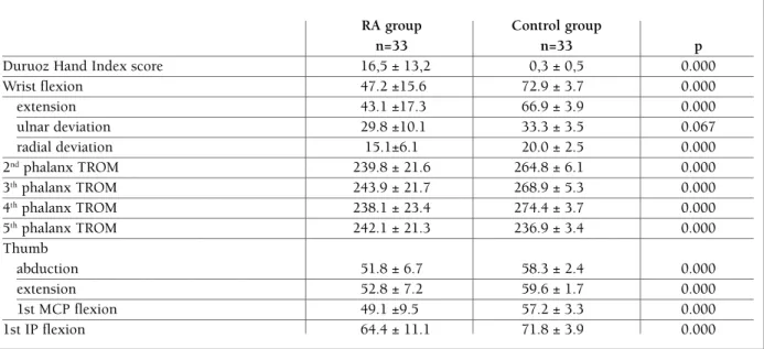
When we evaluated hand grip strength and pinch strength, we found negative correlation with tenosyno -vitis score. Correlation coefficients and p values are shown in Table IV. As the purdue pegboard scores were lower, tenosynovitis scores were significantly higher. Correlation between hand grip and tenosynovitis score is shown in Figure 3. There was a significant posi-tive correlation between DHI scores and tenosyno-tAble II. duruoz hAnd Index score And rAnge of MotIon of rA group
RA group Control group
n=33 n=33 p
Duruoz Hand Index score 16,5 ± 13,2 0,3 ± 0,5 0.000
Wrist flexion 47.2 ±15.6 72.9 ± 3.7 0.000 extension 43.1 ±17.3 66.9 ± 3.9 0.000 ulnar deviation 29.8 ±10.1 33.3 ± 3.5 0.067 radial deviation 15.1±6.1 20.0 ± 2.5 0.000 2ndphalanx TROM 239.8 ± 21.6 264.8 ± 6.1 0.000 3thphalanx TROM 243.9 ± 21.7 268.9 ± 5.3 0.000 4thphalanx TROM 238.1 ± 23.4 274.4 ± 3.7 0.000 5thphalanx TROM 242.1 ± 21.3 236.9 ± 3.4 0.000 Thumb abduction 51.8 ± 6.7 58.3 ± 2.4 0.000 extension 52.8 ± 7.2 59.6 ± 1.7 0.000 1st MCP flexion 49.1 ±9.5 57.2 ± 3.3 0.000 1st IP flexion 64.4 ± 11.1 71.8 ± 3.9 0.000
TROM: Total range of motion
GripstrengthLastpinchTip to tippinchThree jawchuck
RA groupControl group
Grip Strength (kg)45A4005101520253035*p<0.001****RighthandLefthandBothhandsAssemblyPurdue Pegboard Scores35B300510152025****
Grip
strength pinchLast Tip to tippinch Three jawchuck
RA group Control group Gr ip S tr en gt h ( kg ) 45 A 40 0 5 10 15 20 25 30 35 *p<0.001 * * * * Right
hand handLeft handsBoth Assembly
Pu rd u e Pe gb oa rd S co re s 35 B 30 0 5 10 15 20 25 * * * *
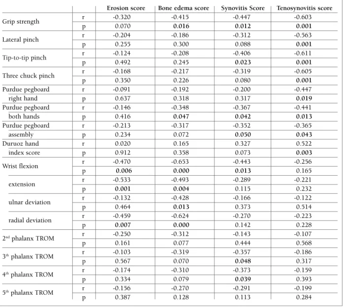
vitis score. Wrist ROM was essentialy correlated nega-tively with bone edema scores. There were no correla-tion between second to fifth finger ROM and MRI scores. Correlation coefficients and p values are shown in Table IV.
dIscussIon
Functional outcome in RA is influenced by the extent of structural damage to joints, bones and tendons plus severity of the joint inflammation18. The joints of the hand are among the first to be affected in RA and hand function is an important aspect of global function19. Pain, decrement in joint ROM, muscle weakness, re-duced grip strength and hand deformities may alter hand function and fine hand skills8. The resulting hand disability affects the activities of daily living and may cause dependency on others, which is a major problem tAble III. MeAn globAl scores of wrIst And
Mcp joInts In pAtIents wIth rA
MRI findings Range of MRI
and location possible scores scores
Bone erosions* 0-30 5.9 (0-19) Carpal joints 0-20 5.1 (0-18) MCP joints 0-10 0.9 (0-5) Bone edema* 0-30 5.2 (0-20) Carpal joints 0-20 4.6 (0-20) MCP joints 0-10 0.6 (0-4) Synovitis ¤ 0-10 2.3 (0-7) Carpal joints 0-8 2.1 (0-6) MCP joints 0-2 0.2 (0-2) Tenosynovitis ¤ 0-18 3.8 (0-14) Flexor tendons 0-6 0.8 (0-6) Extensor tendons 0-12 3.1 (0-11) Note: *n= 33; ¤ n= 31 Data in parentheses are ranges
fIgure 2.MRI scan of the dominant hand in 55-year-old man with confirmed diagnosis of RA. T2 weighted coronal GE
fat-suppressed sequence (a) showing erosion, edema and synovitis in all of the carpal bones, distal radius-ulna and distal metacarpal bones (short arrows). MRI scan of the right hand of the same patient. T2 weighted coronal PD fat-suppressed sequence (b) showing erosions, edema and synovitis in all of the carpal bones, distal radius-ulna and distal metacarpal bones (long arrows). T1 weighted transverse pre- (c) and post-contrast (d) MR images (unenhanced and gadolinium-enhanced) of the dominant hand in 61-year-old man with confirmed diagnosis of RA. MR images showing tenosynovitis of extensor pollicis brevis (first compartment), extensor carpi radialis, longus and brevis (second compartment), extensor carpi ulnaris (fourth compartment) and flexor tendons (stars). Note the presence of bone erosion, edema and synovitis in carpal bones.
tAble Iv. correlAtIon between hAnd evAluAtIon pArAMeters And MrI fIndIngs
Erosion score Bone edema score Synovitis Score Tenosynovitis score
Grip strength r -0.320 -0.415 -0.447 -0.603 p 0.070 0.016 0.012 0.001 Lateral pinch r -0.204 -0.186 -0.312 -0.563 p 0.255 0.300 0.088 0.001 Tip-to-tippinch r -0.124 -0.208 -0.406 -0.611 p 0.492 0.245 0.023 0.001
Three chuck pinch r -0.168 -0.217 -0.319 -0.605
p 0.350 0.226 0.080 0.001 Purdue pegboard r -0.091 -0.192 -0.200 -0.447 right hand p 0.637 0.318 0.317 0.019 Purdue pegboard r -0.146 -0.348 -0.367 -0.441 both hands p 0.416 0.047 0.042 0.013 Purdue pegboard r -0.213 -0.317 -0.352 -0.365 assembly p 0.234 0.072 0.050 0.043 Duruoz hand r 0.020 0.165 0.327 0.522 index score p 0.912 0.358 0.073 0.003 Wrist flexion r -0.470 -0.653 -0.443 -0.256 p 0.006 0.000 0.013 0.165 extension r -0.533 -0.493 -0.289 -0.221 p 0.001 0.004 0.115 0.232 ulnar deviation r -0.132 -0.428 -0.166 -0.122 p 0.464 0.013 0.373 0.514 radial deviation r -0.459 -0.624 -0.270 -0.223 p 0.007 0.000 0.142 0.228 2ndphalanx TROM r -0.250 -0.312 -0.143 -0.107 p 0.161 0.077 0.444 0.568 3th phalanx TROM r -0.103 -0.319 -0.357 -0.186 p 0.567 0.070 0.048 0.317 4thphalanx TROM r -0.174 -0.310 -0.373 -0.159 p 0.334 0.079 0.039 0.393 5thphalanx TROM r -0.156 -0.270 -0.291 -0.199 p 0.387 0.128 0.113 0.284
TROM: Total range of motion
in RA20. Therefore hand involvement and function should be seriously taken into account in the evalua-tion of the patients.
In this study, we evaluated rheumatoid hand by defining deformities and investigated the effect of RA on the grip strength, pinch strength, joint ROM, dexte -rity and functional ability. Hand deformity is an im-portant feature of RA and presence of hand deformities added useful prognostic information, being an early sign of a more severe disease. In this study fifteen pa-tients had at least one deformity (45.5%) and
deformity rate was lower when compared to other stu -dies21,22. This may be due to patients with advanced stage disease could not be included into the study be-cause of limitations in MR imaging (such as difficulties in positioning in MRI machine due to contractures). We compared hand function of the patients with hand deformity with the patients without deformity. Grip strength, tip to tip pinch strength, three jaw chuck strength, purdue pegboard right hand, purdue peg-board both hand, purdue pegpeg-board assembly and DHI scores were significantly lower in the patients with
hand deformities in comparison to the patients with-out deformity.
Grip strength and pinch strength are important in-dices of the functional integrity of the hand. Grip strength and three chuck pinch constitute 14% and 10% of all the daily activities respectively23. In this study, we found grip strength and all subgroups of the pinch strength significantly lower in RA group. Simi-lar to our findings, several studies reported that pa-tients with RA had lower grip strength and pinch strength24,25. ROM measurement can provide a cost effec tive assessment of hand function23. Limited ROM can lead to impaired hand function and difficulty in daily living activities. Decrease in joint ROM, as di sease progress in RA, was reported by several studies26,27. Similar to the previous studies, we also found signifi-cant decrease in wrist and finger ROM in all directions except for ulnar deviation in RA group.
Conventional measures like ROM and grip strength explain only a part of limitations in performing activi-ties of daily life. So we used the Purdue pegboard test for evaluating hand dexterity and similar to previous study done by Jones et al28, we found Purdue scores significantly lower in patients with RA. We used DHI to assess functional handicap and functional disabili-ty15. DHI had better responsiveness than impairment and disease activity measures such as pain, morning stiffness duration, swelling and tenderness8. DHI scores
of our patients were significantly higher when com-pared to control group.
As well as clinical parameters, radiological tech-niques are used in diagnosis and follow up of the pa-tients. Radiographic damage is an important outcome measure in RA, in addition to assessments of physical function and disease activity. Several studies have shown that disease activity is associated with physical disability and radiographic damage29. MRI is an ima -ging method that can reveal erosions as well as synovitis and tenosynovisynovitis early in RA, giving a detailed pi -cture of joint inflammation and damage10. Given these advantages, MRI has a major potential as an outcome measure in RA clinical trials and investigations10,18. In this study we performed MR imaging on the dominant hand of RA patients. We assessed the relationship of MR imaging findings of RA with the clinical parameters we had been evaluated. Disease duration revealed posi -tive correlation with bone marrow edema and synovi-tis scores but not with bone erosion and tenosynovisynovi-tis scores. MRI scores did not differ significantly between RF (+) and RF (-) patients. Though all MRI scores were higher in patients with deformities, the difference was not statistically significant. We found significant corre-lation between wrist motion and bone edema-erosion scores.
We identified significant correlation between tenosynovitis and grip strength, pinch strength, Purdue
-100Tenosynovitis scoreDuruoz hand index score
1614121086420-2102030A40r=0.52250-100Tenosynovitis scoreGrip strength score1614121084620-2-41020B30r=0.522r=-0.60340
-10 0 Te n os yn ov it is s co re
Duruoz hand index score 16 14 12 10 8 6 4 2 0 -2 10 20 30 A 40 r=0.522 50 -10 0 Te n os yn ov it is s co re
Grip strength score 16 14 12 10 8 4 6 2 0 -2 -4 10 20 B 30 r=0.522 r=-0.603 40
pegboard test scores and DHI scores.A study similar to our work at certain points was reported by Benton et al30. They investigated whether MR imaging could be used to predict functional outcome in patients with RA in 6-year follow-up. They found significant correlation between health assessment questionnaire (HAQ), short form 36 (SF-36) and bone erosion score and reported that early MRI evidence of bone marrow edema at the carpus can be a predictor of global function as mea-sured by SF-36. Zheng et al18compared Sollerman hand function test with MRI parameters to investigate the role of early MR imaging of the wrist in predicting functional outcome in RA and suggested that MRI bone edema, bone erosion, and to a lesser extent synovitis and tendonitis detected at the wrist in early RA have prognostic significance in terms of hand and specifi-cally tendon function in the medium term18. Haavard-sholm et al31found significant correlation between grip strength and bone edema score. Eshed et al19identified flexor tenosynovitis in the hands as a risk factor for des -tructive erosion of the joints.
As a result, this study showed that RA effects nega-tively hand function and dexterity and the parameters used in the evaluation of hand function in RA patients were mainly associated with tenosynovitis scores. Tenosynovitis is a common pathology in RA and clear-ly has a significant impact on hand function32. So MRI can be used as a supportive method in early diagnosis of tenosynovitis and may be useful in identification of patients requiring aggressive treatment. Further prospective studies are required to explore these pos-sibilities and clarify the clinical relevance of these pa-rameters with MRI findings.
correspondence to
Asiye Mukaddes Erol
Bursa Sevket Yilmaz Training and Research Hospital E-mail: [email protected]
references
1. Eberhardt KB, Fex E. Functional impairment and disability in early rheumatoid arthritis-development over 5 years. J Rheuma-tol 1995;22:1037-1042.
2. Alamanos Y, Voulgari PV, Drosos AA. Incidence and prevalence of rheumatoid arthritis based on the 1987 American College of Rheumatology criteria: a systematic review. Semin Arthritis Rheum 2006;36:182-188.
3. Boscheinen-Morrin J, Conolly WB (2001) The Hand-Funda-mentals of Therapy, third edition, Reed Elsevier, Oxford:183--201.
4. Durmus D, Uzuner B, Durmaz Y, et al. Michigan Hand Out-comes Questionnaire in rheumatoid arthritis patients: relation-ship with disease activity, quality of life, and hand grip strength. J Back and Musculoskelet Rehabil 2013;26:467–473.
5. Bodur H, Yilmaz O, Keskin D. Hand disability and related vari-ables in patients with rheumatoid arthritis. Rheumatol Int 2006;26:541-544.
6. Dellhag B, Bjelle A. A five-year follow up of hand function and activities of daily living in rheumatoid arthritis patients. Arthri-tis Care Res 1999;12:33-41.
7. Dellhag B, Hosseini N, Bremell T, et al. Disturbed grip function in women with rheumatoid arthritis. J Rheumatol 2001;28:2624-2633.
8. Lefevre-Colau MM, Poiraudeau S, Fermanian J, et al. Respon-siveness of the Cochin rheumatoid hand disability scale after surgery. Rheumatology 2001;40:843-850.
9. Boutry N, Hachulla E, Flipo RM, et al. MR Imaging Findings in hands in early rheumatoid arthritis: comparison with those in systemic lupus erythematosus and primary Sjogren syndrome. Radiology 2005;236: 593–600.
10. McQueen FM, Stewart N, Crabbe J et al. Magnetic resonance imaging of the wrist in early rheumatoid arthritis reveals a high prevalence of erosions at four months after symptom onset. Ann Rheum Dis 1998;57:350-356.
11. Østergaard M, Edmonds J, McQueen F, et al. An introduction to the EULAR-OMERACT rheumatoid arthritis MRI reference image atlas. Ann Rheum Dis 2005;64:3-7.
12. Arnett FC, Edworthy SM, Bloch DA, et al. The American Rheumatism Association 1987 revised criteria for the classifi-cation of rheumatoid arthritis. Arthritis Rheum 1988;31: 315–324.
13. Mathiowetz V, Rennells C, Donahoe L. Effect of the elbow po-sition on grip and key pinch strength. J Hand Surgery (Am) 1985;10:694-697.
14. Buddenberg LA, Davis C. Test-retest reliability of the Purdue Pegboard Test. Am J Occup Ther 2000;54:555-558.
15. Duruöz MT, Poiraudeau S, Fermanian J, et al. Development and validation of a rheumatoid hand functional disability scale that assess functional handicap. J Rheumatol 1996;23: 1167-1172. 16. Ostergaard M, Peterfy C, Conaghan P, et al. OMERACT Rheuma-toid Arthritis Magnetic Resonance Imaging Studies. Core set of MRI acquisitions, joint pathology definitions, and the OMER-ACT RA-MRI scoring system. J Rheumatol 2003;30:1385–1386 17. Haavardsholm EA, Ostergaard M, Ejbjerg BJ, et al. Reliability and sensitivity to change of the OMERACT rheumatoid arthri-tis magnetic resonance imaging score in a multireader, longitu-dinal setting. Arthritis Rheum 2005;52:3860–3867.
18. Zheng S, Robinson E, Yeoman S et al. MRI bone edema predicts eight year tendon function at the wrist but not the requirement for orthopaedic surgery in rheumatoid arthritis. Ann Rheum Dis 2006;65:607-611.
19. Eshed I, Feist E, Althoff CE, et al. Tenosynovitis of the flexor ten-dons of the hand detected by MRI: an early indicator of rheuma-toid arthritis. Rheumatology (Oxford) 2009;48:887-891. 20. Dellhag B, Burckhardt CS. Predictors of hand function in
pa-tients with rheumatoid arthritis. Arthritis Care Res 1995;8:16--20.
21. Mody GM, Meyers OL, Reinach SG. Handedness and deformi-ties, radiographic changes, and function of the hand in rheuma-toid arthritis. Ann Rheum Dis 1989;48:104-107.
22. Johnsson PM, Eberhardt K. Hand deformities are important signs of disease severity in patients with early rheumatoid arthri-tis. Rheumatology (Oxford) 2009;48:1398–1401.
23. Fowler NK, Nicol AC. Functional and biomechanical assess-ment of the normal and rheumatoid hand. Clin Biomech 2001;
16:660-666.
24. Fraser A, Vallow J, Preston A, et al. Predicting ‘normal’ grip strength for rheumatoid arthritis patients. Rheumatology (Ox-ford) 1999;38:521-528.
25. Nordenskiöld UM, Grimby G. Grip force in patients with rheumatoid arthritis and fibromyalgia and in healthy subjects. A study with the Grippit instrument. Scand J Rheumatol 1993;22:14-19.
26. Callahan LF, Pincus T, Huston JW 3rd, et al. Measures of activi -ty and damage in rheumatoid arthritis: depiction of changes and prediction of mortality over five years. Arthritis Care Res 1997;10:381-394.
27. Goodson A, McGregor AH, Douglas J, et al. Direct, quantitative clinical assessment of hand function: Usefulness and repro-ducibility. ManTher 2007;12:144-152.
28. Jones E, Hanly JG, Mooney R, et al. Strength and function in the normal and rheumatoid hand. J Rheumatol 1991;18:1313--1318.
29. Ødegård S, Landewé R, van der Heijde D, Kvien TK, Mowinckel P, Uhlig T. Association of early radiographic damage with im-paired physical function in rheumatoid arthritis: a ten-year, lon-gitudinal observational study in 238 patients. Arthritis Rheum. 2006;54:68-75.
30. Benton N, Stewart N, Crabbe J, et al. MRI of the wrist in early rheumatoid arthritis can be used to predict functional outcome at 6 years. Ann Rheum Dis 2004;63:555-561.
31. Haavardsholm EA, Bøyesen P, Østergaard M, et al. MRI findings in 84 early rheumatoid arthritis patients: Bone marrow oedema predicts erosive progresion. Ann Rheum Dis 2008;67:794-800. 32. McQueen FM, Beckley V, Crabbe J, et al. Magnetic resonance imaging evidence of tendinopathy in early RA predicts tendon rupture at six years. Arthritis Rheum 2005;52:744–751.