OR
IGI
N
A
L
R
E
S
E
A
R
C
H
Production capacity of maximal isometric grip
strength in women with rheumatoid arthritis:
a pilot study
Capacidade de produção de força de preensão isométrica máxima em mulheres com artrite
reumatoide: um estudo piloto
Capacidad de producción de fuerza de prensión isométrica máxima en mujeres con artritis
reumatoide: una investigación experimental
Rodrigo da Rosa Iop1, Ana Paula Shiratori1, Luciana Ferreira2, Noé Gomes Borges Júnior3,
Susana Cristina Domenech4, Monique da Silva Gevaerd4
Correspondence to: Monique da Silva Gevaerd – R: Pascoal Simone, 358 - Coqueiros – CEP: 88080-350 – Florianópolis (SC) – E-mail: [email protected] Presentation: Sept. 2014 – Accepted for publication: Dec. 2014 – Financing source: none – Conlict of interests: nothing to declare.
Study developed at the Multisector Analysis Laboratory (MULTILAB) of the Health and Sport Sciences Center (CEFID) at the State University of Santa Catarina (UDESC) – Florianópolis, Brazil.
1Doctoral Students from the Post-Graduate Program in Human Movement Sciences. State University of Santa Catarina – UDESC. 2Master in Human Movement Sciences. State University of Santa Catarina – UDESC.
3PhD. Professor of the Post-Graduate Program in Human Movement Sciences. Center for Health and Sport Sciences. State University
of Santa Catarina – UDESC.
4Doctors. Professor of the Post-Graduate Program in Human Movement Sciences. Center for Health and Sport Sciences. State
University of Santa Catarina – UDESC.
ABSTRACT | The aim of this study was to verify the ca-pacity of maximum handgrip strength (HGSmax) in women with rheumatoid arthritis (RA) and its relationship with disease activity. Nine women with RA and ten healthy wo-men were selected. The demographics data were recor-ded of both groups, and clinical characteristics of wom- en with RA. The level of disease activity was evaluated by the protocol Disease Activity Score (DAS−28) using C-Reactive Protein (CRP). To measure the HGSmax was used an extensometer dynamometer. The results showed a statistically signiicant diference of HGSmax between the participants (Arthritis and Control: 129.41±52.10 e 192.46±38.98N). In relation to dominance, women with RA showed no signiicant diference in HGSmax. The results of HGSmax for the dominant hand showed a strong linear relationship with the CRP (r=0.751). It also noted that was a moderate non-linear relation of HGSmax for the dominant hand and non-dominant with the number of tender and swollen joints. Thus, it is clear that women with RA have reduced ability to produce HGSmax independent of dom- inance, in addition, this study demonstrated the direct relationship that exists between HGSmax and the level of disease activity.
Keywords | Arthritis Rheumatoid; Muscle Strength Dynamometer, C-Reactive Protein.
11
Descritores | Artrite Reumatoide; Dinamômetro de Força Muscular; Proteína C-Reativa.
RESUMEN | Este artículo tuvo como objetivo veriicar la ca-pacidad de producción de fuerza de prensión manual máxima (FPMmax) en mujeres con artritis reumatoide (AR) y su relación con la actividad de la enfermedad. Han sido elegidas nueve mu-jeres con AR y diez saludables. Se han registrado datos demo-gráicos de los dos grupos, además de las características clínicas de las mujeres con AR. La determinación del nivel de actividad de la enfermedad se realizó por el protocolo Disease Activity Score (DAS−28) a través de la Proteína C-Reactiva (PCR). Para la adquisición de la FPMmax se utilizó el dinamómetro de tipo exten-sómetro. Los resultados muestran una diferencia signiicativa de
la FPMmax entre los participantes (Artritis y Control: 129,41±52,10 e 192,46±38,98 N). Con respeto a la dominación, las mujeres con AR no presentaron diferencias signiicativas en la FPMmax. Se ob-tuvo resultados para la mano dominante en el cual mostró una fuerte relación lineal con la PCR (r=0,751). También se compro-bó una moderada relación no lineal de la FPMmax para la mano dominante y la no dominante con el número de articulaciones dolorosas e inlamadas. De esta manera, resulta evidente que las mujeres con AR presentan reducción en la capacidad de produc-ción de la FPMmax independiente de la dominancia, además de mostrar a través de esta investigación la relación directa entre la FPMmax y el nivel de actividad de la enfermedad.
Palabras clave | Artritis Reumatoide; Dinamómetro de Fuerza Muscular; Proteína C-Reactiva.
INTRODUCTION
Rheumatoid arthritis (RA) is a chronic inlammato-ry disease, associated with functional disabilities result-ing from articular lesions1,2, which mainly afects joints
in the hands3,4.
A method commonly used in order to assess function-ality in patients with RA is the hand grip strength test (HGST)5, which is an efective marker for establishing
the level of dysfunction in RA suferers6,7. his test
pro-vides a quantitative assessment in terms of functionality, which is pertinent because a person’s ability to exert pres-sure with their hands plays an important role in explor-ing and manipulatexplor-ing objects, as well as contributexplor-ing to everyday functions related to the upper limbs and torso8,9.
he HGST has several purposes within the clinical sphere, with it being recommended to provide assistance in the diagnosis, evaluation and comparison of surgical techniques, as well as for recording purposes during the rehabilitation process and in treatment response10,11.
Despite some studies having already veriied the reduction of hand grip strength (HGS)12,13, the
mech-anism that causes this functional loss remains largely misunderstood14. he functional restrictions that occur
in RA can be related to the progressive and systemic characteristic of the disease, mainly to the efect of the inlammatory process that leads to joint lesions15. Some
studies have veriied the relationship between HGS and the level of disease activity by means of the DAS-2816,17.
In this sense, assessing the level of disease activity
provides objective evidence regarding the inlammatory process18, thereby contributing to understanding
con-cerning the inluence of the inlammatory process in reducing HGS in individuals with RA.
Given the aforementioned points, the aim of this study was to test the production capacity for maximum hand grip strength (HGSmax) in female RA suferers and its relationship with the disease’s activity.
METHODOLOGY
Study participants
Nine women with RA (arthritis group=AG) and ten healthy women (control group=CG) were selected and paired according to age (±2 years). he AG was referred by rheumatologists, in accordance with classiication cri-teria from the American College of Rheumatology19, with
all being active arthritis suferers (DAS-28≥2.6)20. All
participants with other immune disorders, cognitive dei-ciencies, sensory or physical of the upper limbs, blindness, estrogen, antibiotic or anticonvulsant users, active smokers and pregnant women were excluded from the study.
Clinical examinations
in addition to the clinical characteristics of the women with RA.
he level of disease activity was determined by means of the Disease Activity Score (DAS−28) Protocol, where the number of painful joints (NPJ) and number of swollen joints (NSJ) (shoulders, el-bows, wrists, metacarpophalangeals, proximal inter-phalangeal joints and knees) are bilaterally counted. he patient’s own perception regarding their state of health (SOH) was also veriied, this was done us-ing a unidimensional scale with a 0-100mm mark21.
Together with the aforementioned parameters, the serum level of the C-reactive protein (CRP) was used in order to perform the inal DAS−2822 calculation.
he inal score from the DAS-28 ranged from 0 to 1023, where the higher the value, the greater the level
of disease activity.
he CRP serum concentration was determined by a turbidimetric method, using a CRP-turbilátex kit (Biotécnica) and a Spectrophotometer (BTLyzer 100®),
at a 540nm wavelength. he values were expressed in mg/L, with values up to 6mg/L being considered as normal.
In order to determine the dominant hand, the Edinburgh Handedness Inventory24,25 was used.
In order to ind the HGSmax, an extensome-ter-type dynamometer was used. he dynamometer component responsible for measuring strength was an S-beam load cell with four strain gauges (Kyowa, Japan). he dynamometer had a load limit of 1000N (Newtons) and an excellent linearity coeicient (r2=0.9999)26.
Procedure for collecting the HGS
maxFirstly, hand size was measured in order to individ-ually calculate the dynamometer grip, in accordance with that as described by Ruiz-Ruiz27. he guidelines
from the American Society of Hand herapists28 were
adopted for the tests. he test’s beginning and end were determined through visual feedback, where the partici- pants observed a “press” command on their computer screens and performed a voluntary muscle contraction for a maximum of 10 seconds, in accordance with the Protocol set out by Ronningen and Kjeyen29. hree
repetitions were alternately performed by each hand, beginning with the dominant hand28. he time
inter-val between the repetitions was 1 minute, based on the protocol from Ikemoto, et al30.
HGS
maxdata processing
he data were processed through a speciic routine, programmed in Scilab v.4.1.2 Software (INRIA, ENPS, France), which provided the mean from the three maxi- mum strength curves31. he HSG
max was deined as the
highest value from the strength curve vs time32. he
ob-tained signals were recorded on a hard drive and iltered using a irst order low-pass ilter with a 25Hz cutof frequency.
Statistical analysis
AG characterization was achieved through descrip-tive statistics, using reladescrip-tive frequency distributions for the categorical variables and mean and standard devia-tion for the continuous variables.
In order to determine the diference of HGSmax between AG and CG, an independent t-test was per-formed. A paired Student’s t-test was conducted in or-der to identify the diferences in the groups depending on dominance. Finally, a Pearson correlation test was performed between the HGSmax vs DAS-28, SOH and CRP. he relationship between the HGSmax, NPJ and NSJ was veriied by means of the Spearman test.
he analyses were done using the Statistical Package for the Social Sciences (SPSS−20.0 Version) software, while adopting a 5% signiicance level.
RESULTS
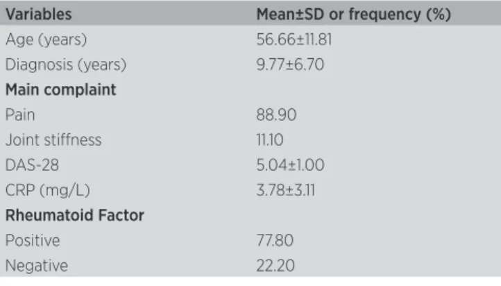
19 women were evaluated, with an average age of 56.31 years, 9 of these being from the AG, 10 from the CG. Among the clinical characteristics from the AG, all the participants who were found to have active ar-thritis presented a positive rheumatoid factor with pain being the complaint most made (Table 1).
Table 1. Clinical characteristics of the AG participants (n=9)
Variables Mean±SD or frequency (%)
Age (years) 56.66±11.81 Diagnosis (years) 9.77±6.70
Main complaint
Pain 88.90
Joint stifness 11.10
DAS-28 5.04±1.00
CRP (mg/L) 3.78±3.11
Rheumatoid Factor
Positive 77.80
Table 2. Relationship between the HGSmax of the dominant and non-dominant hand with the parameters for disease activity in the AG
HGSmax NAD NAE EGS PCR DAS28
Dominant rho=−0.542* p=0.00
rho=−0.537* p=0.00
r=−0.527** p=0.01
r=0.751** p=0.01
r=0.336 p=0.18
Non-Dominant rho=−0.528* p=0.01
rho=−0.435* p=0.03
r=−0.681** p=0.00
r=0.533 p=0.07
r=0.000 p=0.50
* Signiies p<0.05 in Spearman’s Correlation (rho); ** Signiies p<0.05 in the Pearson correlation coeicient (r), (p<0.05). NPJ: number of painful joints; NSJ: number of swollen joints; SOH: state of health
While assessing the HGSmax, a statistically signiicant diference was observed between the AG and CG in both hands. he HGSmax from the AG was 129.41±52.10N, whereas that from the CG was 192.46±38.98N (Chart 1). Regarding dominance, women from the AG did not show any signiicant diference in the HGSmax. However, there was a signiicant diference found in the CG for the HGSmax, as regards to dominance (Chart 1).
#
#
*
Dominant hand Non-dominant hand
AG CG
Maximal isome
tric hand grip (N)
350
300
250
200
150
100
50
0
Chart 1. Maximal isometric hand grip strength of the dominant and non-dominant hand from the AG and CG.
# Signiies p<0.05 in the comparison between the AG and the CG. Independent Student’s t-test. * Signiies p<0.05 in the comparison between the dominant and non-dominant hand in the CG. Paired Student’s t-test
Based on the correlation tests, in both hands, the HGSmax showed no linear relationship with the total score from the DAS−28. However, the results from the HGSmax for the dominant hand showed a moderate-ly linear relationship with SOH and a strong linear relationship with CRP. Whereas, for the non-domi-nant hand, there was a strong linear relationship be-tween the HGSmax and the SOH, while there was no signiicant linear relationship with CRP. A moderate non-linear relationship of the HGSmax was found for the dominant and non-dominant hand with NPJ and NSJ (Table 2).
DISCUSSION
he HGSmax results from the tests corroborate with those from published data, thereby reiterating the
inding that women with RA have lesser HGSmax when compared with healthy women33-36.
Concerning the diference in the HGSmax in rela-tion to dominance, data from the literature are contro-versial. Fraser, et al.37 found that the dominant hand of
patients with RA was on average 20% weaker than the contralateral hand. Whereas, in healthy individuals, the dominant hand was stronger than the non-dominant hand. Another study38 found that the dominant hand of
individuals with RA presented greater HGSmax. Due to these contradictory indings, it is diicult to draw con-clusions regarding the inluence of dominance on grip strength39.
In the assessment of the relationship of HGSmax with the disease activity parameter, the HGSmax has been described to present a good association with this variable40. In this sense, Dedeoglu, et al.41 found a
neg-ative correlation between HGSmax and DAS-28. his relationship was strengthened by West and Wallber-Jonsson42, during a longitudinal study in which a
neg-ative relationship was observed between HGSmax with DAS-28 and CRP. Arvidson, et al.43,44 also
demonstrat-ed a negative association between CRP and HGSmax. Despite no correlation having been veriied between HGS and the total DAS−28 score, this study found a negative correlation with NPJ, NSJ and SOH and a positive correlation with CRP.
CRP can serve as a regulatory marker of the in-lammatory pathway and other inin-lammatory mark-ers45. Disease activity plays an important role in the
mechanism that contributes to physical and functional impairment in patients with RA46. he association
be-tween the disease activity parameters and the HGSmax suggests that the inlammatory process can act as cata-bolic mediators in the muscle47.
Häkkinen, et al.48 believed that the reduction in
related to joint destruction, ligament laxity and an im-balance in muscle function in individuals with RA. he progression of this pathological process can generate serious deformities, which results in signiicant func-tional limitations49.
Another mechanism resulting from inlammation that contributes to the loss of motor command, to atro-phy and to muscle weakness is pain50. he process that
triggers pain in RA suferers is related to the peripheral sensitization, where the aferent nerves become hyper-sensitive to movement51,52. Considering that pain was
the main complaint clinic mentioned by patients, as was veriied negative correlation between HGSmax and NPJ, the data from this study reinforce the involvement of the inlammatory process in the reduced HGSmax of in-dividuals with RA.
However, it is important to stress that given the small number of individuals evaluated, where the par-ticipants were selected by convenience, as well as the absence of radiological examination in order to deter-mine the state of the disease, there is a limitation in-herent to this study, thereby damaging the generaliza-tion of the obtained results. Such limitageneraliza-tions occurred due to the diiculty of inding patients with RA who were available to go to the laboratory to participate in the study, as well as the lack of funding to perform radiological examinations. Despite their preliminary nature, the data from is study is considered relevant for characterizing the HGS of patients with RA, partici- pants of studies developed in our laboratory. Finally, it is clear that further work must be performed with a larger number of subjects in order to study and charac-terize the relationship between HGS and the level of disease activity. he information from this study will contribute in terms of improving the quality of life for these individuals; it also highlighted the importance and relevance of the multidisciplinary treatment, and thereby should lead to better management and treat-ment of the pathology.
CONCLUSION
he indings showed that women with RA pre-sented a reduction in their ability to produce HGSmax, regardless of the dominant hand. In addition, they demonstrated that there is a direct relationship between HGSmax and the determining parameters for the level of disease activity.
REFERENCES
1. Oken O, Batur G, Gunduz R, Yorgancioglu RZ. Factors associated with functional disability in patients with rheumatoid arthritis. Rheumatol Int. 2008;29(2):163-6.
2. Eurenius E, Stenström CH. Physical activity, physical itness, and general health perception among individuals with rheumatoid arthritis. Arthritis Rheum. 2005;53(1):48-55.
3. Watanabe K, Tsubota S, Chin G, Aoki M. Diferences in parameters of the explosive grip force test between young and older women. J Gerontol A Biol Sci Med Sci. 2011;66(5):554-8.
4. Bodur H, Yilmaz O, Keskin D. Hand disability and related variables in patients with rheumatoid arthritis. Rheumatol Int. 2006;26(6):541-4. 5. Ferraz MB, Ciconelli RM, Araujo PMP, Oliveira LM, Atra E. The
efect of elbow lexion and time of assessment on the measure-ment of grip strength in rheumatoid-arthritis. J Hand Surg Am. 1992;17A(6):1099-103.
6. Thyberg I, Hass UA, Nordenskiöld U, Gerdle B, Skogh T. Activity limitation in rheumatoid arthritis correlates with reduced grip force regardless of sex: the Swedish TIRA project. Arthritis Rheum. 2005;53(6):886-96.
7. Adams J, Burridge J, Mullee M, Hammond A, Cooper C. Correlation between upper limb functional ability and structural hand impairment in an early rheumatoid population. Clin Rehabil. 2004;18(4):405-13.
8. Hoenig H, Grof G, Pratt K, Goldberg E, Franck W. A randomized controlled trial of home exercise on the rheumatoid hand. J Rheumatol. 1993;20(5):785-9.
9. Haidar SG, Kumar D, Bassi RS, Deshmukh SC. Average versus maximum grip strength: which is more consistent? J Hand Surg Br. 2004;29(1):82-4.
10. Rosén B. Recovery of sensory and motor function after nerve repair. A rationale for evaluation. J Hand Ther. 1996;9(4):315-27.
11. MacDermid JC, Kramer JF, Woodbury MG, McFarlane RM, Roth JH. Interrater reliability of pinch and grip strength measure-ments in patients with cumulative trauma disorders. J Hand Ther. 1994;7(1):10-4.
12. Hakkinen A, Kautiainen H, Hannonen P, Ylinen J, Makinen H, Sokka T. Muscle strength, pain, and disease activity explain individual subdimensions of the Health Assessment Questionnaire disability index, especially in women with rheumatoid arthritis. Ann Rheum Dis. 2006;65(1):30-4.
13. Alomari MA, Keewan EF, Shammaa RA, Alawneh K, Khatib SY, Welsch AA. Vascular Function and Handgrip Strength in Rheumatoid Arthritis Patients. Sci World J. 2012;2012.
14. Rajagopalan A, Burne JA. Stretch relexes and joint dynamics in rheumatoid arthritis. Exp Brain Res. 2010;201(1):37-45.
15. Pollard L, Choy EH, Scott DL. The consequences of rheumatoid arthritis: quality of life measures in the individual patient. Clin Exp Rheumatol. 2005;23(5 Suppl 39):S43-52.
16. Targonska-Stepniak B, Majdan M. Associations between parameters of nutritional status and disease activity in patients with rheuma-toid arthritis. Pol Arch Med Wewn. 2011;121(4):122-8.
17. Dogu B, Kuran B, Yilmaz F, Usen A, Sirzai H. Is hand bone mineral density a marker for hand function in patients with established rheumatoid arthritis? The correlation among bone mineral density of the hand, radiological indings and hand function. Clin Rheumatol. 2013.
18. Fransen J, Van Riel PL. Outcome measures in inlammatory rheu-matic diseases. Arthritis Res Ther. 2009;11(5):244.
for the classiication of rheumatoid arthritis. Arthritis Rheum. 1988;31(3):315-24.
20. Prevoo MLL, Vanthof MA, Kuper HH, Vanleeuwen MA, Vandeputte LBA, Vanriel P. Modiied disease-activity scores that include 28-joint counts - development and validation in a prospective longitudi-nal-study of patients with rheumatoid-arthritis. Arthritis Rheum. 1995;38(1):44-8.
21. Pincus T, Sokka T. Quantitative measures for assessing rheuma-toid arthritis in clinical trials and clinical care. Best Pract Res Clin Rheumatol. 2003;17(5):753-81.
22. Inoue E, Yamanaka H, Hara M, Tomatsu T, Kamatani N. Comparison of Disease Activity Score (DAS)28- erythrocyte sedimentation rate and DAS28- C-reactive protein threshold values. Ann Rheum Dis. 2007;66(3):407-9.
23. Fransen J, Van Riel PL. DAS remission cut points. Clin Exp Rheumatol. 2006;24(6 Suppl 43):S-29-32.
24. Brito G, Brito L, Paumgartten F, Lins M. Lateral preferences in brazilian adults: An analysis with the Edinburgh Inventory. CORTEX. 1989;25(3):403-15.
25. Oldield RC. The assessment and analysis of handedness: the Edinburgh inventory. Neuropsychologia. 1971;9(1):97-113. 26. Borges NGJ, Domenech SC, da Silva ACK, Dias JA. Estudo
com-parativo da força de preensão isométrica máxima em diferentes modalidades esportivas. Rev Bras Cineantropom Desempenho Hum. 2009;11(3):292-8.
27. Ruiz-Ruiz J, Mesa JL, Gutiérrez A, Castillo MJ. Hand size inluen-ces optimal grip span in women but not in men. J Hand Surg Am. 2002;27(5):897-901.
28. Fess EE. Grip strenght. In: Casanova JS, editor. Clinical assessment recommendations. Chicago: American Society of Hand Therapists; 1992. p. 41-5.
29. Ronningen A, Kjeyen I. Efect of an intensive hand exercise pro-gramme in patients with rheumatoid arthritis. Scand J Occup Ther. 2008;15(3):173-83.
30. Trossman PB, Li P-W. The efect of the duration of intertrial rest periods on isometric grip strength performance in young adults. Occup Ther J Res. 1989;9(6):362-73.
31. Mathiowetz V, Weber K, Volland G, Kashman N. Reliability and validity of grip and pinch strength evaluations. J Hand Surg Am. 1984;9(2):222-6.
32. Ikemoto Y, Demura S, Yamaji S, Minami M, Nakada M, Uchiama M. Force-time parameters during explosive isometric grip correlate with muscle power. Sport Sci Healt. 2007;2(2):64-70.
33. Nordenskiöld UM, Grimby G. Grip force in patients with rheumatoid arthritis and ibromyalgia and in healthy subjects: a study with the Grippit instrument. Scand J Rheumatol. 1993;22(1):14-9.
34. Bjork MA, Thyberg ISM, Skogh T, Gerdle BUC. Hand function and activity limitation according to health assessment questionnaire in patients with rheumatoid arthritis and healthy referents: 5-year followup of predictors of activity limitation (the Swedish TIRA project). J Rheumatol. 2007;34(2):296-302.
35. Brorsson S, Nilsdotter A, Pedersen E, Bremander A, Thorstensson C. Relationship between inger lexion and extension force in healthy women and women with rheumatoid arthritis. J Rehabil Med. 2012;44(7):605-8.
36. Speed CA, Campbell R. Mechanisms of strength gain in a hand-grip exercise programme in rheumatoid arthritis. Rheumatol Int. 2012;32(1):159-63.
37. Fraser A, Vallow J, Preston A, Cooper RG. Predicting ‘normal’ grip strength for rheumatoid arthritis patients. Rheumatology. 1999;38(6):521-8.
38. Nordenskiold U, Grimby G. Assessments of disability in women with rheumatoid arthritis in relation to grip force and pain. Disabil Rehabil. 1997;19(1):13-9.
39. Andria G, Attivissimo F, Giaquinto N, Lanzolla AML, Quagliarella L, Sasanelli N. Functional evaluation of handgrip signals for parkinso-nian patients. IEEE Trans Instrum Meas. 2006;55(5):1467-73. 40. Myers DB, Grennan DM, Palmer DG. Hand grip function in
patients with rheumatoid arthritis. Arch Phys Med Rehabil. 1980;61(8):369-73.
41. Dedeoglu M, Gafuroglu U, Yilmaz O, Bodur H. The relationship be-tween hand grip and pinch strengths and disease activity, articular damage, pain, and disability in patients with rheumatoid arthritis. Turk J Rheumatol. 2013;28(2):69-77.
42. West E, Wållberg-Jonsson S. Health-related quality of life in Swedish men and women with early rheumatoid arthritis. Gend Med. 2009;6(4):544-54.
43. Arvidson NG, Larsson A, Larsen A. Simple function tests, but not the modiied HAQ, correlate with radiological joint damage in rheu-matoid arthritis. Scand J Rheumatol. 2002;31(3):146-50.
44. Cesari M, Penninx BW, Pahor M, Lauretani F, Corsi AM, Williams GR, et al. Inlammatory markers and physical performance in older persons: the InCHIANTI study. J Gerontol A Biol Sci Med Sci. 2004;59(3):242-8.
45. Stenholm S, Rantanen T, Heliövaara M, Koskinen S. The mediating role of C-reactive protein and handgrip strength between obesity and walking limitation. J Am Geriatr Soc. 2008;56(3):462-9. 46. Stucki G, Brühlmann P, Stucki S, Michel BA. Isometric muscle
strength is an indicator of self-reported physical functional disability in patients with rheumatoid arthritis. Br J Rheumatol. 1998;37(6):643-8.
47. Roubenof R, Roubenof RA, Ward LM, Holland SM, Hellmann DB. Rheumatoid cachexia: depletion of lean body mass in rheuma-toid arthritis. Possible association with tumor necrosis factor. J Rheumatol. 1992;19(10):1505-10.
48. Häkkinen A, Hannonen P, Häkkinen K. Muscle strength in healthy people and in patients sufering from recent-onset inlammatory arthritis. Br J Rheumatol. 1995;34(4):355-60.
49. Boutry N, Larde A, Lapegue F, Solau-Gervais E, Flipo RM, Cotten A. Magnetic resonance imaging appearance of the hands and feet in patients with early rheumatoid arthritis. J Rheumatol. 2003;30(4):671-9.
50. Riley J, Boulis NM. Molecular mechanisms of pain: a basis for chronic pain and therapeutic approaches based on the cell and the gene. Clin Neurosurg. 2006;53:77-97.
51. Coggeshall RE, Hong KA, Langford LA, Schaible HG, Schmidt RF. Discharge characteristics of ine medial articular aferents at rest and during passive movements of inlamed knee joints. Brain Res. 1983;272(1):185-8.