Delineating the GRIN1 phenotypic
spectrum
A distinct genetic NMDA receptor encephalopathy
ABSTRACT
Objective: To determine the phenotypic spectrum caused by mutations inGRIN1 encoding the NMDA receptor subunit GluN1 and to investigate their underlying functional pathophysiology.
Methods:We collected molecular and clinical data from several diagnostic and research cohorts. Functional consequences ofGRIN1 mutations were investigated in Xenopus laevis oocytes.
Results:We identified heterozygous de novoGRIN1 mutations in 14 individuals and reviewed the phenotypes of all 9 previously reported patients. These 23 individuals presented with a distinct phenotype of profound developmental delay, severe intellectual disability with absent speech, muscular hypotonia, hyperkinetic movement disorder, oculogyric crises, cortical blindness, gen-eralized cerebral atrophy, and epilepsy. Mutations cluster within transmembrane segments and result in loss of channel function of varying severity with a dominant-negative effect. In addition, we describe 2 homozygousGRIN1 mutations (1 missense, 1 truncation), each segregating with severe neurodevelopmental phenotypes in consanguineous families.
Conclusions:De novoGRIN1 mutations are associated with severe intellectual disability with cor-tical visual impairment as well as oculomotor and movement disorders being discriminating pheno-typic features. Loss of NMDA receptor function appears to be the underlying disease mechanism. The identification of both heterozygous and homozygous mutations blurs the borders of dominant and recessive inheritance ofGRIN1-associated disorders.Neurology®2016;86:2171–2178 GLOSSARY
ASD5 autism spectrum disorder; ID 5 intellectual disability; NMDAR 5 NMDA receptor; SNV 5 single nucleotide variant; VPA5 valproate.
NMDA receptors (NMDARs) are tetrameric ligand-gated ion channels permeable to Na
1, K
1,
and Ca
21, composed of 2 glycine binding GluN1 subunits and 2 glutamate binding GluN2/3
subunits (GluN2A, GluN2B, GluN2C, GluN2D, GluN3A, GluN3B).
1–3In contrast to GluN2/3 subunits, which each have specific spatial and temporal expression patterns
throughout the CNS, GluN1 encoded by GRIN1 is a ubiquitous component of the receptor.
4,5Mutations of the NMDAR subunits are associated with a variety of different
neurodevelop-mental phenotypes,
3including intellectual disability (ID),
6–8epilepsy,
9–13and autism spectrum
disorders (ASD), and different psychiatric diseases.
14,15While several studies have focused on the phenotypic spectrum of GRIN2A- and
GRIN2B-associated disorders, little is known about the clinical presentations of individuals with GRIN1
mutations. We reviewed clinical data on individuals with GRIN1 mutations from different
research cohorts as well as from diagnostic laboratories.
In addition, we reviewed available clinical and genetic information from previously
re-ported patients with GRIN1 mutations. Nine heterozygous de novo mutations had previously
been reported with rudimentary phenotypic information: 4 individuals were described with
nonsyndromic and nonspecific ID (mental retardation, autosomal dominant 8, OMIM
Johannes R. Lemke, MD Kirsten Geider, PhD Katherine L. Helbig, MS Henrike O. Heyne, MD Hannah Schütz, MS Julia Hentschel, PhD Carolina Courage, MD Christel Depienne, PhD Caroline Nava, MD, PhD Delphine Heron, MD Rikke S. Møller, PhD Helle Hjalgrim, MD Dennis Lal, PhD Bernd A. Neubauer, MD Peter Nürnberg, PhD Holger Thiele, PhD Gerhard Kurlemann, MD Georgianne L. Arnold, MD Vikas Bhambhani, MD Deborah Bartholdi, MD Christeen Ramane J. Pedurupillay, MS Doriana Misceo, PhD Eirik Frengen, PhD Petter Strømme, MD, PhD Dennis J. Dlugos, MD Emily S. Doherty, MD Emilia K. Bijlsma, MD, PhD Claudia A. Ruivenkamp, PhD Mariette J.V. Hoffer, PhD Amy Goldstein, MD Deepa S. Rajan, MD Vinodh Narayanan, MD Keri Ramsey, BSN Newell Belnap, PA-C Isabelle Schrauwen, PhD Ryan Richholt, BAS Bobby P.C. Koeleman,
PhD
Author list continued on next page
*These authors contributed equally to this work as co-last authors. Authors’ affiliations are listed at the end of the article.
Go to Neurology.org for full disclosures. Funding information and disclosures deemed relevant by the authors, if any, are provided at the end of the article. The Article Processing Charge was paid by the authors.
This is an open access article distributed under the terms of the Creative Commons Attribution-NonCommercial-NoDerivatives License 4.0 (CC BY-NC-ND), which permits downloading and sharing the work provided it is properly cited. The work cannot be changed in any way or used commercially.
614254),
8,16,171 individual had epileptic
encephalopathy of Lennox-Gastaut type,
9and 4 individuals were described with severe
developmental delay and associated
move-ment disorders.
18Our study aimed at a comprehensive
delin-eation of the associated phenotype and at an
investigation of the functional consequences
of the identified GRIN1 mutations.
METHODS Patients.We reviewed clinical and genetic infor-mation on patients in whom GRIN1 mutations were detected through different next-generation sequencing approaches in diagnostic and research settings including targeted panel sequencing19or whole-exome sequencing (table e-1, a and b, on
the Neurology®Web site at Neurology.org). Depending on the
availability of parental samples, the de novo status of each mutation was confirmed through conventional Sanger sequencing.
Standard protocol approvals, registration, and patient consents.Written informed consent for genetic testing was ob-tained for every individual. Genetic testing within research set-tings was approved by the Commission of Medical Ethics of the University of Antwerp, Belgium; the Ethics Committee of Western Zealand, Denmark; the Kantonale Ethikkommission Zürich, Switzerland; and additional local ethics committees of the contributing centers.
In silico prediction. Predictions of the functional impact of identified coding variants were assessed by different in silico anal-ysis programs (PolyPhen2, http://genetics.bwh.harvard.edu/ pph2/ and MutationTaster, http://www.mutationtaster.org) using the GRIN1 transcript number ENST00000371561 (table e-2). Variants were compared to 60,706 controls of the ExAC browser (http://exac.broadinstitute.org/). Conservation of mutated positions was evaluated using sequence alignment of different species (figure e-1). Molecular modeling of the NMDAR subunits was performed according to Endele et al.7
DNA constructs, oocyte expression, and electrophysiology.
For Xenopus laevis oocyte experiments, GluN1 and GluN2B con-structs and capped cRNAs were generated as described previ-ously.11,12Several mutations were introduced into these constructs
using the QuikChange site-directed mutagenesis kit (Stratagene; Agilent Technologies, Santa Clara, CA) and confirmed by DNA sequencing. Oocytes were isolated and maintained as described previously.20Glutamate, glycine, and Zn21dose–response curves
of wild-type GluN1/GluN2B and mutant NMDARs were analyzed by 2-electrode voltage clamp recording as described.21
Concentration–response curves and current traces shown in the figures were drawn using KaleidaGraph (Synergy Software, Reading, PA). Data represent means6 SE. Statistical significance was evaluated using a Student 2-tailed unpaired t test if not otherwise stated. All chemicals used were obtained from Sigma. RESULTS We identified 14 previously unreported patients with heterozygous GRIN1 mutations. In 13 individuals, the mutation was confirmed to be de novo, whereas in patient 22 parental samples were not available for segregation analysis. However, given that the particular mutation in patient 22 was recur-rent in 2 other patients (patients 21 and 23) and
absent in large control datasets, we concluded that the mutation is pathogenic and included this patient in our analysis. In addition to these 14 novel cases, we reviewed phenotypic data of all 9 previously reported cases8,9,16–18and present additional clinical informa-tion on 4 of these patients.8,9,16Thus, we were able to
collectively review the clinical data of 23 patients (table 1). In addition to patients with de novo GRIN1 mutations, we identified 2 homozygous GRIN1 mu-tations segregating in 2 unrelated families with severe neurodevelopmental disorders (1.1–2 and 5.1–3). In both cases, family members carrying heterozygous GRIN1 variants were unaffected.
Phenotypes associated with heterozygous de novo GRIN1 mutations. Almost all patients carrying a de novo GRIN1 mutation presented with profound global developmental delay, usually already apparent in the neonatal period and resulting in severe intellectual disability (21/22; 95%). (table e-1, a and b) Patients usually never acquired the ability to walk and had absent or extremely limited verbal communication skills.
Of the 21 patients in whom information regarding muscular tone could be retrieved, 15 (71%) had severe truncal and initial appendicular hypotonia. Many patients developed corticospinal signs, such as hyperreflexia (6/21; 29%) or spasticity (6/21; 29%), consistent with a diagnosis of spastic quadriparesis. The majority of patients showed choreatic, dystonic, or dyskinetic movement disorders (14/23; 61%), including oculomotor abnormalities such as oculogy-ric crises (5/23; 22%). Nonspecific stereotypic move-ments were noted in 7/21 patients (33%).
A significant proportion of patients (16/23; 70%) had epilepsy. The epilepsy phenotype of GRIN1 muta-tion carriers was variable with respect to age at onset (day of life 1–11 years), seizure semiology (infantile spasms, tonic and atonic seizures, hypermotor seizures, focal dyscognitive seizures, febrile seizures, generalized seizures, status epilepticus), and the associated EEG pattern (hypsarrhythmia, focal, multifocal and general-ized spikes and waves). In addition to the variability of the epilepsy phenotype, the outcome relating to the control of seizures was variable: while at least 5 patients (5/16; 31%) had therapy-resistant epilepsy, 2 patients became seizure-free or had long periods of seizure free-dom on valproate (VPA), whereas 2 additional patients responded well on a combination of topiramate, leve-tiracetam, and clobazam or the introduction of vigaba-trin and clonazepam in addition to VPA. In most other epilepsy patients, clinical data were not available or seizures were not reported as a prominent phenotype (table e-3, a and b). Given this variability, the epilepsy phenotype of GRIN1 mutation carriers usually did not resemble specific forms of epileptic encephalopathies, Joaquim Sá, MD Carla Mendonça, MD Carolien G.F. de Kovel, PhD Sarah Weckhuysen, MD, PhD Katia Hardies, PhD Peter De Jonghe, MD, PhD Linda De Meirleir, MD Mathieu Milh, MD, PhD Catherine Badens, MD Marine Lebrun, MD Tiffany Busa, MD Christine Francannet, MD Amélie Piton, PhD Erik Riesch, MS Saskia Biskup, MD, PhD Heinrich Vogt, MD Thomas Dorn, MD Ingo Helbig, MD Jacques L. Michaud, MD Bodo Laube, PhD* Steffen Syrbe, MD* Correspondence to Dr. Lemke: [email protected] Supplemental data at Neurology.org
which is in contrast to GRIN2A- and some GRIN2B-associated disorders.9–13
At least 8 GRIN1 patients were diagnosed with ASD or ASD-like features (8/23; 35%), acknowledg-ing that the severe level of ID in most patients made a separate diagnosis of ASD challenging. This is in line with previous reports in GRIN2B-associated dis-orders14: 4 individuals presented with aggression and
self-injurious behavior or disturbed pain perception
(4/23; 17%). Eight patients (8/23; 35%) had nonspecified sleep disorders. Several patients had feeding difficulties (9/23; 39%), likely due to their underlying hypotonia and spasticity, requiring tube feeding in some patients. Several patients presented with cortical visual impairment or delayed visual mat-uration (5/23; 22%).
MRI findings were available for review in 19 pa-tients, and nonspecific volume loss or atrophy was
Table 1 Phenotypes of cases with GRIN1 mutations
ID
Mutation (NM_007327)
Mutation
(protein) Zygosity Origin
Protein
domain Functional effect Consequence Phenotype
1.1 c.649C.T p.Arg217Trp Hom Biparental NTD Increased Zn21inhibition LoF (mild) Severe ID, MD 1.2 c.649C.T p.Arg217Trp Hom Biparental NTD Increased Zn21inhibition LoF (mild) Severe ID, MD
2 c.1654A.C p.Ser549Arg Het De novo Pre-M1 Severe ID, MD, Sz
3 c.1656C.A p.Asp552Glu Het De novo Pre-M1 Severe ID, MD, Sz,
CVI
4a c.1656C.A p.Asp552Glu Het De novo Pre-M1 Severe ID, Sz
5.1 c.1666C.T p.Gln556* Hom Biparental Pre-M1 Nonfunctional LoF (complete) Fatal EE 5.2 c.1666C.T p.Gln556* Hom Biparental Pre-M1 Nonfunctional LoF (complete) Fatal EE 5.3 c.1666C.T p.Gln556* Hom Biparental Pre-M1 Nonfunctional LoF (complete) Fatal EE 6a c.1670C.G p.Pro557Arg Het De novo Pre-M1 Reduced maximal agonist-inducible
currents
LoF (complete) Severe ID
7a c.1679_1681dupGCA p.Ser560dup Het De novo Pre-M1 Reduced activity of receptor LoF (complete) Severe ID, Sz, CVI
8 c.1852 G.C p.Gly618Arg Het De novo M2 Nonfunctional LoF (complete) Severe ID 9 c.1858G.C p.Gly620Arg Het De novo M2 Nonfunctional LoF (complete) Severe ID
10a c.1923G.A p.Met641Ile Het De novo M3 Severe ID, MD, Sz
11 c.1933G.T p.Ala645Ser Het De novo M3 Similar to wild-type Unclear Severe ID, Sz, CVI 12a c.1940A.C p.Tyr647Ser Het De novo M3 Reduced maximal agonist-inducible
currents
LoF (intermediate)
Severe ID, Sz
13a c.1950C.G p.Asn650Lys Het De novo M3 Severe ID, MD, Sz
14a c.1984G.A p.Glu662Lys Het De novo S2 Mild ID
15a c.2443G.A p.Gly815Arg Het De novo M4 Reduced maximal agonist-inducible
currents
LoF (complete) Severe ID, MD, Sz
16 c.2443G.A p.Gly815Arg Het De novo M4 Reduced maximal agonist-inducible currents
LoF (complete) Severe ID, MD, Sz, CVI
17 c.2443G.A p.Gly815Arg Het De novo M4 Reduced maximal agonist-inducible currents
LoF (complete) Severe ID, MD, Sz, CVI
18 c.2444G.T p.Gly815Val Het De novo M4 Severe ID, MD, Sz
19 c.2449T.C p.Phe817Leu Het De novo M4 Reduced maximal agonist-inducible currents
LoF (intermediate)
Severe ID
20a c.2449T.C p.Phe817Leu Het De novo M4 Reduced maximal agonist-inducible
currents
LoF (intermediate)
ID, MD
21 c.2479G.A p.Gly827Arg Het De novo M4 Nonfunctional LoF (complete) Severe ID, MD 22 c.2479G.A p.Gly827Arg Het Unknown M4 Nonfunctional LoF (complete) Severe ID, MD, Sz 23 c.2479G.A p.Gly827Arg Het De novo M4 Nonfunctional LoF (complete) Severe ID, MD, Sz 24 c.2530C.T p.Arg844Cys Het De novo CBD Similar to wild-type Unclear Severe ID, MD, Sz 25 c.2530C.T p.Arg844Cys Het De novo CBD Similar to wild-type Unclear Severe ID, MD, Sz Abbreviations: CBD5 Ca21-calmodulin binding domain; CVI5 cortical visual impairment; Het 5 heterozygous; Hom 5 homozygous; ID 5 intellectual
disability; LoF5 loss of function; M1-4 5 transmembrane domains; MD 5 movement disorder; NTD 5 aminoterminal domain; S1-2 5 ligand binding domains; Sz5 seizures.
Summary of phenotypic information of all published (a) and novel cases withGRIN1 mutations. The data comprise 23 patients with heterozygous proven (or
seen in 11/19 (58%) patients. These MRI findings were considered insufficient to explain the presence of quadriplegia or movement disorder seen in pa-tients. Cortical atrophy was apparent already at young ages (e.g., cases 18 at 20 months and 25 at 3 years of age) without evidence for a significant age-dependent progression.
Two patients (2/21; 10%) had a marfanoid habi-tus, whereas several had normal stature or growth retardation or low body weight. Microcephaly was observed in 6/23 (26%) patients. Dysmorphic fea-tures, if present, were subtle. Deep-set eyes, which were previously reported as a common feature,18
could not be consistently seen in our patients. While 21/22 patients had severe ID, one previously reported patient carrying a GRIN1 p.Glu662Lys muta-tion presented with a less severe phenotype.8Patient 14
was able to walk independently at age 18 months and had speech delay with adequate first words at 9 months and 2-word phrases only at 5 years of age. Additionally, the patient did not present with any other comorbid conditions including epilepsy, movement disorder, and abnormalities of muscle tone or vision.
GRIN1 mutations with recessive inheritance.In a consan-guineous family with ID and ASD, a homozygous GRIN1 p.Arg217Trp missense variant was found in 2 affected brothers. Both parents were healthy hetero-zygous carriers. The variant was predicted to be path-ogenic (table e-2) and was absent in ExAC controls.
In a second consanguineous family, we identified a homozygous p.Gln556* truncation mutation in 3 siblings with severe neonatal epileptic encephalopa-thy. All 3 siblings died between 5 days of life and 5 months secondary to intractable seizures. Both pa-rents were unaffected heterozygous carriers of the p.Gln556* mutation.
Among the 110 ExAC-annotated missense and 8 truncating GRIN1 variants, none was reported to be homozygous in 60,706 control samples.
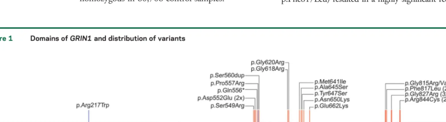
Spectrum and clustering of GRIN1 mutations.All 16 dif-ferent de novo mutations identified in the 23 novel and published cases are missense alterations and cluster within or in close proximity to the transmembrane do-mains forming the intrinsic ion channel pore of the receptor (figure 1). Interestingly, this region is widely spared from genetic variation in controls and shows a high level of conservation in different species (figure e-1). In contrast, there is extensive enrichment of reported single nucleotide variants (SNV) in controls in the N-terminal domain as well as the C-terminus of GRIN1. De novo mutations have not been observed in either domain. The reported mutations do not allow us to assess the genetic variability of S1 and S2 ligand binding sites. However, the S1 domain contains numerous and frequent SNV but no disease-causing mutations so far, whereas S2 showed less variability with only 2 nonrecurrent SNV but one mutation (figure 1). Moreover, this single mutation detected within S2 was associated with the mildest phenotype (patient 14) when compared to all other GRIN1 de novo mutations closely related to the transmembrane domains. Five de novo mutations have been detected recurrently in independent patients within our cohort (p.Asp552Glu, p.Gly815Arg, p.Phe817Leu, p.Gly827Arg, p.Arg844Cys).
The only mutation being far outside the trans-membrane cluster is the p.Arg217Trp missense vari-ant that was identified in a recessive family with unaffected heterozygous carriers.
Functional investigations. Ectopic expression of the GluN1 mutants with wild-type GluN2B subunits revealed that 4 of the mutants gave no response to up to 10 mM glutamate and glycine and were hence rated as nonfunctional (p.Gln556*, p.Gly618Arg, p.Gly620Arg, p.Gly827Arg; n 5 10, each). For all other mutants, changes in maximal inducible currents and agonist affinities were examined. Four mutations (p.Pro557Arg, p.Tyr647Ser, p.Gly815Arg, p.Phe817Leu) resulted in a highly significant reduction
Figure 1 Domains of GRIN1 and distribution of variants
Signal peptide (SP), the extracellular N-terminal domain, and ligand binding sites (S1, S2), the transmembrane domains (M1-4), as well as the intracellular C-terminal domain (CTD) with the proximal Ca21calmodulin binding domain (CBD). De novo mutations (red) cluster within or in very close proximity to
M1-4. In addition, this region is particularly spared from nonsynonymous genetic variation according to the ExAC browser (rare/single variants, gray; repeated/frequent variants, black). The 2 homozygousGRIN1 variants are marked in blue.
of agonist Imax values (p , 0.001, n 5 5–9; figure e-2a). Our model of the GRIN1/GRIN2B NMDAR11
predicts strong intersubunit and intrasubunit interactions between adjacent helical structures M3 and M4 for mutated positions p.Pro557, p.Tyr647, p.Gly815, p.Phe817, and p.Gly827, thereby destabilizing the quaternary structure of the receptor transmembrane domains and disturbing assembly of NMDARs. Consistent with an impaired function of GRIN1 de novo mutations, analysis of the agonist dose-response curves for mutations p.Gly815Arg and p.Phe817Leu additionally revealed a highly significant reduction in the affinities for both glutamate and glycine (figure e-2b). For positions p.Gly618 and p.Gly620, our model predicts a location in the membrane re-entrant loop (the P-loop) of the GluN1 subunit, which determines the narrow constriction and ion selectivity of the channel. Thus, conversion of the pore residues p.Gly618 and p.Gly620 to a charged arginine side chain likely has strong impact on NMDA channel properties. Coexpression of wild-type GluN1 with mutants GluN1 p.Tyr647Ser or p.Phe817Leu resulted in intermediate effects of agonist Imax values (p, 0.01, n 5 5), supporting the idea of a negative impact of the substitutions on the function of hetero-oligomeric NMDARs (figure e-2c). Mutations p.Arg217Trp, p.Ala645Ser, and p.Arg844Cys generated functional receptors with agonist Imax values (p. 0.05, n5 5) similar to wild-type (data not shown). Functional analysis of the missense alteration GluN1 p.Arg217Trp in the N-terminal Zn21-binding domain with wild-type
GluN2A subunits showed a significant increase in Zn21
inhibition (figure e-2d), suggesting impaired activation in vivo due to increased tonic inhibition of GRIN1Arg217Trp–GRIN2A receptors at physiologic
concentrations of Zn21. However, functional analysis of mutations p.Ala645Ser and p.Arg844Cys revealed no significant effects on agonist Imax values or affinity, suggesting that these 2 substitutions may alter NMDAR function through other mechanisms. For example, since the recurrent substitution p.Arg844Cys is located in the intracellular Ca21-calmodulin-binding
domain, disturbed interactions with intracellular proteins may impair receptor function. Our analyses of de novo mutations are consistent with a dominant-negative effect resulting in a significant loss of receptor function.
DISCUSSION We reviewed 14 novel and 9 previ-ously published individuals carrying heterozygous GRIN1 de novo mutations associated with neurode-velopmental phenotypes.
Profound developmental delay associated with severe ID and lack of speech development are pre-dominant features, and muscular hypotonia leading
to spastic quadriplegic cerebral palsy, hyperkinetic movement disorders including dystonia and chorea, and oculomotor abnormalities as well as cortical visual impairment appeared to be recurrent findings in individuals with GRIN1 encephalopathy. While some of these features can be seen in other genetic epileptic encephalopathies such as SCN2A and SCN8A encephalopathies (OMIM 613721, 600702), these findings are novel in the context of NDMAR en-cephalopathies. Seizures occurred in about two-thirds of cases. However, there was no obvious epilepsy pattern with respect to age at onset, seizure semiology, EEG features, or outcome. Fur-thermore, the frequency of epilepsy in GRIN1 patients may be an overestimate due to inclusion of a high proportion of epilepsy patients in the screening cohorts.
GRIN1 encodes GluN1 and autoantibodies pri-marily targeting an epitope within the N-terminal domain of GluN1 result in the increasingly recog-nized clinical entity of NMDAR encephalitis.22,23
NMDAR encephalitis is an acute paraneoplastic or parainfectious neurologic disorder where decay of NMDARs is considered to be the underlying patho-mechanism, partially paralleling the pathophysiology of GluN1 loss of function seen in our patients with GRIN1 encephalopathy. While the acuity of the clin-ical presentation is vastly different, with NMDAR encephalitis presenting as an acute acquired condition while GRIN1 encephalopathy is a chronic neurode-velopmental disease, we would like to emphasize a shared group of symptoms including choreatic and dystonic movements, seizures, and sleep cycle dysregulation, which we observe in our patient cohort. Further research will be able to address the question whether the GRIN1 encephalopathy pheno-type is specific enough to hypothesize a spectrum of NMDAR impairment in human disease.
GRIN1 de novo mutations cluster within or in direct proximity to the transmembrane domains of GRIN1 (figure 1). These regions are largely spared from genetic variation, underlining the crucial impor-tance of these domains. The sole mutation outside this cluster is p.Arg217Trp, which appears to be only pathogenic when present homozygously and thus might mediate its effect through different effects com-pared to the heterozygous variants near the trans-membrane domains.
All reported de novo mutations are missense var-iants. In contrast to GRIN2A and GRIN2B, hetero-zygous truncation of GRIN1 apparently does not result in a neurologic phenotype. Furthermore, both deletions encompassing GRIN1 as well as truncating or splice-site variants are seen in control databases, suggesting that haploinsufficiency in GRIN1, albeit rare, is tolerated in the human population.
In addition to 16 different heterozygous de novo mutations, we describe 2 recessive GRIN1 mutations. The homozygous loss-of-function p.Arg217Trp mutation segregated with severe ID, ASD, and move-ment disorders in 2 affected siblings in a consanguin-eous family (family 1), and a homozygous p.Gln556* truncation mutation was found in 3 individuals with fatal epileptic encephalopathy in family 5 (table 1).
As the NMDAR obligatorily contains 2 GluN1 subunits, truncation or lack of both GRIN1 alleles would result in a knock-out and complete deprivation of the NMDAR. The almost continuous seizure activity with suppression-burst EEG and early death of all 3 affected siblings in family 5 underlines the vital role of GluN1 in NMDAR functioning.
This disease mechanism as well as functional data point towards a dominant-negative effect. In contrast to the gain of function frequently detected in other GRIN-associated neurodevelopmental disorders,11,12
GRIN1 de novo mutations p.Pro557Arg, p.Ser560d-up, p.Gly618Arg, p.Gly620Arg, p.Tyr647Ser, p.Gly815Arg, p.Phe817Leu, and p.Gly827Arg result in a loss of function.
The extent and nature of the loss of NMDAR function due to de novo GRIN1 mutations corre-sponded only marginally to the uniformly severe phe-notypes. One possible explanation for this lack of genotype–phenotype correlation may be effects on the subunit composition or trafficking of the NMDAR that potentially lead to a shared secondary mechanism compensating GluN1 defects indepen-dent of the individual severity of the respective GRIN1 mutation eventually escaping visualization using standard artificial in vitro models.
Given the multiple observations of heterozygous de novo GRIN1 loss-of-function mutations, GRIN1-associated disorders are a recurrent cause of severe and complex neurodevelopmental disorders. Additionally, homozygous GRIN1 mutations segregating with severe phenotypes display phenotypic overlap with individuals carrying heterozygous GRIN1 de novo mutations. This observation blurs the borders of autosomal-dominant and autosomal-recessive inheritance. The constellation of severe ID and associated findings including promi-nent hypotonia, movement disorders, oculogyric crises, cortical visual impairment or blindness, absent speech, behavioral issues, sleep disorder, and seizures may allow for a phenotypic differentiation from other frequent forms of severe ID. Together with the catastrophic phe-notype seen in homozygous truncation mutation car-riers, our findings underscore the essential role of the NMDAR subunits in neurodevelopment.
AUTHOR AFFILIATIONS
From the Institute of Human Genetics (J.R.L., H.O.H., J.H.) and Inte-grated Research and Treatment Center (IFB) Adiposity Diseases (H.O.H.), University of Leipzig Hospitals and Clinics; Department of
Neurophysiology and Neurosensory Systems (K.G., H.S., B.L.), Techni-cal University Darmstadt, Germany; Division of CliniTechni-cal Genomics (K.L.H.), Ambry Genetics, Aliso Viejo, CA; Division of Human Genetics (C.C.), University Children’s Hospital, Inselspital, Bern, Switzerland; Folkhälsan Institute of Genetics (C.C.), Helsinki, Finland; INSERM (C.D., C.N., S.W.), U 1127, Sorbonne Universités, UPMC Université Paris 06, CNRS, UMR 7225, Institut du Cerveau et de la Moelle Épi-nière (ICM); Département de Génétique (C.D., C.N., D.H.) and Centre de Reference Épilepsies Rares, Epilepsy Unit (S.W.), AP-HP, Hôpital de la Pitié-Salpêtrière, Paris, France; Danish Epilepsy Centre (R.S.M., H.H.), Dianalund; Institute for Regional Health Services (R.S.M., H.H.), University of Southern Denmark, Odense; Cologne Center for Genomics (CCG) (D.L., P.N., H.T.) and Center for Molecular Medicine Cologne (CMMC) (P.N.), University of Cologne, Germany; Psychiatric and Neurodevelopmental Genetics Unit (D.L.), Massachusetts General Hospital and Harvard Medical School, Boston; Program in Medical and Population Genetics (D.L.) and Stanley Center for Psychiatric Research (D.L.), Broad Institute of MIT and Harvard, Cambridge, MA; Depart-ment of Neuropediatrics (B.A.N.), University Medical Center Giessen and Marburg, Giessen; Department of Neuropediatrics (G.K.), Univer-sity Medical Center Münster, Germany; Division of Medical Genetics, Department of Pediatrics, University of Pittsburgh School of Medicine (G.L.A.), and Division of Child Neurology (A.G., D.S.R.), Children’s Hospital of Pittsburgh, PA; Department of Medical Genetics (V.B.), Children’s Hospitals and Clinics of Minnesota, Minneapolis; Institute of Clinical Genetics (D.B.), Klinikum Stuttgart, Germany; Department of Medical Genetics (C.R.J.P., D.M., E.F.) and Department of Clinical Neurosciences for Children, Women and Children’s Division (P.S.), Oslo University Hospital and University of Oslo, Norway; Division of Neurology (D.L.D., I.H.), The Children’s Hospital of Philadelphia, PA; Section of Clinical Genetics (E.S.D.), Carilion Clinic Children’s Hospi-tal, Roanoke, VA; Department of Clinical Genetics (E.K.B., C.A.R., M.J.V.H.), Leiden University Medical Center, the Netherlands; The Center for Rare Childhood Disorders (V.N., K.R., N.B., I.S., R.R.), Translational Genomics Research Institute (TGen), Phoenix, AZ; Department of Medical Genetics (B.P.C.K., C.G.F.d.K.), UMCU, Utrecht, the Netherlands; Centro Hospitalar do Algarve (J.S., C.M.), Faro, Portugal; Neurogenetics Group, VIB-Department of Molecular Genetics (S.W., K.H., P.D.J.), and Laboratory of Neurogenetics, Insti-tute Born-Bunge (S.W., K.H., P.D.J.), University of Antwerp; Division of Neurology (P.D.J. and S.W.), Antwerp University Hospital, Belgium; Department of Paediatric Neurology (L.D.M.), University Hospital, Vrije Universiteit Brussel, Bruxelles, Belgium; Aix Marseille Université (M.M.), INSERM, GMGF UMR_S 910; APHM, Departement de Ge-netique Medicale (C.B.), and Département de Génétique Médicale (T. B.), Hopital d’Enfants de la Timone, Marseille; Génétique Clinique (M. L.), Chromosomique et Moléculaire, CHU Hôpital Nord, St Pirest en Jarez; Service de Génétique Médicale (C.F.), Centre Hospitalier Univer-sitaire de Clermont-Ferrand; Département de Médicine Translationnelle et Neurogénétique (A.P.), IGBMC, CNRS UMR 7104/INSERM U964/ Université de Strasbourg, Illkirch; Laboratoire de Diagnostic Génétique (A.P.), Hôpitaux Universitaires de Strasbourg, France; CeGaT GmbH (E.R., S.B.), Tübingen, Germany; Swiss Epilepsy Center (E.R., H.V., T.D.), Zurich, Switzerland; Department of Neuropediatrics (I.H.), Uni-versity Medical Center Schleswig-Holstein, Kiel Campus, Germany; Cen-tre de Recherche du CenCen-tre Hospitalier Universitaire Sainte-Justine and Departments of Pediatrics and Neurosciences (J.L.M.), Université de Montréal, Canada; and the Department of General Paediatrics (S.S.), Division of Child Neurology and Inherited Metabolic Diseases, Centre for Paediatrics and Adolescent Medicine, University Hospital Heidelberg, Germany.
AUTHOR CONTRIBUTIONS
Study concept and design: Johannes R. Lemke, Bodo Laube, Steffen Syrbe. Acquisition of data: Johannes R. Lemke, Kirsten Geider, Katherine L. Helbig, Hannah Schütz, Carolina Courage, Christel Depienne, Caroline Nava, Delphine Heron, Rikke S. Møller, Helle Hjalgrim, Dennis Lal, Bernd A. Neubauer, Peter Nürnberg, Holger Thiele, Gerhard Kurlemann, Georgianne L. Arnold, Vikas Bhambhani, Deborah Bartholdi, Christeen Ramane J. Pedurupillay, Doriana Misceo, Eirik Frengen, Petter Strømme, Dennis J. Dlugos, Emily S. Doherty, Emilia K. Bijlsma,
Claudia A. Ruivenkamp, Mariette J.V. Hoffer, Amy Goldstein, Deepa S. Rajan, Vinodh Narayanan, Keri Ramsey, Newell Belnap, Isabelle Schrau-wen, Ryan Richholt, Bobby P.C. Koeleman, Joaquim Sá, Carla Mendonça, Carolien G. de Kovel, Sarah Weckhuysen, Katia Hardies, Peter De Jonghe, Linda De Meirleir, Mathieu Milh, Catherine Badens, Marine Lebrun, Tiffany Busa, Christine Francannet, Amélie Piton, Erik Riesch, Saskia Biskup, Heinrich Vogt, Thomas Dorn, Jacques L. Michaud. Analysis and interpretation: Johannes R. Lemke, Kirsten Geider, Katherine L. Helbig, Henrike O. Heyne, Hannah Schütz, Julia Hentschel, Erik Riesch, Saskia Biskup, Ingo Helbig, Bodo Laube, Steffen Syrbe. Writing of manuscript: Johannes R. Lemke, Katherine L. Helbig, Ingo Helbig, Bodo Laube, Stef-fen Syrbe. Critical revision of manuscript for intellectual input: all authors. Study supervision: Johannes R. Lemke, Bodo Laube, Steffen Syrbe.
ACKNOWLEDGMENT
The authors thank the patients and family members for their participation in this study.
STUDY FUNDING
Johannes R. Lemke (32EP30_136042/1) and Peter De Jonghe (G.A.136.11.N and FWO/ESF-ECRP) received financial support within the EuroEPINOMICS-RES network (www.euroepinomics.org) within the Eurocores framework of the European Science Foundation (ESF). Saskia Biskup and Henrike Heyne received financial support from the German Federal Ministry for Education and Research (BMBF IonNeurONet: 01 GM1105A and FKZ: 01EO1501). Katia Hardies is a PhD fellow of the Institute for Science and Technology (IWT) Flanders. Ingo Helbig was sup-ported by intramural funds of the University of Kiel, by a grant from the German Research Foundation (HE5415/3-1) within the EuroEPINOMICS framework of the European Science Foundation, and additional grants of the German Research Foundation (DFG, HE5415/5-1, HE 5415/6-1), German Ministry for Education and Research (01DH12033, MAR 10/012), and grant by the German chapter of the International League against Epilepsy (DGfE). The project also received infrastructural support through the Institute of Clinical Molecular Biology in Kiel, supported in part by DFG Cluster of Excellence“Inflammation at Interfaces” and “Future Ocean.” The project was also supported by the popgen 2.0 network (P2N) through a grant from the German Ministry for Education and Research (01EY1103) and by the Inter-national Coordination Action (ICA) grant G0E8614N. Christel Depienne, Caroline Nava, and Delphine Heron received financial support for exome analyses by the Centre National de Génotypage (CNG, Evry, France).
DISCLOSURE
J. Lemke and K. Geider report no disclosures relevant to the manuscript. K. Helbig is employed by and receives a salary from Ambry Genetics. H. Heyne, H. Schütz, J. Hentschel, C. Courage, C. Depienne, C. Nava, D. Heron, R. Møller, H. Hjalgrim, D. Lal, B. Neubauer, P. Nürnberg, H. Thiele, G. Kurlemann, G. Arnold, V. Bhambhani, D. Bartholdi, C. Pedurupillay, D. Misceo, E. Frengen, P. Strømme, D. Dlugos, E. Doherty, E. Bijlsma, C. Ruivenkamp, M. Hoffer, A. Goldstein, D. Rajan, V. Narayanan, K. Ramsey, N. Belnap, I. Schrauwen, R. Richholt, B. Koeleman, J. Sá, C. Mendonça, C. de Kovel, and S. Weckhuysen report no disclosures relevant to the manuscript. K. Hardies, since mid-October 2015, is under employment of UCB Pharma (Braine-l’Alleud, Belgium). The company had no part in this study. P. De Jonghe, L. De Meirleir, M. Milh, C. Badens, M. Lebrun, T. Busa, C. Francannet, and A. Piton report no disclosures relevant to the manuscript. E. Riesch is employed by and receives a salary from CeGaT GmbH. Exome sequencing is among commercially available tests of both institutions S. Biskup is employed by and receives a salary from CeGaT GmbH. Exome sequencing is among commercially available tests of both institutions. H. Vogt, T. Dorn, I. Helbig, J. Michaud, B. Laube, and S. Syrbe report no disclosures relevant to the manuscript. Go to Neurology.org for full disclosures.
Received September 23, 2015. Accepted in final form March 1, 2016. REFERENCES
1. Laube B, Kuhse J, Betz H. Evidence for a tetrameric struc-ture of recombinant NMDA receptors. J Neurosci 1998; 18:2954–2961.
2. Paoletti P, Bellone C, Zhou Q. NMDA receptor subunit diversity: impact on receptor properties, synap-tic plassynap-ticity and disease. Nat Rev Neurosci 2013;14: 383–400.
3. Yuan H, Low CM, Moody OA, Jenkins A, Traynelis SF. Ionotropic GABA and glutamate receptor mutations and human neurologic diseases. Mol Pharmacol 2015; 88:203–217.
4. Hardingham GE, Bading H. Synaptic versus extrasynap-tic NMDA receptor signalling: implications for neuro-degenerative disorders. Nat Rev Neurosci 2010;11: 682–696.
5. Paoletti P. Molecular basis of NMDA receptor functional diversity. Eur J Neurosci 2011;33:1351–1365. 6. de Ligt J, Willemsen MH, van Bon BW, et al. Diagnostic
exome sequencing in persons with severe intellectual dis-ability. N Engl J Med 2012;367:1921–1929.
7. Endele S, Rosenberger G, Geider K, et al. Mutations in GRIN2A and GRIN2B encoding regulatory subunits of NMDA receptors cause variable neurodevelopmental phenotypes. Nat Genet 2010;42:1021–1026.
8. Hamdan FF, Gauthier J, Araki Y, et al. Excess of de novo deleterious mutations in genes associated with glutamater-gic systems in nonsyndromic intellectual disability. Am J Hum Genet 2011;88:306–316.
9. Allen AS, Berkovic SF, Cossette P, et al. De novo muta-tions in epileptic encephalopathies. Nature 2013;501: 217–221.
10. Carvill GL, Regan BM, Yendle SC, et al. GRIN2A muta-tions cause epilepsy-aphasia spectrum disorders. Nat Genet 2013;45:1073–1076.
11. Lemke JR, Hendrickx R, Geider K, et al. GRIN2B mutations in West syndrome and intellectual disability with focal epilepsy. Ann Neurol 2014;75: 147–154.
12. Lemke JR, Lal D, Reinthaler EM, et al. Mutations in GRIN2A cause idiopathic focal epilepsy with rolandic spikes. Nat Genet 2013;45:1067–1072.
13. Lesca G, Rudolf G, Bruneau N, et al. GRIN2A muta-tions in acquired epileptic aphasia and related child-hood focal epilepsies and encephalopathies with speech and language dysfunction. Nat Genet 2013; 45:1061–1066.
14. O’Roak BJ, Deriziotis P, Lee C, et al. Exome sequencing in sporadic autism spectrum disorders identifies severe de novo mutations. Nat Genet 2011;43:585–589. 15. Tarabeux J, Kebir O, Gauthier J, et al. Rare mutations in
N-methyl-D-aspartate glutamate receptors in autism spec-trum disorders and schizophrenia. Transl Psychiatry 2011; 1:e55.
16. Redin C, Gerard B, Lauer J, et al. Efficient strategy for the molecular diagnosis of intellectual disability using tar-geted high-throughput sequencing. J Med Genet 2014;51: 724–736.
17. Zhu X, Petrovski S, Xie P, et al. Whole-exome sequencing in undiagnosed genetic diseases: interpreting 119 trios. Genet Med 2015;17:774–781.
18. Ohba C, Shiina M, Tohyama J, et al. GRIN1 mutations cause encephalopathy with infantile-onset epilepsy, and hyperkinetic and stereotyped movement disorders. Epilepsia 2015;56:841–848.
19. Lemke JR, Riesch E, Scheurenbrand T, et al. Targeted next generation sequencing as a diagnostic tool in epileptic disorders. Epilepsia 2012;53:1387–1398.
20. Laube B, Hirai H, Sturgess M, Betz H, Kuhse J. Molec-ular determinants of agonist discrimination by NMDA receptor subunits: analysis of the glutamate binding site on the NR2B subunit. Neuron 1997;18:493–503. 21. Laube B, Kuhse J, Rundstrom N, Kirsch J, Schmieden V,
Betz H. Modulation by zinc ions of native rat and re-combinant human inhibitory glycine receptors. J Physiol 1995;483:613–619.
22. Dalmau J, Tuzun E, Wu HY, et al. Paraneoplastic anti-N-methyl-D-aspartate receptor encephalitis associated with ovarian teratoma. Ann Neurol 2007;61:25–36. 23. Gleichman AJ, Spruce LA, Dalmau J, Seeholzer SH,
Lynch DR. Anti-NMDA receptor encephalitis antibody binding is dependent on amino acid identity of a small region within the GluN1 amino terminal domain. J Neurosci 2012;32:11082–11094.
New! AAN Transforming Leaders Program
Are you an established AAN member neurologist 101 years out of residency who wants to move your career to the next level? Do you want to become a future leader at the AAN and in the field of neurology? Apply for the new, elite AAN Transforming Leaders Program by visiting AAN.com/view/TransformingLeaders. Application deadline is July 1, 2016.
20 Minutes Pack a Punch
Neurology®Podcasts
• Interviews with top experts on new clinical research in neurology • Editorial comments on selected articles
• Convenient—listen during your commute, at your desk, or even at the gym • On demand—it’s there when you want it
• Fun and engaging • New topic each week • FREE
Listen now at www.aan.com/podcast
Save These Dates for AAN CME Opportunities!
Mark these dates on your calendar for exciting continuing education conferences by the American Academy of Neurology.Learn more at AAN.com/conferences.
Sports Concussion Conference
• July 8–10, 2016, Chicago, IL, at the Hilton Chicago Fall Conference
DOI 10.1212/WNL.0000000000002740
2016;86;2171-2178 Published Online before print May 6, 2016
Neurology
Johannes R. Lemke, Kirsten Geider, Katherine L. Helbig, et al.
encephalopathy
phenotypic spectrum: A distinct genetic NMDA receptor
GRIN1
Delineating the
This information is current as of May 6, 2016
Services
Updated Information &
http://n.neurology.org/content/86/23/2171.full including high resolution figures, can be found at:
Supplementary Material
740.DC1
http://n.neurology.org/content/suppl/2016/05/06/WNL.0000000000002 Supplementary material can be found at:
References
http://n.neurology.org/content/86/23/2171.full#ref-list-1
This article cites 23 articles, 4 of which you can access for free at:
Subspecialty Collections
http://n.neurology.org/cgi/collection/ion_channel_gene_defects
Ion channel gene defects
http://n.neurology.org/cgi/collection/developmental_disorders
Developmental disorders
http://n.neurology.org/cgi/collection/all_movement_disorders
All Movement Disorders
http://n.neurology.org/cgi/collection/all_genetics
All Genetics
http://n.neurology.org/cgi/collection/all_epilepsy_seizures
All Epilepsy/Seizures
following collection(s):
This article, along with others on similar topics, appears in the
Permissions & Licensing
http://www.neurology.org/about/about_the_journal#permissions its entirety can be found online at:
Information about reproducing this article in parts (figures,tables) or in
Reprints
http://n.neurology.org/subscribers/advertise
Information about ordering reprints can be found online:
rights reserved. Print ISSN: 0028-3878. Online ISSN: 1526-632X.
1951, it is now a weekly with 48 issues per year. Copyright © 2016 American Academy of Neurology. All ® is the official journal of the American Academy of Neurology. Published continuously since Neurology