Mutations in
MAB21L2
Result in Ocular
Coloboma, Microcornea and Cataracts
Brett Deml1,2, Ariana Kariminejad3, Razieh H. R. Borujerdi4, Sanaa Muheisen1, Linda M. Reis1, Elena V. Semina1,2*
1Department of Pediatrics and Children’s Research Institute at the Medical College of Wisconsin and Children’s Hospital of Wisconsin, Milwaukee, Wisconsin, United States of America,2Department of Cell Biology, Neurobiology and Anatomy, Medical College of Wisconsin, Milwaukee, Wisconsin, United States of America,3Kariminejad-Najmabadi Pathology and Genetics Center, Tehran, Iran,4Qom Welfare
Organization, Qom, Iran
Abstract
Ocular coloboma results from abnormal embryonic development and is often associated with additional ocular and systemic features. Coloboma is a highly heterogeneous disorder with many cases remaining unexplained. Whole exome sequencing from two cousins af-fected with dominant coloboma with microcornea, cataracts, and skeletal dysplasia identi-fied a novel heterozygous allele inMAB21L2, c.151 C>G, p.(Arg51Gly); the mutation was present in all five family members with the disease and appeared de novo in the first affect-ed generation of the three-generational paffect-edigree.MAB21L2encodes a protein similar to C. elegansmab-21 cell fate-determining factor; the molecular function of MAB21L2 is
large-ly unknown. To further evaluate the role ofMAB21L2, zebrafish mutants carrying a p.
(Gln48Serfs*5) frameshift truncation (mab21l2Q48Sfs*5) and a p.(Arg51_Phe52del) in-frame
deletion (mab21l2R51_F52del) were developed with TALEN technology. Homozygous
zebra-fish embryos from both lines developed variable lens and coloboma phenotypes:
mab21l2Q48Sfs*5embryos demonstrated severe lens and retinal defects with complete
le-thality whilemab21l2R51_F52delmutants displayed a milder lens phenotype and severe
colo-boma with a small number of fish surviving to adulthood. Protein studies showed decreased stability for the human p.(Arg51Gly) and zebrafish p.(Arg51_Phe52del) mutant proteins and predicted a complete loss-of-function for the zebrafish p.(Gln48Serfs*5) frameshift
trunca-tion. Additionally, in contrast to wild-type humanMAB21L2transcript, mutant p.(Arg51Gly)
mRNA failed to efficiently rescue the ocular phenotype when injected intomab21l2Q48Sfs*5
embryos, suggesting this allele is functionally deficient. Histology, immunohistochemistry, and in situ hybridization experiments identified retinal invagination defects, an increase in cell death, abnormal proliferation patterns, and altered expression of several ocular markers in themab21l2mutants. These findings support the identification ofMAB21L2as a novel
factor involved in human coloboma and highlight the power of genome editing manipulation in model organisms for analysis of the effects of whole exome variation in humans.
OPEN ACCESS
Citation:Deml B, Kariminejad A, Borujerdi RHR, Muheisen S, Reis LM, Semina EV (2015) Mutations inMAB21L2Result in Ocular Coloboma, Microcornea and Cataracts. PLoS Genet 11(2): e1005002. doi:10.1371/journal.pgen.1005002
Editor:Gregory S. Barsh, Stanford University School of Medicine, UNITED STATES
Received:May 9, 2014
Accepted:January 14, 2015
Published:February 26, 2015
Copyright:© 2015 Deml et al. This is an open access article distributed under the terms of the
Creative Commons Attribution License, which permits unrestricted use, distribution, and reproduction in any medium, provided the original author and source are credited.
Data Availability Statement:All relevant data are within the paper and its Supporting Information files.
Author Summary
Coloboma is a hole or gap in one or more of the structures of the eye. Coloboma occurs when the eye is not formed properly during prenatal development. It is often associated with additional eye abnormalities and can result in significant loss of vision. Identification of the genetic causes of coloboma provides more information about how the eye develops. We used whole exome sequencing in an affected family to identify mutations in a new gene associated with dominant coloboma in humans,MAB21L2. We used genome editing to disrupt themab21l2gene in zebrafish, which led to similar eye conditions in affected fish, providing additional evidence for the role of this gene in eye development. The func-tion of MAB21L2 is largely unknown; further study of the pathways affected by MAB21L2 deficiency and study of the zebrafish generated by this project will provide more informa-tion about the role of this gene in eye development.
Introduction
Coloboma is a congenital segmental ocular defect which can affect one or more structures of the eye; typical coloboma results in an inferior deficiency of iris, chorioretinal, and/or optic nerve tissue [1–3]. Ocular coloboma is believed to result from failure of normal closure of the optic fissure during embryonic eye development, termed optic fissure closure defect (OFCD) [1,2]. Coloboma can occur as an isolated anomaly (simple coloboma) but in most cases it is as-sociated with additional ocular defects, including microphthalmia, cataract, retinal detach-ment, and ocular motility disorders [1–4]. Microcornea, a reduction in the diameter of the cornea, is common in colobomatous eyes and can be associated with normal or even increased (macrophthalmic) axial length [2,3,5]. Additional systemic anomalies are present in a large proportion of patients with OFCDs including brain, skeletal, cardiac, or urogenital anomalies [3,4].
A number of genes have been associated with coloboma including transcriptional regula-torsSOX2,OTX2,PAX2,PAX6,CHD7andSALL2;secreted signaling factor-encoding gene SHH; members of the transforming growth factor-beta (TGF-beta) superfamilyGDF6and GDF3;members of the retinoic acid synthesis pathwaySTRA6andALDH1A3;as well as membrane porphyrin transporterABCB6, and a member of the HIPPO growth control path-way,YAP1[6–19]. Due to the number of genes involved and the large proportion of unex-plained cases, whole exome sequencing has been used with increasing frequency to screen families with coloboma and other ocular conditions [16–21]. Since whole exome sequencing generates a large number of variants and often involves genes with unexplored/unknown function, studies in animal models can be invaluable in providing further insight into the functional roles of candidate genes/variants and their possible involvement in the studied phenotype [17,22]. Recent advances in genomic editing technologies using engineered nucle-ases empowered researchers with tools for the functional exploration of these genes/variants of interest [23].
In this manuscript we present evidence for the role ofMAB21L2in human coloboma and further investigate its role in vertebrate ocular development by generation and analysis of zeb-rafishmab21l2mutants.
Results
Identification of a
MAB21L2
mutation in a human pedigree affected with
ocular disease
The proband (Patient 1) was diagnosed with bilateral microcornea, iris and chorioretinal coloboma, corectopia, nystagmus and cataract (Fig. 1); the cataractous lens of his right eye was removed at age 24. Based on ultrasound measurements at age 32, the axial length was 29.02 mm (right eye) and 24.67 mm (left eye), thus showing no signs of microphthalmia
Fig 1. Ocular Images of Patient 1.Photographs of the right (A, C, E, G) and the left (B, D, F, H) eyes showing bilateral iris coloboma with microcornea and corectopia (C-F) as well as chorioretinal coloboma (G, H) in the proband (Patient 1).
(normal axial length for adult males is approximately 23 mm) [24]. Evaluation of other systems identified mild contracture in the knees and elbows and rhizomelia of the upper and lower limbs: the lengths of the femur (40.3 cm, left and 40.6 cm, right), tibia (34.5cm, left and 34.3 cm, right), humerus (26 cm, left and 26.3 cm, right), ulna (23.5 cm, left and 23.4 cm, right) and radius (22.2 cm, left and 22.6 cm, right) were determined by scanography and the observed femur/tibia (1.18) and humerus/radius (1.17) ratios were found to deviate from the normal reference numbers of 1.23 and 1.31, respectively. No signs of cardiac, brain or other ab-normalities were identified. Analysis of family history identified a similarly affected sister, brother, and two nephews, while his father, mother, and two additional sisters were unaffected (Fig. 2A).
Independent whole exome sequencing of DNA samples from the proband’s two affected nephews (III-1 and III-2,Fig. 2A) yielded a mean coverage of 63.9X ± 4.0 for the two samples with 96.25% ± 0.49 of bases having>10X coverage. Initial analysis of the whole exome data
excluded mutations in 72 known factors involved in anophthalmia, microphthalmia, and/or coloboma [25]. Comparison of whole exome data of the two nephews identified 16 shared het-erozygous variants that were novel or rare (<1%): one splicing, one frameshift indel, one
inframe indel, and 13 missense alleles (three novel and ten rare) that were found to be damag-ing by at least 4 functional effect prediction programs (S1 Table). The five novel alleles and one rare frameshift variant were tested for cosegregation in the pedigree; only the heterozygous MAB21L2missense mutation c.151 C>G, p.(Arg51Gly), was present in all five affected
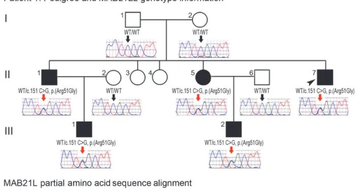
indi-viduals (II-1, -5, -7 and III-1, -2) and absent in the unaffected parents (a low‘G’peak at posi-tion c.151 was seen in the mother’s sample (I-2) in addition to the wild-type‘C’on sequencing of two independent PCR reactions, suggesting that she is likely to have low-level mosaicism for this mutation) (Fig. 2A). TheMAB21L2variant was further identified as a strong candidate based on its absence in all control populations (EVS, dbSNP, and 1000 genomes) and having the highest Genomic Evolutionary Rate Profiling (GERP++) score, as well as enrichment of both humanMAB21L2and mouseMab21l2transcripts in ocular structures based in BioGPS (http://biogps.org). The mutation was predicted to be damaging by SIFT, Polyphen2, Mutation Taster and MutationAssessor, and demonstrated high conservation scores for the affected nu-cleotide with a GERP++ score of 6.16 and a PhyloP score of 2.94 (of note, the highest possible GERP++ score is 6.17 (most conserved) [26]).
Analysis of additional human patients affected with ocular disease
To further explore the role ofMAB21L2in human coloboma phenotypes, we examined 276 pa-tients with developmental ocular conditions from our collection as well as whole exome data from 125 cases derived from the UK10K_Rare_Coloboma (EGA Study ID: EGAS00001000127) project of the UK10K Consortium study [18,31]. We identified no additional mutations in our population that included 39 patients affected with A/M and coloboma, 16 patients with
Fig 2.MAB21L2mutations and protein sequence conservation. A.Three-generation pedigree of Patient 1 withMAB21L2genotype information. DNA chromatograms for all tested family members are shown with c.151C position indicated with black (WT allele) or red (heterozygous mutant allele) arrows. The proband (Patient 1) is indicated with a black arrowhead. Please note the presence of the mutant allele in all affected individuals, its absence in unaffected family members, and the presence of a low‘G’peak in addition to the normal‘C’nucleotide at the mutant position in the proband’s unaffected mother. B.Amino acid alignment of the MAB21L1-3 and mab-21 regions surrounding the arginine at position 51; amino acids identical between different homologs are highlighted with a light grey color, three invariant residues are shown in dark grey; the glycine (G) predicted to replace arginine 51 in Patient 1 is shown in red font; the positions and predicted effects of the zebrafishmab21l2mutations involving the same region are also shown in red font. Accession numbers for
sequences utilized in the alignment are provided in Methods.
coloboma and normal eye size, 104 patients with A/M without coloboma, 50 with aniridia, 37 with various anterior segment dysgenesis conditions, 12 with cataract, and 18 with other devel-opmental ocular conditions. Analysis of the UK10K_Rare_Coloboma cohort identified a hetero-zygous mutation affecting the same arginine residue described above (Fig. 2B), c.152 G>A, p.
(Arg51His), in two samples: UK10K_COL5001067 (the c.152 G>A change was present in 10
out of 23 high quality reads) and UK10K_COL5001068 (the c.152 G>A change was seen in 21
out of 32 high quality reads). While the publicly available UK10K data does not specify family re-lationships, the presence of identical mutations in consecutively numbered samples suggests that these individuals may be family members. Similar to the c.151 C>G, p.(Arg51Gly) allele, this
mutation was not reported in the Exome Variant Server (0/13,006 alleles), the 1000 Genomes Browser (0/2,194 alleles), or dbSNP, and is predicted to be damaging by SIFT, Polyphen2, Muta-tion Taster and MutaMuta-tion Assessor. These data indicate a small contribuMuta-tion ofMAB21L2 muta-tions to human coloboma phenotypes (<2%) and suggest no role forMAB21L2mutations in the
other ocular disorders that were examined in this study.
Zebrafish
mab21l2
expression studies
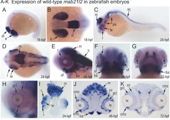
To investigate the role ofMAB21L2/mab21l2in ocular development, we performed careful evaluation ofmab21l2expression in zebrafish using in situ hybridization. At 18-hpf, expression was seen in the presumptive eye field and midbrain (Fig. 3A, B). At 24-hpf,mab21l2expression was seen in the retina (ventral and dorsal periphery), lens, spinal cord, midbrain and pharyn-geal arch region (Fig. 3C, D, I). In 48–72-hpf, the transcripts could be detected in the ciliary marginal zone (CMZ) (or germinal zone) of the retina that contains multipotent retinal pro-genitors, in the inner nuclear and ganglion cell layers of the retina, and around the optic fissure in the eye, as well as the midbrain, hindbrain, developing fins, and branchial arches (Fig. 3E-H, J, K).
Generation and gross morphological analysis of
mab21l2
zebrafish
mutants
To determine the effect ofmab21l2deficiency on ocular development, themab21l2gene was dis-rupted using TALEN genome editing technology. DNA sequencing ofmab21l2mutant fish iden-tified the following alleles: c.141_153delCCAAGAGCCCCGT, p.(Gln48Serfs5); c.150_156del
CCGTTTC, p.(Arg51Serfs4); c.151delC, p.(Arg51Valfs6); c.151dupC, p.(Arg51Profs14); and
c.155delT, p.(Phe52Serfs5) frameshift mutations (all predicted to result in truncation of the
mab21l2 protein at ~14% of its total length) and an in-frame deletion of two amino acids includ-ing the arginine at position 51, c.151_156delCGTTTC, p.(Arg51_Phe52del) (Fig. 3L,S1 Fig.).
Zebrafish carrying heterozygous frameshift alleles were crossed to generate homozygous or compound heterozygousmab21l2mutants and heterozygous fish with the
c.155delT, p.(Phe52Serfs5) allele (3), homozygous for the c.150_156delCCGTTTC, p.(Arg51
Serfs4) allele (2), homozygous for c.141_153delCCAAGAGCCCCGT p.(Gln48Serfs5)
muta-tion (9) or homozygous for the c.151delC, p.(Arg51Valfs6) allele (3) by restriction digest and/
or Sanger sequencing. Crosses involving heterozygous carriers of the c.151_156delCGTTTC, p. (Arg51_Phe52del) allele produced 12 out of 57 (21%) abnormal embryos, also consistent with
Fig 3. Expression and mutations of zebrafishmab21l2. A-K.Expression pattern ofmab21l2in zebrafish 18–72-hpf embryos. Whole mount images (A-H) and sections (I-K) are shown.A, B.At 18-hpf, expression in the presumptive eye field (e) and midbrain (m) is observed.C, D, I.At 24-hpf,mab21l2expression is seen in the periphery of the retina (r), lens (le), spinal cord (sc), midbrain (m) and pharyngeal arch region (pa).E-H, J, K.At 48–72-hpf, expression in the ciliary marginal zone (cmz), inner nuclear layer (inl) and ganglion cell layer (gcl) of the retina, and the region of the optic fissure (of) in the eye as well as the midbrain (m), hindbrain (h), developing fins (f), and branchial arches (ba) is shown with arrows.L.Distribution and expression ofmab21l2mutations induced by TALENs. On the left, a schematic of the zebrafish mab21l2 protein is shown as a light grey box with the mab-21 domain (amino acids 62–346) indicated in dark grey color; the positions of the zebrafish mutations identified in the progeny of TALEN-injected fish are shown at the top of the box and the position of the human mutation identified in Patient 1 is indicated at the bottom; the positions of the p.(Gln48Serfs*5) and p.(Arg51_Phe52del) mutations are shown with red arrows. On the
right, a graph summarizing results of semi-quantitative RT-PCR analysis of wild-type and mutantmab21l2transcript levels in 48-hpf homozygous embryos is shown.
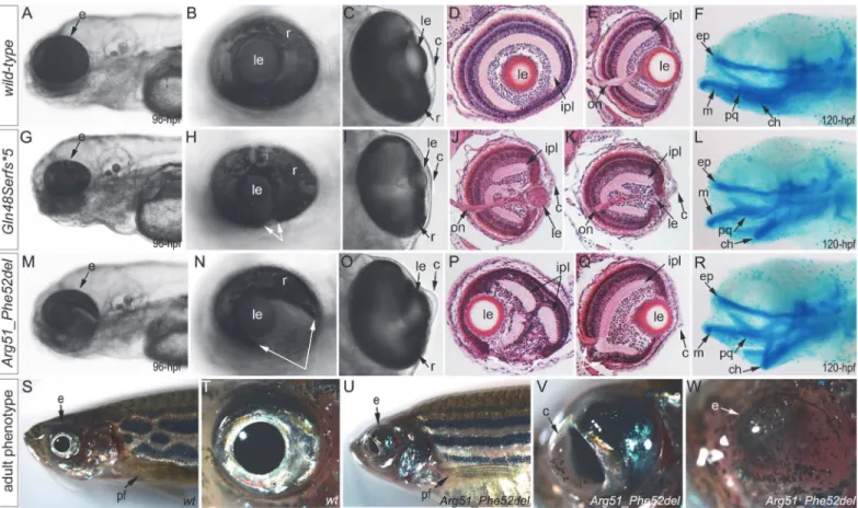
a recessive mode of inheritance. The abnormal phenotype was first evident at 72-hpf and in-volved severe ocular coloboma in all affected embryos and corneal defects in 42% (5/12), while lenses and eye size appeared to be only mildly affected (Fig. 4M-O).
Mutant lines were established for the c.141_153delCCAAGAGCCCCGT, p.(Gln48Serfs5)
frameshift mutation (mab21l2Q48Sfs5) and the c.151_156delCGTTTC, p.(Arg51_Phe52del) in-frame deletion (mab21l2R51_F52del) alleles (Fig. 3LandS1 Fig.) and were further characterized. Expression of mutantmab21l2transcripts was examined by RT-PCR using RNA extracted from 48-hpf embryos. Both mutant transcripts were found to be expressed at slightly higher levels than wild-type transcript, but this increase was not statistically significant (Fig. 3L,S1 Fig.).
Histological analysis of the 96–120-hpf homozygous embryos withmab21l2Q48Sfs5and other frameshift mutations demonstrated a small degenerative (Fig. 4J) or absent (Fig. 4K) lens, disorganized retina (particularly the inner plexiform layer), and irregular cornea (Fig. 4J, K).
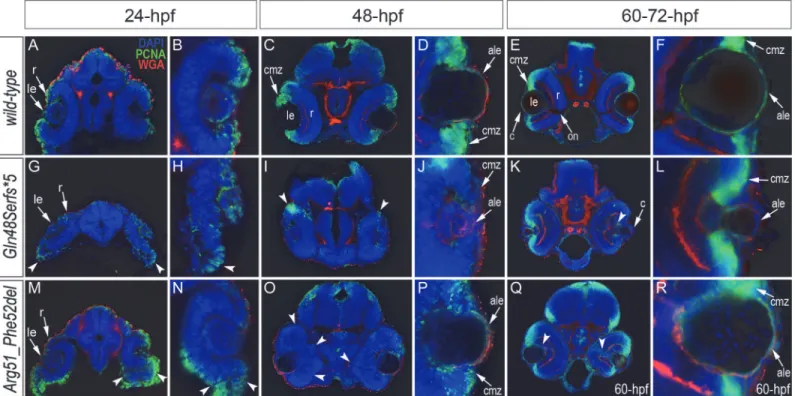
Fig 4. Phenotypic analysis of homozygous embryos carrying mutantmab21l2alleles encoding p.(Gln48Serfs*5) truncation (mab21l2Q48Sfs*5) and
p.(Arg51_Phe52del) in-frame deletion (mab21l2R51_F52del) proteins. A-R.Embryonic phenotypes. Images of wild-type larvae at 96-hpf (A-E) and 120-hpf (F), as well as embryos carrying the p.(Gln48Serfs*5) frameshift (G-L) or p.(Arg51_Phe52del) in-frame deletion (M-R) alleles are shown. Whole mount images (A-C, G-I, and M-O), as well as frontal (E, J, K, Q) and sagittal (D, P) ocular sections are presented. Please notice reduced eye size (G-K), coloboma (white arrows in H), degenerative (H-J) or absent (K) lens, disorganized retina and irregular cornea (J, K) in embryos with frameshift mutations, as well as severe coloboma (white arrows in N) with disorganized retina, discontinuous RPE (white arrows in P, Q), and corneal defects (O) but overall comparable to wild-type eye size and lenses (M-Q) in embryos that are homozygous for the in-frame deletion. Alcian blue stain of wild-type (F) and mutant embryos (L, R) identified defects in craniofacial development with primary defects in the development of the ceratohyal cartilage of the hyoid arch (or the second pharyngeal arch).S-W.Adult phenotype. Images of adult wild-type (S, T) and Arg51_Phe52del mutant (U-W) fish: please notice microphthalmic highly disorganized eye with pigmented cornea (V) and anophthalmic contralateral eye with residual abnormal pigmented tissue (W). Please also note normal appearance of pectoral fins in the mutant fish (U). ch, ceratohyal; ep, ethmoid plate; m, Meckel's cartilage; pq, palatoquadrate; e, eye; c, cornea; le, lens; ipl, inner plexiform layer; on, optic nerve; pf, pectoral fins; r, retina.
Histological analysis of themab21l2R51_F52delhomozygous embryos confirmed severe coloboma, abnormally formed retina (inner plexiform layer) and cornea, discontinuous RPE (retinal pig-mented epithelium), as well as generally normal eye size and lens appearance (Fig. 4P, Q). Alcian blue staining identified craniofacial defects in somemab21l2Q48Sfs5(3/7; 43%) and
mab21l2R51_F52del(3/6; 50%) 120-hpf homozygous fish: the cartilage elements of the pharyngeal arches, particularly the ceratohyal cartilage of the hyoid arch (or the second pharyngeal arch), were malformed and displaced (Fig. 4L, R) while the ethmoid plate cartilage of the upper jaw ap-peared to be largely unaffected. Analysis of the pectoral fin did not identify any obvious malfor-mations inmab21l2Q48Sfs5ormab21l2R51_F52del120-hpf embryos ormab21l2R51_F52deladult fish (Fig. 4U).
Homozygousmab21l2Q48Sfs5embryos demonstrated 100% lethality with no embryos sur-viving into adulthood while two out of fifteen (13%)mab21l2R51_F52delhomozygous larvae de-veloped into adults. Gross morphological examination of adult fish revealed a severe ocular phenotype with anophthalmia of one eye and microphthalmia of the contralateral eye (see Fig. 4S, Tfor wild-type and 4U-W for mutant fish phenotypes). In addition to small size, the ocular structures appeared to be highly disorganized with an expanded anterior segment and pigmented cornea (Fig. 4V).
Comparative analysis of apoptosis, proliferation and ocular markers in
wild-type and
mab21l2
embryos
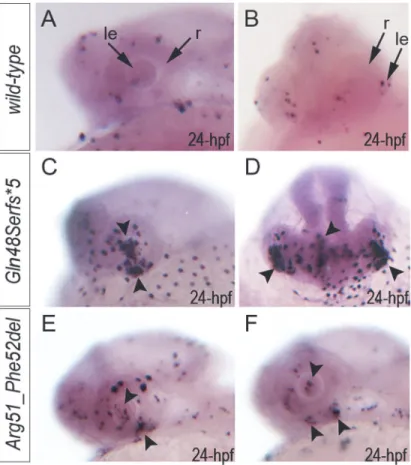
Analysis of apoptosis with TUNEL assay (that detects terminal deoxynucleotidyl transferase dUTP nick end labeling) identified an excessive number of TUNEL-positive cells in 24–72-hpf homozygousmab21l2Q48Sfs5embryos in comparison to wild-type (Fig. 5A-D;S3 Fig.): at 24- hpf, the increased number of TUNEL-positive cells was observed in the developing lens and ventral retina (Fig. 5C, D); at 48–72-hpf clusters of TUNEL-positive cells continued to be found in lens, margins of the open optic fissure and other regions of the retina (possibly the outer nucle-ar layer) (S3 Fig.). Inmab21l2R51_F52delmutants carrying the in-frame deletion allele, a moderate increase in TUNEL-positive cells was observed: at 24-hpf an increase in TUNEL-positive cells was detected in the ventral region of the retina with some staining in other retinal regions and in the lens (Fig. 5E, F); at 48–72-hpf, TUNEL staining continued to be seen primarily the retina and not in the lens (S3 Fig.). A moderate increase in TUNEL staining was also observed in the brain and other embryonic tissues in both mutants (Fig. 5D,S3 Fig.).
Immunohistochemistry with PCNA (Proliferating Cell Nuclear Antigen) was performed to compare proliferation patterns in 24–72-hpf wild-type and bothmab21l2Q48Sfs5and
cells of anterior lens epithelium in both 48- and 60-hpfmab21l2R51_F52delembryos (Fig. 6P, R). ZL-1 positive cells were observed in wild-type embryos starting from 24-hpf (S5 Fig.); this staining was not noted in the abnormal eyes ofmab21l2Q48Sfs5mutants at 24-hpf but was de-tected in 48- and 72-hpfmab21l2Q48Sfs5embryos and 72-hpfmab21l2R51_F52delmutants (S5
Fig.).
Both mutants demonstrated abnormal retinal shape at 24-hpf (Fig. 6G, H, M, N) suggesting a possible invagination defect; a shallow optic cup, particularly at its ventral region, was de-tected in themab21l2Q48Sfs5frameshift mutants (Fig. 6G, H) and a smaller, more constricted, optic vesicle was present in themab21l2R51_F52deldeletion mutants (Fig. 6M, N); abnormal reti-nal folding was evident in both mutants at 24- and 48-hpf (arrowheads inFig. 6G, H, M-O).
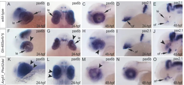
Expression of thepax6b,pax2.1andfoxe3genes was examined in wild-type and mutant fish (Fig. 7,S5 Fig.). Expression ofpax6bin wild-type embryos was detected throughout the devel-oping eye at 24-hpf (Fig. 7A, B) with a more restricted pattern in the retinal ganglion cells and inner nuclear layer neurons at 48-hpf (Fig. 7C). Inmab21l2Q48Sfs5mutants,pax6bexpression was detected in both the developing lens and retina but appeared to be downregulated in the ventral/temporal versus dorsal/nasal domain (arrowhead inFig. 7F, G); at 48-hpfpax6b ex-pression was not present in the region of the coloboma (arrowhead inFig. 7H). In the mab21l2R51_F52delmutants,pax6bexpression was detected throughout the abnormally folded
Fig 5. Summary of TUNEL assays in zebrafish wild-type andmab21l2mutant embryos.TUNEL results in 24-hpf wild-type (A-B),mab21l2Q48Sfs*5embryos (C-D) andmab21l2R51_F52delmutants (E-F) are shown. An increase in TUNEL staining was observed in bothmab21l2mutants with remarkably high levels in the
mab21l2Q48Sfs*5embryos, particularly in the lens and ventral retina (C-D), and moderately increased levels in
themab21l2R51_F52delembryos (E-F); arrowheads indicate sites of increased TUNEL staining in the eye and
brain; le, lens; r, retina.
retina at 24-hpf (Fig. 7K, L); at 48-hpf, 50% (2/4) of the embryos showed patchy pattern of pax6bexpression (Fig. 7M) while others (2/4) demonstrated a grossly normalpax6b distribu-tion (Fig. 7N). We also examined zebrafishpax2.1expression, which normally shows robust staining at the opposite edges of the open fissure at early embryonic stages (24-hpf) (Fig. 7D) and becomes diminished and more restricted to the optic nerve and the site of choroid fissure as the fissure begins to close (by 48-hpf) (Fig. 7E,S5 Fig.). Inmab21l2Q48Sfs5homozygous mu-tants,pax2.1expression appears to be unaffected at 24-hpf (Fig. 7I) but an altered pattern is observed at later stages with broad and intense expression continuing in the region of optic fis-sure (Fig. 7J,S5 Fig.) and abnormal areas ofpax2.1-positive cells being noticeable in the central retina (arrowheads inFig. 7JandS5 Fig.). In themab21l2R51_F52delmutants,pax2.1expression is expanded in 48-hpf embryos and continues to be observed at the edges of the retina at the site of coloboma at 72-hpf (Fig. 7O,S5 Fig.); nopax2.1positive patches were detected in other retinal regions. Expression offoxe3(which marks the lens epithelial layer) in the developing 48-hpf lens seemed to be somewhat downregulated in both mutants with mutant lenses also appearing smaller than wild-type lenses (S5 Fig.).
Analysis of wild-type and mutant MAB21L2/mab21l2 proteins
To examine the effect of the p.(Arg51Gly) mutation on the MAB21L2 protein, we tested the ex-pression, localization and protein stability of the wild-type and mutant proteins via transfec-tions of the corresponding FLAG-tagged constructs into human lens epithelial B3 (HLE-B3)
Fig 6. Analysis of proliferation patterns and differentiation markers in wild-type and mutant embryos.Immunostaining with PCNA (Proliferating Cell Nuclear Antigen) (green), DAPI (blue), wheat germ agglutinin (WGA) (red) was performed using 24–72-hpf wild-type embryos (A-F) as well asmab21l2Q48Sfs*5 (G-L) andmab21l2R51_F52del(M-R) mutant tissues. Embryonic stages are indicated above; overlay fluorescence images are shown and single immunoreactivity
data is available inS4 Fig. White arrowheads point to defects in retinal invagination and regions of abnormal retinal folding (H,M,N) observed in both
mab21l2Q48Sfs*5andmab21l2R51_F52delembryos (please see text), as well as areas of aberrant PCNA labeling (I,K,O,Q). Please also note the absence of PCNA staining in the anterior lens epithelium of 48- and 72-hpfmab21l2Q48Sfs*5mutants (J,L). ale, anterior lens epithelium; c, cornea; cmz, ciliary marginal zone; le,
lens; on, optic nerve; r, retina.
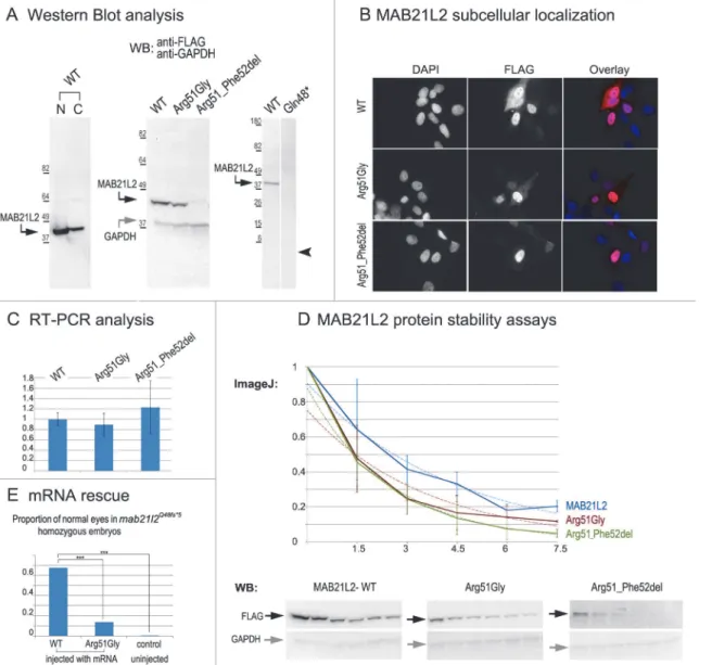
cells [32];MAB21L2was also found to be endogenously expressed in these cells (S1 Fig.). Mab21l proteins were previously shown to localize primarily to the nucleus [33]. Western Blot analysis of nuclear and cytoplasmic fractions confirmed the presence of ~41kDa wild-type MAB21L2 protein in both cellular compartments with greater nuclear localization (Fig. 8A). Western blot analysis of whole-cell extracts detected a decreased amount of the p.(Arg51Gly) mutant in comparison to wild-type protein (Fig. 8A); the mutant protein was found to be pres-ent at 31.97% ± 9.36% of wild-type based on three independpres-ent experimpres-ents. Cellular immuno-fluorescence analysis demonstrated no significant alteration in the localization pattern between wild-type and mutant protein with predominant nuclear localization and some cytoplasmic staining for both forms (Fig. 8B). Since the levels of recombinantMAB21L2transcript encod-ing for wild-type and p.(Arg51Gly) mutant proteins detected by RT-PCR did not show a signif-icant difference (Fig. 8C,S1 Fig.), we proceeded to perform protein stability assays using a series of cycloheximide treatments of HLE-B3 cells transfected with MAB21L2 constructs (Fig. 8D). Protein stability assays showed a more rapid decrease in the amount of p.(Arg51Gly) mutant in comparison to wild-type protein at all examined time points, with 11% of the mutant protein being present after 7.5 hours of cycloheximide treatment in comparison to 20% of the wild-type MAB21L2 (Fig. 8D). Calculation of the proteins’half-life identified a statistically sig-nificant difference (p<0.05) in stability of wild-type (2.64±0.25 hours) versus the p.(Arg51Gly)
mutant (1.91±0.36). These data suggest that the substitution of the arginine at position 51 to glycine affects MAB21L2 stability.
Functional analysis of the p.(Gln48) (to represent the zebrafish p.(Gln48Serfs5) mutant)
and p.(Arg51_Phe52del) proteins was also undertaken in HLE-B3 cells. The p.(Gln48)
mu-tant was found to be highly unstable as no detectable level of this protein (estimated to have a
Fig 7. Analysis ofpax6b,pax2.1andfoxe3expression in wild-type,mab21l2Q48Sfs*5andmab21l2R51_F52delembryos.Wild-type (A-E) and mutant
(F-O) zebrafish embryos at 24–48-hpf were analyzed as indicated in the right bottom corner of each image. Please note a change inpax6btranscript distribution
at 24-hpf and 48-hpf inmab21l2Q48Sfs*5(arrowheads in F-H), retinal folding defect inmab21l2R51_F52delembryos at 24-hpf (arrowheads in K, L) and visibly abnormalpax6bpattern at 48-hpf in some (arrowheads in M) but not all (N)mab21l2R51_F52delembryos.pax2.1expression seems to be unaffected in 24-hpf frameshift mutant embryos (I) but shows an abnormal pattern in both mutants at 48-hpf (J,O). At 48-hpf, in addition to more broad and intensepax2.1
expression in the region of optic fissure, abnormalpax2.1staining was detected in central retina inmab21l2Q48Sfs*5embryos (arrowheads in J). le, lens; of,
optic fissure; retina.
molecular weight of ~5.5 kDa) was identified via Western blot analysis (Fig. 8A). The p.(Arg51_Phe52del) mutant also demonstrated a reduction in the amount of detected protein to 2.57% ± 2.20% of wild-type MAB21L2 protein based on three independent experiments
Fig 8. MAB21L2 wild-type and mutant protein studies. A.Western Blot analyses of MAB21L2 wild-type and mutant proteins (with N-terminal FLAG tag) in HLE-B3 cells. Please note the presence of wild-type MAB21L2 in both nuclear (N) and cytoplasmic (C) fractions (left), decreased expression of Arg51Gly and Arg51_Phe52del mutants (center) and the absence of Gln48*mutant protein (right; arrowhead indicates region corresponding to 5.5kDa, the predicted molecular weight for this peptide); predicted molecular weight for MAB21L2 is ~41kDa and for GAPDH is ~36 kDa; positions and corresponding molecular weights of protein ladder are shown for every blot.B.Cellular immunofluorescence analysis demonstrated no significant alteration in the localization pattern between wild-type and mutant (Arg51Gly and Arg51_52del) proteins with predominant nuclear localization and some cytoplasmic staining for all forms. C.RT-PCR analysis of MAB21L2 wild-type and mutant transcripts in human lens epithelial cells transfected with corresponding expression constructs. D.Protein stability assays with cycloheximide showed a more rapid decrease in the amount of Arg51Gly and Arg51_Phe52del mutant proteins in comparison to wild-type protein. Western blots signals were measured using ImageJ software, and obtained values were graphed to produce degradation curves represented by solid blue (wild-type), red (Arg51Gly) and green (Arg51_Phe52del) lines (standard deviations for every time point are indicated as thin vertical lines and exponential decay curves fitted into each graph are shown as dotted lines of corresponding colors); representative Western blot (WB) images are shown on the bottom (stability assays were performed in triplicate; 0, 1.5, 3, 4.5, 6, and 7.5 correspond to hours of exposure to cycloheximide).E.Summary of mRNA rescue experiments. Proportions of embryos with normal eyes in homozygousmab21l2Q48fs*5embryos injected with wild-type humanMAB21L2
mRNA, mRNA encoding the p.(Arg51Gly) mutant protein, and uninjected control larvae are shown; statistically significant (p<0.0005) differences are indicated with asterisks (***).
(Fig. 8A); cellular immunofluorescence analysis demonstrated predominantly nuclear staining similar to wild-type distribution (Fig. 8B). Protein stability assays confirmed increased instabil-ity of the p.(Arg51_Phe52del) protein with less than 5% of mutant protein being detected after 7.5 hours of cycloheximide exposure versus 20% for the wild-type (Fig. 8D). The half-life of the p.(Arg51_Phe52del) mutant was estimated to be 1.91±0.27 hours versus 2.64±.25 for wild-type, which represented a statistically significant difference (p<0.05). The half-life values of the
p.(Arg51_Phe52del) and p.(Arg51Gly) proteins were not significantly different from each other despite the observed reduced level of p.(Arg51_Phe52del) in comparison to p.(Arg51Gly) in replicate Western blots of untreated cells (see above,Fig. 8A); this suggests that there may be additional factors affecting the stability of the p.(Arg51_Phe52del) mutant which may require alternative assays to uncover [34].
To further test the pathogenicity of the human p.(Arg51Gly) allele, we injected embryos generated by heterozygousmab21l2Q48Sfs5crosses with either wild typeMAB21L2mRNA or mRNA encoding for the p.(Arg51Gly) mutant; a small number of embryos were left uninjected as a control. The embryos were carefully examined for ocular anomalies at 72-hpf, divided into affected and normal groups, and then genotyped. In total, out of 104 surviving embryos in-jected with wild typeMAB21L2, 68% (21/31) of homozygous fish developed normal eyes and lenses while out of 108 surviving embryos injected with the mutant mRNA encoding for p. (Arg51Gly), only 14% (3/22) of homozygous fish developed normal eyes and lenses; in the con-trol group, none of the 9 uninjected homozygous embryos (out of 36 surviving) had normal eyes and lenses. The difference in the proportion of normal phenotypes seen in the homozy-gousmab21l2Q48Sfs5fish injected with wild-type mRNA versus embryos injected with either mutant mRNA or left uninjected was statistically significant (p<.0005), while the difference
be-tween the mutant RNA- injected and the uninjected embryos was not statistically significant (p>.20) by chi-squared analysis (Fig. 8E). The efficient rescue by wild typeMAB21L2mRNA
confirms that the ocular phenotype in the mutant line is caused by themab21l2deficiency and demonstrates functional conservation between zebrafish and human mab21l2/MAB21L2 pro-teins. The absence of robust rescue by mRNA encoding for the p.(Arg51Gly) mutant supports the pathogenic role of thisMAB21L2allele in human disease.
To investigate the possibility of dominant-negative effects for the mutant allele,MAB21L2 wild type mRNA or mRNA encoding for the p.(Arg51Gly) mutant were injected (at the same concentrations as in rescue experiments) into wild-type embryos and observed for phenotypes up to 120-hpf. Among the 39 surviving embryos that were injected with wild-typeMAB21L2, no embryos displayed an eye phenotype (9 fish showed moderate-severe overall malforma-tions). Similarly, among the 61 surviving embryos that were injected with the mutant mRNA encoding for the p.(Arg51Gly) protein, no embryos showed an eye phenotype (10 fish showed moderate-severe overall malformations). Thus, these experiments identified no dominant-neg-ative effect for the p.(Arg51Gly) mutant allele in zebrafish at concentrations that are sufficient for phenotypic rescue of themab21l2-deficient phenotype.
Discussion
involving double mutants ofmab-21andsmamembers (SMADfamily) inC.elegansindicated thatmab-21is positioned downstream ofsmaand is negatively regulated bydbl-1, a homolog of humanBMP5, thus placingmab-21in the TGF-βsignaling cascade [35,36]. The TGF-β
signaling cascade plays an important role in ocular development and mutations in many asso-ciated factors, such asBMP4,BMP7,GDF3, andGDF6, lead to an overlapping spectrum of phenotypes in humans [13,14,37–39]. Additionally, mab-18 (PAX6 ortholog) mutants demon-strate a phenotype that is highly similar to mab-21C.elegansmutants [27] andMab21l2 ex-pression was found to be upregulated in embryonic mouse lenses heterozygous for aPax6 loss-of-function allele [40] suggesting a possible conserved genetic interaction.
In mouse,Mab21l2ocular expression was reported in the dorsal optic vesicle and head surface ectoderm in E8.5–9.5 embryos and the neural retina, optic nerve and RPE at E12 [41,42]; homozygousMab21l2knockout mice demonstrated a rudimentary retina and aphakia due to improper invagination of the optic vesicle as well as ventral body wall defects, improper forma-tion of the heart and liver, and embryonic lethality [41,42]. Previously published reports of lens expression in animal models are somewhat inconsistent: four studies reported lack of lens ex-pression forMab21l2/mab21l2(with many showing the presence of lens expression for its close homolog,Mab21l1/ mab21l1) [41,43–45] while three other manuscripts notedMab21l2/mab21l2 transcripts in the developing lens [33,46,47], which is consistent with the results of our study. In zebrafish, morpholino-mediated knockdown ofmab21l2resulted in microphthalmia and incom-plete retinal development including discontinuous inner and outer plexiform layers [47]. In the zebrafish genetic mutants that we developed, severe defects in the development of both the lens and retina were observed consistent with strong expression ofmab21l2in both tissues. While the phenotypes ofmab21l2morphants and mutants show overlap, some ocular features, including severe lens degeneration, coloboma, and early retinal invagination defects, appear to be more pronounced or observed primarily in genetic mutants; this may be due to incomplete disruption of mab21l2 function via morpholino, the transient nature of morpholino-induced effects, and/or genetic background differences.
Both of themab21l2mutants generated in this study demonstrated variable retinal invagi-nation defects. In zebrafish, retinal invagiinvagi-nation, which occurs upon contact of the optic vesicle (formed as an extension of the forebrain neuroepithelium) with the surface ectoderm, is com-pleted with formation of the cup-shaped optic vesicle by ~22-hpf. Inmab21l2Q48Sfs5embryos, a shallow misshapen optic cup, especially at its ventral part, was detected in 24-hpf embryos; the lens mass (similar to the mammalian lens vesicle) was also visibly smaller than normal at this stage. In themab21l2R51_F52delin-frame deletion mutant, a more constricted optic cup with some ventral and dorsal retinal folding was observed, while the developing lens appeared to have normal size and shape in most embryos. The observed defects in early optic cup forma-tion indicate a possible conserved role formab21l2in this process in vertebrates.
seem more likely to be caused by the severe disorganization of mutant ocular tissues rather than direct regulation of expression of those markers by mab21l2.
The distinct phenotypes associated with themab21l2Q48Sfs5frameshift truncation and
mab21l2R51_F52delin-frame deletion mutations suggest different molecular mechanisms. The mab21l2R51_F52delin-frame deletion demonstrated milder features in comparison to the mab21l2Q48Sfs5mutant, such as less affected lens development, less pronounced cell death and incomplete embryonic lethality. These observations support the possibility that this mutation results in a hypomorphic allele retaining some of its normal function, particularly in relation to lens development. The severe coloboma observed inmab21l2R51_F52delembryos may point to the particular importance of domains located in the N-terminal region of mab21l2 for its proper functioning in the developing retina. Since the zebrafish p.(Arg51_Phe52del) in-frame deletion and the human p.(Arg51Gly) missense alleles affect homologous regions of the MAB21L2/mab21l2 proteins, the mechanism(s) of these mutations may be similar to each other. Understanding of these mechanisms requires additional studies into the domain struc-ture and function of MAB21L2/mab21l2 protein.
Analysis of wild-type and mutant proteins in human lens epithelial cells identified a range of protein stability defects among mutant forms with the human p.(Arg51Gly) mutant stability mildly reduced, the zebrafish p.(Arg51_Phe52del) mutant stability highly affected, and the p.(Gln48) truncation protein (similar to the p.(Gln48Serfs5) mutant) being entirely unstable.
This analysis suggested that the zebrafish p.(Gln48Serfs5) mutation results in a complete
loss-of-function allele, since the similar mutant protein p.(Gln48) was completely unstable and
any protein produced would be missing the entire mab-21 domain. These experiments also indicated the likely importance of arginine 51 and the immediately adjacent amino acids for normal conformation and stability of the MAB21L2/mab21l2 protein because disruptions of this region resulted in decreased stability for both the human p.(Arg51Gly) and the zebrafish p.(Arg51_Phe52del) proteins. Further studies demonstrated that, in contrast to wild-type humanMAB21L2mRNA, mutant mRNA encoding for the p.(Arg51Gly) protein failed to efficiently rescue the ocular phenotype of homozygousmab21l2Q48Sfs5embryos, suggesting functional deficiency for the identified human mutation. In addition, overexpression of mutantMAB21L2mRNA in zebrafish embryos resulted in no obvious ocular phenotype, im-plying absence of a dominant-negative effect. However, it is possible that the full impact of the p.(Arg51Gly) mutation was not discernable due to insufficient dosage, its acting later in devel-opment, or other factors; this needs to be further investigated by additional approaches, includ-ing development of zebrafish mutants carryinclud-ing this specific allele.
No abnormal phenotype was seen in heterozygous zebrafish for either the frameshift trunca-tion or in-frame deletrunca-tion mutatrunca-tions, while a dominant phenotype was observed in human pa-tients with the p.(Arg51Gly) alteration. This may be explained by variability in gene dosage requirements between species [61] or a different mechanism for the human mutation. The pres-ence of mild rhizomelia/ contractures in human patients withMAB21L2mutations suggests its involvement in skeletal development. Additional phenotypes such as shortened body/curved tail, craniofacial malformations and lethality were present in the zebrafishmab21l2mutants while heart, liver, embryonic lethality and other anomalies were previously reported in mouseMab21l2 mutants [41,42]. Considering these observations and the fact that multiple pleiotropic effects of mab-21 mutations have been previously noted [27], additionalMAB21L2-associated phenotypes are likely to be identified and may present with broad interfamilial variability.
resulting in similar phenotypes (anophthalmia or colobomatous microphthalmia with rhizomelic skeletal dysplasia in two families); one of the reported families includes the UK10K_COL5001067 and UK10K_COL5001068 cases with the p.(Arg51His) mutation that we presented in results from the publically available data. In addition, one family with a reces-siveMAB21L2mutation affecting the C-terminal region of the protein was identified. Func-tional assays of the mutant alleles using an inducible HEK293 cell system expressing wild-type or mutant MAB21L2 as GFP fusion proteins suggested a possible gain-of-function effect for the dominant mutations based on observed increased protein stability; an increase in phospho-ERK1 was detected with induction of either wild-type or p.(Arg51His) mutant MAB21L2 pro-tein [62]. This differs from the proposed mechanism of the mutation identified in our study which revealed a decrease in protein stability for the p.(Arg51Gly) mutant tagged with FLAG epitope and absence of a dominant phenotype in zebrafish embryos injected with mRNA en-coding for this protein. This may be due to diverse effects of the studied amino acid substitu-tions (Arg51Gly versus Arg51His, Arg51Cys, and Glu49Lys) on MAB21L2 protein structure, different effects of tags on protein stability (GFP versus FLAG), or other factors, and needs to be investigated further. Rainger and co-authors (2014) also suggested an involvement of MAB21L2 in ssRNA binding [62]. In our manuscript, we developed an allelic series of zebrafish mab21l2mutants, uncovered an early onset optic cup apoptosis phenotype in affected embry-os, and demonstrated a decrease in protein stability, as well as functional deficiency and no dominant-negative effect for the p.(Arg51Gly) mutation in zebrafish embryos.
Taken as a whole, the complete co-segregation of the identifiedMAB21L2allele with the ocular phenotype in the family, thede novoappearance of the mutation in the affected proge-ny of unaffected parents with low-level mosaicism present in the mother, the in silico and experimental indications of the functional importance of the arginine at position 51, the de-velopment of ocular defects inmab21l2mutant zebrafish embryos, as well as rescue of the oc-ular phenotype of homozygousmab21l2Q48Sfs5embryos with injection of wild-type but not the p.(Arg51Gly)-encoding mutant mRNA, provide strong support for the pathogenicity of the identifiedMAB21L2allele and the involvement ofMAB21L2in human ocular develop-ment and disease. The developed zebrafishmab21l2mutants will help to further explore the developmental roles and molecular function of this conserved ocular factor.
Materials and Methods
Ethics statement
The human study was approved by the Children’s Hospital of Wisconsin Institutional Re-view Board (protocol number CHW 03/56) with written informed consent obtained from each participant and/or their legal representative, as appropriate. This study also utilized data generated by the UK10K Consortium, derived from samples from UK10K_Rare_Colo-boma (EGA Study ID: EGAS00001000127); a full list of the investigators who contributed to the generation of the data is available fromhttp://www.UK10K.org. Access to data generated by the UK10K_Rare_Coloboma (EGA Study ID: EGAS00001000127) project of the UK10K Consortium study (http://www.UK10K.org) was obtained through a Data Access Agreement. The animal protocol was approved by the Institutional Animal Care and Use Committee at the Medical College of Wisconsin (protocol number AUA00000351_AR_5).
Human sequencing studies
Axeq Technologies (Rockville, MD). The obtained data were aligned using the Burrows-Wheeler Aligner (BWA) and variants were called using the SAMTOOLS analysis pipeline available through Axeq. Exome data were analyzed using the SNP & Variation Suite (Golden Helix, Bozeman, MT) to identify variants that are shared between the affected individuals and then prioritized based on their absence/rarity in the general population (as reported in publicly available databases dbSNP (http://www.ncbi.nlm.nih.gov/snp), NHLBI Exome Sequencing Project Exome Variant Server (EVS;http://evs.gs.washington.edu/EVS/), and 1000 Genomes (http://www.1000genomes.org/data), as well as our own data) and possible effect on protein function; for missense variants, functional profiling was performed using SIFT, Polyphen2, Mutation Taster, MutationAssessor, and FATHMM as well as nucleotide conservation scores GERP++ and PhyloP as accessed through dbNSFP [63].
To confirm the identifiedMAB21L2changes and perform cosegregation analysis, DNA from all available family members was amplified using the primers and conditions provided in S2 Table. PCR products were sequenced bidirectionally using Big Dye Terminator chemistry and the ABI 3730XL sequencer (Applied Biosystems/Life Technologies, Carlsbad, CA, USA). Sequences were reviewed manually and using Mutation Surveyor (SoftGenetics, State College, PA) and compared to the NM_006439.4 transcript.
Variant Call Format (VCF) files from the UK10K_Rare_Coloboma (EGA Study ID: EGAS00001000127) project of the UK10K Consortium study (http://www.UK10K.org) were analyzed using the SNP & Variation Suite (Golden Helix, Bozeman, MT) with filtering for the MAB21L2gene only. DNA samples from this cohort were not available for independent confir-mation of the identified variants.
Additional human samples were screened using Sanger sequencing of the 1362-bp PCR product encompassing the fullMAB21L2coding region using PCR primers and two extra in-ternal primers (S2 Table). Sequences were analyzed by manual inspection and Mutation Sur-veyor software as described above.
MAB21L2 protein studies
Protein sequences were aligned using the Kalign multiple sequence alignment tool (http:// www.ebi.ac.uk/Tools/msa/kalign). The accession numbers for the MAB21L sequences used in the protein alignment are as follows: human MAB21L2 (AF155219.1), murine Mab21l2 (AF223425.1), zebrafish mab21l2 (AY038031.1), human MAB21L1 (NM_005584), murine Mab21l1 (AF228913.1), zebrafish mab21l1 (NM_152974.2), human MAB21L3 (NM_152367.2), murine Mab21l3 (NM_172295.4), zebrafish mab21l3 (NM_001110025.1),Drosophila melanoga-stermab-21 (NP_651971.2),Caenorhabditis elegansmab-21 (NP_497940.2).
The expression construct for MAB21L2 wild-type protein with N-terminal FLAG (Cat. #EX-V1703-M11) was purchased from GeneCopoiea (Rockville, MD). The mutations were in-troduced using the Agilent QuikChange Lightning (Cat. #210519) site directed mutagenesis kit (Santa Clara, CA). Mutagenesis primers were designed using the online QuikChange Primer Design tool. HPLC purified mutagenesis primers were ordered from Thermo Fisher Scientific (Waltham, MA) and sequences were as follows: MAB21L2-p.R51G-Sense—5’-gaggtgcag gagcctggcttcatcagct-3’, MAB21L2-p.R51G-Antisense—5’-agctgatgaagccaggctcctgcacctc-3’, MAB21L2-p.51_52del-Sense—5’-gctcaaggagctgataggctcctgcacctc-3’, MAB21L2-p.51_52del-Antisense—5’- gaggtgcaggagcctatcagctccttgagc-3’, MAB21L2- Q48X-Sense—5’- gaagcgagg ctcctacacctccacttcct-3’, MAB21L2- Q48X-Antisense—5’- aggaagtggaggtgtaggagcctcgcttc-3’.
performed by transfecting HLE-B3 cells with 15μg of DNA and 30μl of Lipofectamine 2000 (Life Technologies) in Opti-MEM in 100 mm petri dishes.
For Western blot analysis, nuclear and cytoplasmic fractions were generated using the Cel-Lytic NuCLEAR Extraction kit (Cat#NXTRACT Sigma-Aldrich,). 48 hours after transfection, cells were washed with PBS, collected, and resuspended in 0.5 ml of hypotonic lysis buffer and incubated on ice for 15 minutes, 30μl of 10% IGEPAL was added to the cell suspension. Cells were then vortexed and spun at 10,000 xg for 30 seconds at 4°C. The supernatant (cytoplasmic fraction) was decanted and saved. The nuclear fraction was generated by adding 70μl of nucle-ar extraction buffer to the pellet, incubating on ice for 25 minutes, spun at 20,000 xg for 5 min-utes at 4°C and the supernatant was saved (nuclear fraction). Molecular weights for wild-type and mutant proteins were estimated using Protein Molecular Weight calculator (http:// bioinformatics.org/sms/prot_mw.html). The BenchMark Pre-stained Protein Ladder (Cat. #10748-010) (Life Technologies) was utilized as the protein molecular weight standard.
The stability of the MAB21L2 wild type and mutant proteins were characterized using a modified protocol previously described by Alur et al. 2010 [64]. In these experiments, cyclo-heximide, which is known to block translational elongation and thus to inhibit protein biosyn-thesis in eukaryotic organisms, was used to treat cells transfected with MAB21L2 expression constructs in a time-course analysis to allow for protein degradation rates to be revealed in the absence of new protein production. Briefly, cells were split 24 hours after transfection into 35 mm petri dishes. After an additional 24 hours, cells were treated with 100μg/ml cyclohexi-mide (Cat. #C4859; Sigma-Aldrich) and collected at the indicated time points (0-, 1.5-, 3-, 4.5-, 6, and 7.5-hours after exposure). Whole cell lysates were generated by washing with PBS, cells were scraped in PBS and collected in 1.5 ml tubes, cells were spun at 1,000 xg for 1.5 minutes and PBS was removed. Cells were then resuspended in 40μl of 1x RIPA buffer (50 mM Tris-HCL pH 8.0, 150 mM NaCL, 0.1% SDS, 0.5% sodium deoxycholate, 1% Triton X-100) and in-cubated on ice for 15 minutes, vortexing every 5 minutes. Cells were spun down for 10 minutes at 20,000 xg at 4°C and the supernatant was used for Western Blot analysis (see below). West-ern blots signals for MAB21L2 were quantified using ImageJ software and normalized to GAPDH levels (also measured by ImageJ) and the obtained values were graphed to produce degradation curves. Stability assays were performed in triplicate. Exponential decay curves were fitted to each replicate stability assay and the half-lives were determined. A two-tailed stu-dent's t-test was used to determine if the groups were different.
Lysates were denatured by adding equal volume of 2x sample buffer (65.8 mM Tris-HCl, pH 6.8, 26.3% (w/v) glycerol, 2.1% SDS, and 0.01% bromophenol blue) (Cat. #161-0737) (Bio-Rad, Hercules, CA) followed by boiling for 4 minutes at 95°C and run on a 10% Criterion Tris-HCl Gel (Cat. #345-1018) (Bio-Rad). Gels were immunobloted with anti-FLAG (Cat. #F1804) (Sigma-Aldrich) antibody at a 1:1000 dilution or anti-GAPDH (Cat#ab8245) (Abcam, Cam-bridge, MA) antibody at 1:5000 dilution. The secondary antibody was goat anti-mouse IgG HRP conjugate (Cat. #12-349) (Upstate Cell Signaling Solutions) at a 1:2500 dilution. The blots were developed using the Chemiluminescent Nucleic Acid Detection Module Kit (Thermo Fisher Scientific).
for 5 minutes at room temp with 1:1000 DAPI in PBS, washed twice with PBS and mounted on glass slides.
Expression studies, histology, apoptosis assays and
immunohistochemistry in zebrafish
Zebrafish (Danio rerio) maintenance and developmental staging were performed as previously described [65]. The protocol was approved by the Institutional Animal Care and Use Commit-tee at the Medical College of Wisconsin. The expression ofmab21l2,pax6b,pax2andfoxe3was studied in 18–72-hpf zebrafish embryos using transcript-specific antisense riboprobes and pre-viously described protocols [65]. For themab21l2probe, a 698-bp fragment covering the se-quence between nucleotides 788 and 1485 (GenBank# AY038031.1; primers provided inS2 Table) was utilized; clones forpax6b(AL915181) andpax2.1(AL906738) were purchased from Open Biosystems (Huntsville, Alabama) and linearized with EcoRv and BamHI, respectively; for thefoxe3probe, a full length 1369-bp fragment covering the sequence between nucleotides 16 and 1384 (GenBank #BC163348.1) was utilized.
Gross morphological and histological analysis of zebrafish embryos was performed as previ-ously described [65]. For apoptosis analysis, whole mount TUNEL assay as described by Eimon was followed [66]. For immunohistochemistry, immunohistochemical staining of zeb-rafish frozen sections was performed with DAPI (Cat. #62247) (Thermo Scientific), PCNA at 1:100 (Cat. #P8825) (Sigma-Aldrich), ZL-1 at 1:100 (ZIRC), and wheat germ agglutinin at 5μg/ml (Cat. #W32464) (Life Technologies) following the previously published protocols [67–70].
Generation of
mab21l2
mutant lines
TALENs were constructed following the Sanjana et al (2012) protocol [23]. Briefly,mab21l2 TALENs were designed to target the region surrounding the nucleotide corresponding to the mutation site in human patients (left TALEN—5’-GAAGGAGGTGGAGGTCCAA-3’and right TALEN—5’-CTATCTCGCTCAGGGAGCT-3’). Interruption of themab21l2target se-quence was predicted to affect the BanII restriction site (GRGCYC) located between the left and right TALENs; thus BanII digestion of the PCR-amplified genomic fragment involving this region was utilized to determine TALEN cutting efficiencies as well as to identify mutant embryos/adult fish and was followed by DNA sequencing for confirmation. Zebrafish embryos at the 1–4 cell stage were injected with TALEN RNA; initial analysis of injected larvae con-firmed genome editing events and ~200 injected embryos were raised to adulthood to generate mosaic founders; these fish were then bred to produce embryos carrying germlinemab21l2 mutations that were raised to adulthood, genotyped using the primers provided inS2 Table, and bred to generate homozygous/compound heterozygous embryos.
RNA isolation, RT-PCR and mRNA injections
were used as a loading control: GAPDH-F 5’- CCAAGGTCATCCATGACAACT-3’and GAPDH-R 5’- GAGGCAGGGATGATGTTCTG-3’(PCR product equal 148 bp). Endogeneous expression ofMAB21L2transcript in the human lens epithelial cell line was detected with the following primers MAB21L2-endogenous-F, 5'-CCAGGTGGAAAACGAGAGTG-3' and MAB21L2-rt-R, 5’-GGTAGAGCACCACCTCAAATTC-3’(PCR product equal 384 bp).
To detectmab21l2transcript levels in zebrafish RNA, three independent samples, each con-taining two embryos of either homozygousmab21l2Q48Sfs5andmab21l2R51_F52delmutants or wild type, were isolated using the RNAqueous-Micro Total RNA Isolation Kit (Cat. #AM1931) (Life Technologies). Each sample was reverse transcribed as described previously. Samples were amplified formab21l2using the following primers:mab21l2-zf-T-F 5’-TCTTTTCCT GGGAGTTGTGC-3’andmab21l2-zf-T-R 5’- CCCCATCTGGTTCAGGTAAA-3’(PCR prod-uct equal 350 bp). BecauseMAB21L2/mab21l2represents a single exon gene, a minus-reverse transcriptase control was included in each RT-PCR experiment to assure absence of contami-nating DNA in each RNA sample. Amplification of the zebrafish gene for translation elonga-tion factor 1 alpha (loading control) was performed with the following primers:ef1ax1-2F 5’ -TCTCTCAATCTTGAAACTTATCAATCA-3’andef1ax3R 5’- AACACCCAGGCGTACTT GAA-3’(PCR product equal 205 bp). The 1 Kb Plus DNA Ladder was utilized as nucleic acid standard (Cat. #10787-018) (Life Technologies). The PCR products were analyzed by electro-phoresis and bands were quantitated using Image J software.
For rescue and overexpression experiments, mRNA was generated using the T7 mMES-SAGE mMACHINE Transcription kit (Cat. #AM1344) (Life Technologies). 300 pg of mRNA was injected into 1–4 cell stage embryos produced by wild-type or c.141_153delCCAAGAG CCCCGT heterozygous mutant crosses. The injected embryos were observed for up to 5 days of fertilization, separated into groups based on observed presence/absence of ocular phenotype and then genotyped.
Supporting Information
S1 Table. Summary of novel or rare heterozygous variants predicted to be functionally sig-nificant.
(DOC)
S2 Table. PCR conditions and oligonucleotides utilized for amplification of gene regions in this study.
(DOCX)
S1 Fig. (A) DNA chromatograms that illustrate sequences of c.141_153delCCAA-GAGCCCCGT, p.(Gln48Serfs5) and c.151_156delCGTTTC, p.(Arg51_Phe52del) alleles
of 205 and 148 bp, respectively. (TIF)
S2 Fig.mab21l2compound heterozygous embryos generated by the cross of
c.150_156delCCGTTTC, p.(Arg51Serfs4) and c.151dupC, p.(Arg51Profs14)
heterozy-gous carriers.A wild-type fish from the same progeny is shown at the top and followed by 7 compound heterozygous embryos. Please note small eye (black arrowheads) in all mutant embryos and shortened tail (black arrows) in 4 out of 7 fish.
(TIF)
S3 Fig. TUNEL results in 48–72-hpf wild-type (A-D),mab21l2
Q48Sfs5embryos (E-H) and
mab21l2R51_F52delmutants (I-L) are shown.An increase in TUNEL staining was observed in bothmab21l2mutants with remarkably high levels in themab21l2Q48Sfs5embryos (E-H) and moderately increased levels in themab21l2R51_F52delembryos (I-L); arrowheads indicate sites of increased TUNEL staining in the eye and brain; le, lens; r, retina; m, midbrain.
(TIF)
S4 Fig. Immunostaining with PCNA (Proliferating Cell Nuclear Antigen) and DAPI in 24–72-hpf wild-type (A-F),mab21l2Q48Sfs5(A1-F1), andmab21l2R51_F52del(A2-F2) embry-os.Overlay images of the PCNA and DAPI immunostaining are shown inFig. 6. The arrow-heads in A1 and A2 indicate abnormal retinal folding;le, lens; m, midbrain; r, retina.
(TIF)
S5 Fig. Immunostaining with ZL-1 (red) and in situ hybridization withpax6b,pax2.1and
foxe3antisense riboprobes in wild-type (A-F),mab21l2Q48Sfs5(A1-F1) andmab21l2R51_F52del (C2-F2) embryos.The ZL-1 staining is absent in 24-hpf (A1) but present in 48–72-hpf mutant embryos (B1, C1, C2),pax2.1pattern is abnormal in 72-hpf mutant embryos (D1, E1, D2, E2), arrowhead in D1 shows abnormal areas ofpax2.1-positive cells in the central retina and arrow-heads in E1 show broad and intense expression in the region of optic fissure;foxe3expression is decreased in 48-hpf mutants (F1, F2); le, lens; of, optic fissure.
(TIF)
Acknowledgments
The authors gratefully acknowledge the patients and their families for their participation in research studies.
Author Contributions
Conceived and designed the experiments: BD EVS. Performed the experiments: BD SM. Ana-lyzed the data: BD SM EVS. Contributed reagents/materials/analysis tools: AK RHRB LMR. Wrote the paper: BD LMR EVS. Oversaw human subject recruitment and ethical approvals: LMR. Performed clinical evaluation of patients and participated in enrollment: AK RHRB.
References
1. Morrison D., FitzPatrick D., Hanson I., Williamson K., van Heyningen V., et al. (2002). National study of microphthalmia, anophthalmia, and coloboma (MAC) in Scotland: Investigation of genetic aetiology. J. Med. Genet. 39, 16–22. PMID:11826019
3. Skalicky S.E., White A.J., Grigg J.R., Martin F., Smith J., et al. (2013) Microphthalmia, anophthalmia, and coloboma and associated ocular and systemic features: understanding the spectrum. JAMA Ophthalmol., 131, 12, 1517–1524. doi:10.1001/jamaophthalmol.2013.5305PMID:24177921 4. Nakamura K.M., Diehl N.N., Mohney B.G. (2011) Incidence, ocular findings, and systemic associations
of ocular coloboma: a population-based study. Arch Ophthalmol. 129(1), 69–74. doi:10.1001/ archophthalmol.2010.320PMID:21220631
5. Toker E., Elcioglu N., Ozcan E., Yenice O., Ogut M. (2003). Colobomatous macrophthalmia with micro-cornea syndrome: report of a new pedigree. Am J Med Genet A. 121A, 25–30. PMID:12900897 6. Schneider A., Bardakjian T., Reis L.M., Tyler R.C., Semina E.V. (2009). Novel SOX2 mutations and
genotype-phenotype correlation in anophthalmia and microphthalmia. Am. J. Med. Genet. A 149A, 2706–2715. doi:10.1002/ajmg.a.33098PMID:19921648
7. Schilter K.F., Schneider A., Bardakjian T., Soucy J.F., Tyler R.C., et al. (2011). OTX2 microphthalmia syndrome: four novel mutations and delineation of a phenotype. Clin. Genet. 79,158–168. doi:10. 1111/j.1399-0004.2010.01450.xPMID:20486942
8. Sanyanusin P., Schimmenti L.A., McNoe L.A., Ward T.A., Pierpont M.E., et al. (1995). Mutation of the PAX2 gene in a family with optic nerve colobomas, renal anomalies and vesicoureteral reflux. Nat. Genet. 9, 358–64. PMID:7795640
9. Azuma N., Yamaguchi Y., Handa H., Tadokoro K., Asaka A., et al. (2003). Mutations of the PAX6 gene detected in patients with a variety of optic-nerve malformations. Am. J. Hum. Genet. 72, 1565–1570. PMID:12721955
10. Vissers L.E., van Ravenswaaij C.M., Admiraal R., Hurst J.A., de Vries B.B., et al. (2004). Mutations in a new member of the chromodomain gene family cause CHARGE syndrome. Nature Genet., 36, 955– 957. PMID:15300250
11. Wang L., He F., Bu J., Zhen Y., Liu X., et al. (2012) ABCB6 mutations cause ocular coloboma. Am.J. Hum.Genet., 90, 1, 40–48. doi:10.1016/j.ajhg.2011.11.026PMID:22226084
12. Schimmenti L.A., de la Cruz J., Lewis R.A., Karkera J.D., Manligas G.S., et al. (2003). Novel mutation in sonic hedgehog in non-syndromic colobomatous microphthalmia. Am. J. Med. Genet. A 116A, 215– 221. PMID:12503095
13. Asai-Coakwell M., French C.R., Berry K.M., Ye M., Koss R., et al. (2007). GDF6, a novel locus for a spectrum of ocular developmental anomalies. Am. J. Hum. Genet. 80, 306–15. PMID:17236135 14. Ye M., Berry-Wynne K.M., Asai-Coakwell M., Sundaresan P., Footz T., et al. (2010). Mutation of the
bone morphogenetic protein GDF3 causes ocular and skeletal anomalies. Hum. Mol. Genet. 19,287– 98. doi:10.1093/hmg/ddp496PMID:19864492
15. Casey J., Kawaguchi R., Morrissey M., Sun H., McGettigan P., et al. (2011) First implication of STRA6 mutations in isolated anophthalmia, microphthalmia, and coloboma: a new dimension to the STRA6 phenotype. Hum. Mutat. 32, 1417–1426. doi:10.1002/humu.21590PMID:21901792
16. Fares-Taie L., Gerber S., Chassaing N., Clayton-Smith J., Hanein S., et al. (2013). ALDH1A3 mutations cause recessive anophthalmia and microphthalmia. Am. J. Hum. Genet. 92, 265–270. doi:10.1016/j. ajhg.2012.12.003PMID:23312594
17. Kelberman D., Islam L., Lakowski J., Bacchelli C., Chanudet E., et al. (2014) Mutation of SALL2 causes recessive ocular coloboma in humans and mice. Hum.Mol.Genet., 23, 10, 2511–2526, doi:10.1093/ hmg/ddt643PMID:24412933
18. Williamson K.A., Rainger J., Floyd J.A., Ansari M., Meynert A., et al (2014). Heterozygous loss-of-func-tion mutaloss-of-func-tions in YAP1 cause both isolated and syndromic optic fissure closure defects. Am. J. Hum. Genet. 94, 295–302. doi:10.1016/j.ajhg.2014.01.001PMID:24462371
19. Williamson K.A., FitzPatrick D.R. (2014). The genetic architecture of microphthalmia, anophthalmia and coloboma. Eur. J. Med. Genet. 57, 369–80. doi:10.1016/j.ejmg.2014.05.002PMID:24859618 20. Kondo Y., Koshimizu E., Megarbane A., Hamanoue H., Okada I., et al. (2013). Whole-exome
sequenc-ing identified a homozygous FNBP4 mutation in a family with a condition similar to microphthalmia with limb anomalies. Am. J. Med. Genet. A 161A, 1543–1546. doi:10.1002/ajmg.a.35983PMID:23703728 21. Zahrani F., Aldahmesh M.A., Alshammari M.J., Al-Hazzaa S.A., Alkuraya F.S. (2013). Mutations in
c12orf57 cause a syndromic form of colobomatous microphthalmia. Am. J. Hum. Genet. 92, 387–391. doi:10.1016/j.ajhg.2013.01.008PMID:23453665
22. Manzini M.C., Tambunan D.E., Hill R.S., Yu T.W., Maynard T.M., et al (2012) Exome sequencing and functional validation in zebrafish identify GTDC2 mutations as a cause of Walker-Warburg syndrome. Am J Hum Genet. 91(3), 541–7. doi:10.1016/j.ajhg.2012.07.009PMID:22958903
24. Hashemi H., Khabazkhoob M., Miraftab M., Emamian M.H., Shariati M., et al. (2012) The distribution of axial length, anterior chamber depth, lens thickness, and vitreous chamber depth in an adult population of Shahroud, Iran. BMC Ophthalmol. 12, 50–57 doi:10.1186/1471-2415-12-50PMID:22988958 25. Deml, B., Reis, L.M., Maheshwari, M., Griffis, C., Bick, D., et al. (2014). Whole exome analysis identifies
dominant COL4A1 mutations in patients with complex ocular phenotypes involving microphthalmia. Clin. Genet. [Epub ahead of print]
26. Davydov E.V., Goode D.L., Sirota M., Cooper G.M., Sidow A., et al. (2010). Identifying a high fraction of the human genome to be under selective constraint using GERP++. PLoS Comput. Biol. 6, e1001025. doi:10.1371/journal.pcbi.1001025PMID:21152010
27. Baird S.E., Fitch D.H., Kassem I.A., Emmons S.W. (1991). Pattern formation in the nematode epider-mis: determination of the arrangement of peripheral sense organs in the C. elegans male tail. Develop-ment 113, 515–526. PMID:1782863
28. Finn R.D., Bateman A., Clements J., Coggill P., Eberhardt R.Y., et al. (2014) Pfam: the protein families database. Nucleic Acids Res. 42, D222–D230. doi:10.1093/nar/gkt1223PMID:24288371
29. Kranzusch P.J., Lee A.S., Berger J.M., Doudna J.A. (2013). Structure of human cGAS reveals a con-served family of second-messenger enzymes in innate immunity. Cell Rep. 3, 1362–1368. doi:10. 1016/j.celrep.2013.05.008PMID:23707061
30. Sun L., Wu J., Du F., Chen X., Chen Z.J. (2013). Cyclic GMP-AMP synthase is a cytosolic DNA sensor that activates the type I interferon pathway. Science. 339, 786–791. doi:10.1126/science.1232458 PMID:23258413
31. Muddyman D., Smee C., Griffin H., Kaye J., the UK10K Project. (2013). Implementing a successful data-management framework: the UK10K managed access model. Genome Med. 5, 100. doi:10. 1186/gm504PMID:24229443
32. Andley U.P., Rhim J.S., Chylack L.T. Jr., Fleming T.P. (1994). Propagation and immortalization of human lens epithelial cells in culture. Invest. Ophthalmol. Vis. Sci. 35, 3094–3102. PMID:8206728 33. Mariani M., Baldessari D., Francisconi S., Viggiano L., Rocchi M., et al. (1999). Two murine and human
homologs of mab-21, a cell fate determination gene involved in Caenorhabditis elegans neural develop-ment. Hum. Mol. Genet. 8, 2397–2406. PMID:10556287
34. Alvarez-Castelao B., Ruiz-Rivas C., Castaño J.G. (2012) A critical appraisal of quantitative studies of protein degradation in the framework of cellular proteostasis. Biochem Res Int. 2012, 823597, 1–11. doi:10.1155/2012/823597PMID:23119163
35. Morita K., Chow K.L., Ueno N. (1999). Regulation of body length and male tail ray pattern formation of Caenorhabditis elegans by a member of TGF-beta family. Development 126, 1337–1347. PMID: 10021351
36. Baldessari D., Badaloni A., Longhi R., Zappavigna V., Consalez G.G. (2004). MAB21L2, a vertebrate member of the Male-abnormal 21 family, modulates BMP signaling and interacts with SMAD1. BMC Cell Biol. 5, 48. PMID:15613244
37. Reis L.M., Tyler R.C., Schilter K.F., Abdul-Rahman O., Innis J.W., et al. (2011). BMP4 loss-of-function mutations in developmental eye disorders including SHORT syndrome. Hum. Genet. 130, 495–504. doi:10.1007/s00439-011-0968-yPMID:21340693
38. Wyatt A.W., Osborne R.J., Stewart H., Ragge N.K. (2010). Bone morphogenetic protein 7 (BMP7) mu-tations are associated with variable ocular, brain, ear, palate, and skeletal anomalies. Hum. Mutat. 31, 781–787. doi:10.1002/humu.21280PMID:20506283
39. French C.R., Stach T.R., March L.D., Lehmann O.J., Waskiewicz A.J. (2013). Apoptotic and prolifer-ative defects characterize ocular development in a microphthalmic BMP model. Invest. Ophthalmol. Vis. Sci. 54, 4636–4647. doi:10.1167/iovs.13-11674PMID:23737474
40. Wolf L.V., Yang Y., Wang J., Xie Q., Braunger B., et al. (2009). Identification of pax6-dependent gene regulatory networks in the mouse lens. PLoS One 4, e4159. doi:10.1371/journal.pone.0004159PMID: 19132093
41. Yamada R., Mizutani-Koseki Y., Koseki H., Takahashi N. (2004). Requirement for Mab21l2 during mu-rine retina and ventral body wall. Dev. Biol. 274, 295–307. PMID:15385160
42. Saito Y., Kojima T., Takahashi N. (2012). Mab21l2 is essential for embryonic heart and liver develop-ment. PLoS One. 7,e32991. doi:10.1371/journal.pone.0032991PMID:22412967
43. Wong R.L., Chow K.L. (2002). Depletion of Mab21l1 and Mab21l2 messages in mouse embryo arrests axial turning, and impairs notochord and neural tube differentiation. Teratology. 65, 70–77. PMID: 11857508