Universidade de Lisboa
Faculdade de Farmácia
Caloric Restriction and Consequences in
Metabolism
Miguel de Gouveia Quadrado Mora Marques
Monografia de Mestrado Integrado em Ciências Farmacêuticas apresentada à Universidade de Lisboa através da Faculdade de Farmácia
Orientador:
Prof. Auxiliar Margarida Maria Fernandes Baptista Silva
5
Table of contents
Abstract ... 7 Abbreviations ... 9 List of figures ... 12 Objectives ... 13Caloric restriction in ageing ... 13
Ageing ... 15
Mitochondrial biogenesis in ageing ... 16
IGF-1R (Insulin-like growth factor 1 receptor) ... 18
TOR (Target of rapamycin) ... 19
AMPK (AMP-dependent protein kinase) ... 21
AMPK and mTORC1 ... 24
Sirtuins ... 24
Caloric restriction and mitochondrial biogenesis ... 29
How might mitochondrial biogenesis protect against damage? ... 32
Caloric restriction mimetics ... 33
Rapamycin ... 34
Metformin ... 36
Polyphenols - resveratrol ... 37
Conclusion ... 40
7
Abstract
Caloric restriction (CR) may contribute for improving multiple metabolic processes, generating benefits throughout the whole organism. In this context, the potential delay of ageing progression and of development of age-related diseases has been widely studied. To explain the ageing process, the “mitochondrial theory of aging” is regarded as one of the most promising concept, which establishes a link between an impairment of mitochondria and consequent production and accumulation of ROS. Several studies suggest that improvements in mitochondrial function induced by CR are related with an increase in mitochondrial biogenesis and turnover.
The present monograph summarizes the main biochemical mechanisms associated with CR and aims to review the major research reports concerning the respective consequences to mitochondrial metabolism. This review will focus on CR-dependent role on health promotion by modulating crucial pathways associated with mitochondrial biogenesis. These are controlled by AMPK, sirtuins, IGF-1, TOR and PGC-1α, leading to lower endogenous ROS production.
However in recent years, some risks associated with CR and with implementation of long-term severe diets in humans have been defined. The search for safer and more practical alternative compounds, known as “CR mimetics”, has emerged. Metformin, rapamycin and resveratrol are examples among the most promising compounds, with potential for future therapeutic application. Therefore, the mechanisms underlying the physiological benefits of CR mimetics will also be reviewed in present work.
Key-words: Caloric restriction, ROS, Mitochondrial biogenesis, AMPK, Sirtuins, IGF-1, TOR, PGC-1α, Caloric restriction mimetics, Metformin, Rapamycin, Resveratrol
8
Resumo
A restrição calórica (RC) pode contribuir para melhorar múltiplos processos metabólicos, gerando benefícios em todo o organismo. Neste contexto, o potencial atraso na progressão do envelhecimento e no desenvolvimento de doenças relacionadas à idade têm sido amplamente estudados. Para explicar o processo de envelhecimento, a “teoria mitocondrial do envelhecimento” é considerada como um dos conceitos mais promissores, que estabelece uma relação estreita entre o comprometimento da mitocôndria e a consequente produção e acumulação de espécies reativas de oxigénio (ROS). Vários estudos sugerem que uma melhoria na função mitocondrial induzida por RC está relacionada com um aumento na biogénese e turnover mitocondrial.
A presente monografia resume os principais mecanismos bioquímicos associados à RC e tem como objetivo principal rever os trabalhos científicos mais significativos sobre as inerentes consequências para o metabolismo mitocondrial. Esta revisão irá focar-se no papel da RC na promoção da saúde, modulando vias cruciais associadas à biogénese mitocondrial. Estes processos são controlados por AMPK, sirtuinas, IGF-1, TOR e PGC-1α, levando a uma menor produção endógena de ROS.
No entanto, nos últimos anos, alguns riscos para o Homem associados à RC e à implementação de dietas drásticas a longo prazo, foram também estabelecidos. A pesquisa de compostos alternativos mais seguros e práticos, conhecidos como “miméticos de RC”, tem vindo a desenvolver-se. A metformina, rapamicina e resveratrol constituem exemplos entre os compostos mais promissores, com potencialidades para futura aplicação terapêutica. Por este motivo, os mecanismos subjacentes aos benefícios fisiológicos dos miméticos de RC também serão abordados no âmbito deste trabalho.
Palavras-chave: Restrição calórica, ROS, Biogénese mitocondrial, AMPK, Sirtuinas, IGF-1, TOR, PGC-1α, Miméticos de restrição calórica, Metformina, Rapamicina, Resveratrol
9
Abbreviations
2DG
– 2-deoxy-D-glucose4E-BP1
– eukaryotic translation initiation factor 4E-binding protein 1ADP
– adenosine diphosphateAkt
– protein kinase BAKT1S1
– proline-rich AKT1 substrate 1AMBRA1
– activating molecule in BECN1-regulated autophagy protein 1AMP
– adenosine monophosphateAMPK
– AMP-activated protein kinaseATG1
– autophagy related 1ATP
– Adenosine triphosphateBECN1
– beclin-1CaMKKβ
– calcium calmodulin dependent kinase kinase βCBS
– cystathionine β-synthaseCNTF
– ciliary neurotrophic factorCR
– caloric restrictionCRM
– caloric restriction mimeticDeptor
– DEP-domain-containing mTOR-interacting proteinDR
– dietary restrictioneNOS
– endothelial nitric oxide synthaseEpac1
– Rap Exchange factor directly activated by cAMP 1FKBP12
– cytoplasmic protein FK-binding protein 12FOXO
– forkhead box OGAP
– GTPase activating proteinGCN5
–General control nonderepressible 5HGPS
– Hutchinson–Gilford progeria syndrome10
IGF-1R
– insulin-like growth factor 1 receptorIRS-1
– Insulin receptor substrate 1 0LKB1
– Liver kinase B1LXR
– Liver X receptorMDM2
– mouse double minute 2 homologMO25
– mouse protein-25mSIN1
– stress-activated protein kinase interacting protein 1mtDNA
– mitochondrial DNAmTORC
– mammalian target of rapamycin complexNampt
– Nicotinamide phosphoribosyltransferaseNF-κB
– nuclear factor kappa BNRF
– nuclear respiratory factorOXPHOS
– oxidative phosphorylationPDPK1
– phosphoinositide-dependent protein kinase 1PGC-1α
– Peroxisome proliferator-activated receptor gamma coactivator-1 alfaPI(3,4,5)P
2– phosphatidylinositol-3,4,5-bisphosphatePI(3,4,5)P
3– phosphatidylinositol-3,4,5-trisphosphatePI3K
– phosphatidylinositol 3-kinasePPARα
–peroxisome proliferator-activated receptor alfaPPARγ
– peroxisome proliferator-activated receptor gammaPRAS40
– proline-rich Akt substrate of 40 kDaPRC
– PGC related coactivatorProtor-1
– protein observed with rictor-1Raptor
– regulatory-associated protein of TORRictor
– rapamycin-insensitive companion of mTORROS
– reactive oxygen species11
S1P
1–sphingosine 1-phosphate receptor 1S6K
– ribosomal 6 kinaseSRC-3
– nuclear receptor coactivator 3SREBP1c
–sterol regulatory element binding protein 1cSirt (1 to 7)
– Sirtuin (1 to 7) (Mouse or rat)SIRT (1 to 7)
– Sirtuin (1 to 7) (Human)STAC
– sirtuin activating compoundSTAT
– signal transducer and activator of transcriptionSTRAD
– sterile-20-related adaptorTFAM
– transcription factor A of the mitochondriaTKR
– tyrosine kinase receptorTOR
– target of rapamycinULK1
– Unc-51-Like Kinase 112
List of figures
Figure 1 | The effects of mitochondrial biogenesis disruption in ageing. Source: Yuan et al., 2016.[15] ... 16 Figure 2 | Main mechanisms affected by caloric restriction. Source: López-Lluch and Navas, 2015.[37] ... 18 Figure 3 | mTOR activation pathway by growth factors and main downstream targets. Source: Perl 2015.[57] ... 20 Figure 4 | AMPK main modulators and main targets. Source: Mihaylova 2011.[73] ... 22 Figure 5 | Domain Structure of α, β and γ Subunits of AMPK and Their Isoforms. Source: Hardie et al., 2007. [74] ... 23 Figure 6 | mTORC1 activation pathways by Insulin and AMPK. Source: Hardie et al., 2007.[74] ... 24 Figure 7 | The NAD+-dependent Sirt1 deacetylase reaction. Source: Cantó and Auwerx, 2012. [89] ... 25 Figure 8 | Targets of SIRT1 in mammalian cells. Mitochondrial uncoupling protein 2 (UCP2). Liver X receptor (LXR). Source. Guarante 2007.[90] ... 26 Figure 9 | Sirt1 mediates metabolic benefits in various tissues. CREB-regulated transcription coactivator 2 (CRTC2), phosphoglycerate mutase-1 (PGAM-1), peroxisome proliferator-activated receptor α (PPAR α), sterol regulatory element binding protein 1c (SREBP1c). Targets that are directly activated by Sirt1 are shown in green. Those repressed or inhibited by Sirt1 are shown in pink. Source: Chang and Guarente, 2013. [100] ... 27 Figure 10 | AMPK and Sirt1 promotion of mitochondrial biogenesis via PGC-1α. Source: Yuan et al., 2016.[15] ... 30 Figure 11 | Influence of SRC-3 and GCN5, AMPK and Sirt1 in the expression levels of PGC-1α. Source: Cantó and Auwerx, 2009.[123] ... 31 Figure 12 | Relationship between mitochondrial biogenesis and ROS production. Source: Guarente, 2007. [90] ... 32 Figure 13 | Targets of the main caloric restriction mimetics and their downstream effects. Source: Gillespie et al., 2016. [138] ... 34 Figure 14 | Resveratrol indirectly activates AMPK and Sirt1, promoting mitochondrial biogenesis and lip oxidation gene expression. CI-V represent mitochondrial respiratory complexes I to V. Source: Cantó and Auwerx, 2012. [89] ... 39
13
Objectives
Nowadays, caloric restriction is the most powerful intervention to delay ageing progression and the development of age-related chronic diseases, affecting several pathways in most species.
This review will focus on the main pathways targeted by caloric restriction and their influence on the mitochondrial biogenesis, which in its turn has been associated with ageing through the “mitochondrial theory of aging”, one of the main theories regarding this topic.
Nevertheless, despite the effectiveness and benefits of this intervention, its severity and potential risks have led the scientific community to search for more practical and safer treatments, such as caloric restriction mimetics that can reproduce the restriction’s effect without the need for dramatic changes in an individual’s nutrition.
Therefore, this review will also explore these alternatives to caloric restriction, gathering the available information, in order to provide a better understanding of the mechanisms underlying their effect and usability.
Hopefully, this review will serve as a tool to future investigations, gathering information and compiling the most significant research studies in the field.
Caloric restriction in ageing
Calorie restriction (CR) is the reduction of calorie intake by 30% to 60%, without incurring malnutrition or a reduction in essential nutrients.(1)
The nature of the cause of the extended longevity induced by CR has always been in question since initial experimental reports, whether it was due to the reduction of protein intake or to that of calories.(2) Reduced calorie intake was considered the main factor for a long time, however, a novel approach, the “geometric framework” or “nutritional geometry”(3), provided a helpful tool for dissecting the consequences of composite nutritional manipulations of the diet on different phenotypic parameters including lifespan.
Works by Partridge et al.(4), Solon-Biet et al.(5) and Speakman et al.(6) have shed a new light on CR, suggesting that it does not deal simply with quantitative reduction in energy, but also with a qualitative change in macronutrients, as in the ratio between protein and non-protein components of the diets.(7)
14
This leads us to the distinction between CR and DR (dietary restriction). CR implies a decreased total calorie intake, namely glucose dilution for yeast, dilution of bacterial density for worms, simple food dilution for fruit fly or food restriction for mammals, whereas DR involves the reduced intake of specific nutrients, each characterized by its respective caloric value, having an important role in experimental studies, clarifying the relevance of specific nutrients.(8) The benefits for the entire organism associated with CR are undisputed, inducing improvements on multiple metabolic pathways. In particular, it has been regarded as the most powerful and non-genetic intervention for extending healthspan and lifespan in multiple animal models, including yeast, fruit flies, worms, rodents and nonhuman primates.(9)
The first evidence that CR extends mean and maximum lifespan was published in 1935 by McCay et al. (2) Since then, numerous studies have reported that lifelong CR, extends maximum lifespan and delays age-associated diseases. However, the biological mechanisms of CR remain poorly understood, despite several hypothesis having been proposed, including inflammatory processes, oxidative damage, mitochondrial dysfunction, apoptosis and body fat composition.(10) The involvement of a single gene or pathway is highly unlikely to be responsible for the overall effect of CR, but there are some conflicting studies. (11) For example, removal of ethanol and/or acetic acid extends the chronological longevity (the survival of a population of nondividing cells) of yeast, used as the model organism, whereas their replicative lifespan (the number of daughter cells generated from a single mother cell) is more sensitive to glucose restriction. (12)
In general, an increase in nutrient uptake unbalances metabolism and requires the interaction of several regulatory pathways to reach a new equilibrium. A new balance is obtained by inhibiting processes involved in cell proliferation and glycolysis, via the regulation of growth-associated pathways, such as insulin-like growth factor 1 receptor (IGF-1R)-dependent pathways and TOR (target of rapamycin)-dependent activities. CR also decreases the production of reactive oxygen species (ROS) and increases mitochondrial biogenesis through different pathways (such as sirtuins, and eNOS) leading to an improved mitochondrial health.(13)
The diet-reduced calorie intake increases metabolic efficiency. As a consequence, this regimen leads to higher protection against cellular damage and remodeling mechanisms,
15
avoiding the useless expenditure of energy, thereby reducing less efficient metabolism and synthetic pathways.
Ageing
According to World Health Organization (WHO), it is estimated that the number of people aged 60 years and older will outnumber children younger than 5 years, by the year of 2020.(14) This is due to a steady increase in life expectancy in developed countries for over 150 years, through improvements in public health and in lifestyle.
But what is ageing and why do we age? Aging is a degenerative process, associated with metabolic activities, manifested by the progressive decline in physiological functions in biological systems (15). Characteristic phenotypes that include changes in body composition, energy availability and demand imbalance, homeostatic dysregulation and finally neurodegeneration and loss of neuroplasticity may develop. (16) These physiological changes are controlled by many factors, fueling over 300 theories (17). Although none of these are fully recognized or accepted worldwide, most of these theories will fall into one of two different categories, genetic or stochastic. The genetic ones, also known as programmed theories, propose that longevity is already genetically determined and programmed by an internal clock. On the other hand, stochastic theories, also referred to as damage theories, propose that chance error and accumulation of damage over time causes aging (18).
Among the stochastic theories, the “mitochondrial theory of aging” (previously named “free radical theory of aging”) presented by Denham Harman, gains relevance. This well-known theory is based on the idea that, as an organism grows, mitochondria impairment contributes to the production of free radicals, in particular, ROS from normal metabolism, accumulating oxidative and mitochondrial DNA damage, caused by the toxicity of the latter.(19) This oxidative damage might be the primary cause of ageing. In fact, the two parameters that have shown strong correlation with longevity are the increase in ROS production at mitochondria and the degree of fatty acid unsaturation in membranes.(20) Consistently, an increase in mitochondrial biogenesis has been proved to decrease ROS production and accumulation, indicating that mitochondrial health might be essential for extended longevity.
16
Mitochondrial biogenesis in ageing
Mitochondria are the most dynamically responsive sensing organelles in eukaryotic cells, acting to satisfy metabolic energy demands, supply biosynthetic precursors, and consequently regulate numerous processes, including proliferation (21), immune responses (22), apoptosis (23), and cell viability (24).
Within all cells, either dividing or differentiated, damaged mitochondria are degraded, mainly through mitophagy. Under appropriate conditions, functional mitochondria are stimulated to proliferate, through mitochondrial biogenesis.(25) Mitochondrial biogenesis is a complex process consisting of both growth and division of pre-existing mitochondria.(26) The steady state number of this organelle represents an equilibrium between mitochondrial biogenesis and eventual degradation. (27)
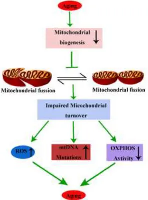
The decline of mitochondrial function during the process of ageing is recognizable by distinct morphological changes, such as abnormal rounded shape (28), reduction of mitochondrial DNA but increase of mutation rate (29), reduction of respiratory complex activity (30) as well as impaired mitochondrial biogenesis. Disruption of biogenesis will reduce mitochondrial turnover, resulting in the accumulation of ROS, oxidized lipids, proteins, mutant DNA and impairment of the OXPHOS (oxidative phosphorylation) activity, further aggravating the aging process (Fig.1).(15)
17
Therefore, it is believed that maintenance of mitochondrial biogenesis capacity during ageing is a key factor in preventing the progression of ageing-related diseases.
Mitochondrial biogenesis involves the transcription of both nuclear and mtDNA-encoded genes and is orchestrated by the Peroxisome proliferator-activated receptor Gamma Coactivator-1 (PGC-1) family of transcriptional coactivators (31). Although this family consists of three members, namely, PGC-1α, PGC-1β and the PGC-related coactivator (PRC), the first one is often cited as a master regulator of the biogenesis process, inducing the expression of nuclear respiratory factors (NRF-1 and NRF-2) and of transcription factor A in mitochondria (TFAM). These respiratory factors control the expression of nuclear-encoded mitochondrial genes and the delivery of these proteins to mitochondria.(32) TFAM is critical in the control of replication, transcription and maintenance in mitochondrial biogenesis.(33)
The expression and activity of PGC-1α can be altered by cold exposure, physical activity, or fasting. The protein PGC-1α can be reversibly activated through phosphorylation, methylation or deacetylation.(34) CR contributes for the activation of many pathways that lead to mitochondrial turnover, which significantly increases mitochondrial biogenesis in multiple tissues in mice (35) and in human muscles (36).
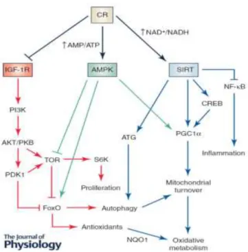
In the following section, the main molecular mechanisms involved in the CR effect on lifespan and healthspan will be briefly discussed, focusing on their impact on mitochondrial health. These mechanisms are orchestrated by the insulin-like growth factor 1 receptor (IGF-1R), the AMP-activated protein kinase (AMPK) and the Sirtuin family (Fig.2).
18
Figure 2 | Main mechanisms affected by caloric restriction. Source: López-Lluch and Navas, 2015.(37)
IGF-1R (Insulin-like growth factor 1 receptor)
The function of IGF-1 (insulin-like growth factor 1) is exerted primarily through binding to its specific receptor, the IGF-1R. To explain the mechanisms underlying CR-induced slowing in ageing, several studies indicate that changes in the IGF-1R signaling system are involved in the modulation of ageing in vertebrates (38,39), as well as in invertebrates (40), since CR reduces plasma levels of IGF-1, insulin and glucose in rodents (41) and also in humans (42).
Binding of IGF-1 to IGF1R leads to intracellular recruitment and tyrosine phosphorylation of the adaptor protein IRS-1 (Insulin receptor substrate 1), stimulating tyrosine kinase receptors (TKRs). Phosphorylated IRS-1, in turn, binds to the src homology 2 domains of the 85-kDa subunit of phosphatidylinositol 3-kinase (PI3K), leading to its activation(43). Following receptor engagement, PI3K phosphorylates phosphatidylinositol 4,5-bisphosphate [PI(3,4,5)P2] to generate
phosphatidylinositol-3,4,5-trisphosphate [PI(3,4,5)P3] that recruits and activates the serine/threonine protein
kinase Akt (also known as protein kinase B) via phosphorylation on threonine 308 by phosphoinositide-dependent protein kinase 1 (PDPK1). (44,45)
Activated AKT can phosphorylate a wide range of cellular proteins, mammalian target of rapamycin complex (mTORC), forkhead box O (FOXO) transcription factors, nuclear
19
factor kappa-light-chain-enhancer of activated B cells (NF-κB), and mouse double minute 2 homolog (MDM2)(46).
These are important regulatory elements in the effect of CR on lifespan, since FoxO is involved in the induction of several stress response genes (47) and its inhibition decreases lifespan in C. elegans and D. melanogaster (48). Consistently, a genetic variation of the FoxO3A gene has shown benefits in cardiovascular diseases and insulin sensitivity in long-lived and healthy men.(49) Inhibition of mTORC also extends lifespan in several organisms, due to its influence on the metabolic pathways associated with longevity, through its main targets.
TOR (Target of rapamycin)
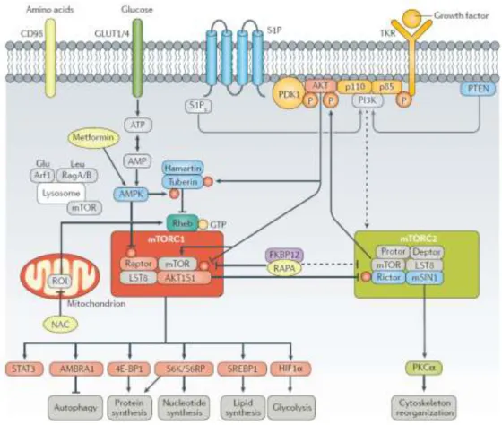
Target of Rapamycin (TOR) is a highly conserved protein kinase and a central controller of cell growth and metabolism, that responds to stress (ATP levels), nutrients (primarily glucose and amino acids) and growth factors (such as insulin and IGF-1).(50,51) TOR was originally discovered in yeast but is conserved in all eukaryotes, including plants, worms, flies, and mammals (mTOR) in 2 functionally and structurally distinct multiprotein complexes, TORC1 and TORC2 (mTORC1 and mTORC2 in mammals), each one responsible for a set of different downstream pathways.
The protein complex mTORC1 is composed of mTOR, regulatory-associated protein of TOR (Raptor, which seems to be an adaptor that presents substrates to the mTOR catalytic subunit), TORC subunit LST8, DEP-domain-containing mTOR-interacting protein (deptor) and proline-rich AKT1 substrate 1 (AKT1S1). On the other hand, mTORC2 is comprised of mTOR, rapamycin-insensitive companion of mTOR (rictor), stress-activated protein kinase interacting protein 1 (mSIN1, also known as TORC2 subunit MAPKAP1), protein observed with rictor-1 (protor-1), deptor and LST8 (Fig.3). (52)
The activity of TOR results on cell growth activation by positively and negatively regulating several anabolic and catabolic processes, respectively, phosphorylating four signature substrates: (1) ribosomal 6 kinase (S6K), which controls protein translation via ribosome biogenesis (53); (2) eukaryotic translation initiation factor 4E-binding protein 1 (4E-BP1), which regulates mRNA translation(54); (3) signal transducer and activator of transcription (STAT)3 (on Ser727) in response to amino acid excess (55); (4) and
20
activating molecule in BECN1 (beclin-1)-regulated autophagy protein 1 (AMBRA1), which prevents serine/threonine-protein kinase ULK1/ATG1 from binding to membranes and starting autophagosome formation (Fig.3). (56)
The anabolic processes controlled by TOR include transcription, protein synthesis, ribosome biogenesis, nutrient transport, and mitochondrial anabolism. Conversely, TOR negatively regulates catabolic processes such as mRNA degradation, ubiquitin-dependent proteolysis, autophagy, and apoptosis. (57)
When food is scarce, cell growth is not stimulated by TOR as opposed to when food is available, in which TOR inhibits autophagy and stimulates protein synthesis and cell proliferation(50), leading to an increase in lifespan, as noticed in a CR-induced down-regulation. This effect has been demonstrated in C. elegans and D. melanogaster, as well as in mice.(58,59)
Figure 3 | mTOR activation pathway by growth factors and main downstream targets. Source: Perl 2015.(57)
Following the previously mentioned Akt activation, the latter phosphorylates mTOR, which forms the two previously mentioned interacting complexes, mTORC1 and mTORC2. Signaling through sphingosine 1-phosphate receptor 1 (S1P1) also activates
21
mTOR complex 1 (mTORC1) via PI3K. Akt also phosphorylates and inactivates two different proteins that inhibit mTORC1, the tumor suppressor TSC2 and PRAS40 (proline-rich Akt substrate of 40 kDa). TSC2 is part of a TSC1–TSC2 heterodimer that is a GTPase activating protein (GAP) for the small GTPase Rheb. Thus, inactivation of TSC1–TSC2 maintains Rheb GTP-bound and active. Rheb binds and activates mTORC1 directly. PRAS40 binds and inhibits mTORC1 directly, and Akt/PKB-mediated phosphorylation of PRAS40 blocks this inhibition, promoting either proliferation when conditions are favorable or autophagy when otherwise. Growth factors also activate mTORC2, but the mechanism remains to be elucidated. (57,60)
When mTORC1 is inhibited, the activity of the previously mentioned S6K decreases, extending lifespan and delaying the progression of age-related diseases (59), whereas FoxO-dependent autophagy, which is essential for lifespan extension in yeast (61) and in
C. elegans (62), is increased. It is also interesting to note that 4E-BP1, which is inhibited
upon mTOR activation (54), is induced upon CR in flies and mediates the effects of CR on mitochondrial biogenesis and longevity (63), revealing the importance of the attenuation of mTOR signaling in the impact of CR in the extension of lifespan.
Although CR can activate mTORC1 through various pathways, nutrients are the dominant TOR input. Despite the fact that the mechanism responsible for the activation remains elusive, it is known that high levels of amino acids can compensate for an absence of the other mTORC1 inputs but not vice versa, and only nutrients activate TOR in unicellular organisms. Amino acid availability leads to activation of Rag, which binds mTORC1, delivering it to Rheb. Growth factor and energy-activated Rheb then activate mTORC1. mTORC1 still responds to amino acids in cells deficient for TSC1–TSC2, suggesting that the nutrient input into mTORC1 is a downstream process in relation to TSC1–TSC2. Interestingly, AMPK activation is the best described intracellular trigger for mTOR inhibition. (60)
AMPK (AMP-dependent protein kinase)
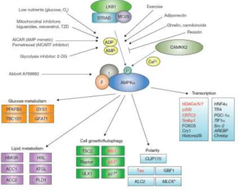
One of the central regulators of cellular and organismal metabolism in eukaryotes is AMPK, which is activated when intracellular concentration of AMP increases in relation to ATP, due to either limited ATP production (glucose deprivation or hypoxia, for example), or increased energy expenditure, such as in muscle contraction.(64)
22
It is also modulated by cytokines that regulate whole-body energy balance (65), including leptin, adiponectin, ghrelin, cannabinoids, interleukin-6 (66) and ciliary neurotrophic factor (CNTF)(67). Drugs used to treat type 2 diabetes (including metformin (68) and thiazolidinediones (69)), or natural plant products such as berberine (70), resveratrol (71) (present in grapes and red wine) and (–)epigallocatechin3-gallate(72) (present in green tea) may also interfere significantly with AMPK cascade.
Figure 4 | AMPK main modulators and main targets. Source: Mihaylova 2011.(73)
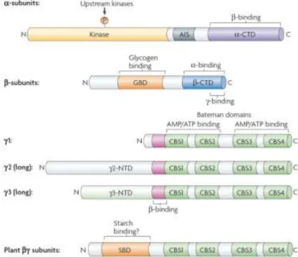
AMPK is a very sensitive energy heterotrimeric Ser/Thr kinase, composed of an α, β and γ subunits (74), capable of rapidly phosphorylating metabolic enzymes, such as acetyl-CoA carboxylase (ACC) for fatty acid biosynthesis, or phosphorylating proteins that regulate gene expression, such as PGC-1α (75) and FoxO3a (76).
There are two different forms of α (α1 and α2) and β (β1 and β2) subunits, while three
different γ isoforms (γ1, γ2 and γ3) exist (74). The α subunits are the catalytic subunits of
the functional heterotrimer and contain the Thr172 residue, whose phosphorylation is required for full enzymatic activity (77). The β subunit is responsible for binding the α and γ subunits (78), integrating an evolutionary conserved carbohydrate binding domain, which allows AMPK to interact with glycogen particles, resulting in inhibition of AMPK when glycogen stores are loaded (79). The γ subunits are responsible for the sensitivity
23
of AMPK to increases in the cellular AMP/ATP ratio, characterized by four tandem repeats known as cystathionine β-synthase (CBS) motifs, which form an interface for interaction with two AMP or ATP molecules in a mutually exclusive way and with a third AMP molecule in a non-exchangeable mode (80).
Figure 5 | Domain Structure of α, β and γ Subunits of AMPK and Their Isoforms. Source: Hardie et al., 2007. (74)
AMPK is only fully active after phosphorylation of Thr172 within the activation loop of
the α subunit catalytic domain, whereas in basal conditions, the activity of the enzyme is low due to ATP binding. (81) Liver kinase B1 (LKB1) specifically activates the α2 subunit (82) whereas calcium calmodulin dependent kinase kinase β (CaMKKβ) is specific for the α1 subunit.(83)
The main upstream kinase in most cell types is the LKB1/STRAD/MO25 complex (Fig. 4). Although this complex seems to be constitutively active and phosphorylates AMPK continuously, in basal conditions, the phosphate is immediately removed by protein phosphatases. However, when AMP concentrations increase, such as in energy stress conditions, AMP binds to the AMPKγ subunits, promoting a conformational change that renders AMPK a poorer substrate for dephosphorylation.(81) Then, the increased phosphorylation levels of the Thr172 residue results in the full activation of the enzyme
24
(84), which will restore ATP concentrations by activating energy-producing processes, such as the oxidation of fatty acids and mitochondrial biogenesis.
Since AMPK is a master regulator of mitochondrial biogenesis and lipid metabolism, its influence in lifespan has been studied and observed in several organisms from yeast to mammals, having been demonstrated that an increase in AMPK activity is associated with a longer lifespan while its inhibition shortens it.(85,86)
Although some studies suggest that its activity is not affected by CR (87) Others indicate an increase in AMPK activity in heart and skeletal muscle(75,88). However, according to López-Lluch and co-authors, “these discrepancies could be due to differences in the period of time under CR or the degree of CR which can play an important role in nutrient balance”(37).
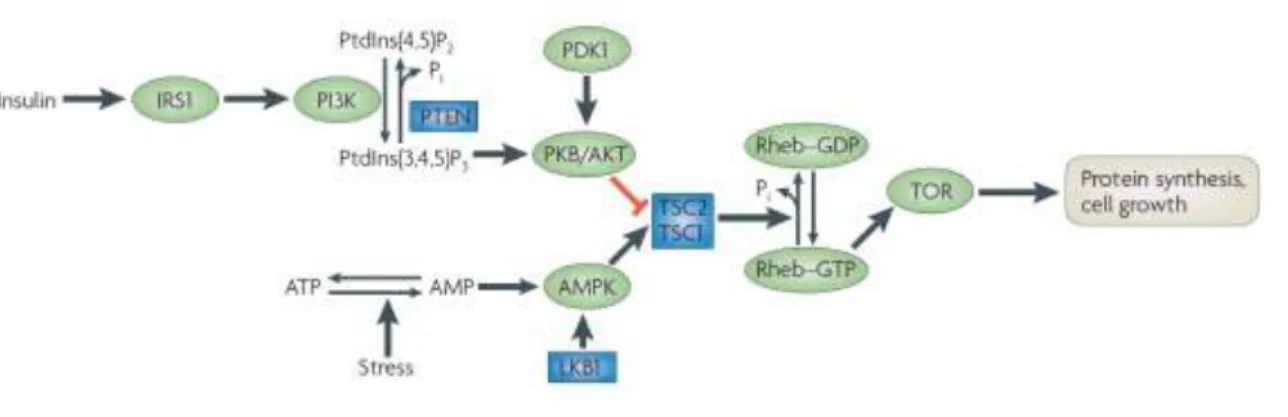
AMPK and mTORC1
As mentioned before, a low energy status activates AMPK, which will restore cellular energy levels by inhibiting energy-consuming anabolic pathways, as is the case of the mTORC1 pathway. AMPK phosphorylates both Raptor, inhibiting mTORC1 directly and TSC2, activating TSC1-TSC2 to inhibit Rheb and ultimately mTORC1. It is not known whether AMPK also controls mTORC2.(74)
Figure 6 | mTORC1 activation pathways by Insulin and AMPK. Source: Hardie et al., 2007.(74)
Sirtuins
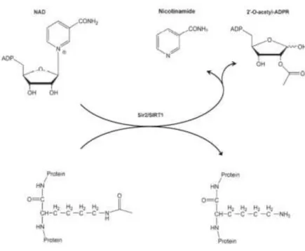
Mammalian sirtuins are nicotinamide adenine dinucleotide (NAD+) - dependent class III
histone deacetylases, controlling cell survival, energy regulation, metabolism, inflammation and ageing in all organisms.(34)
25
The first identified sirtuin protein was silent information regulator 2 (Sir2) from
Saccharomyces cerevisiae. Sir2 was originally characterized as a chromatin-silencing
component, shown to regulate transcriptional silencing at cell-mating loci, telomeres and ribosomal DNA (rDNA) in yeast, through deacetylation of the epsilon-amino groups of lysine in the amino-terminal domains of histones.(9)
Sir2 cleaves the glycosidic bond between nicotinamide and adenosine diphosphate (ADP)– ribose in NAD and this reaction requires the presence of acetylated lysine. Thus, one molecule of NAD and one molecule of acetyl-lysine (in a protein) are converted to one molecule each of deacetylated protein, nicotinamide and 2’-O-acetyl-ADP-ribose (Fig.7). (89)
Figure 7 | The NAD+-dependent SIRT1 deacetylase reaction. Source: Cantó and Auwerx, 2012. (89)
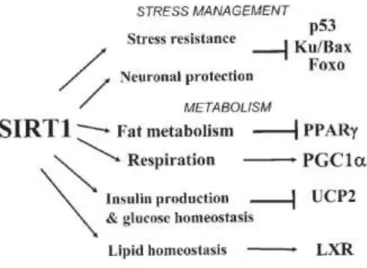
Despite their relevance in histone deacetylation in yeast, a large number of nonhistonic nuclear or cytosolic proteins, which tend to be transcription factors, cofactors and enzymes, have been identified as Sirt1 protein substrates. Proteins targets of Sirt1, may control a number of important pathways in mammals and can be divided into two categories.
26
Figure 8 | Targets of SIRT1 in mammalian cells. Mitochondrial uncoupling protein 2 (UCP2). Liver X receptor (LXR). Source. Guarante 2007.(90)
The p53 and FOXO proteins, which are deacetylated by Sirt1, will be major regulators conferring resistance to cellular stress and mediating DNA repair and apoptosis. The second category is involved in metabolism. This group includes PPARγ (peroxisome proliferator-activated receptor)γ and PGC-1α, which are regulators of adipocyte biology.(90)
In addition to deacetylating several protein substrates, sirtuins are also responsible for other enzymatic activities, such as ADP ribosylation, demalonylation, desuccinylation, depropionylation and debutyrylation. (91)
In humans, this family of ubiquitously expressed proteins comprises seven members (Sirt1-7), sharing the sequence homology of catalytic domain with Sir2, which function to regulate metabolism and coordinate cellular responses throughout the various tissues. In human cells, Sirt3, Sirt4 and Sirt5 are localized in the mitochondria, Sirt1, Sirt6 and Sirt7 are primarily localized in the nucleus, whereas Sirt1 and Sirt2 are localized in the cytosol. (89)
Sirtuin family members can also be distinguished by their different enzymatic activities. Sirt1 and Sirt5 (also described to demalonylate and desuccinylate proteins) act as deacetylases (92,93), whereas Sirt4 seems to be a mono-ADP-ribosyl transferase (94). On the other hand, both activities are displayed by Sirt2, Sirt3 and Sirt6. The activity of Sirt7 is yet unclear, altought it has been hypothesized to act also as a deacetylase. (95–98) From all sirtuins, Sirt1 is the mammalian ortholog most highly related to Sir2, and the most studied one as well.
27
However, unlike the intra-nuclear localization of yeast Sir2, Sirt1 is not tightly bound to chromatin but, instead, shuttles between cytoplasm and nucleus. (99) The presence of Sirt1 is determined by two nuclear localization signals as well as two nuclear exportation signals, explaining why Sirt1 location may differ depending on the cell type or tissue. (9)
Figure 9 | Sirt1 mediates metabolic benefits in various tissues. CREB-regulated transcription coactivator 2 (CRTC2), phosphoglycerate mutase-1 (PGAM-1), peroxisome proliferator-activated receptor α (PPAR α), sterol regulatory element binding protein 1c (SREBP1c). Targets that are directly activated by Sirt1 are shown in green. Those repressed or inhibited by Sirt1 are shown in pink. Source: Chang and Guarente, 2013. (100)
It is known that limited glucose availability, induced by calorie restriction, in the growth medium of the budding yeast Saccharomyces cerevisiae leads to the activation of Sir2 and the extension of replicative lifespan, due to a shift from fermentation to respiration. (101) When calories are restricted, more carbons are oxidized in mitochondria via the electron transport chain-mediated cellular respiration, which produces NAD+ from NADH, resulting in elevated NAD+ or decreased NADH levels. On the other hand, when cells have high levels of calories, a substantial fraction of the NAD pool is recruited into a high carbon flow of glycolysis by the enzyme glyceraldehyde-3-phosphate dehydrogenase. (102,103)
As mentioned above, NAD+ levels are generally increased in situations of energy/nutrient stress, such as exercise or calorie restriction, as is the activity of Sir2/Sirt1. Since sirtuins depend on NAD availability, it would be expected that the activity of Sir2 and Sirt1 would
28
be modulated by physiological alterations in the NAD/NADH ratio. However, according to Anderson and coauthors, NAD levels do not correlate with the lifespan of yeast(104), since they are actually decreased during calorie restriction, indicating that Sir2 is not primarily regulated by the availability of NAD (101). It was also unsuccessfully proposed for such influence to derive from NADH levels. As the latter is a competitive inhibitor of Sir2, the overexpression of NADH oxidase or alternative oxidase, both of which increase NADH oxidation, could alter the lifespan of wild type yeasts. However, this was not observed. (12)
These information lead the scientific community to believe that increased respiration plays a major role in lifespan extension by caloric restriction in yeast, and that Sir2 acts to facilitate this process through detoxification of oxidized macromolecules. (104) In this regard, it is important to notice that the inner membrane of mitochondria is impermeable to NADH and NAD, being the malate-aspartate shuttle used for translocating electrons produced during glycolysis for oxidative phosphorylation. This important shuttle allows the hydrogen ions of NADH produced in the cytosol to reach the electron transport chain in the mitochondria. Thus, overexpression of mitochondrial NADH shuttle components and the overexpression of mitochondrial NADH dehydrogenase, which specifically lowers NADH levels, extends yeast replicative lifespan in a Sir2-dependent manner. (105)
Alternatively, caloric restriction may activate Sir2 by regulating the levels of nicotinamide, which is an inhibitor of Sirt1.(104,106,107) Crystal structures of the conserved sirtuin catalytic domains reveal that NAD and the peptide containing an acety-lated lysine residue enter the active site from opposite sides of a cleft between a large Rossmann fold domain and a small Zn-binding domain. During the formation of an alkylimidate intermediate between the ADP-ribose 1’ position and the acetyl oxygen, nicotinamide dissociates from NAD and occupies a so-called C-pocket. If nicotinamide binds to the C-site before alkylimidate conversion, it will inhibit the deacetylation reac-tion. Thus, removal of nicotinamide may be as important for the activation of Sir2/Sirt1 as the production of NAD. (108)
Soon after the discovery that Sir2 extended the replicative lifespan of yeast, it was proposed that the ortholog of Sir2 could carry out the same lifespan-prolonging effects in
Caenorhabditis elegans (109) and in Drosophila melanogaster (110), as well as in
mammals, and to mediate beneficial effects of calorie restriction (CR) on health and longevity.
29
Indeed, Sirt1 overexpression exhibits similar effects to those of Sir2 in yeast via mechanisms involving an increase in whole-body metabolic efficiency via mitochondrial biogenesis and a reduction of high fat diet induced metabolic damage, delaying the onset of age-associated diseases, including cancer, atherosclerosis and diabetes (111), and also decreasing expression of the ageing-associated protein p16Ink4a and lowering levels of DNA damage. (112)
Mice lacking both copies of Sirt1 failed to show an extended lifespan in response to caloric restriction, but displayed a shorter median lifespan than wild type mice (113,114), whereas mice with elevated Sirt1 expression exhibited a beneficial phenotype resembling that of caloric restriction (leaner, more metabolically active, more glucose tolerant, and have reduced levels of circulating cholesterol, proinflammatory adipokines, insulin, and fasting glucose). (115) Consistently, mice lacking one allele of Sirt1 still showed identical lifespan to that observed in wild type mice, when subjected to caloric restriction. (116)
Caloric restriction and mitochondrial biogenesis
As previously mentioned, PGC-1α is considered the main orchestrator of mitochondrial biogenesis and it is tightly connected to the activity of AMPK, SirT1 and IGF-1.
Cytoplasmic Sirt1 and PGC-1α are partly co-localized in the mitochondria, which is associated with D-loop region of mtDNA, the original site for replication and transcription.(15)
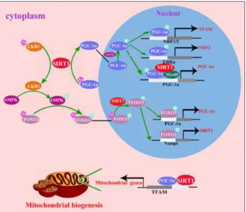
AMPK and Sirt1 promotion of mitochondrial biogenesis via PGC-1α occurs both in the nucleus and cytoplasm, involving transcription of both nuclear and mtDNA-enconded genes, and it is initiated with Sirt1-mediated LKB1 deacetylation, phosphorylating AMPK, causing its activation. Activated AMPK phosphorylates FOXO3, leading to the translocation of the latter to the nucleus. In the nucleus, Sirt1 deacetylates FOXO3, which will induce transcription of PGC-1α and Nampt (Nicotinamide phosphoribosyltransferase). Nampt will increase NAD+ levels required for Sirt1-dependent deacetylation and thus activation of PGC-1α.(15) In conjunction with the activation of AMPK, Sirt1 is required for the deacetylation and synchronized activation of PGC-1α that is translocated to the nucleus.(117) In the nucleus, PGC-1α is phosphorylated at Thr177 and Ser538 by AMPK, enhancing the expression of NRF-2 and TFAM, which in turn upregulate the mitochondrial biogenesis process. PGC-1α also
30
forms a complex with Sirt1 and MyoD, which drives the transcription/replication of mtDNA and consequently improving mitochondrial biogenesis (Fig.10).(15)
This complex has been discovered in purified mitochondrial extracts of mouse skeletal muscle (118), where it binds to the promoter of PGC-1α creating a positive feedback and auto-regulatory loop for PGC-1α expression.(119)
Figure 10 | AMPK and SirT1 promotion of mitochondrial biogenesis via PGC-1α. Source: Yuan et al., 2016.(15)
To balance the deacetylase activity of SirT1, acetylation and consequent inhibition of PGC-1α is achieved by the acetyltransferase GCN5 (or general control nonderepressible 5). (120) GCN5 is upregulated in a high-fat diet, hyperacetylating PGC-1α and recruiting its transcriptional repressor receptor interacting protein 140 to the site, through SirT1 and MyoD. This results in a decrease in the expression of PGC-1α gene, leading to the depression of mitochondrial biogenesis.(121) Accordingly, SRC-3 (nuclear receptor coactivator 3)-knock-out mice display increased mitochondrial function and are protected against obesity (Fig.11), since SRC-3 activates GCN5 gene expression.(122)
31
Figure 11 | Influence of SRC-3 and GCN5, AMPK and SIRT1 in the expression levels of PGC-1α. Source: Cantó and Auwerx, 2009.(123)
On the other hand, Akt/PKB, a previously mentioned downstream target of IGF-1 is known to phosphorylate PGC-1α and, hence, decrease its stability and transcriptional activity.(124)
Consistently, in diet-induced-obese mice, treatment with SirT1 agonists, such as small molecules as SRT1720 (125) or resveratrol (126) results in an increased mitochondrial function in skeletal muscle and improved insulin resistance(125), through deacetylation and activation of PGC-1α. These findings suggest that the latter plays a key role in the induction of mitochondrial biogenesis through the AMPK-Nampt-SirT1 pathway. However, despite the strong influence of this pathway on PGC-1α-induced mitochondrial biogenesis, one must not ignore the importance of the degradation of damaged mitochondria in the overall balance of the mitochondrial turnover. Mitophagy, a mitochondrial specific form of autophagy, is vital for homeostatic control of this organelle, being essential for the maintenance of mitochondrial genomic integrity (127) and control of mitochondrial homeostasis in response to cellular stressors.(128)
Although the process is yet unclear in mammals, it has been shown to be of utmost importance in the maintenance of mitochondrial content and integrity in yeast (127), having potential implications in ageing and age-related diseases such as neurodegeneration.(129,130)
32
How might mitochondrial biogenesis protect against damage?
Although the impact of ROS in ageing and ageing-related diseases is yet unclear, it is known that CR reduces the production of ROS and increases mitochondrial biogenesis, even though it is not certain whether the latter alone is sufficient to reduce ROS production and consequent accumulation.
Therefore, one hypothesis is that the natural progressive loss of mitochondria over time might be dampened by having more of these organelles, hence, mitigating deleterious effects on age-related diseases.(131)
Another possibility is that mitochondrial biogenesis reduces the production of ROS, which are formed when electrons are stalled on complexes I and III of the electron transport chain, and combined with local oxygen to produce ROS.(132)
Works from Barja 2007 and Guarente 2007 outlined two possible mechanisms, represented in Figure 12, for a causal relationship between mitochondrial biogenesis and reduced ROS.(90,132)
Figure 12 | Relationship between mitochondrial biogenesis and ROS production. Source: Guarente, 2007. (90)
According to Model 1, electrons can stall when the charge gradient across the mitochondrial membrane generated by proton extrusion becomes too steep. This condition makes the electron transport chain more difficult, namely to pump more protons out of mitochondria against this hyperpolarized gradient. This happens under conditions of excess of energy and high levels of NADH. According to Guarente 2007, “an expanded mitochondrial surface would reduce the steepness of the charge gradient by spreading the
33
charges over a larger area and, in this way, may prevent electron stalling and ROS production”(90).
A second possible mechanism, represented by Model 2, involves an increase in respiration that might reduce local oxygen levels and thus reduce the generation of ROS by complexes I and III. This way, the binding of oxygen to these complexes might be decreased, disfavoring ROS production, but not impacting on respiration itself.(90,132) However, further research and evidence are still required to fully validate these theories.
Caloric restriction mimetics
Even though caloric restriction may extend the human lifespan, the severity of such regimen and the difficulty to maintain long-term diets in humans has led to the search of a more practical treatment, with less drastic diets and safe drugs or methods that can reproduce the effect of CR without limiting the amount of food.
CR mimetics (CRMs) are compounds or methods (such as bariatric surgery or exercise) that provide the physiological benefit of CR without the need for restriction of calories.(133) The concept is relatively new and was proposed by Lane et al.(134) in a study of 2-deoxy-D-glucose (2DG) effects, which showed bioactivity in rats. Generally CRMs should have the following key features: they should mimic the metabolic, hormonal and physiological effects of CR; they should not significantly decrease long-term food intake; they should activate stress–response pathways, as observed in CR, and protect against a variety of stressors; and they should reduce inflammation and autoimmunity. (135)
There are authors who attempt to group CRMs according to their targets, as upstream or downstream, however, it is difficult to classify all CRMs accordingly, and so, a molecular definition is required.(136) Upstream-type CRMs use a mechanism of action targeting the energy metabolism system and transmitting a signal in the upstream direction to mimic CR, whereas downstream-type act on an intracellular signaling system and exert the same effect as CR on downstream pathways.(137) The benefit of CRMs could be significant and hence, many of these molecules received received attention from governments, pharmaceutical and private research organizations, with special regard to rapamycin, resveratrol and metformin, which are the most widely studied compounds.
34
Figure 13 | Targets of the main caloric restriction mimetics and their downstream effects. Source: Gillespie et al., 2016. (138)
Rapamycin
As mentioned before, mTOR activity has been shown to be down-regulated by caloric restriction, resulting in increased autophagy (139) and decreased protein translation(140), both associated with promoting health and lifespan. However, mTOR inhibition can be achieved by either directly or via activation/inhibition of up-stream or down-stream factors of mTOR complexes. Among all of them, rapamycin is the most well-known, characterized and used inhibitor.(140)
Rapamycin is a macrocyclic lactone-based compound produced by the bacterium Streptomyces hygroscopicus that was first found in the soil of Easter Island in 1972. (141)
Rapamycin is bound between cytoplasmic protein FK-binding protein 12 (FKBP12) and the mTOR kinase subunits of mTORC1, causing functional inhibition of mTOR and the mTOR pathway.(142) Consequently, interleukin-2 signaling and other cytokine-receptor-dependent signaling pathways are inhibited, preventing the activation of T-cells and B-cells. Due to its ability of inhibiting immune responses, rapamycin has been used clinically to prevent transplant rejection and treat autoimmune diseases.(143)
35
Rapamycin has been documented to attenuate age related-disease phenotypes of numerous cell-based models, including cardiovascular diseases (144), premature aging disease HGPS (Hutchinson–Gilford progeria syndrome) (145)and neurodegenerative diseases (146). This attenuation leads to an extension of lifespan in various model organisms such as S. cerevisiae (147), D. megalonaster (148) and M. muscularis (149), possibly due to the fact that mTOR inhibition by rapamycin leads to increased autophagy of accumulated cells in senescence, which have been extensively linked to aging and the aging phenotype (150). It has been demonstrated that the clearance of these cells may improve symptoms of diseases associated with age (151).
Numerous studies have shown that inhibition of the TOR signaling pathway extends lifespan in yeast, nematodes and flies(152–154) in response to rapamycin treatment(147). Then, it was reported that rapamycin extends the median and maximum lifespan of 20-month-old mice, which corresponds to approximately 60 years of age in humans, by 28-38% as compared with control animals. Moreover, this finding was complemented with a decrease in TOR activity (58), suggesting that life expectancy can be extended in elderly humans. The phosphorylation levels of ribosomal protein subunit S6 (rpS6), a target of S6K in the mTOR signaling pathway(155), were also evaluated, having rapamycin-fed mice shown a significant reduction in the levels of phosphorylated rpS6, when compared with control mice in adipose tissue. These findings are consistent with data reported by Selman et al.(59), sustaining that S6K inhibition increased lifespan.
In another report, the average lifespan of mature mice increased by 60%, when treated with rapamycin for three months, and the therapeutic benefit of rapamycin was also observed in a mouse model of mtDNA-driven mitochondrial disease.(156) Additionally, some improvements were associated with decreased expression of proteins involved in oxidative stress, AMPK activation and cell cycle control.(157)
However, long-term side effects in rats have been reported in other studies, including hyperglycemia, impaired glucose tolerance and insulin resistance(158), which question the rational use of rapamycin as a CRM.
36
Metformin
Metformin is another CRM of interest to gerontologists. Metformin is a biguanide recommended as the first-line drug for type 2 diabetes in the guidelines of the American Diabetes Association and European Association for the Study of Diabetes(159,160) Metformin was identified in a screening assay of drugs showing similar transcriptional profiles to that of caloric restriction in mice(161) and was shown to have CR-related longevity benefits mediated by the activation of AMPK in C. elegans.(162)
Metformin transiently inhibits the mitochondrial respiratory chain (specifically, complex I), increases the intracellular AMP/ATP ratio, and activates AMPK, through the binding of either AMP or ADP.(137) Metformin also promotes activation of AMPK by increasing phosphorylation at Thr-172 of the α subunit.(163) However, LKB1 is required for the activity of AMPK, since its deletion resulted in almost complete loss of the latter’s activity.(164)It was observed that 8 weeks of metformin treatment had more pronounced effects on CR-like gene expression than 8 weeks of CR, which leads to the possibility that metformin might optimize the activity or responses of the LKB1-AMPK pathway, depending on the energy requirements.(161)
Administration of metformin to experimental animals results in different effects depending on the species. In the nematode Caenorhabditis elegans, metformin may extend the lifespan through a mitochondrial process and the production of metformine-induced ROS triggers a signaling cascade that increases overall lifespan.(165) Metformin lifespan-extending effect was also found to be dependent of both TORC1 inhibition and AMPK activation, which is consistent with the genetic screening conducted by Wu et al.(165) that revealed that metformin was associated with inactivation of TORC1. In Drosophila, in spite of not prolonging lifespan(166), administration of metformin caused activation of AMPK and the reduction of adipose tissue weight. Curiously, long-term intake of metformin with a starch-supplemented diet shortened the lifespan of
Drosophila melanogaster, when compared to the control group fed with starch
alone.(167)
In C57BL/6 mice, lower doses of metformin increased the survival rate significantly, whereas a higher dose reduced the survival rate.(168) This may be due to the fact that excessive binding of metformin to AMPK subunits at higher concentrations may interfere with the association of AMPK subunits, blocking the formation of the AMPK heterotrimeric complex, because the -NH groups in metformin are able to form hydrogen
37
bonds with amino acid residues in these subunits.(163) Consistently, results obtained by Meng et al. showed that low metformin concentrations drastically increase the phosphorylation of α1 subunit by LKB1 and reduced the dephosphorylation of the α subunit at Thr-172 by protein phosphatase PP2C, but only in the presence of both β and γ subunits.
The lack of longevity benefits observed in both these species has led to uncertainty as to whether metformin is a true CRM, despite having beneficial effects on other aspects of the aging process. However, a systematic review and meta-analysis of 260 articles by Campbell et al.(169) concluded that administration of metformin lowered all-cause mortality in diabetics when compared to nondiabetics and diabetics under non-metformin therapies. The decrease in aging associated with metformin suggests that it acts as an antioxidant when improving health.
Polyphenols - resveratrol
Polyphenols are compounds, often portrayed as nutraceuticals in the media, characterized by the presence of one or more hydroxyl groups bound to an unsaturated aromatic hydrocarbon ring such as benzene and are produced by plants in response to stress or fungal infections.(170)
Among these compounds, resveratrol (or 3,5,4′-trihydroxystilbene) has emerged as the compound that best mimics the CR effect on several organisms(171). It has been demonstrated that it can mimic the benefits associated with CR and that CR did not further extend the lifespan in yeast grown in a resveratrol supplemented medium.(172) Resveratrol is a sirtuin activating compound (STAC), firstly indentified by Sinclair through screening of small molecular libraries for compounds that could activate sirtuins and extend lifespan in a yeast model(172). This well-known polyphenol is commonly found in a variety of foods, including a number of berries, peanuts, grapes and red wine(173). STACs are plant-derived metabolites, such as flavones, stilbenes, chalcones, and anthocyanidins that activate SIRT1(172) and among these, resveratrol is still the strongest natural STAC(174) and the best CR mimetic(171). The effects of resveratrol include a decrease in insulin secretion and insulin level, an increase in insulin sensitivity, a decrease in fat mass, an increase in mitochondrial biogenesis and oxidative phosphorylation, an increase in the NAD+/NADH ratio and the subsequent activation of sirtuins, an increase in AMPK activity and increase in mitophagy and autophagy.(171)
38
The biochemical effects of Resveratrol are pleiotropic, where one of its main targets is the direct activation of AMPK(175), which is supported by Um et al. that showed that AMPK deficient mice were fairly resistant to the metabolic improvement effect of resveratrol.(176) Furthermore, it has been recently demonstrated that obese humans treated with resveratrol for thirty days produce similar effects to those induced by CR, activating AMPK, increasing Sirt1 and PGC-1α and improving muscle mitochondrial respiration based on fatty acids.(177) Similarly, resveratrol also increased lifespan on mice kept on a high fat diet and the same was observed with synthetic activators such as SRT1720 and SRT2104, which prolonged the lifespan of mice fed either a high-calorie or low-calorie diet.(178) However, it is not clear whether the effect of resveratrol on PGC-1α and mitochondrial biogenesis is Sirt1-dependent, leading to many studies contradicting the importance of the latter and its relation to resveratrol. Interestingly, the work of Dasgupta and Milbrandt also indicates that resveratrol could directly activate AMPK in a manner that is completely independent of Sirt1 activity, whereas Sinclair’s group, using an inducible knockout model, showed that intact Sirt1 in mice is required for resveratrol’s activity on mitochondrial biogenesis and function(179). This might due to the fact that Sirt1 is essential for resveratrol action but as a downstream consequence of AMPK activation, rather than as a direct molecular target of resveratrol. AMPK activation by resveratrol is very fast and prominent after a few minutes, leading to a gradual increase in NAD+ levels and, thus, accumulation, as a consequence to the activation of fatty acid oxidation, and subsequently activating Sirt1(123). Consistently, Sirt1 becomes detectable only after a few hours, indicating that AMPK activation is an earlier event. Being the mechanism unclear, it is also possible that the connection between Sirt1, AMPK and PGC-1α is nonlinear and definitely cell/tissue-type dependent and influenced by energy and nutrient status.
39
Figure 14 | Resveratrol indirectly activates AMPK and SirT1, promoting mitochondrial biogenesis and lip oxidation gene expression. CI-V represent mitochondrial respiratory complexes I to V. Source: Cantó and Auwerx, 2012. (89)
Another of the known effects of resveratrol is the inhibition of cAMP degrading phosphodiesterases.(180) This inhibition leads to elevated cAMP levels and that to activation of AMPK and cAMP-inducible factors such as the Rap Exchange factor directly activated by cAMP 1 (Epac1), which is required for resveratrol mediated increases in NAD+ levels, which, in turn, were abolished by silencing of Epac1.
Resveratrol was subsequently shown to extend longevity in worms, flies, fish and nematodes and fruit flies in a sirtuin-dependent manner.(71,181) However, recent studies added some controversy by showing that resveratrol had no effect on the longevity of mice fed a normal diet (182) and on mitochondrial proteins in the muscle(183), while high levels of resveratrol activated AMPK in C2C12 myotubes in culture, which did increase mitochondrial protein levels. Despite the uncertainty concerning the effect of resveratrol in the extension of longevity, epidemiological studies have demonstrated that consumption of resveratrol can improve health and prevent age-related diseases.(184) In clinical studies, intake of resveratrol improved the memory capacity of elderly subjects and improved the blood lipid levels and glucose control in obese and adult diabetic subjects.(177,185,186)
These data may explain why red wine reduces the health risks regarding unhealthy diets, thus explaining the French paradox.
40
Conclusion
Overall, the results from multiple studies showed that reduced calorie intake has a broad effect on healthspan and longevity, through multiple mechanisms that involve most of the metabolic pathways in tissues and organs.
This review was focused on the metabolic consequences of CR, aiming to outline what is known about the main mechanisms affected by CR and their potential advantages on delaying ageing progression and on the development of age-related chronic diseases. AMPK, sirtuins, IGF-1, TOR and PGC-1α have proved to be essential as major effectors by inhibiting processes involved in cell proliferation and glycolysis and increasing autophagy and mitochondrial biogenesis and turnover These crucial processes are required to maintain an active and “healthy” cellular homeostasis, in order to lower endogenous ROS production, regarded as one of the main causes of ageing. However, investigating whether the reduction of ROS during CR is the result of an increase in mitochondrial biogenesis per se is a worthy goal for future research.
These physiological effects were also found to be modulated by some nutraceuticals and compounds, acting as CR mimetics. This review also approached those which are considered as the most promising ones, namely resveratrol, rapamycin and metformin. Information gathered confirmed their effectiveness in stimulating beneficial metabolic responses throughout the whole organism, featuring a positive influence on the mitochondrial health, despite the uncertainty concerning some steps of their mechanisms of action, which will require further studies for full comprehension of the complex dynamic interactions.
Although these mechanisms are still unclear, the scientific community has gained a more accurate understanding of the overall processes at molecular level, which could potentially contribute for the development of more practical and safer treatments.
To conclude, despite the fact that the introduction of new drugs as CRMs is most likely coming in a near future, the responsibility of maintaining a healthy lifestyle should be emphasized. The availability of such drugs will almost surely not fully replace the need for careful control of dietary habits, the need of regular exercise, especially in patients with metabolic syndrome or diabetes.
41
References
1. Mercken EM, Carboneau BA, Krzysik-Walker SM, De Cabo R. Of mice and men: The benefits of caloric restriction, exercise, and mimetics. Ageing Res Rev. 2012;11(3):390–8.
2. McCay CM, Crowell MF, Maynard LA. The effect of retarded growth upon the length of life span and upon the ultimate body size. 1935. Nutrition. 1935;1(1):63– 79.
3. Simpson SJ, Raubenheimer D. Caloric restriction and aging revisited: the need for a geometric analysis of the nutritional bases of aging. journals Gerontol. 2007 Jul;62(7):707–13.
4. Mair W, Piper MDW, Partridge L. Calories do not explain extension of life span by dietary restriction in Drosophila. PLoS Biol. 2005 Jul;3(7):1305–11.
5. Solon-Biet SM, Raubenheimer D, Huang X, McMahon AC, Cooney GJ, Simpson SJ, et al. The Ratio of Macronutrients, Not Caloric Intake, Dictates Cardiometabolic Health, Aging, and Longevity in Ad Libitum-Fed Mice. Cell Metab. 2014;19(3):418–30.
6. Speakman JR, Mitchell SE, Mazidi M. Calories or protein? The effect of dietary restriction on lifespan in rodents is explained by calories alone. Vol. 86, Experimental Gerontology. 2016. p. 28–38.
7. Raubenheimer D, Simpson SJ, Mayntz D. Nutrition, ecology and nutritional ecology: Toward an integrated framework. Funct Ecol. 2009 Feb;23(1):4–16. 8. Kapahi P, Kaeberlein M, Hansen M. Dietary restriction and lifespan: Lessons from
invertebrate models. Vol. 39, Ageing Research Reviews. 2017. p. 3–14.
9. Wang Y. Molecular links between caloric restriction and Sir2/SIRT1 activation. Diabetes Metab J. 2014;38(5):321–9.
10. Masoro EJ. Biochemical and molecular mechanisms of aging: from model systems to human longevity. Biochimica et Biophysica Acta - General Subjects. 2009 Oct;1790(10):949–50.
11. Fontana L, Partridge L, Longo VD. Extending healthy life span-from yeast to humans. Vol. 328, Science. 2010. p. 321–6.
12. Wei M, Fabrizio P, Madia F, Hu J, Ge H, Li LM, et al. Tor1/Sch9-regulated carbon source substitution is as effective as calorie restriction in life span extension. PLoS Genet [Internet]. 2009 May;5(5):e1000467. Available from: http://www.ncbi.nlm.nih.gov/pubmed/19424415
13. Picca A, Pesce V, Lezza AMS. Does eating less make you live longer and better? An update on calorie restriction. Clin Interv Aging. 2017;12:1887–902.