Characterization of partial
Hox
gene sequences in annual fish of the
subfamily Cynolebiatinae (Cyprinodontiformes, Rivulidae)
Verónica Gutiérrez
1, María José Arezo
2and Graciela García
11
Sección Genética Evolutiva, Facultad de Ciencias, Universidad de la República, Montevideo, Uruguay.
2
Sección Biología Celular, Facultad de Ciencias, Universidad de la República, Montevideo, Uruguay.
Abstract
Hox genes encode a family of transcription factors implicated in conferring regional identity along the anteroposterior axis in developing animal embryos. These genes are organized in genomic clusters, expressed collinearly and highly conserved in vertebrates. Among teleost, South American annual killifishes of the Cynolebiatinae subfamily repre-sent an excellent model in development studies because their embryos are capable of undergoing reversible devel-opmental arrest (diapause) at three well-defined morphological stages. They are also an excellent model for evolutionary studies due to the high rates of mutation of their mitochondrial genome, their karyotypic divergence and their morphological variability. In this study, three partial homeobox sequences were isolated from different species of the Cynolebiatinae subfamily. Phylogenetic analyses and sequence comparisons revealed that they belong to the anterior Hox complex group, specifically to paralogue groups 1 and 3. This is the first time that partialHox genes have been described in species of the Cynolebiatinae subfamily.
Key words:subfamily Cynolebiatinae, annual killifishes,Hoxgenes.
Received: April 17, 2006; Accepted: November 6, 2006.
Introduction
The homeobox genes play important roles in the de-velopmental processes of many multicellular organisms. The homeobox was originally described as a conserved protein-coding sequence of about 180 base pairs, in many developmental control genes ofDrosophila. The encoded proteins are evolutionarily conserved transcription factors that share a 60 amino acids DNA-binding domain (the homeodomain, or HD) and regulate axial patterning seg-ment or cell identity and proliferation (Lewis, 1978; McGinniset al., 1984a, b). Homeodomain proteins regu-late diverse developmental programs by modulating ex-pression patterns of target genes in a temporal, spatial and tissue-specific manner (Gehringet al., 1994a). X-ray crys-tallographic and NMR spectroscopic analyses on several members of this family revealed that the structure consists of a triα-helical core and an N-terminal arm that becomes partially ordered in the presence of DNA (Qianet al., 1989; Kissingeret al., 1990; Gehringet al., 1994a; Wolberger, 1996; Passneret al., 1999). The N-terminal region, just up-stream of helix 1, contacts the minor groove of DNA, whereas helix 3, known as the “recognition helix”, binds to
the major groove. The mode of HD-DNA interaction ap-pears to be highly conserved within HD-containing pro-teins (Gehringet al., 1994b; Chauvetet al., 2000).
In vertebrates, theHox genes (a class of homeobox genes) are organized in linked chromosomal clusters and show a striking colinearity in their 5’-3’ chromosomal posi-tion. They also show remarkably similar expression pat-terns along the anteroposterior (AP) axis, specifying the identities of different body regions during embryogenesis (McGinnis and Krumlauf, 1992). While invertebrates stud-ied to date have a single Hox cluster, tetrapod vertebrates have four separated Hox clusters (termed clusters A to D) lying on four different chromosomes. Within each cluster, the genes have been classified into 13 paralogue groups ac-cording to sequence homology and location in the genome (Krumlauf, 1994). The expression pattern of Hox genes during development and homeodomain sequence compari-sons indicate relatedness between anterior (Hox1-3), me-dial (Hox4-8) and posterior (Hox9-13) paralogue groups (Ruddleet al., 1994). Some paralogue groups can be recog-nized by the homeodomain sequence alone, others only by using characteristic residues outside the homeodomain (Sharkeyet al., 1997).
Although it is widely assumed that vertebrates have four Hox clusters, studies on ray-finned fish have shown that they have more Hox clusters than tetrapods. These
Send correspondence to Graciela García. Facultad de Ciencias, Iguá 4225, 11400 Montevideo, Uruguay. E-mail: ggarcia@fcien. edu.uy.
extranumeral Hox clusters result from a genome duplica-tion event that is specific for the fish (actinopterygian) lin-eage. Sets of seven Hox clusters have been described in
Danio rerio(Amoreset al., 1998; Princeet al., 1998), in three pufferfish species, Takifugu rubripes, Tetraodon nigroviridis and Sphenoides nephelus (Aparicio et al., 1997, 2002; Jaillonet al., 2004; Amoreset al., 2004) and in
Oryzias latipes(Naruseet al., 2000). However, preliminary data indicates fourHoxclusters in the non-annual killifish
Fundulus heteroclitus (Misof and Wagner, 1996). The four-cluster situation is also retained in the sarcopterygian lineage (Kohet al., 2003), in basal ray-finned fishes, such as the bichirPolypterus senegalus(Ledjeet al., 2002), and in the horn sharkHeterodontus francisci(Kimet al., 2000). The study of Hox clusters, therefore, provides the opportu-nity to understand the relationship between molecular evo-lution, genome organization and gene expression.
The Cynolebiatinae subfamily (Cyprinodontiformes: Rivulidae) is a specious group of annual fish distributed from northeastern Brazil to northeastern and southern Ar-gentina, Uruguay and Paraguay (Costa, 1995). They live in temporary ponds and each generation completes a full life cycle within one year. The population survives dry seasons in the form of eggs buried in the mud. During the subse-quent rainy season, the ponds refill, the eggs hatch and the larvae rapidly grow to sexual maturity and reproduce (Wourms, 1967, 1972a). The developmental pattern of an-nual killifish embryos is unique compared to other teleosts, and is characterized by dispersion and subsequent reaggregation of blastomeres and the occurrence of embry-onic diapause. Diapause is a state of developmental arrest that precedes the onset of unfavorable environmental con-ditions, is promoted by genetic and environmental factors and typically occurs as part of the natural developmental program. Thus, embryos may enter diapause even under conditions considered optimal for development. Annual killifish embryos can undergo reversible developmental diapause at one or all of three well-defined morphological stages (diapause I, II and III) (Wourms, 1967, 1972a, b; Hand and Podrabsky, 2000). Studies in diapausing em-bryos of the annual killifish Austrofundulus limnaeus
reveal that during diapause II protein synthesis is
substan-tially diminished due to depressed metabolism. At this stage embryos appear to be more resistant to environmental stresses such as anoxia and dehydration (Podrabsky and Hand, 1999, 2000; Podrabskyet al., 2001).
To date, early development has been studied in some
Austrolebias species (subfamily Cynolebiatinae, sensu
Costa 1995; 1998) and available data show that members of this genus display a dispersion-reaggregation process and that they undergo facultative arrest at diapause I and II and obligate arrest at diapause III (Wourms, 1972a; Carter and Wourms, 1991; Arezoet al., 2005).
The development of annual fish embryos exhibits sev-eral characteristics that are of interest and there is a lack of useful information about their developmental genetic trol. The study presented here is the first description con-cerningHoxgenes in different teleost fish species belonging to the Cynolebiatinae. We established their cluster affiliation and investigated if amino acid changes in homeodomain pri-mary sequence affect its ternary structure and the homeo-domain-DNA interaction. Our results suggest that the sequences isolated by us are highly homologues to the ante-rior genes of the Hox complex of other vertebrates.
Materials and Methods
Specimens
We studied fourAustrolebiasspecies (A. bellottii, A. charrua, A. cheradophilus and A. viarius) from temporary ponds in Uruguay and A. adloffi from the southernmost Brazilian state of Rio Grande do Sul (which borders Uru-guay) kindly supplied by L. Malabarba (1991). A specimen ofBrevoortia aurea(Clupeiformes: Alosinae) was also ex-amined (Table 1). Tissues and voucher specimens are de-posited in the Evolutionary Genetics Section, Science Faculty, University of the Republic (Sección Genética Evo-lutiva, Facultad de Ciencias, Universidad de la República), Montevideo, Uruguay.
Genomic DNA extraction, amplification, cloning and sequencing
Genomic DNA was isolated from ethanol-fixed liver tissue using sodium chloride protein precipitation followed
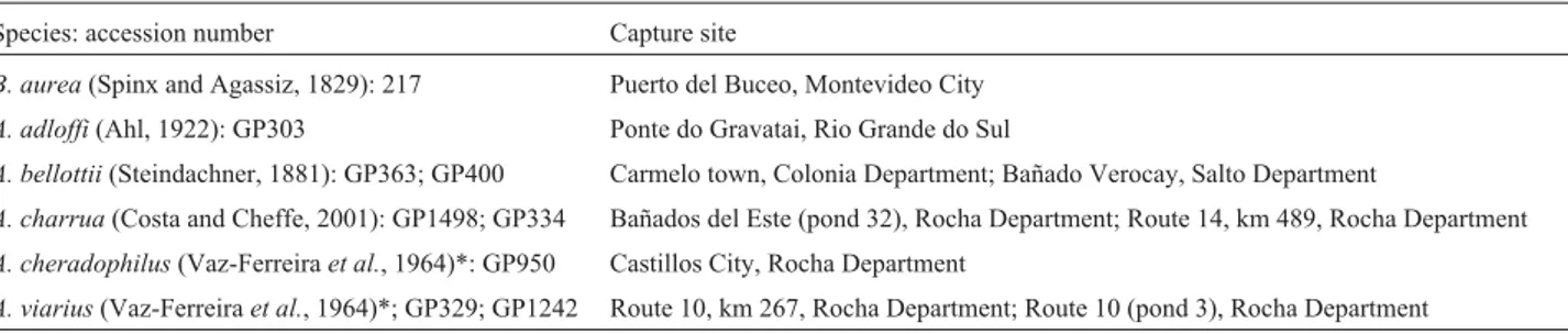
Table 1- Capture sites of theAustrolebias(A) andBrevoortia(B) specimens analyzed by us. TheA. adloffispecimens came from Brazil, all other specimens came from Uruguay.
Species: accession number Capture site
B. aurea(Spinx and Agassiz, 1829): 217 Puerto del Buceo, Montevideo City
A. adloffi(Ahl, 1922): GP303 Ponte do Gravatai, Rio Grande do Sul
A. bellottii(Steindachner, 1881): GP363; GP400 Carmelo town, Colonia Department; Bañado Verocay, Salto Department
A. charrua(Costa and Cheffe, 2001): GP1498; GP334 Bañados del Este (pond 32), Rocha Department; Route 14, km 489, Rocha Department
A. cheradophilus(Vaz-Ferreiraet al., 1964)*: GP950 Castillos City, Rocha Department
A. viarius(Vaz-Ferreiraet al., 1964)*; GP329; GP1242 Route 10, km 267, Rocha Department; Route 10 (pond 3), Rocha Department
by ethanol precipitation of total DNA (modified from Me-dranoet al., 1990). To ascertain the quality of DNA, an aliquot of the extract DNA was run in ethidium bromide stained 1% (w/v) agarose gel using 1XTAE (Tris-Acetate-EDTA).
Amplification of a homeodomain region was per-formed by the polymerase chain reaction (PCR) with the specific primers Sog1 (5’-TGAGCTGGAAAAGGAGTT C-3’) and Sog2 (5’-CTACGGTTTTGGAACCAG-3’). These primers were designed from a partialHoxsequence previously isolated in our laboratory from Austrolebias gymnoventris(Pereiro and García, unpublished data) using the degenerate primers SO1 and SO2 (Bayascas et al., 1997). The PCR was carried out in a 10µL total volume
us-ing 6.4µL of H2O, 1µL of 10X buffer, 0.3µL of MgCl2
(50 mM), 0.2µL of dNTPs (10 mM), 0.5µL of each primer
(10µM), 0.1µL ofTaqDNA polymerase (5 U/µL)
(Invi-trogen) and 1µL of DNA, under the following conditions:
one denaturation step at 94 °C for 5 min, followed by 30 cy-cles of 94 °C for 45 s, 48 °C for 45 s and 72 °C for 1 min, and a final elongation step at 72 °C for 7 min. The PCR products were separated by electrophoresis in a 6% (w/v) non-denaturing polyacrylamide gel and visualized by silver staining (Sanguinettiet al., 1994).
Every PCR fragment was subsequently cloned into the pGEM®-T Easy vector using the reagents and protocols supplied by the manufacturer (Promega). The recombinant DNA was obtained by DNA minipreparation of individual clones by alkaline lysis (Sambrooket al., 1989). The clones were screened for correctly sized inserts by electrophoresis in ethidium bromide stained 0.8% (w/v) agarose gel using 1XTAE, followed by a PCR with the specific Sog1 and Sog2 primers. Sequencing reactions were performed on each template using the Sog2 primer and were analyzed in an automated ABI PRISM 377 DNA Sequencer. Nucleo-tide sequences fromA. bellottii,A. cheradophilusandA. viarius were conceptually translated using the commer-cially available computer software Gene Runner 3.05 for Windows (Hastings Software). The amino acid sequences were compared against the National Center for Biotechnol-ogy Information (NCBI) protein database by using the BLASTp program on the Basic Local Alignment Search Tool (BLAST) network service (Altschulet al., 1990).
Southern blot analysis
To verify the existence ofHoxsequences in the Cyno-lebiatinae subfamily we first performed a Southern blot analysis of genomic DNA from twoAustrolebiassister taxa (A. viarius and A. charrua) using the A. cheradophilus
homeobox amplified fragment as a probe to screen for ho-mologous sequences. We also carried out a Southern blot analysis of genomic DNA from three other Austrolebias
species (A. adloffi, A. bellottii and A. charrua) andB. aurea
using theA. bellottiiamplified fragment as a probe.
For Southern blotting analysis genomic DNA was isolated as describe above from different species of
Austrolebias and the distantly related taxon B. aurea
(Table 1). Approximately 10µg of DNA was digested with Hinf-I (10000 U/mL) (Promega) at 37 °C for 24 h. After di-gestion, the DNA was separated using 0.5X TBE (Tris-Borate-EDTA) and 1.5% (w/v) agarose gel stained with ethidium bromide and transferred onto Hybond-N+ nylon membranes (Amersham Life Science) and the blot hybrid-ized consecutively with two different probes using the ECL direct nucleic acid labeling and detection systems (cata-logue no. RPN3001; Amersham Biosciences). The probes used were the vectors containing the amplified A. cheradophilusandA. bellottiifragment, both digested with 20000 U mL-1of the Nde-I restriction enzyme (Biolabs). Probe hybridization was carried out overnight in 0.5 M NaCl at 42 °C and the hybridized bands visualized after 2 h exposure according to the instructions of the kit.
Phylogenetic analyses
Homeodomain amino acid sequences from A. bellottii,A. viarius,A. cheradophilusand partial homeo-domain sequences for anterior (1-3), medial (4-8) and pos-terior (9-13) Hox paralogue groups from other metazoans obtained from GenBank, were aligned by using the CLUSTALX 1.8 program (Thompson et al., 1997) (Table 2). Partial homeodomain sequences from A. gymnoventrisandA. luteoflammulatus, previously isolated in our laboratory (Pereiro and Garcia, unpublished data) were also included in our alignment. The MEGA 3 program (Kumar et al., 2004) was used to construct a neighbor-joining (NJ) tree (Saitou and Nei, 1987) using a p-distance model and complete deletion. Bootstrap values for the nodes were determined by analyzing 1000 bootstrap repli-cates to estimate the strength of the groupings.
Nucleotide substitution, structural and functional analyses
For nucleotide substitution analysis we used the DnaSP 4.00 computer program (Rozaset al., 2003) to com-pare synonymous (Ks) and non-synonymous (Ka) sites, to calculate ratio rates (ω= Ka/Ks) and to evaluate the codon
usage bias between Cynolebiatinae species and their re-spective paralogue groups, in order to detect positive selec-tion in highly conservedHoxgene fragments.
and Peitsch, 1997) was then used to analyze and visualize the structures.
Results
PCR products and Southern blotHoxgenes analyses
The PCR amplifications using the Sog1 and Sog2 specific primers detected fragments of 105 bp in A. bellottii, 115 bp in A. cheradophilus and 115 bp in A. viarius. The deduced amino acid sequences analyses (BLASTp and sequence comparison) identified them as
Hoxgenes. TheA. cheradophilusandA. viariusamino acid sequence alignments were identical, so we will not differ-entiate between theseHoxgenes in the text. The nucleotide and deduced amino acid sequences of these subfamily spe-cies have been deposited in the GenBank database (Table 2).
The Southern blot analysis detected a single band in bothA. viariusandA. charruausing theA. cheradophilus
homeobox amplified fragment as a probe to screen for ho-mologousHoxsequences (Figure 1a). A single band was
also detected in B. aurea, A. adloffi, A. bellottii and A.
charruausing theA. bellottiiamplified fragment as a probe (Figure 1b).
Phylogenetic analyses
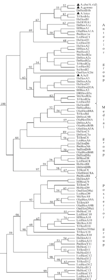
To determine the cluster affiliation of the isolated par-tialHoxgenes, we performed a combined NJ-phylogenetic tree for anterior (Hox1-3), medial (Hox4-8) and posterior (Hox9-13) paralogue groups, using partial homeodomain (HD) sequences of known vertebratesHoxgenes (Table 2, Figure 2). As shown in Figure 2, A. cheradophilus, A. viarius,A. gymnoventrisandA. luteoflammulatusgrouped with members of the paralogue group 1 (PG1) with 69% bootstrap support. It is interesting to note that A.
cheradophilus,A. viariusandA. gymnoventris Hoxgenes appear closely related toDanio rerioHoxB1b (55%) and that ofA. luteoflammulatustoTakifugu rubripesHoxB1a (82%). On the other hand, theA. bellottii Hoxgene grouped within the paralogue group 3 (PG3) with 78% bootstrap support, specifically with members from clusters A and D but without robust statistical support.
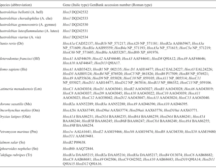
Table 2- Abbreviations and GenBank accession numbers of species used in Figures 2 and 3.
Species (abbreviation) Gene (Italic type) GenBank accession number (Roman type)
Austrolebias bellottii(A. bell) Hox3DQ242532
Austrolebias cheradophilus(A. che) Hox1DQ242533
Austrolebias gymnoventris(A. gymno) Hox1DQ242530
Austrolebias luteoflammulatus(A. luteo) Hox1DQ242531
Austrolebias viarius(A. via) Hox1DQ242534
Danio rerio(Dr) HoxA1aCAD52137;HoxB1bNP_571217;HoxA2bNP_571181; HoxB2aAAH65967;HoxA3a
NP_571609;HoxB3aAAH95559;HoxB4aNP_571193;HoxA5aNP_571615;HoxC5aNP_571219;
HoxC6bNP_571605;HoxB8aAAH53287;HoxB8bXP_691976.
Heterodontus francisci(Hf) HoxA1AAF44639;HoxA2AAF44640;HoxA3AAF44641;HoxD8Q9IA12;HoxA9AAF44646;
HoxA10AAF44647;HoxD13Q9IA17.
Homo sapiens(Hs) HoxA1AAB35423;HoxB1NP_002135;Hox D1AAH14477;HoxA2EAL24227;HoxA3EAL24226;
HoxD3AAH05124;HoxB4NP_076920;HoxC5NP_061826;HoxB6P17509;HoxB8NP_076921;
HoxA9AAP35636;HoxD9NP_055028;HoxC10NP_059105;HoxA11NP_005514;HoxC11
NP_055027;HoxD11AAH14477;HoxD12NP_067016;HoxB13NP_006352;HoxC13NP_059106.
Latimeria menadoensis(Lm) HoxC1AAO43034;HoxD1AAO43041;HoxB2AAO43027;HoxB3AAO43028;HoxA6AAO43019;
HoxC8AAO43037;HoxD9AAO43045;HoxA10AAO43022;HoxC10AAO43039;HoxA11
AAO43023;HoxC12AAO38042;HoxD12AAO43047;HoxA13AAO43024;HoxC13AAO43040.
Morone saxatilis(Ms) HoxB2aAAN52289;HoxB3aAAN52288;HoxA9AAD46396;HoxA10AAD46395.
Oncorhynchus mykiss(Om) HoxA2biAAX63749;HoxD9aiAAX63774;HoxD9aiiAAX63776;HoxD10aiAAX63771.
Oryzias latipes(Olat) HoxA1ABAA86231;HoxD3ABAA86235;HoxB4ABAA86239;HoxD4ABAA86241;HoxA5A
BAA86244;HoxB5BBAA86245;HoxB6BBAA86247;HoxC8ABAA86248;HoxA9ABAA86255;
HoxA9BBAA86254.
Petromyzon marinus(Pm) Hox1wAAL61641;HoxE2AAM19466;HoxN6AAM19474;HoxR8AAC04330;HoxX10AAM19480;
HoxY11AAM19481.
Salmon salar(Ss) HoxB2P09638.
Sphoeroides nephelus(Sn) HoxB6bAAQ72844.
Takifugu rubripes(Tr) HoxB1aDAA05215;HoxB2aDAA05216;HoxB3aDAA05217;HoxB4O13074;HoxC6AAB68682;
HoxC8AAB68681;HoxA9O42506;HoxC9O42502;HoxA10AAB68683;HoxD10Q9IA14;HoxD11
Synonymousvs.Non-synonymous nucleotide substitutions
Although amino acid substitution rates increase, they never exceed those of synonymous substitutions because relaxed purifying selection is acting (Hartl, 2000). How-ever, the non-synonymous (Ka) to synonymous (Ks) sub-stitution ratio (ω) was significantly larger than 1, if positive
selection is acting. Our analysis of synonymous nucleotide substitutions reveals that the species analyzed had a Ks value of 26, whereas the other PG1 species had a Ks value of 23. For non-synonymous substitutions, we obtained a Ka value of 89 for the Cynolebiatinae subfamily species and 81 for the remaining PG1 species (Table 2). In both cases, Cynolebiatinae subfamily species presented higher values with respect to other species of the paralogue group 1. Fur-thermore, when we compared the ratio rates (= Ka/Ks) be-tween the Cynolebiatinae subfamily and other PG1 species we found that they varied from 0.14 to 1.88, indicating that a weak footprint of positive selection was acting (Hartl, 2000).
We also calculated the codon bias index (CBI) to esti-mate the codon usage bias. When the Cynolebiatinae subfamily sequences were excluded, the CBI was 0.670 for the PG1 species, 0.736 for PG3 species and 0.846 for
Cynolebiatinae subfamily species. Additionally, it is worth noting that the G+C3s value for theAustrolebiasspecies was 0.913, whereas 0.703 was obtained for the other PG1 species.
Structural and functional analyses
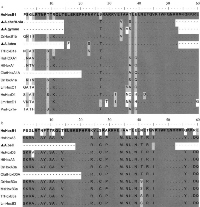
To ascertain which homeodomain (HD) amino acids are conserved within species of this annual fish subfamily, we compared Hox protein sequences from species that grouped with them (Figure 3). The alignment of PG1 partial HD sequences revealed that, as compared with the human HoxB1 sequence, the sequences for the fish studied had the following substitutions: A. cheradophilus and A. viarius
had Thr-27, Val-34 and Ser-37 substitutions; A. gymnoventrishad Thr-27, Val-34, Val-36 and Ser-37 sub-stitutions; andA. luteoflammulatushad Phe-16, Ser-23 and Thr-27 substitutions (Figure 3a). Furthermore, the compar-ison of the primary structures of Austrolebias Hox1 se-quences revealed some differences between them. WhileA. cheradophilus shared high sequence identity with A. gymnoventris, sequence similarity was considerably lower when compared with A. luteoflammulatus. Interestingly, these sequences shared some unexpected residues with other PG1 species. For example, the Thr-27 present inA.
Figure 1- a) Southern blot analysis ofAustrolebias viarius(lane 1) andAustrolebias charrua(lane 2) genomic DNA digested with Hinf-I. The probe used was the amplifiedHoxfragment inAustrolebias cheradophilus. Lane 3: 1 kb DNA ladder molecular marker (GIBCO). b) Southern blot analysis of
cheradophilus,A. gymnoventris and A. luteoflammulatus
was present in almost all sequences analyzed, except in HsHoxB1 and LmHoxD1, whereas the Val-34 present inA. cheradophilus and A. gymnoventris was only present in DrHoxB1b sequence while Ser-37 was present in both HsHoxA1 and DrHoxA1a. On the other hand, Ser-23 pres-ent in theA. luteoflammulatussequence only was also pres-ent in TrHoxB1a. However, it is worth noting that conserved and paralogue-characteristic amino acids were present in all the sequences we analyzed.
The analysis of the PG3 HD sequences revealed that
A. bellottiihad Arg-24, Cys-27, Pro-29, Met-34, Asn-36, Leu-37, Asn-39, Thr-41 and Arg-43 substitutions pared to the human HoxB1 sequence. However, when com-pared to human HoxA3 we founded that the only difference was a Val-45 substitution (Figure 3b). In this sequence, conserved and paralogue-characteristic amino acids were also present. The HD 3D structure modeling ofA. bellottii,
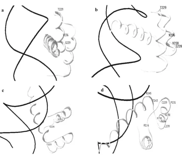
A. cheradophilus,A. gymnoventrisandA. luteoflammulatus
Hox proteins showed that the ternary structure of the HD and the HD-DNA interaction had not been altered (Figure 4).
Discussion
AlthoughHoxgenes have been characterized in many species of teleost fish, this study is the first report about par-tial Hox gene isolation and characterization in annual killifishes belonging to the subfamily Cynolebiatinae.
We employed PCR amplification with specific prim-ers to isolate partial homeodomain sequences from three
Austrolebiasspecies,A. bellottii, A. cheradophilusandA. viarius. These sequences and those previously isolated fromA. gymnoventrisandA. luteoflammulatus(Pereiro and García, unpublished data) represent the first homeobox fragments reported for Cynolebiatinae subfamily species. The isolated sequences show a high percentage of similar-ity with those of other vertebrates. Accordingly with the homeobox sequence conservation in virtually all metazo-ans (McGinniset al., 1984a), our Southern blot analyses re-vealed that homeobox sequences are conserved not only among species of the Cynolebiatinae subfamily but also in the unrelated clupeid fishBrevoortia aurea(Figure 1).
Because of homeodomain amino acid conservation the phylogenetic tree topology (Figure 2) followed by com-parative analyses (Figure 3) allowed us to predict the distri-bution of Austrolebias species partial Hox sequences within the anterior group ofHox genes. So far, we have identified partial homeodomain sequences of threeHox1
sequences and aHox3 sequence in annual fish. Interest-ingly, even though homeodomain sequences are extraordi-narily conserved throughout evolution, comparative analyses have shown unexpected amino acid shifts among Cynolebiatinae subfamily species. The alignment of the HD sequences from the PG1 group reveals that two of these residues reside in two different Austrolebias species, Figure 2- Neighbor-joining tree using partial homeodomain amino acid
Val-36A in A. gymnoventris and Phe-16L in A. luteoflammulatus(Figure 3a). Note that Leu-16 is a con-served and paralogue-characteristic amino acid present in members of the PG1 (Sharkeyet al., 1997). Similarly, anal-ysis of the HD sequences from the PG3 group revealed that
A. bellottiiHD shared high sequence identity with members of clusters A and D, although this species presents a Val-45I residue (Figure 3b).
Although some residues distinguish Hox paralogue groups from one another, it has been shown that the HD
structures are remarkably similar and that the mode of HD-DNA interaction has been extraordinarily well con-served (Sharkeyet al., 1997). Because of this and the fact that the homeodomain of the human HoxB1 protein and those of Cynolebiatinae subfamily species share a greater percentage of amino acid identity (Figure 3), we used the three-dimensional structure of HoxB1 as a template for homology modeling (Figure 4). We found no ternary struc-ture changes even if primary sequences diverge. Neverthe-less,in vivoassays in which the function of homologous
Figure 3- a) Alignment of partial homeodomain amino acid sequences from species that clustered with theAustrolebiasspecies (A. cheradophilus, A. gymnoventris, A. luteoflammulatus and A. viarius) b) Alignment of partial homeodomain amino acid sequences from species that clustered with
proteins is tested in the context of site-directed mutagenesis (Golding and Dean, 1998) may resolve the issue.
As we expected, Hox genes from Cynolebiatinae subfamily species showed the signature of adaptive amino acid replacements since non-synonymous substitutions greatly exceeded the synonymous substitution sites among distantly related taxa of the paralogue group 1. Although they were not statistically significant, the rates of both syn-onymous and non-synsyn-onymous nucleotide substitutions were higher in the Cynolebiatinae subfamily species than in the other PG1 species. Despite the fact that positive selec-tion was acting, the substituselec-tion pattern detected among Cynolebiatinae subfamily and PG1 species could be indi-cating that for more distantly related species two or more independent mutations (multiple hits) might have occurred at the same site. Thus, in a molecular phylogenetic context, amino acid sites were identical not because of identity by descent from a common ancestor but because different
types of mutation (e.g.parallel, convergent or reverse mu-tations) could arise at the same site.
Our analyses based on a restricted highly conserved region of the HD showed that both synonymous and non-synonymous rates increased in many comparisons among Cynolebiatinae subfamily species and other PG1 Hox se-quences. However, the highest codon usage bias value among them (CBI = 0.846) could be indicating that some synonymous substitutions were also constrained for these
Hox genes. Further substitution pattern analysis among CynolebiatinaeHoxgenes could be important in detecting the characteristic evolutionary signature of duplicated di-vergent evolution among theHoxgenes, as has previously been found in the ray-finned fish HoxA cluster (Wagneret al., 2005).
of mitochondrial sequence divergence and high rates of amino acid replacements among coding regions have been previously detected (Garcíaet al., 2000, 2002). In this case,
Hoxgenes were assigned to a series of physiological and life-history variables such as generation time, life span, age of first reproduction, rate of population increase and meta-bolic rate that are certainly not exclusively confined to mi-tochondria.
While the limited homeobox sequences described in the present paper allowed us to assign them to specific paralogue groups, theAustrolebiaspartial Hox sequences could not be assigned to specific Hox clusters.. More se-quence data, e.g., from the entire homeobox region and flanking regions, are needed to assign these genes to their correct clusters. Furthermore, it would be interesting to de-termine the number of Hox clusters in even one species of the Cynolebiatinae subfamily, since at least seven Hox clusters have been described for many teleosts (Amoreset al., 1998, 2004; Aparicioet al., 2002; Naruseet al., 2004) and only four in the killifishFundulus heteroclitus(Misof and Wagner 1996). Given that the development of annual killifishes shows reversible arrest at three well-defined stages, it would also be interesting to compareHoxgene ex-pression between species of the subfamily Cynolebiatinae.
Acknowledgments
We wish to thank the following people: L. Malabarba for kindly donate individuals ofA. adloffifrom RS, Brazil (1991), E. Castillo for kindly provided the degenerate prim-ers SO1 and SO2, G. Bedó and Y. Panzera for their techni-cal assistance, F. Panzera for his helpful corrections and critical reading and an anonymous reviewer for useful com-ments on an earlier draft of this article. The authors are also grateful to the Japanese government for the donation of equipment.
References
Altschul SF, Gish W, Miller W, Myers EW and Lipman DJ (1990) Basic local alignment search tool. J Mol Biol 215:403-410. Amores A, Suzuki T, Yan Y-L, Pomeroy J, Singer A, Amemiya C and Postlethwait JH (2004) Developmental roles of pufferfish Hox clusters and genome evolution in ray-finned fish. Genome Res 14:1-10.
Amores A, Force A, Yan YL, Joly L, Amemiya C, Fritz A, Ho RK,et al.(1998) Zebrafish hox clusters and vertebrate ge-nome evolution. Science 282:1711-1714.
Aparicio S, Chapman J, Stupka E, Putnam N, Chia JM, Dehal P, Christoffels A,et al.(2002) Whole-genome shotgun assem-bly and analysis of the genome ofFugu rubripes. Science 297:1301-1310.
Aparicio S, Hawker K, Cottage A, Mikawa Y, Zuo L, Venkatesh B, Chen E,et al.(1997) Organization of theFugu rubripes
Hox clusters: Evidence for continuing evolution of verte-brate Hox complexes. Nat Genet 16:79-83.
Arezo MJ, Pereiro L and Berois N (2005) Early development in the annual fish Cynolebias viarius. J Fish Biol 66:1357-1370.
Bayascas JR, Castillo E, Muñoz-Mármol AM and Saló E (1997) Planarian Hox genes: Novel patterns of expression during regeneration. Development 124:141-148.
Carter CA and Wourms JP (1991) Cell behaviour during early de-velopment in the South American annual fishes of the genus
Cynolebias. J Morphol 210:247-266.
Chauvet S, Merabet S, Bolder D, Scott MP, Pradel J and Graba Y (2000) Distinct Hox protein sequences determine specificity in different tissues. Proc Natl Acad Sci USA 97:4064-4069. Costa WJE (1998) Phylogeny and classification of Rivulidae
re-visited: Origin and evolution of annualism and miniaturiza-tion in rivulid fishes (Cyprinodontiformes, Aplocheiloidei) J Comp Biol 3:33-92.
Costa WJEM (1995) Pearl Killifishes: The Cynolebiatinae: Sys-tematics and Biogeography of a Neotropical Annual Fish Subfamily (Ciprinodontiformes, Rivulidae). T.F.H. Publi-cations, Neptune City, 128 pp.
García G, Alvarez-Valin F and Gómez N (2002). Mitochondrial genes: Signals and noise in the phylogenetics reconstruction of the annual killifish genus Cynolebias (Cyprinodonti-formes, Rivulidae). Biol J Linn Soc Lond 76:49-59. García G, Wlasiuk G and Lessa EP (2000). High levels of
mito-chondrial cytochrome b divergence in annual killifishes of de genusCynolebias(Cyprinodontiformes, Rivulidae). Zool J Linn Soc 129:93-110.
Gehring WJ, Affolter M and Burglin T (1994a) Homeodomain proteins. Annu Rev Biochem 63:487-526.
Gehring WJ, Qian YQ, Billeter M, Furokubo-Tokunaga K, Schier AF, Resendez-Perez D, Affolter M,et al.(1994b) Homeo-domain-DNA recognition. Cell 78:211-223.
Golding GB and Dean AM (1998) The structural basis of molecu-lar adaptation. Mol Biol Evol. 15:355-369.
Guex N and Peitsch MC (1997) SWISS-MODEL and the Swiss-PdbViewer: An environment for comparative protein modeling. Electrophoresis 18:2714-2723.
Hand SC and Podrabsky JE (2000) Bioenergetics of diapause and quiescence in aquatic animals. Thermochim Acta 349:31-42.
Hartl DL (2000) A Primer of Population Genetics. 3rd edition. Sinauer Associates, Sunderland, 221 pp.
Jaillon O, Aury JM, Brunet F, Petit JL, Stange-Thomann N, Mauceli E, Bouneau L,et al.(2004) Genome duplication in the teleost fishTetraodon nigroviridisreveals the early ver-tebrate proto-karyotype. Nature 431:946-957.
Kim CB, Amemiya C, Bailey W, Kawasaki K, Mezey J, Miller W, Minoshima S,et al.(2000) Hox cluster genomics in the horn shark,Heterodontus francisci. Proc Natl Acad Sci USA 97:1655-1660.
Kissinger CR, Liu BS, Martin-Blanco E, Kornberg TB and Pabo CO (1990) Crystal structure of an engrailed homeodomain-DNA complex at 2.8 A resolution: A framework for under-standing homeodomain-DNA interactions. Cell 63:579-590. Koh EG, Lam K, Christoffels A, Erdmann MV, Brenner S and Venkatesh B (2003)Hox gene clusters in the Indonesian coelacanth, Latimeria menadoensis. Proc Natl Acad Sci USA 100:1084-1088.
Kumar S, Tamura K and Nei M (2004) MEGA3: Integrated Soft-ware for Molecular Evolutionary Genetics Analysis and Se-quence Alignment. Brief Bioinform 5:150-163.
Ledje C, Kim CB and Ruddle FH (2002) Characterization ofHox
genes in the bichir,Polypterus palmas. J Exp Zool 294:107-111.
Lewis EB (1978) A gene complex controlling segmentation in
Drosophila. Nature 276:565-570.
McGinnis W and Krumlauf R (1992) Homeobox genes and axial patterning. Cell 68:283-302.
McGinnis W, Levine MS, Hafen E, Kuroiwa A and Gehring WJ (1984a) A conserved DNA sequence in homeotic genes of theDrosophilaAntennapedia and bithorax complexes. Na-ture 308:428-433.
McGinnis W, Garber RL, Wirz J, Kuroiwa A and Gehring WJ (1984b) A homologous protein-coding sequence in
Drosophilahomeotic genes and its conservation in other metazoans. Cell 37:403-408.
Medrano JF, Aasen E and Sharrow L (1990) DNA extraction from nucleated red blood cells. Biotechniques 8:43.
Misof BY and Wagner GP (1996) Evidence for four Hox clusters in the killifish, Fundulus heteroclitus (Teleostei). Mol Phylogenet Evol 5:309-322.
Naruse K, Tanaka M, Mita K, Shima A, Postleithwait J and Mitani H (2004) A medaka gene map: The trace of ancestral verte-brate proto-chromosomes revealed by comparative gene mapping. Genome Res 14:820-828.
Naruse K, Fukamachi S, Mitani H, Kondo M, Matsuoka T, Kondo S, Hanamura N,et al. (2000) A detailed linkage map of Medaka,Oryzias latipes: Comparative genomics and ge-nome evolution. Genetics 154:1773-1784.
Passner JM, Ryoo HD, Shen L, Mann RS and Aggarwal AK (1999) Structure of a DNA-bound ultrabithorax-extraden-ticle homeodomain complex. Nature 397:714-719. Podrabsky JE, Carpenter JF and Hand SC (2001) Survival of
wa-ter stress in annual fish embryos: Dehydration avoidance and egg envelope amyloid fibers. Am J Physiol Regul Integr Comp Physiol 280:R123-R131.
Podrabsky JE and Hand SC (2000) Depression of protein synthe-sis during diapause in embryos of the annual killifish
Austrofundulus limnaeus. Physiol Biochem Zool 73:799-808.
Podrabsky JE and Hand SC (1999) The bioenergetics of embry-onic diapause in an annual killifish, Austrofundulus limnaeus. J Exp Biol 202:2567-2580.
Prince VE, Joly L, Ekker M and Ho RK (1998) Zebrafishhox
genes: Genomic organization and modified colinear expres-sion patterns in the trunk. Development 125:407-420. Qian YQ, Billeter M, Otting G, Müller M, Gehring WJ and
Wüthrich K (1989) The structure of the Antennapedia
ho-meodomain determined by NMR spectroscopy in solution: Comparison with prokaryotic repressors. Cell 59:573-580. Rozas J, Sánchez-Delbarrio JC, Messeguer X and Rozas R (2003)
DnaSP, DNA polymorphism analyses by the coalescent and other methods. Bioinformatics 19:2496-2497.
Ruddle FH, Bentley KL, Murtha MT and Risch N (1994) Gene loss and gain in the evolution of the vertebrates. Develop-ment Suppl:155-161.
Saitou N and Nei M (1987) The neighbor-joining method: A new method for reconstructing phylogenetic trees. Mol Biol Evol 4:406-425.
Sambrook J, Fritsch EF and Maniatis T (1989) Molecular Clon-ing. A Laboratory Manual. 2nd edition. Cold Spring Harbor Laboratory Press, New York.
Sanguinetti CJ, Dias Neto E and Simpson AJ (1994) Rapid silver staining and recovery of PCR products separated on polya-crylamide gels. Biotechniques 17:914-921.
Schwede T, Kopp J, Guex N and Peitsch MC (2003) SWISS-MODEL: An automated protein homology-modeling server. Nucleic Acids Res 31:3381-3385.
Sharkey M, Graba Y and Scott MP (1997)Hoxgenes in evolution: Protein surfaces and paralog group. Trends Genet 13:145-151.
Thompson JD, Gibson TJ, Plewniak F, Jeanmougin F and Higgins DG (1997) The CLUSTAL_X windows interface: Flexible strategies for multiple sequence alignment aided by quality analysis tools. Nucleic Acids Res 25:4876-4882.
Wagner GP, Takahashi K, Lynch V, Prohaska SJ, Fried C, Stadler PF and Amemiya C (2005) Molecular evolution of dupli-cated ray finned fish HoxA clusters: Increased synonymous substitution rate and asymmetrical co-divergence of coding and non-coding sequences. J Mol Evol 60:665-676. Wolberger C (1996) Homeodomain interactions. Curr Opin Struct
Biol 6:62-68.
Wourms JP (1972a) The developmental biology of annual fishes. I. Stages in the normal development of Austrofundulus myersiDahl. J Exp Zool 182:143-168.
Wourms JP (1972b) The developmental biology of fishes. III. Pre-embryonic diapause of variable duration in the eggs of annual fishes. J Exp Zool 182:389-414.
Wourms JP (1967) Annual fishes. In: Wilt FW and Wessels NK (eds) Methods in Development Biology. Thomas Y. Crowell, New York, pp 123-137.
Internet Resources
Gene Runner software, http://www.generunner.com.
Basic Local Alignment Search Tool (BLAST), http://www.ncbi. nlm.nih.gov/BLAST (August 1, 2005).
Swiss Model software, http://swissmodel.expasy.org.