Immune response and
tissue cytoprotection
Two sides of the same coin in
immunopathology
Ivo Marguti
Dissertation presented to obtain the Ph.D degree in Biochemistry
Instituto de Tecnologia Química e Biológica | Universidade Nova de Lisboa
Research work coordinated by:
Ph.D Supervisor: Dr. Thiago Lopes Carvalho
The work presented on this Thesis has been funded by the grant
Index
Acknowledgments 1
Preface 2
Sumário 3
Summary 6
Chapter 1: General Introduction 9
1.The immune system 10
1.1 Innate immunity 12
1.2 Adaptive immunity 19
1.2.1 T cell activation 19
1.2.2 Regulation of immune responses by T cells 24
1.3 Tolerance to infection 27
2. Heme oxygenase-1 31
2.1 Heme catabolism and its by-products 31
2.2 Regulation of HO-1 expression 34
2.3 Immunoregulatory effects of HO-1 36
2.3.1 Effects of the heme/HO-1 system on
innate immunity 36
2.3.2 Effects of the heme/HO-1 system on
adaptive immunity 40
2.3.3 Cytoprotective effects of HO-1 43
3. Iron metabolism and immunity 46
4. Sepsis and malaria as models of immune-mediated
diseases 52
4.1 Sepsis 52
Chapter 2: Expansion of CD4+CD25+Foxp3+ T Cells by
Bone Marrow-Derived Dendritic Cells 61
1. Abstract 62
2. Introduction 63
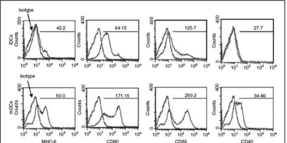
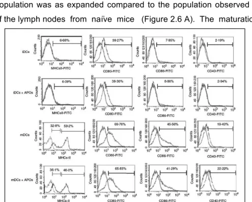
3. Results 65
3.1 Characterization of bone-marrow-derived
dendritic cells 65
3.2 Phagocytosis of apoptotic thymocytes by
dendritic cells 66
3.3 Effect of apoptotic cells on dendritic cell
maturation 67
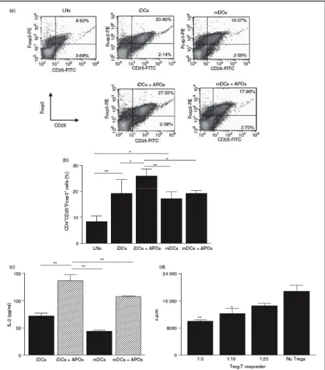
3.4 Evaluation of the CD4+CD25+Foxp3+ T cell
population after co-culture of lymph-node cells
with DC 67
3.5 Expansion of the CD4+CD25+Foxp3+ T cell
population in CD4+CD25+ depleted cultures 69
4. Discussion 72
5. Methods 76
7. Acknowledgments 80
8. References 80
Chapter 3: A Central Role for Free Heme in the
Pathogenesis of Severe Sepsis 85
1. Abstract 86
2. Introduction 87
3. Results 88
polymicrobial infection 88
3.2 HO-1 prevents free heme from eliciting severe
sepsis 93
3.3 Free heme is a critical component in the
pathogenesis of severe sepsis 98
3.4 Free heme elicits programmed cell death 100
3.5Heme triggers HMGB1 release in vitro and
in vivo 102
3.6 HPX neutralizes the cytotoxic effect of free
heme 104
3.7 Low HPX serum concentration is associated
with organ dysfunction and fatal outcome in septic
shock patients 106
4. Discussion 108
5. Methods 112
6. Acknowledgments 123
7. References 124
Chapter 4: Sickle Hemoglobin Confers Tolerance
to Plasmodium infection 127
1. Abstract 128
2. Introduction 129
3. Results 130
3.1 Sickle Hb Confers a Survival Advantage
against Malaria in Mice 130
3.2 Sickle Hb Confers Tolerance to Plasmodium
Infection in Mice 133
3.4 Induction of HO-1 by Sickle Hb Inhibits the
Production Of Chemokines Involved in the
Pathogenesis of ECM 138
3.5 Sickle Hb Confers Tolerance to Plasmodium
Infection via HO-1 Expression in Hematopoietic Cells 139
3.6 Sickle Hb Inhibits the Activation/Expansion of
CD8+ T Cells Recognizing Antigens Expressed
by Plasmodium 142
3.7 Sickle Hb Induces HO-1 Expression via a
Mechanism Involving Nrf2 143
3.8 Sickle Hb Confers Tolerance to Plasmodium Infection via a Mechanism Involving CO
Produced through Heme Catabolism by HO-1 145
4. Discussion 152
5. Tables 157
6. Methods 159
7. Acknowledgments 166
8. References 168
Chapter 5: General Discussion 171
References 185
Acknowledgments
I would like to express my deepest gratitude to Dr. Thiago Lopes Carvalho, who became my supervisor under turbulent circumstances and made everything in his power to ensure the completion of this Thesis in the best possible way. I am also in great debt with Dr. Bruno Silva-Santos and Dr. Lars Jansen, who were caught in the turmoil and were utterly understanding and supportive. The help of the three of you was indispensable for the completion of this Thesis.
My gratitude also extends to the IGC Direction for the opportunity to work at this Institute for four years, and for making sure its students are supported whenever needed.
I would also like to thank my dear friend Dr. Luiz Vicente Rizzo for his constant availability to support and advise a former student. Many times he has offered the right word at the right time, and I feel very fortunate to have him as a friend.
Many thanks to the people that generously spent their time making this Thesis better. A very special thanks to Alê, Lu, Virgínia and Jorge Carneiro.
My appreciation is extended to the co-authors of the published work that made this Thesis possible.
Many thanks to Rui Gardner and Telma Lopes for endless hours spent sorting cells.
Preface
Sumário
O sistema imune é fundamental para a manutenção da viabilidade do hospedeiro em casos de infecção. No entanto, os mecanismos usados pelo sistema imune para controlar infecções podem também causar destruição tecidual, levando ao desenvolvimento de imunopatologia. Para sobreviver a infecções o hospedeiro precisa controlar a proliferação do agente infeccioso e, ao mesmo tempo, evitar os efeitos deletérios da resposta imune em seus tecidos. Para alcançar este objetivo, o hospedeiro utiliza as seguintes estratégias: aumento da capacidade tecidual de suportar danos infligidos pelo sistema imune e/ou controle da ativação exacerbada do sistema imune. Nesta Tese buscou-se aprofundar o conhecimento sobre os mecanismos utilizados pelo hospedeiro para empregar de maneira bem sucedida tais estratégias.
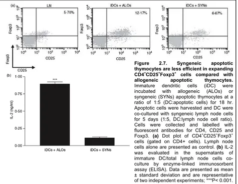
Para estudar o papel de células do sistema imune na regulação da magnitude de respostas imunológicas, analisamos a capacidade das células dendríticas (DC) de expandir células T reguladoras (TREG). Utilizando um sistema de co-cultura de
células in vitro, demonstrou-se que as DC são capazes de induzir a expanão da população de TREG com capacidade
supressora. É de realçar que, a expansão de TREG foi maior
quando as DC apresentavam antígenos não-próprios. Estes resultados sugerem que a expansão da população de TREG
Os mecanismos envolvidos na prevenção da imunopatologia através do aumento da capacidade tecidual de suportar danos causados pelo sistema imune, foram estudados em dois modelos experimentais de infecção, malária e sepse. Utilizando o modelo experimental de sepse, estudou-se o efeito da enzima heme oxigenase-1 (HO-1) na proteção dos tecidos contra danos causados pela resposta imune. Para tal, foram utilizados ratinhos com o gene da enzima HO-1 intacto (Hmox1+/+) e ratinhos em que este gene foi inativado (Hmox1-/-). A indução de sepse em ratinhos Hmox1+/+ acarretou modesto dano tecidual a orgãos viatis, enquanto o mesmo procedimento em animais Hmox1-/- causou severo dano tecidual a orgãos vitais levando à falência dos mesmos. É importante notar que apesar da diferença observada na severidade dos danos aos orgãos vitais, a resposta imune à infecção induzida pela sepse foi muito similar em ambos os genótipos. Esses dados sugerem que na ausência da expressão de HO-1 há um aumento na susceptibilidade dos orgãos a danos causados pelo sistema imune. A maior suscetibilidade observada em animais Hmox1
-/-ocorre, ao menos parcialmente, devido ao maior nível de heme em circulação nestes animais. É de realçar que, a administração de heme a animais Hmox1+/+ após a indução de sepse desencadeou severo dano tecidual a orgãos vitais, enquanto a retirada de heme da circulação preveniu o desenvolvimento de sepse severa. Esses resultados reforçam a noção de que o heme atua como uma molécula capaz de potencializar danos teciduais a orgãos vitias em animais com sepse.
transgênicos que expressam moléculas de hemoglobina mutadas (HbSAD), o que acarreta uma forma leve de anemia falciforme, não desenvolvem ECM, enquanto animais com hemoglobina normal (HbWT) sucumbem à doença. É de realçar que, a proteção conferida pela HbSAD possui dois componentes: o aumento da capacidade dos tecidos em suportar danos infligidos pelo sistema imune e, o controle da responsta imunológica evitando o desenvolvimento de uma resposta exacerbada. Como observado no modelo experimental de sepse, a expressão de HO-1 em animais com HbSAD é essencial para manter baixos níveis de heme durante a infecção e, assim, evitar a potencialização dos danos causados pelo sistema imune. Por outro lado, a HbSAD possui um efeito immunoregulatório, ainda sem mecanismo definido, que é independente da expressão de HO-1. Esse efeito induz uma drástica redução na expansão e ativação de células T CD8+, essenciais para o desenvolvimento da ECM.
Summary
The immune system is fundamental to maintaining the host’s viability upon infection. Nonetheless, the mechanisms used to control pathogens may also cause tissue damage, leading to the development of immunopathology. The host’s capacity to survive infections depends on its ability to control the pathogen burden, while avoiding the deleterious effect of immune responses on its own tissues. To achieve this goal, the host may apply the following strategies: increasing tissue resilience to immune-mediated insult and/or controlling exacerbated immune activation. This Thesis sought insight into the mechanisms used by the host to successfully employ both strategies.
To study the role of immune cells in regulating the extent of immune activation, we analyzed the capacity of dendritic cells (DC) to expand regulatory T cells (TREG). By using an in vitro
co-culture system, it was demonstrated herein that DC can induce the expansion of TREG with suppressive activity. Importantly, TREG
expansion is greater when DC are loaded with non-self antigens. These findings suggest that the expansion of the TREG
compartment during an immune response might serve as a mechanism to avoid unfettered immune activation that could result in immunopathology. In this way, the interactions among the cells of the immune system might, per se, be sufficient to prevent the development of immunopathology.
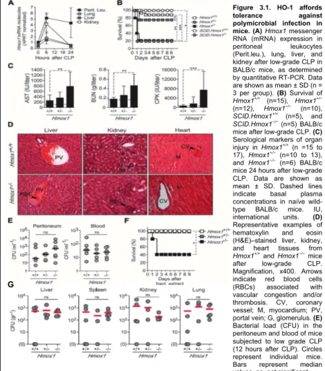
damage and organ failure, Hmox1-/- mice suffered from extensive tissue damage leading to multi-organ failure. Importantly, this occurred despite apparently similar overall levels of immune activation in WT and Hmox1-/- mice, suggesting that in the absence of HO-1 tissues become less resilient to insults perpetrated by the immune system and, therefore, more prone to immune-mediated damage. The diminished resilience to immune insult is at least partially due to increased heme levels in Hmox1
-/-mice. In line with the pathological effect of heme in precipitating tissue damage in sepsis, its administration caused mild sepsis to become lethal in WT type animals, while heme scavenging rescued mice from developing severe sepsis.
The action of increased tissue cytoprotection acts together with the control of unfettered immune response to afford protection against experimental cerebral malaria (ECM) by mutations in hemoglobin. Transgenic animals carrying a mutated form of hemoglobin (HbSAD), leading to a mild sickle cell disease phenotype, were protected from ECM development while animals with normal hemoglobin (HbWT) succumbed. The protection afforded by HbSAD derives from a composite effect of increased tissue cytoprotection in addition to immunoregulation. As in sepsis, HO-1 protects against ECM development by decreasing heme levels upon infection, thereby protecting tissues from the highly deleterious effects of combined exposure to pro-inflammatory mediators and heme molecules. On the other hand, the immunoregulatory effect of HbSAD works by a yet unidentified mechanism, independent of HO-1 expression, and leads to the marked suppression of CD8+ T cells proliferation and activation.
1. The immune system
The immune system is comprised of a wide range of molecules and cell types spread along the body surface and strategically placed to detect and rapidly respond to invading pathogens and/or insulting agents. Its importance in maintaining host viability is strikingly demonstrated in the rare cases of natural mutations affecting molecules of the immune system in human subjects, leading to increased susceptibility to infections1-5 or to the development of autoimmune diseases6.
The normal course of a protective immune response is often characterized by the concerted action of the innate and adaptive immune system (see sections 1.1 and 1.2). The initial steps in this process require activation of the innate immune system through the recognition of evolutionarily conserved molecules expressed by microorganisms, termed broadly P
athogen-Associated Molecular Patterns (PAMP), and/or host-derived molecules associated with tissue damage or malfunction, termed broadly Danger-Associated Molecular Patterns (DAMP). This recognition process occurs through a multitude of germ-line encoded receptors, generally termed Pattern Recognition
Receptors (PRR), and leads to innate immune cell activation. Upon activation, cells of the innate immune system termed
antigen presenting cells (APC) (see section 1.1), migrate to secondary lymphoid organs, carrying processed antigenic peptides. The recognition of peptides in the context of major
characterized by the ability to produce large quantities of cytokines and/or kill target cells upon antigenic re-stimulation. B cells can be directly activated by PRR stimulation alone or in combination with B cell receptor ligation, but normally require help from T cells in order to achieve full differentiation. After activation B cells differentiate into plasma cells and start producing large quantities of antibodies.
innate immune activation to adaptive immune effector function and memory acquisition, need to be tightly regulated to avoid excessive tissue damage and/or development of autoimmunity (see sections 1.1, 1.2).
A more detailed description of the innate and adaptive immune response will be given below.
1.1 Innate immunity
The innate immune system is the host’s first line of defense against invading pathogens and/or harmful insults. The cellular components of the innate immune system include myeloid cells, such as macrophages and dendritic cells (DC), neutrophils, mast cells, basophils, and natural killer (NK) cells. More recently, previously uncharacterized cell populations with innate characteristics, and mainly involved in inducing T helper 2 responses (TH2) (see section 1.2), have been described and
The activation of the innate immune system can be depicted schematically as a process that relies on detection of pathogens and/or insults, with the subsequent activation of intracellular signaling cascades leading to gene expression of effector molecules. These effector molecules will, in turn, act to eliminate the insult through a multitude of mechanisms.
The process of detecting pathogens relies on the recognition of evolutionarily conserved molecular structures that are essential for microorganism survival, such as components of the cell wall or nucleic acids, and, therefore, are unlikely to suffer major modifications without consequent loss of pathogen fitness. These conserved structures are recognized by germ-line encoded receptors expressed in a variety of cell types, broadly termed PRR12. Importantly, PRR can also recognize or respond to concentration changes in endogenous molecules produced or released upon cellular stress or damage, i.e. ATP13, uric acid14,15,
serum amyloid A16, amyloid-β17 , endogenous nucleic acids18-21,
and high-mobility group box-1 (HMGB1)22.
Four families of PRR have been described so far: Toll-like receptor (TLRs), Nucleotide-binding oligomerization domain-like
receptors (NLRs), retinoic acid inducible gene I
(
RIG-I)-likeThe signals initiated by PAMP recognition via PRR culminate in the activation of key transcription factors that are primarily responsible for the transcription of effector molecules. Among these transcription factors are nuclear factor kappa B (NF-κB), activating protein-1 (AP-1), nuclear factor of activated
T-cells (NFAT) and interferon regulatory factor (IRF) 3 and 723-26. The intensity of the signaling pathways leading to the activation of these transcription factors will dictate the specificity of the resultant response27. Importantly, in the case of pathogens, most often more than one PRR will be triggered upon recognition. The overall production of effector molecules will, therefore, depend on the net effect of activation and inhibitory signals received by the cell. As a general principle, PRR stimulation will lead to NF-κB, NFAT, and AP-1 activation that will, in turn, initiate the transcription of proinflammatory cytokines, chemokines and molecules involved in direct pathogen killing such as antimicrobial peptides, nicotinamide adenine dinucleotide phosphate (NADPH) oxidase subunits, and nitric oxide synthase (iNOS). The production of type I interferons will occur through the activation of IRF3 and IRF728.
and TRIF-related adaptor molecule (TRAM)33 are also involved in TLR signaling, and distinct combinations among them upon stimuli generate specific patterns of gene transcription.
CLRs signal through tyrosine-based motifs such as immunoreceptor tyrosine-based activation motifs (ITAM), hemITAM (resembling half of normal ITAM motifs) and immunoreceptor tyrosine-based inhibitory motifs (ITIM). While the latter might work by increasing the threshold for innate cell activation34-36, the first two activate innate cells. Importantly, not all CLRs have an intracellular domain capable of transducing signal and are, therefore, coupled with adaptors, such as the Fc
receptor γ chain, that contain tyrosine-based motifs and are responsible for signal transduction upon PAMP stimulation26. The ITAMs and hemITAMs will recruit and activate spleen tyrosine kinase (Syk) and the ITIM will recruit phosphatases, i.e. SHP-1 and SHP-226. Upon Syk activation, recruitment of the adaptor molecule CARD9 normally ensues and leads to the activation of downstream transcription factors, such as NF-κB37,38.
NLRs form a large family of cytoplasmic receptors that have a particular structure containing a nucleotide-binding domain (NBD) and leucine-rich repeat domain (LRR). NOD-1 and NOD-2 induce the production of proinflammatory cytokines using
lysosomal contents into the cytoplasm17,44, i.e. cathepsin B. The activation of inflammatory caspases follows inflammasome assembly and may lead to different outcomes such as the
conversion of pro-IL-1β and pro-IL-18 to active forms or cell death39.
RLRs constitute the smallest family of PRR. It is composed of the cytoplasmic RNA helicases retinoic acid inducible gene 1 (RIG-I), melanoma differentiation associated factor 5 (MDA5), and laboratory of genetics and physiology 2 (LGP2). Unlike the other PRR families, RLRs seem to be restricted in the array of pathogens they recognize, being limited to viral detection, and more specifically, to viral RNA detection25. Upon RNA binding, RLRs associate with the adaptor protein MAVS and induce NF-kB-dependent transcription of proinflammatory genes and IRF3 and IRF7-dependent transcription of type I interferons25. Some of the characteristics and ligands of the different PRR families are summarized in table 1.1.
To avoid exacerbated inflammation and potential tissue damage, innate immune responses are regulated by a myriad of regulatory mechanisms. Some examples are discussed in further detail below.
Animals deficient for IRAK-M (Irak-m-/-) have increased intestinal inflammation upon Salmonella thyphimurium infection45 and succumb to influenza infection due to an uncontrolled inflammatory response46. Accordingly, Irak-m-/- cells present increased NF-kB45, p38 mitogen-activated protein (MAP) kinase
45,47, c-Jun N-terminal kinase (JNK)45 and extracellular-signal
PAMP.
Table 1.1. Characteristics of PRR from different families.
PRR Cellular localization Ligands
Proximal adaptor molecule
TLR
TLR2 Plasma membrane Lipoprotein MyD88 TLR3 Endolysosome Double Stranded RNA TRIF TLR4 Plasma membrane LPS MyD88 and TRIF TLR5 Plasma membrane Flagelin MyD88 TLR7 Endolysosome Single stranded RNA MyD88
TLR9 Endolysosome CpG-DNA MyD88
TLR10 Endolysosome Profilin-like molecule MyD88
NLR
NOD1 Cytoplasm
γ -D-glutamyl-mesodiaminopimelic
acid RIP2
NOD2 Cytoplasm muramyl dipeptide RIP2, MAVS
CLR
Dectin-1 Plasma membrane β-Glucan Syk/CARD9 Dectin-2 Plasma membrane β-Glucan Syk/CARD9 Mincle Plasma membrane SAP130 Syk/CARD9 DNGR-1 Plasma membrane Unknown content of dead cells Syk
RLR
RIG-I Cytoplasm Short double stranded RNA MAVS MDA5 Cytoplasm Long double stranded RNA MAVS
Note: Adapted from references 23-26,29,39.
was later described. A20-deficient mice (A20-/-) die shortly after birth showing severe multi-organ inflammation, an effect at least partially attributable to uncontrolled NF-κB activation50.
Subsequently, A20 was shown to modulate signaling by TLRs51,52, NLRs53 and RLRs54 as well. Importantly, A20
expression is induced upon NF-κB activation and, therefore, acts in a negative feedback loop to contain exacerbated activation of the former48. Other ubiquitin-editing enzymes such as deubiquitinating enzyme A (DUBA)55 and CYLD56,57 have also been shown to act as negative modulators of innate immune activation.
There are several other mechanisms described to limit innate immune cell activation such as, but not limited to, the expression of molecules with RNase activity inducing rapid decay of inflammatory cytokine RNA58, microRNAs59, inhibitory receptors36,60,61, and suppression of innate immune cells by specific subsets of T lymphocytes62-64. Of note, heme oxygenase-1 (HO-oxygenase-1) is proposed to be one of the molecules involved in controlling activation of innate immune cells65. Data supporting this notion is described in detail in section 2. The existence of many diverse systems implicated in controlling excessive inflammation highlights its importance in maintaining host viability. Importantly, one common feature of deregulated innate immune cell activation is the consequent unfettered activation of the adaptive immune compartment58,60, a process thought to be mainly driven by activated dendritic cells (DC).
Later, data using transgenic mice allowing for transient DC depletion confirmed that DC are essential in vivo for T cell priming71-75 and re-stimulation76,77. This unique capacity relies on DC’s ability to provide T cells with adequate amounts of MHC-peptide complex, costimulation and cytokines, when properly activated by PAMP78,79.
The cytokines produced by DC during T cell activation play a major role in instructing which sort of T cell response will be generated80,81. Notwithstanding their role in initiating adaptive immune responses, DC also play a predominant role in central82,83 and peripheral immunological tolerance84-86 (see section 1.2 and 1.3). The mechanisms employed by DC to generate and maintain peripheral tolerance are discussed in further detail below.
1.2 Adaptive Immunity
1.2.1 T cell activation
are generically termed “central tolerance” and, although essential to maintain host viability, fall out of the scope of this thesis and will not be described in further detail. The focus will be given to mechanisms inducing effector function on the major T cell subsets, i.e. CD4+ and CD8+, and on mechanisms involved in maintaining immunological tolerance in the periphery and avoiding immunopathology.
Both major T cell subsets, i.e. CD4+ and CD8+, are activated via a similar process that is comprised of signals given to the T cell by the APC. The first signal inducing T cell activation is triggering of the T cell receptor (TCR) by cognate recognition of antigenic peptides presented by APC, particularly DC, in the context of MHC class I and II molecules, for CD8+ and CD4+ T cells respectively. Together with TCR stimuli, APC will deliver costimulatory signals by engaging several pairs of receptors on the APC and the T cell surface such as CD80/86 and CD2887,88, 4-IBB and 4-IBBL89,90, CD70 and CD2791,92. The costimulatory signals will convey proliferative, survival and, in some cases, instructive signals to T cells. Cytokines are the third signal provided by APC to T cells. They work as instructive signals that transmit to T cells information regarding the kind of pathogen encountered and, therefore, gear T cells to differentiate into the appropriate effector phenotype79. The net effect of available antigen amount, costimulation and instructive cytokines will ultimately lead to the generation of the adequate T cell response. As described above, cytokines are the distinctive signal required for the differentiation of CD4+ T cells into different effector TH cell lineages, although some costimulatory molecules
have also been reported to provide signals that drive TH cell
differentiation of the main TH cell subsets, i.e. TH1, TH2 and TH17,
are well defined. Briefly, TH1 cells rely on IL-12 signaling for their
differentiation95, TH2 rely on IL-496, and TH17 relies on TGF-β and
IL-697,98. Each of these subsets will produce a determined set of signature cytokines that will mediate their effector function (see table 1.2). Importantly, the main cytokines produced by each TH
cell lineage, will reinforce their own phenotype while inhibiting the conversion of naïve cells into any other TH cell phenotype80.
Signaling through cytokine receptors will lead to activation of specific signal transducers and activators of transcription (STAT) transcription factors. STAT activation, in turn, will lead to expression of lineage specification transcription factors that reinforce TH lineage commitment, largely through epigenetic
modifications of target genes99. An overall view of the cytokines and transcription factors involved in TH cell differentiation can be
found on table 1.2.
Table 1.2. Minimal cytokine requirements for TH cell lineage
commitment.
Lineage
Inducer cytokines
Cytokine signal transduction
Lineage transcription
factor
Effector cytokines/molecules
TH1 IL-12 STAT4 T-bet IFNγ, TNF
TH2 IL-4 STAT6 GATA-3 IL-4, IL-5, IL-13
TH17 IL-6+TGF-β STAT3 RORγT IL-17, IL-22, IL-21
TREG TGF-β+IL-2 Smad3/
STAT5
Foxp3 IL-10, TGF-β,
CLTA4
Note: Cytokine requirements based on in vitro differentiation of naïve T cells in the presence of TCR triggering and costimulation80.
response to signals received by innate immune cells via their PRR. Therefore, upon recognition of PAMP from fungi APC will produce higher amounts of IL-6 and IL-2337,38 and induce TH17
responses. On the other hand, Gram-positive bacteria95 or PAMP from protozoan parasites, such as Toxoplasma gondii100, will induce IL-12 production and the consequent generation of TH1
cells. In the context of helminth infection IL-4, most probably derived from innate cells101-103, has been shown to play a predominant role and will orchestrate the development of a TH2
response.
The requirements of TCR stimulation, costimulation and instructive cytokines are also necessary for CD8+ T cell activation. In the case of CD8+ T cells, antigen is recognized coupled to MHC class I molecules and delivered together with costimulation by APC. It is also possible for CD8+ T cells to become producers of cytokines analogous to those produced by TH2 and TH17 cells however, the relevance of these
subpopulations is currently poorly understood104-107. In fact, the cytokine milieu present during CD8+ T cell activation is thought to influence more dramatically the acquisition of effector versus
memory potential108,109. When fully activated, CD8+ T cells will become IFNγ and TNF producers and will express cytotoxic molecules, i.e. perforin and granzyme B.
Type I IFN and IL-12 are largely produced upon viral infection and, together with IL-2, drive expansion and generation of fully activated short-lived cytotoxic CD8+ T cells expressing
the generation of memory CD8+ T cells rather than short-lived effector cells, as observed by the abnormal accumulation of memory CD8+ T cells in mice overexpressing IL-21116 and the inability to control chronic viral infection when IL-21 signaling is disrupted117-119. Activation of the mammalian target of rapamycin (mTOR) pathway seems to be critically involved in driving CD8+ T cell differentiation towards an effector versus a memory phenotype. IL-12-induced expression of the transcription factor T-bet is blocked by rapamycin with a concomitant increase in expression of the transcription factor Eomesodermin120. Importantly, while T-bet promotes effector CD8+ T cell response, Eomesodermin is associated with memory CD8+ T cell generation121,122.
1.2.2 Regulation of immune responses by T cells
Regulatory T cells (TREG) are the main cell type involved
in regulating immune reactivity in homeostatic conditions. Unlike TH cells, TREG cells exit the thymus already committed to this
lineage, and with a phenotype resembling that of activated/effector T cells134. Mice that had their thymus removed shortly after birth, allowing TH but impeding TREG migration to the
periphery, develop autoimmune disease135. The master transcription factor implicated in TREG differentiation is forkhead
box P3 (Foxp3) and animals and humans deficient in this transcription factor show loss of immunological tolerance and develop severe autoimmunity136-139. Importantly, TREG are
fundamental throughout life as its conditional ablation in adult animals leads to severe autoimmunity64.
Many different mechanism(s) have been described via which TREG suppress immune responses. These include inhibition
of stable contact formation between DC and naïve TH cells in the
lymph-node140,141, production of the anti-inflammatory cytokines TGF-β142,143 and IL-10144,145, and use of membrane bound
molecules such as cytotoxic T lymphocyte antigen-4 (CTLA-4)146,147. Most likely, all the mechanisms described above, and possibly others, will act in concert to maintain homeostasis. Importantly, during infections the control of ongoing immune responses to pathogens by TREG needs to be tightly regulated to
ensure pathogen clearance and, at the same time, avoid immunopathology148-151.
Although TREG were at first thought to be exclusively of
and in vivo, towards a TREG phenotype. In vitro, TCR stimulation
with concomitant IL-2 and TGF-β signaling induce naïve CD4+ T cells to express the transcription factor Foxp3 and to acquire suppressive function152,153. In vivo, DC were shown to be an important cell type in maintaining peripheral tolerance, partially through de novo generation and maintenance of TREG in the
periphery. Several different mechanisms used by DC to maintain and generate peripheral tolerance have been demonstrated. Non-immunogenic antigen presentation by DC was shown to delete CD4+84 and CD8+ T85,86 cell clones in the periphery, leading to antigen-specific unresponsiveness. DC were also shown to recirculate into the thymus carrying peripherally acquired antigens and inducing deletion of T cell clones reactive to these antigens82.
The induction and expansion of T cells with regulatory properties in the periphery by DCs, including IL-10 producing type-1 T regulatory cells (Tr1)154,155 and TREG156-159, was also
demonstrated. Until recently the differences between thymic derived TREG and peripheral induced TREG cells were very poorly
understood. However, in 2010 the transcription co-factor homeodomain-only protein (Hopx) was described as being fundamental for the suppressor function of TREG generated by DC
in the periphery, without interfering with the function of thymic-derived TREG160. Importantly, a specific DC subpopulation present
in the gut was described to be especially tailored to induce TREG
via a pathway that involves TGF-β and retinoic acid161-163. Most recently, it was demonstrated that in order to avoid TH
necessary, presumably because the peripheral conversion of naïve T cells expands the TCR repertoire of TREG164.
Of note, recent reports have suggested that TREG cells
can themselves be subdivided into different lineages capable of suppressing distinct types of immune responses. It was demonstrated that TREG expressing the transcription factor IRF4,
involved in TH2 differentiation, were more efficient in suppressing
TH2 responses165. That was also the case for the expression of
STAT3 in TREG involved in suppressing TH17 responses166, and
T-bet in TREG suppressing TH1 responses167. These findings
suggest that TREG would be generated or instructed outside the
thymus as an immune response begins, and that the cues involved in TH differentiation could also serve to endow TREG with
specific capacity of controlling the TH response in question.
An interesting feedback loop controlling DC and TREG
numbers has been described in vivo. Adult mice engineered to express the diphtheria toxin receptor along with Foxp3 show a sharp increase in the number of DC upon TREG ablation64. This
increase in DC numbers is thought to be partially responsible for the deregulated TH cell response that followns TREG ablation. It
was later demonstrated that the mechanism underscoring the increase in DC numbers relies on the fact that TREG ablation
increases the amount of available Flt3L, a cytokine that induces the proliferation of DC precursors in secondary lymphoid organs168. Interestingly, loss of DC also induces loss of TREG cells
and consequent T cell activation169. Conversely, increasing DC numbers also increases the number of TREG, to the extent that
regulatory loop to avoid unregulated immune activation and immunopathology.
Interestingly, not only cells with clear regulatory phenotypes, such as TREG and Tr1 cells are able to restrain
immune activation. CD8+ and CD4+ T cells were shown to regulate innate immune activation after PAMP administration, in vivo and in vitro, in an antigen and TREG-independent manner62.
Strikingly, effector/memory CD4+ T cells specifically block NLRP1 and NLRP3 activation in macrophages via signaling pathways initiated by ligation of receptors of the TNF family63.
1.3 Tolerance to infection
In animals, the most studied mechanism to survive infections is resistance. It relies on the ability of the host to prevent disease by diminishing pathogen load. Many mechanisms used by innate and adaptive immunity work by reducing pathogen load and are, therefore, involved in host resistance. Acting in concert with resistance, tolerance to infection encompasses mechanisms that limit the decrease in host health status to a given pathogen load. Tolerance has been well documented and studied in plants, but is still poorly understood in animals170,171.
H
HS= u
HS+ b
H.I
HHS: host health status or fitness
uHS: health status of an uninfected host
bH: slope of the relationship between HHS and I, or
tolerance
I: pathogen load
Given the above, it is possible to conclude that HHS upon
infection depends on host’s health status when uninfected (UHS)
and on how much its health status declines at a given pathogen load during infection (BH.I).
An example of how one can compare tolerance among hosts is shown in figure 1.1. Figure 1.1A shows two genotypes that share the same health status when uninfected. However, upon infection the decline in health status as a function of parasite load is greater in the green genotype, i.e. the slope is steeper, than in the red genotype. In this case the red genotype is more tolerant to infection, by the specific pathogen in question, than the green genotype.
Figure 1.1. Variation in host tolerance. (A) Two different genotypes, depicted as red and green lines, are subjected to infection and tolerance is determined by the slope of the decline in health status as a function of pathogen load. In this case, the red genotype shows increased tolerance to the hypothetical pathogen infection, since its health status decline less than the one of the green genotype to any given pathogen load. (B) An extra genotype, depicted as a blue line, has been added to the picture depicted in
In figure 1.1B a somewhat more complex scenario is depicted. In this case a third genotype (blue) is added which has an uninfected health status that differs from genotypes red and green. Upon infection, however, the health status decline of the blue genotype follows the same slope as that of the red genotype, which is flatter than the green slope. In this case, although through a great extent of the pathogen load gradient, the total health status of the blue genotype is lower than the green and red, the blue genotype shows increased tolerance as compared to the green genotype and the same tolerance as the red one. The difference in health status observed is given by a lower uninfected health status of the blue genotype.
There is data in the literature suggesting that mechanisms that alter tolerance to infection operate during mouse models of infectious diseases. Nonetheless, this data is frequently generated in studies addressing resistance to infection. Some examples of molecules possibly involved in increasing or decreasing tolerance to infection are given below.
Molecules involved in increasing tolerance to infection can be uncovered by studies where deficiency of such molecules induces a worst disease outcome without changes in parasite load. Lack of IL-10 causes a massive and deleterious inflammatory response in mice infected with Toxoplasma gondii, causing premature death of IL-10 deficient mice (Il-10-/-) animals due to immunopathology, while parasite loads remained unchanged176. Furthermore, Il-10-/- mice, despite presenting significantly lower parasitemias than wild type animals when infected with Trypanosoma cruzi, die earlier due to a shock-like syndrome caused by excessive TNF production177. Using a mouse model of malaria infection, it was also shown that mice lacking HO-1 are more susceptible to infection with Plasmodium berghei ANKA as compared to WT mice without changes in parasite load178. This mechanism seems to operate also to protect mice from non-cerebral forms of severe malaria179.
On the other hand, some cytokines and PRR can reduce tolerance to infection. This fact is uncovered when deletion of genes encoding such molecules are beneficial to the host without altering pathogen load. In a model of oral infection with
compared to WT mice180. Lethal outcomes of dengue virus infection can be avoided by blocking the CLR CLEC5A, thus avoiding unfettered proinflammatory cytokine production and vascular permeability, without changing host’s ability to clear viral infection181. Lack of inflammasome components was shown to be protective in Schistosoma mansoni182 and Mycobacterium marinum183 infection by diminishing immunopathology associated with infection, not by altering pathogen load.
Although these studies suggest a role for tolerance to infection in determining disease outcome, since they have not been designed to specifically address tolerance to infection, i.e. determining the slope of health status in relation to varying pathogen load, it is difficult to unequivocally conclude that these molecules alter uniquely tolerance instead of, or in addition to, overall health status.
2.Heme Oxygenase-1
Part of section 2 was adapted from “Immunoregulatory effects of HO-1: How does it work?” Soares, MP, Marguti I, Cunha A, Larsen R, published in Current Opinion in Pharmacology, August, 2009, volume 9, issue 4, pages 482-48965.
2.1 Heme catabolism and its by-products
Heme oxygenases (HO) are heme (iron protoporphyrin IX) catabolizing enzymes. Two isoforms of HO are present in human and mice, heme oxygenase-1 (HO-1, encoded by the
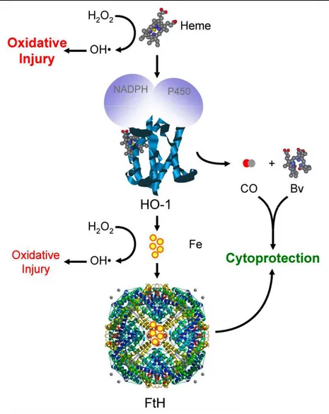
Figure 1.2. The heme/HO-1 system. Free heme is highly pro-oxidant, as its Fe atom can participate in the Fenton reaction via which hydrogen peroxide (H2O2) is converted into the highly reactive hydroxyl radical (OH). This pro-oxidant effect is controlled by several mechanisms including heme catabolism by
HO-1, a reaction assisted by NADPH and Cytochrome P450. Contrary to heme-Fe, labile Fe (yellow circles) can be neutralized by ferritin (FtH).
oxidative stress, hypoxia, heat shock, cytokines, and TLR ligation185. The reaction leading to heme catabolism by HO enzymes opens the protoporphyrin ring leading to the generation of equimolar amounts of carbon monoxide (CO), biliverdin and labile iron (Fe2+) (Figure 1.2). Biliverdin is further converted into the potent anti-oxidant bilirubin by the enzymatic action of biliverdin reductase (BVR), and labile iron is secluded by ferritin multimers preventing its pro-oxidant activity186.
A major effect of heme catabolism by HO-1 is the avoidance of unfettered oxidative damage generated by heme molecules once they have been uncoupled from hemoproteins. The iron molecule inside the protoporphyrin ring of heme can generate large amounts of hydroxil radicals (HO•) from hydrogen peroxide (H2O2), through the Fenton reaction187. The extremely
reactive HO• can, in turn, cause DNA breaks, lipid peroxidation and protein denaturation188. As HO-1 cleaves the protoporphyrin ring, the iron molecule is freed and becomes available to be taken up and stored intracellularly by ferritin molecules, which has its expression increased in response to iron. Ferritin molecules oxidize iron to its ferric (Fe3+) state, thus impairing its pro-oxidant activity, and store the iron inside its cage-like structure189. In addition, with the opening of the protoporphyrin ring, biliverdin and subsequently bilirubin, a potent anti-oxidant, are generated, providing further protection against oxidative damage.
Several lines of evidence suggest that many of the effects of CO are mediated via the modulation of the p38 MAP kinase pathway. CO can prevent cell death by modulating the p38 MAP kinase pathway190,192. The anti-inflammatory effect of CO on macrophages, i.e CO-mediated inhibition of TNF production, is also dependent on p38 MAP kinase 191, and on an initial burst of ROS production by the mitochondria induced by CO193. Furthermore, it is proposed that the protective effects of CO rely on its capacity to interact with heme inside hemoproteins preventing their oxidation and release from heme pockets, a process that would render heme pro-oxidant178,188,194.
Biliverdin/bilirubin also have been demonstrated to induce protection against cell death and inflammation, presumably by their anti-oxidant properties195. Cell death mediated by either NO or H202 has been shown to be prevented by bilirubin196,197.
Biliverdin/bilirubin are also implicated in regulating immune responses, as discussed below (see section 2.3). Finally, the labile iron generated by heme catabolism is thought to afford cytoprotection by inducing the expression of ferritin, an iron storage molecule198. The iron storage capacity of ferritin also underlies its ability to prevent TNF-mediated cell death. Iron sequestration by ferritin prevents excessive ROS production and sustained JNK activation, therefore preventing cell death in this system199.
2.2 Regulation of HO-1 expression
factors (HSF), and Activator Protein-1 (AP-1) in its promoter and enhancer regions200. The broad array of transcription factors capable of regulating HO-1 expression might explain the induction of HO-1 expression by a variety of stimuli.
Under homeostasis, the transcriptional repressor Bach1 inhibits Hmox1 transcription by, in conjunction with small Maf proteins, blocking accessibility of transcription factors to Hmox1
promoter. Both heme and oxidative stress can induce conformational changes in Bach1 leading to its export from the nucleus, ubiquitination and degradation188. Concomitantly, ROS can also activate Nrf2. When cells are under normal conditions, Nrf2 is found in the cytoplasm bound to Kelch-like ECH-associating protein 1 (Keap1) that blocks Nrf2 migration to the nucleus and also facilitates Nrf2 polyubiquitination and degradation. Under oxidative stress conditions, alterations in Keap1 key cysteine residues liberate Nrf2 from sequestration in the cytoplasm and degradation. Furthermore, oxidative stress can also modify cysteine residues on Nrf2 nuclear export signals (NES) facilitating its concentration in the nucleus201. Of note, environmental stress, such as the ones posed by cigarette smoke and xenobiotics, also leads to Nrf2 activation and transcription of other detoxifying and cytoprotective enzymes besides HO-1202.
suggest that HO-1 expression is key to control of immune responses and/or the resulting immunopathology.
2.3 Immunoregulatory effects of HO-1
The notion that HO-1 is an immunoregulatory molecule is supported by the observation that Hmox1 deficiency leads, in mice and humans, to the spontaneous development of a chronic inflammatory pathology characterized by increased blood leukocyte count, serum immunoglobulin M (IgM), accumulation of polymorphonuclear (PMN) cells, and monocyte/macrophages in the spleen, as well as in nonlymphoid tissues, and widespread oxidative tissue injury208-210. Data supporting the notion that the immunoregulatory effects of HO-1 are exerted via its expression on innate and adaptive immune cells, as well as by its expression in non-lymphoid tissues where it affords cytoprotection against oxidative injury, will be discussed below.
2.3.1 Effects of the heme/HO-1 system on innate immunity
Under homeostasis, heme exists essentially as a prosthetic group in several hemoproteins. However, under pathologic conditions noncovalently bound heme can be released from those hemoproteins178,194. The heme produced in this manner can be recognized by TLR4, triggering the production of proinflammatory cytokines by macrophages211. Furthermore, heme can induce neutrophil migration and ROS production by directly activating a G-protein-coupled receptor212 and/or by inducing the production of leukotriene B4213. Although this is the
heme, it remains to be established whether this is the case in more physiological settings, since heme can bind to several proteins194 and lipids188 in plasma and, therefore, might have its ability to activate TLR4 or G protein-coupled-receptors212 altered. Interestingly, the anti-inflammatory effect of immunoregulatory molecules such as IL-10214 and
15-deoxy-Δ12,14-prostaglandin J2 (15D-PGJ2)215 on innate immune cells,
appears to be exerted via the induction of HO-1 in macrophages. Consistent with a role for HO-1 in dampening inflammatory responses after stimulation of the innate immune system, is the observation that stimulation of Hmox1-/-splenocytes with LPS resulted in increased production of pro-inflammatory cytokines, as compared to Hmox1+/+ splenocytes216. Importantly, evidence that HO-1 does not control the inflammatory response in macrophages is also available. For instance, peritoneal macrophages isolated from Hmox1-/- or Hmox1+/+ mice have a similar proinflammatory response to LPS in vitro207,209, raising the possibility that HO-1 may differentially affect macrophage populations and/or other immune cells.
supported by the observation that Hmox1 deficiency, in mice and humans, is associated with widespread neutrophil activation and tissue infiltration, as well as with oxidative tissue injury208-210,218. In addition, pharmacologic induction of HO-1 inhibits the activity of the p47phox, p67phox, and gp91phox subunits of NADPH oxidase, thereby reducing the production of reactive oxygen species by activated neutrophils and macrophages219,220, contributing further to limit oxidative tissue injury.
CO can also modulate TLR signaling. This inhibition can occur via two independent non-exclusive pathways: one that involves enhanced caveolin-1 binding to TLR, and one that is dependent on CO-mediated inhibition of ROS formation by NADPH oxidase. Upon LPS stimulation HO-1 colocalizes with the caveolin-1/TLR4 complex, a process dependent on p38 MAPK activity. Importantly, HO-1-generated CO was shown to enhance caveolin-1 interaction with TLR4, inhibiting downstream signaling and production of pro-inflammatory cytokines221. A broader role for CO in inhibiting TLR function has been shown to depend on its ability to decrease ROS formation, via modulation of NADPH oxidase activity, upon TLR 2, 4, 5 and 9, stimulation and consequently inhibition of TLR recruitment (specifically TLR4 recruitment) towards lipid rafts222. This data shows that CO generated by HO-1 can interfere with the molecular machinery involved in proximal TLR signaling.
was shown to induce the production of TGF-β, a potent anti-inflammatory cytokine, by macrophages, through a pathway that involves stabilization of hypoxia-inducible factor-1 α
(
HIF-1α) viamitochondrial-generated ROS223. Generation of ROS by the mitochondria is also involved in CO-mediated upregulation of peroxisome proliferator-activated receptor-γ (PPAR-γ), an event
shown to be crucial for the anti-inflammatory effects of CO in macrophages224. Most importantly, in an in vivo model of LPS-mediated shock, exogenous CO blocks pro-inflammatory cytokine production and promotes survival of mice challenged with a lethal dose of LPS225.
major cause of pathology is immune-mediated.
2.3.2 Effects of the heme/HO-1 system on adaptive immunity
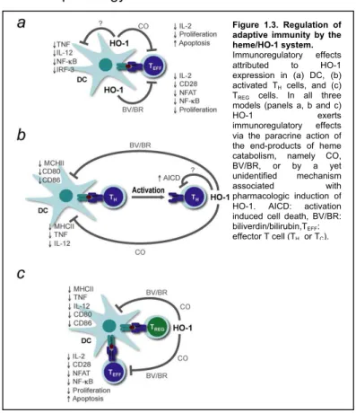
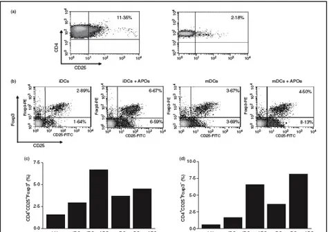
Several in vitro observations suggest that HO-1 exerts immunoregulatory effects by inhibiting T cell activation, proliferation and/or acquisition of effector function. First, the activation of human CD4+ T cells is associated with a significant induction of HO-1 expression231 (Figure 1.3b). Second, pharmacologic induction of HO-1 inhibits human CD4+ and CD8+ T cell activation231. Third, CO inhibits CD4+ T cell activation231 and induces apoptosis in Jurkat T cells232 (Figure 1.3b). Fourth, biliverdin/bilirubin inhibits mouse and human CD4+ T cell activation230 (Figure 1.3b). In keeping with these observations,
Figure 1.3. Regulation of adaptive immunity by the heme/HO-1 system.
Immunoregulatory effects attributed to HO-1 expression in (a) DC, (b) activated TH cells, and (c) TREG cells. In all three models (panels a, b and c)
HO-1 exerts
pharmacologic induction of HO-1 in vivo can drive activated CD4+ T cells to undergo apoptosis via activation-induced cell death (AICD)233 (Figure 1.3b), and can promote dominant peripheral T cell tolerance against transplanted organs234. This latter effect is mimicked by biliverdin/bilirubin, which suppresses T cell-driven inflammatory pathologies such as the rejection of transplanted organs230,235 and autoimmune neuroinflammation43. The immunoregulatory effects of biliverdin/bilirubin are mediated via inhibition of the transcription factors, NFAT and NF-κB, which
suppresses IL-2 production by CD4+ T cells230,235. Whether these effects of BV/BR are mediated by its anti-oxidant activity is still unclear, although data suggests that this might not be the case230. Of notice, both biliverdin and bilirubin are endogenous ligands for the aryl hydrocarbon receptor (AHR)236, which can determine the differentiation of CD4+ T cells towards an anti-inflammatory TREG phenotype or a proinflammatory TH17 effector
phenotype237,238. Although speculative, it is possible that the immunoregulatory effects of HO-1 might be exerted, to at least some extent, through the production of these endogenous AHR ligands.
More recently, it was shown that HO-1 expression in myeloid lineages has protective effects in a model of autoimmune neuroinflammation. This study shows that lack of HO-1 in the
myeloid lineages leads to decreased production of IFN-β, a cytokine that blocks the development of pathogenic IL-17 producing CD4+ T cells. Consequently, animals in which Hmox1
cell populations through the activity of Cre-recombinase. It is important to highlight that in this system the authors did not observe the pathologic features associated with total Hmox1
deletion, i.e. diminished fertility and progressive inflammatory disease, allowing for a more clear evaluation of the mechanisms responsible for exacerbating disease239 in the absence of HO-1.
HO-1 has been suggested to exert immunoregulatory effects by modulating TREG cell function240 (Figure 1.3c), a notion
supported by the following set of observations. First, human and mouse TREG cells express HO-1 constitutively241,242 (Figure 1.3c).
Second, pharmacologic inhibition of HO-1 suppresses human TREG cell function in vitro243. Third, the suppressive activity of
Hmox1+/+ TREG cells is compromised, in vitro, by the presence of
Hmox1-/- DC244. This finding, however, was not reproduced when total irradiated splenocytes were used as APCs242. While these observations suggest that HO-1 regulates TREG cell function in
vitro, the finding that TREG cell development as well as peripheral
maintenance and function, in vitro and in vivo, are normal in
Hmox1-/- mice242, questions the physiologic relevance of the effects attributed to pharmacologic modulation of HO-1 in TREG
cells. Of note, the function of Hmox1-/- TREG has not yet been
tested in conditions where there is strong innate and adaptive immune activation, such as in an infectious disease model. It would be interesting to address whether the increased production of proinflammatory cytokines by Hmox1-/- spleen cells upon stimulation216 might, somehow, affect TREG function.
suggests that heme might control B cell differentiation into plasma cells by interacting with the transcriptional repressor Bach2 and inducing its degradation. Bach2 degradation, in turn, leads to increased expression of the transcription factor B lymphocyte-induced maturation protein 1 (Blimp-1), diminished class switch recombination and somatic hypermutation, and increased plasma cell differentiation245.
2.3.3 Cytoprotective effects of HO-1
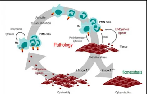
Figure 1.4. Cytoprotective effect of the heme/HO-1 system. Activation of innate immune cells, including Mø and PMN cells is associated with the production of ROS and proinflammatory cytokines, leading to oxidative stress. Expression of HO-1 in response to oxidative stress (i.e. Hmox1+/+) is
cytoprotective and prevents the release of endogenous proinflammatory ligands from injured cells, thus promoting the resolution of inflammation and the return to homeostasis. When expression of HO-1 is impaired (i.e. Hmox1
-/-) oxidative stress leads to widespread programmed cell death resulting in impaired tissue function as well as release of endogenous proinflammatory ligands. By engaging PRR in innate immune cells, endogenous ligands sustain the activation of innate immune cells (inflammation) and exacerbate tissue injury, an effect that might promote the development of immune-mediated inflammatory diseases.
When challenged by a proinflammatory agonist such as LPS, Hmox1-/- mice succumb to unfettered oxidative stress, associated with widespread oxidative tissue injury and end-stage multi-organ failure209,249. This suggests that HO-1 prevents the pathologic outcome of inflammatory responses by affording cytoprotection against oxidative stress. In subsequent studies, the cytoprotective effect of HO-1 was associated with protection against many other immune-mediated inflammatory diseases186.
cardiac myocytes, astrocytes and neurons, among others185,186,188,250. This broad cytoprotective effect might account for the equally broad protective effects of HO-1 against the development of vascular diseases, diabetes, liver dysfunction, kidney failure, myocardial infarction as well as diseases of the central nervous system186.
Presumably, the cytoprotective effect of HO-1 has a dual salutary role in that it sustains tissue/organ function while inhibiting the release of endogenous proinflammatory ligands from injured cells (Figure 1.4). In keeping with this notion,
Hmox1-/- mice produce high levels of circulating endogenous proinflammatory ligands, for example HMGB1, in response to LPS251, and cecal ligation and puncture207, an effect that promotes the development of septic shock251-253. Moreover, pharmacologic induction of HO-1 affords tissue cytoprotection and inhibits the production of extracellular HMGB1 in response to LPS252.
3. Iron metabolism and immunity
Iron is a fundamental trace element for organisms, being implicated in the function of crucial proteins such as cytochrome
c and hemoglobin254. Nonetheless, iron molecules, present or not in heme groups, that are not inside a functional protein moiety can be extremely toxic. As such, iron levels need to be tightly regulated both systemically and intracellularly. Importantly, mammals are not proficient in excreting iron, increasing the relevance of a delicate regulation of systemic iron levels. The regulation of systemic iron levels occurs mainly by controlling dietary iron absorption and mobilization from cellular iron stores, while intracellular iron status is regulated by iron storage and import/export189.
hemoproteins, especially hemoglobin189. The regulation of systemic iron status in mammals is subject to modulation by different stimuli such as anemia, inflammation, hypoxia and changes in erythropoietic rate255,256.
Intracellularly, iron levels are regulated by two major mechanisms: a) storage by ferritin molecules and, b) modifying mRNA stability of proteins involved in import/export of cellular iron256. Ferritin molecules normally contain the majority of the intracellular iron pool. Ferritin is composed by the assembly of multiple L and H-ferritin chains, encoded by different genes, forming a cage-like structure capable of oxidizing and subsequently store iron molecules in a non-reactive state189. On the other hand, ferroportin (FPN) controls cellular iron efflux, and regulation of its levels determines the amount intracellular iron189. Interestingly, mRNA from iron transport and storage molecules, e.g. ferritin, transferrin receptor 1 (TfR1), ferroportin, contain, in its 3´ and/or 5´ untranslated regions, iron responsive elements (IREs) that are responsive to iron regulatory proteins (IRPs) that, in turn, respond to iron levels. The interaction between IRPs and IREs can either stabilize or destabilize the mRNA of target molecules and thus regulate intracellular iron availability257-259.
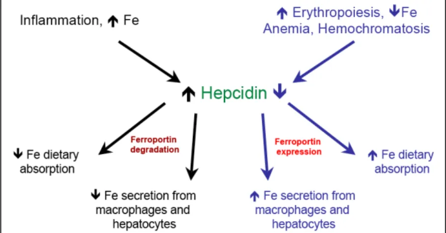
Tf)262. In situations where there is anemia, iron deficit, hypoxia and/or increased erythropoiesis, hepcidin expression is decreased, allowing increased iron absorption from dietary sources and iron mobilization from cellular stores. The reverse is true in cases of iron excess and inflammation263.
The fundamental role of hepcidin in iron regulation is demonstrated by the severe iron overload phenotype that occurs when the hepcidin gene is disrupted264,265 and, on the other hand, by the severe iron deficiency associated with hepcidin overexpression266,267. Induction of hepcidin expression is thought to occur by at least two different pathways, one involved in homeostatic hepcidin expression and another involved in the fluctuations in hepcidin levels during inflammation. In homeostasis, hepcidin transcription responds to transferrin (Tf)- bound iron levels. Tf-bound iron is sensed by Tf receptor-1 (TfR1) that, under normal iron levels, is coupled with the hemochromatosis gene (HFE), a non-classical MHCI molecule268, on the surface of hepatocytes. When serum Tf-bound iron levels increase, its binding to TfR1 dislodges HFE from the complex allowing the association of the latter with the Tf receptor-2 (TfR2). These induced changes initiate hepcidin transcription via a still uncharacterized pathway256. An alternative pathway can induce SMAD4-dependent hepcidin transcription via activation of the bone morphogenetic protein (BMP) receptor in complex with its co-receptor hemojuvelin269,270.
the hepcidin promoter and hepcidin transcription272-274. This response is thought to be a defense strategy used by the host to limit the amount of iron in circulation, thus limiting pathogens access to it275.
Molecularly, hepcidin acts by binding to FPN expressed on membranes of enterocytes, macrophages and hepatocytes, and targeting it for degradation, thus impairing iron export from cells. As FPN is targeted for degradation, these cells become incapable of exporting iron, leading to a decrease in the extracellular iron pool276. Given the above, when hepcidin levels are elevated there is a decrease in the expression of FPN and a consequent decrease in extracellular and increase in intracellular iron content. The reverse happens in cases of low hepcidin expression (see Figure 1.5).
The major protein involved in carrying iron in serum is transferrin (Tf). Tf is mainly produced by the liver and assures, by
devoid of its pro-oxidant activity. Tf carries up to two iron molecules and is responsible for carrying it to where it is needed, i.e. the liver for storage or the bone marrow for red blood cell (RBC) generation255. The majority of the iron present in the body is used by the erythroid bone marrow in the generation of heme groups to be used in hemoglobin molecules in emerging RBC. The delivery of iron to erythroblasts, nucleated precursors of RBC, is mediated by transferrin that, once bound to TfR1, is taken up by erythroblasts. The Tf-iron/TfR1 complex is internalized and degraded, iron is reduced to its Fe2+ form and becomes ready to be used in the confection of new heme groups255. Disruption of the TfR1 gene leads to defective erythropoiesis277,278.
Deregulation of hepcidin production by mutations in hepcidin, TfR2, HFE or hemojuvelin, leads to the development of hemochromatosis a condition characterized by iron overload. In the four cases, hepcidin production is diminished. Mutations in FPN can also lead to a hemochromatosis-like disease. In the latter case, iron overload is found predominantly inside macrophages262. The study of the effects of iron status in immune functions greatly benefited from the occurrence of natural mutations causing hemochromatosis, and from the development of mouse models to mimic these pathologies262.
Iron has been shown to modulate the activation of transcription factors involved in the inflammatory response, such
as NF-κB and HIF-1α, in opposing ways. While high iron
contents might increase NF-κB activation279-282, it inhibits
be regulated by ROS levels and, therefore, might respond to changes in intracellular iron levels, since iron can foster ROS production.
Importantly, the study of animal models of hemochromatosis, especially the model induced by targeted disruption of the hemochromatosis gene (Hfe), has provided insight on the molecular mechanisms involved in the regulation of immune responses by intracellular iron levels. Hfe-deficient mice (Hfe-/-)have lower hepcidin production leading to increased FPN expression by cells and, consequently, low intracellular iron level. Macrophages with low intracellular iron content present
diminished production of IL-6, TNF and IFN-β upon stimulation via TLR4 and Salmonella infection. Importantly, the decreased production of pro-inflammatory cytokines led to decreased intestinal inflammation upon Salmonella infection, but also to increased bacterial load285,286. Furthermore, administration of compounds able to block hepcidin production recapitulate the protective effects against excessive inflammation seem on Hfe
-/-animals285. The mechanisms responsible for these changes rely on diminished translation of inflammatory genes286 and on impairment of the TRIF signaling pathway downstream of TLR4285.