Cytotoxicity and genotoxicity of stilbene derivatives in CHO-K1 and HepG2
cell lines
Cassia Suemi Mizuno
1, Winnifred Ampomaah
1, Fernanda Ribeiro Mendonça
2, Gabriela Carvalho Andrade
2,
Ariel Maria Nazaré da Silva
2, Mirian Oliveira Goulart
1and Raquel Alves dos Santos
21
Department of Pharmaceutical Sciences, University of New England - College of Pharmacy, Portland, ME,
USA.
2
Universidade de Franca, Franca, SP, Brazil.
Abstract
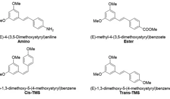
The cytotoxicity and genotoxicity of the stilbenes (E)-methyl-4-(3-5-dimethoxystyryl)benzoate (ester), (E)-4-(3-5-dimethoxystyryl)aniline (amino), (Z)-1,3-dimethoxy-5-(4-methoxystyryl)benzene (cis-TMS) and (E)-1,3-dimethoxy-5-(4-methoxystyryl)benzene (trans-TMS) were investigated in this work. Structural modifications of resveratrol, a naturally occurring stilbene, have been previously performed, including the replacement of hydroxyl by different func-tional groups. Such modifications resulted in significant improvement of target-specific effects on cell death and antiproliferative responses. The parameters were evaluated using XTT assay, clonogenic survival assay and the cytokinesis-block micronucleus assay in CHO-K1 and HepG2 cell lines. The results showed thatcis-TMS is
approxi-mately 250-fold more cytotoxic than the amino and ester, and 128-fold more cytotoxic thantrans-TMS. When
genotoxicity was evaluated, only thetrans-TMS did not significantly increase the frequency of micronucleus (MN).
While thecis-TMS induced a mean of 5.2 and 5.9 MN/100 cells at 0.5M in CHO-K1 and HepG2, respectively, the
amino and ester induced 3.1 and 3.6 MN/100 cells at 10M in CHO-K1, respectively, and 3.5 and 3.8 in HepG2.
Trans-TMS is genotoxic only in HepG2 cells. Based on these results, the cis-TMS was the most cytotoxic and genotoxic compound in both cell lines.
Keywords: stilbene, cytotoxicity, genotoxicity, micronucleus assay.
Received: August 19, 2016; Accepted: January 29, 2017.
Introduction
Stilbenes comprise a class of plant-derived secondary metabolites that are produced in response to fungal infec-tions (Flaminiet al., 2016). Resveratrol is one of the most investigated stilbenes and has been found in many species of plants that are part of our diet such as grapes, peanuts, pomegranade and berries. Resveratrol became the focus of intense research after its presence in wine was associated with the French paradox in which a wine-drinking popula-tion with a high intake of saturated fat showed low inci-dence of cardiovascular diseases. The cardioprotective effects of this compound occur through different mecha-nisms including inhibition of platelet aggregation (Shenet al., 2007), vasorelaxation effects by upregulating NOS ex-pression (Wallerathet al., 2002), and inhibition of LDL ox-idation and subsequent atherosclerosis (Belguendouzet al., 1997). Resveratrol is also a scavenger of H2O2, a reactive oxygen species involved in oxidative stress (Ungvariet al., 2007).
Other naturally occurring stilbenes with similar thera-peutic potential include pterostilbene and piceatannol with anticancer (Seyedet al., 2016), anti-virus (Leeet al., 2016) antioxidant (Mikstacka et al., 2010), anti-inflammatory (Mikstackaet al., 2010), and lipid-lowering (Rimandoet al., 2005) effects among others. Stilbenes reportedly inhib-ited the growth of several cancer cell lines such as colon, breast, prostate, pancreas, melanoma, lung and others. This effect is elicited through different mechanisms including cell cycle arrest and apoptosis induction via caspase activa-tion (Aggarwalet al., 2004). Several animal studies support the evidence provided by thein vitroassays. Resveratrol prevented the formation of colon and intestinal tumors by 70% in Min mice (Schneider et al., 2001). 3,5,4’trime-thoxystilbene (trans-TMS, Figure 1), a natural compound, significantly reduced tumor growth in Colo 205 xenograft model of colon cancer (Panet al., 2008) and pterostilbene reduced the formation of azoxymethane-induced colonic aberrant crypt foci (ACF) (Suhet al., 2007). In a work by Paul et al. (2010), two stilbene derivatives (amino and
trans-TMS, Figure 1) significantly decreased tumor vol-ume and weight in HT-29 xenografts model of colon can-cer. Based on the promising and pleiotropic effects of the
DOI: http://dx.doi.org/10.1590/1678-4685-GMB-2016-0214
Send correspondence to Raquel Alves dos Santos. Laboratory of Genetics and Molecular Biology, Universidade de Franca, Av. Dr. Armando Salles de Oliveira 201, 14404-600 Franca, SP, Brazil. E-mail: [email protected]
stilbenes, several clinical trials have investigated the effects of resveratrol and pterostilbene in patients with cardiovas-cular diseases, type 2 diabetes, chronic obstructive pulmo-nary disease and metabolic syndrome.
With the consideration of stilbenes as potential drug candidates, it is very important to determine the toxicity of these compounds. The genotoxicological analysis is of fun-damental importance during toxicological evaluation of therapeutic drug candidates. Genotoxic compounds are ca-pable of interacting with the DNA molecule, leading to ge-netic damage in essential regions for cycle-control and apoptosis, with the subsequent initiation of neoplastic pro-cesses (Santos and Takahashi 2008). Genotoxicity tests for screening substances (e.g.drug candidates, medicinal plant extract, chemical substance, etc) officially approved by the Organization for Economic Co-operation and Develop-ment (OECD) guidelines includein vitromicronucleus test to detect both clastogenic and aneugenic effects of potential drug candidates (Fenech, 2000).
The cytotoxicity and genotoxicity of the stilbenes (E)-methyl-4-(3-5-dimethoxystyryl)benzoate (ester), (E)-4-(3-5-dimethoxystyryl)aniline (amino), (Z )-1,3-dime-thoxy-5-(4-methoxystyryl)benzene (cis-TMS) and (E )-1,3-dimethoxy-5-(4-methoxystyryl)benzene (trans-TMS) (Figure 1) have not been reported in non-tumoral cell lines. Therefore, in the present study we investigated the cyto-toxicity and genocyto-toxicity of these stilbene derivatives using the CHO-K1 and HepG2 cell lines.
Material and Methods
Chemicals and reagents
Dimethyl sulfoxide (DMSO, CAS 67-68-5), cyto-chalasin B (CytB, CAS 14930-96-2), doxorubicin hydro-chloride (DOX, CAS 25316-40-9) and reagents for cell culture and micronucleus tests were purchased from Sigma-Aldrich (St. Louis, USA). Cell Proliferation Kit (XTT) was purchased from Roche (Mannheim, Germany).
All solvents were anhydrous and all reactions were per-formed under an inert atmosphere, unless aqueous. All round-bottom flasks were kept dried in an oven under 100 °C. Flash chromatography used to purify the compounds was done using a Teledyne Isco CombiFlash and LC-MS were obtained from Waters Micromass Quattro LC. The purity of all stilbenes tested was above 95%.
General procedure for the synthesis of stilbene derivatives
To a cold solution (–78 °C) of phosphonium salt (1.0 equivalent) in THF was addedn-butyllithium (1.6 mol in hexanes, 1.0 equivalent) and the resulting solution was stirred under inert atmosphere for 2 h. A solution of alde-hyde (1.0 equivalent) in THF was added dropwise, and the mixture was stirred for 12 h at room temperature. The re-sulting suspension was poured into water and extracted with dichloromethane. The organic phase was combined and dried over MgSO4, and concentrated under reduced pressure. The crude product was purified through auto-mated flash purification elution with hexanes/ethyl acetate.
(E)-Methyl 4-(3,5-dimethoxystyryl)benzoate (ester): Reaction of (3,5-dimethoxybenzyl) triphenylphosphonium 4 (300 mg, 0.608 mmol) and methyl 4-formylbenzoate (100 mg, 0.608 mmol) afforded the ester as a white solid: 32 mg (17%).1H NMR (CDCl3, 300 MHz):3.83 (s, 6H); 3.92 (s, 3H); 6.42 (s, 1H); 6.68 (s, 2H); 7.04–7.16 (m, 2H); 7.53 (d, 2H,J= 8.4 Hz); 8.01 (d, 2H,J= 8.4 Hz).13C NMR (CDCl3, 75 MHz):52.1, 55.4 (2C), 100.6, 104.9 (2C), 126.4 (2C), 128.1, 129.0, 130.1 (2C), 131.3, 138.8, 141.7, 161.1 (2C), 166.9. LCMS m/z 299.28. (M+H)+
(E)-4-(3,5-Dimethoxystyryl) aniline (amino). Reac-tion of (3,5 dimethoxybenzyl) triphenylphosphonium (300 mg, 0.608 mmol) and 4-nitrobenzaldehyde (92 mg, 0.608 mmol) afforded 1,3-dimethoxy-5-(4-nitrostyryl)benzene. A solution of the latter (100 mg, 0.35 mmol) in acetone/wa-ter (10:5 mL) was heated to 50 °C for 30 min. Sodium dithionite (1526 mg, 8.76 mmol) was slowly added and the
mixture was heated to reflux for 1 h. After cooled to room temperature the mixture was poured into water and ex-tracted with ethyl acetate. The organic phase was combined and dried over MgSO4, and solvent was removed under re-duced pressure. The crude mixture was purified using auto-mated flash chromatography eluting with hexanes giving the amine as a yellow powder: 29 mg (25% yield)1H NMR (CDCl3, 300 MHz):3.82 (s, 6H); 6.36 (s, 1 H); 6.63–6.69 (m, 4H); 6.85 (d, 1H,J= 16.2 Hz); 7.00 (d, 1H,J= 16.2 Hz); 7.33 (d, 2H,J= 8.4 Hz).13C NMR (CDCl3, 75 MHz):
55.4 (2C), 99.4, 104.3 (2C), 115.2 (2C), 125.1, 127.8, 127.9, 129.3, 140.1, 146.4, 161.0 (2C). LCMS m/z 256.23 (M+H)+
1,3-dimethoxy-5-(4-methoxystyryl)benzene- cis and trans-TMS. Reaction of (3,5-dimethoxybenzyl)triphenyl-phosphonium (300 mg, 0.608 mmol) and 4-methoxyben-zaldehyde (75 L, 0.608 mmol) afforded the cis and trans-TMS. Trans-TMS as a white solid: 15 mg (9%).1H NMR (CDCl3, 300 MHz):3.83 (s, 9H); 6.37 (t, 1H,J= 2.1 Hz); 6.65 (d, 2H,J= 1.8 Hz); 6.87-6.89 (m, 2H); 6.92 (d, 1H,J= 5.1 Hz); 7.04 (d, 1H,J= 16.2 Hz); 7.45 (d, 2H,J= 6.9 Hz).13C NMR (CDCl3, 75 MHz): 55.2 (3C), 99.7, 104.2 (2C), 114.2 (2C), 126.5, 127.8 (2C), 129, 130, 139.6, 159.4, 161.0 (2C). LC-MSm/z270.19. (M+H).+Cis-TMS as a viscous liquid: 62 mg (37%).1H NMR (CDCl3, 75 MHz):3.68 (s, 6H); 3.78 (s, 3H); 6.34 (t, 1H,J= 2.4 Hz); 6.44–6.46 (m, 3H); 6.55 (d, 1H,J= 12.3 Hz); 6.78 (d, 2H,J
= 8.7 Hz); 7.24 (d, 2H,J= 8.4 Hz).13C NMR (CDCl3, 75 MHz): 55.3 (3C), 99.7, 106.7 (2C), 113.6 (2C), 128.7, 129.6, 130.2, 130.3 (2C), 139.5, 158.8, 160.6 (2C). LC-MS
m/z271.28 (M+H)+.
Cell culture and treatment conditions
CHO-K1 and HepG2 cells were obtained from the Cell Bank of Rio de Janeiro (BCRJ code 0069 and 0103, re-spectively) and cultured in complete medium containing DMEM+F10 nutrient mixture (1:1, v/v), supplemented with fetal bovine serum (10%, v/v) and penicillin/strepto-mycin stabilized solution (10 mL/L). Cell cultures were in-cubated at 37 °C with atmosphere saturated with 5% of CO2. All experiments were conducted between the 3rdand the 8th passages and three experimental repetitions were performed.
The ester, amino,cis-TMS andtrans–TMS were dis-solved in DMSO in a stock concentration of 0.1 M. The fi-nal concentration of DMSO during treatments did not exceed 0.1% (v/v) in cell cultures. Doxorubicin (DOX) was dissolved in ultrapure sterile water in a stock solution of 900M. All reagents were dissolved prior to use and pro-tected from light. For all experiments, cells were submitted to 24 h treatment with different concentrations of the tested substance and DOX was used as positive control in CHO-K1 cells. Benzo(a)pyrene [B(a)P] at 25M was used as positive control in HepG2 cells.
Cell viability assay – XTT
To determine cell viability, 104 cells/well were seeded on a 96-well plate. After 24 h the cells were treated with ester, amino,cis-TMS andtrans–TMS in a concentra-tion ranging from 7.8M to 1000M and incubated for an additional 24 h. The plates were washed twice in phosphate buffer saline (PBS 1X) and incubated for 4 h at 37 °C with DMEM without phenol red and supplemented with the re-agents of the Cell Proliferation Kit (XTT) as recommended by the manufacturer. Total absorbance was measured at 492 and 690 nm (reference) using a microplate reader (ASYS, Eugendorf, Salzburg, Austria). Results of total absorbance were considered directly proportional to the number of viable cells as a percentage of the negative con-trol (100% of cell viability).
Clonogenic survival assay
The clonogenic assay was performed according to Frankenet al.(2006). Briefly, 200 cells/well were plated in 6 well plates containing 3 mL of complete medium. After 4 h of incubation at 37 °C, the cells were treated with differ-ent concdiffer-entrations of the testing stilbenes for 24 h, then each well was washed with PBS 1X and complete medium was added. The cells were allowed to grow for 7–14 days at 37 °C and 5% of CO2, when colonies were visible. The col-onies formed were fixed with methanol/acetic acid/water (1:1:8 v/v/v) and stained with Giemsa 5% (v/v) in Sorensen phosphate buffer (pH 6.8). The colonies were counted, and the cell survival fraction was calculated as percent colonies relative to the untreated control.
Micronucleus test
Statistical analysis
Statistical analysis of experimental data was per-formed using the software GraphPad Prism 5.0. The IC50 was calculated by linear regression analysis and the com-parative analysis among experimental groups was done with ANOVA, followed by Tukey’s post-hoc test when nificant differences among treatments were found. The sig-nificance was set at p < 0.05 and the results were reported as means and standard deviations (SD) of three independent experimental samples assayed in triplicates.
Results
The cytotoxicity of stilbene derivatives in CHO-K1 and HepG2 cells
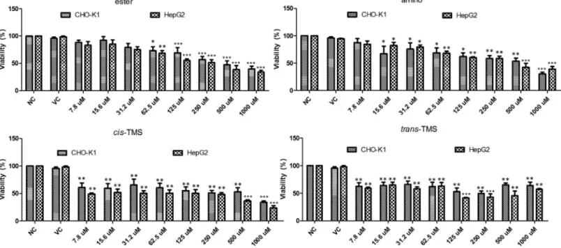
The cytotoxicity of the stilbenes was determined by the XTT assay. Cells were treated with different concentra-tions of the tested stilbenes for 24 h and the results are pre-sented in Figure 2. Cell viability was significantly reduced (p < 0.05) by the ester at 62.5 M (73.3 and 68.6% in CHO-K1 and HepG2, respectively). Significant reduction in cell viability by the amino was observed at 15.6M (67.2%) and 62.5M (68.2%) in CHO-K1 and HepG2, re-spectively, and at all concentrations of thecis-TMS and
trans–TMS in both cell lines. At the lowest concentration (7.8M) of thecis- andtrans-TMS, the cell viability was 60.7% and 62.5%, respectively in CHO-K1, and 49% and 59.1% in HepG2 (p < 0.05). Tukey’s test demonstrated that the ester and amino induced a concentration-dependent re-duction in cell viability, differently from what was ob-served for thecisandtrans-TMS in both cell lines.
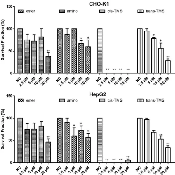
Subsequently, the long-term cytotoxic effects of stilbene derivatives were evaluated by the clonogenic
sur-vival assay. Based on the XTT results, all stilbenes were tested in concentrations ranging from 2.5M to 80M. However, no stilbene could form countable colonies at 40 and 80M (data not shown). According to the results ex-hibited in Figure 3, in CHO-K1, the ester significantly re-duced (p < 0.05) the frequency of survival fraction at 20M (37.8%) and the amino at 10 and 20M (67.1 and 59.6% re-spectively). Similarly, in HepG2, 20M ester significantly reduced the survival fraction (46.1%) while significant re-sponse to amino treatment was observed at 5M (59.3%). Thetrans-TMS showed a significant concentration-depen-dent reduction in the frequency of survival fraction in both cell lines. Interestingly, thecis-TMS showed much higher cytotoxicity than the other tested stilbenes and killed 100% of the cells in all concentrations tested in CHO-K1 cells and at 2.5, 5 and 10M in HepG2. Therefore, this compound was tested at lower concentrations than the other stilbenes (Figure 4). The survival fraction was significantly reduced by thecis-TMS to 45.8, 36.8 and 13.9% at 0.078, 0.156 and 0.3125M, respectively in CHO-K1 cells and 41.3, 54.3, 16.9 and 6.2% at 0.078, 0.156, 0.3125 and 0.625M in HepG2.
Genotoxicity of stilbene derivatives detected by cytokinesis-blocking micronucleus assay (CBMN)
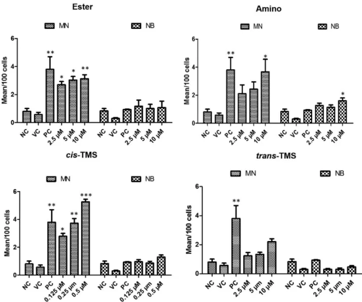
For the genotoxicity analysis, CBMN assay was per-formed to detect micronucleus frequency (MN) and nuclear bridges (NB) in both CHO-K1 and HepG2 cells. According to the results presented in Figure 5, the ester, amino and
cis-TMS increased MN frequency in CHO-K1 cells. How-ever, no significant MN frequency (p < 0.05, compared to NC) occurred in cells treated withtrans-TMS at all concen-trations. The ester increased the mean of MN to 2.7, 3 and
3.1 at 2.5, 5 and 10M, respectively, in comparison to the NC (Figure 5). Similar effect was observed with the amino only at 10M, where 3.6 MN were detected per 100 cell (p < 0.05, Figure 5). All tested concentrations of cis-TMS in-creased the mean of MN in CHO-K1 cells. Even the lowest
concentration tested (0.125M) increased the mean to 2.8
MN/100 cells (p < 0.05). At the highest concentration (0.5
M), the frequency of MN was 5.2/100 cells (p < 0.001). Nuclear bridges were only observed in cells treated with the amino at 10M.
The analysis of genotoxicity in HepG2 revealed that all tested stilbenes increased the frequency of MN when compared to the negative control (p < 0.05) (Figure 6). The
Figure 3- Clonogenic assay showing the survival fraction of CHO-K1 and HepG2 cell lines after 24 h treatment with different concentrations of ester, amino,cis-TMS andtrans-TMS stilbenes. Cells were treated for 24 h and allowed to grow during seven consecutive days. NC: negative control. *p < 0.05 and **p < 0.01 compared to negative control.
lowest concentration of cis-TMS (0.125 M) induced a mean of 3.5 MN/100 cells. At 0.5M ofcis-TMS the fre-quency of MN observed was 6.1/100 cells. Treatments with ester increased the frequency of MN to 2.9 (2.5M), 3.3 (5
M) and 3.5 (10M)/100 cells, while amino induced 2.4, 2.9 and 3.8 MN/100 cells at 2.5, 5 and 10M, respectively. Interestingly, the frequency of MN was significantly in-creased bytrans-TMS treatments in all tested concentra-tions. No significant induction of nuclear bridges was observed in response to treatments with ester, amino,cis
andtrans-TMS in HepG2 cells.
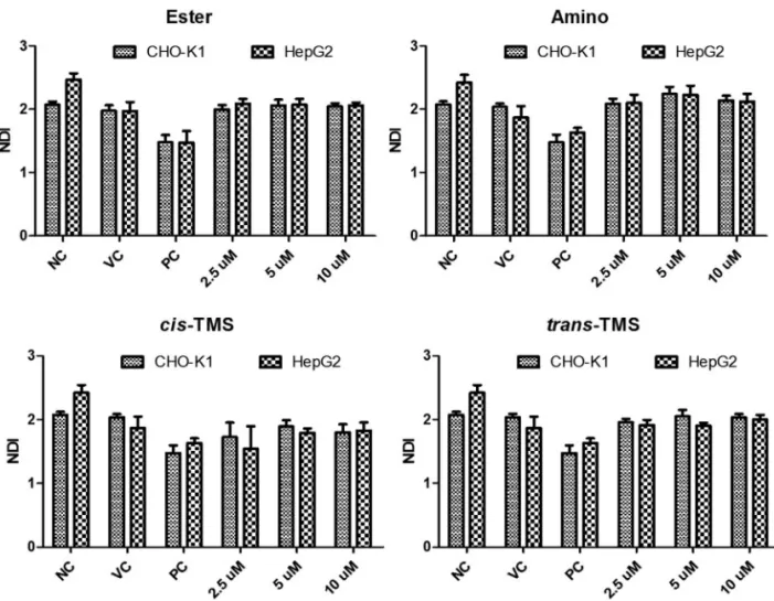
No alteration in the nuclear division (NDI) was ob-served in response to any treatment when compared to the NC in both cell lines (p > 0.05, Figure 7).
Discussion
In the present study, the cytotoxicity and genotoxicity of the ester, amino,cis-TMS andtrans-TMS stilbenes were investigated in CHO-K1 and HepG2 cell lines. CHO-K1 is recommended by the OECD for genotoxicity screening (OECD, 2014). However, HepG2 is more sensitive towards
some genotoxins and enables detection of genotoxic car-cinogens, which give negative results in other currently used bioassays, suggesting that in some cases, they might be more suitable than cell lines currently used for routine screening (Knasmülleret al., 2004). The cytotoxic (XTT) assay was performed to stablish the non-cytotoxic concen-trations of the tested compounds to be used in the evalua-tion their genotoxic potential. The determinaevalua-tion of the non-cytotoxic concentration is important because accord-ing to the OECD (2014) MN assay, cell proliferation must occur in both control and treated cultures to assess the ex-tent of chemical-induced cytotoxicity or cytostasis in all of the cell cultures in which micronuclei are detected. Cyto-toxicity of stilbene derivatives was determined using both the XTT and clonogenic survival assay, which evaluate the short and long term effects of a drug on cell viability and proliferation, respectively, and confirmed by the analysis of nuclear division index through the MN assay.
According to the XTT assay (Figure 2), the most cytotoxic compounds were thecisandtrans-TMS, showing a significant reduction of cell viability at 7.8M in both cell lines. The ester was the least cytotoxic compound with
a significant reduction of cell viability observed at 62.5M in CHO-K1 and HepG2. The reduction of cell viability may be associated with necrosis or late stages of apoptosis, when the metabolic activity is severely reduced (Frankenet al., 2006). Moreover, the surviving cells may be unable to proliferate because of reproductive integrity loss (Munshi
et al., 2005). Therefore, the cell ability to form a clone and produce a viable colony needs to be evaluated to measure the long-term cytostatic/cytotoxic effects of the tested agent (Frankenet al., 2006).
Once found that ester and amino were the least cyto-toxic compounds as evidenced by XTT assay, and that in both cell lines cytotoxicity of ester and amino was detected from 62.5M, clonogenic survival was initially assayed in a concentration ranging from 2.5 to 80M, but at 40 and 80
M no colony formation was detected. Moreover, in CHO-K1 no countable colony was detected after treatment withcis-TMS at all concentrations. Thetrans-TMS, ester and amino significantly reduced the colony formation start-ing at 5, 10 and 20M, respectively (Figure 3) in both cell lines. The ester was the least cytotoxic in this assay. Sup-pression of colony formation by stilbenes may be essen-tially irreversible, raising the possibility that interactions between stilbenes and the molecular entities and events they target may be permanent rather than transient above a certain threshold concentration (Hsieh et al., 2011). Re-garding the stereochemistry of the compounds, studies have shown that thecis-TMS is more cytotoxic than the
transisomer (Aldawsari and Velázquez-Martinez, 2015). According to Hsiehet al.(2011), experimental data about cellular signaling of PSA antigen in LNCaP cells, sug-gested that the stilbene core chemical structure instead of the side chain substitutions has a more critical role in con-trolling proliferative regulatory events.
Due to its high cytotoxicity, thecis-TMS was tested at lower concentrations in the clonogenic survival assay (Fig-ure 4). Only at the 0.3125M concentration and lower it was possible to detected survival fractions in CHO-K1, while in HepG2 colonies were detect at 0.625M. Based on the results from this assay, thecis-TMS is almost 250-fold more cytotoxic than the amino and ester, and 128-250-fold more cytotoxic thantrans-TMS. In the CBMN assay the most genotoxic compound was the cis-TMS, which in-duced a mean of 5.2 MN/100 cells at its highest concentra-tion of 0.5 M in CHO-K1 and 6.1 MN/100 cells in HepG2. This concentration was 5 times lower than the low-est concentration of the other stilbenes (Figure 5).
Both the cytotoxic and genotoxic effects ofcis-TMS are in accordance with the results of in vitrostudies re-ported in the literature. Schneider et al. (2003) demon-strated that thetrans-TMS was 100-fold less effective in in-hibiting the growth of Caco-2 cells than the cis isomer. Similar results were observed whencisandtransstilbenes were tested against five different cancer cell lines (Cushmannet al., 1991). In the work of Paulet al.(2010), thecis-TMS was the most cytotoxicin vitrobut it was not activein vivo. The lack of activity was associated with its
low bioavailability. Thetrans-TMS significantly reduced tumor volumein vivo, showing much higher serum levels than thecis-TMS. Our results clearly demonstrated that the
cis-is more genotoxic thantrans-TMS and the other stil-benes tested. However, interestingly, CHO-K1 cells did not respond totrans-TMS treatment, while this stilbene was genotoxic in HepG2 cells, demonstrating the need for meta-bolization to exhibit genotoxicity. The cis-TMS also in-creased the frequency of MN, which occurs when a whole chromosome or a fragment forms from a chromosome break and is not incorporated into one of the daughter nu-clei during cell division. Genotoxic agents can damage DNA directly or indirectly by interacting with non-DNA targets leading to genotoxic effects, essentially through lipid peroxidation and protein adducts (Kirsch-Volderset al., 2003). As far as proteins are concerned, the literature reports inhibition of DNA repair enzymes, cell cycle con-trol proteins, apoptosis-related gene products, nuclear la-mins, defense proteins against oxidative damage, metabo-lization enzymes, and tubulins of the mitotic/meiotic spindle as mechanisms by which genotoxicants exert their effects (Kirsch-Volderset al., 2003).
Schneideret al.(2003) demonstrated that the growth inhibition of Caco-2 cells bycis-TMS was not related to a cytotoxic effect, but it was associated with the arrest of the
cell cycle progression at the G2/M transition phase. They postulated that the ability of a drug to block cells in G2/M is consistent with a disruption of the mitotic spindles by inter-action with tubulin. The authors also demonstrated that
cis-TMS inhibited tubulin polymerization in a dose-depen-dent manner.
Despite the biological promising effects of stilbenes, little is known about their genotoxicological effects. The present study showed for the first time, that ester, amino,
cis- andtrans-TMS stilbenes exhibit genotoxic effects. The
cis-TMS is significantly more cytotoxic and genotoxic than the other stilbenes, and the genotoxicity oftrans-TMS re-quires metabolic activation. Further studies may elucidate the mechanisms involved in the genotoxicity of the stil-benes evaluated in the present work.
Acknowledgments
The authors acknowledge financial support by the Fundação de Amparo à Pesquisa do Estado de São Paulo (FAPESP grant 2014/12465-0 and fellowship 2015/16909-8).
References
Aggarwal BB, Bhardwaj A, Aggarwal RS, Seeram NP, Shishodia S and Takada Y (2004) Role of resveratrol in prevention and therapy of cancer: Preclinical and clinical studies. Anti-cancer Res 24:2783-2784.
Aldawsari F and Velázquez-Martínez CA (2015) 3,4’,5-trans -Trimethoxystilbene; a natural analogue of resveratrol with enhanced anticancer potency. Invest New Drugs 33:775-786.
Belguendouz L, Fremont L and Linard A (1997) Resveratrol in-hibits metalion-dependent and independent peroxidation of porcine low-density lipoproteins. Biochem Pharmacol 53:1347-1355.
Cushmann M, Nagarathnam D, Gopal D, Chakraborti AK, Lin CM and Hamel E (1991) Synthesis and evaluation of stil-bene and dihydrostilstil-bene derivatives as potential anticancer agents that inhibit tubulin polymerization. J Med Chem 34:2579-2588.
Fenech M (2000) The in vitro micronucleus technique. Mutat Res 455:81-95.
Flamini R, Zanzotto A, Rosso M, Lucchetta G, Vedova AD and Bavaresco L (2016) Stilbene oligomer phytoalexins in grape as a response toAspergillus carbonariusinfection. Physiol Mol Plant Pathol 93:112-118.
Franken NAP, Rodermond HM, Stap J, Haveman J and van Bree C (2006) Clonogenic assay of cells in vitro. Nat Protoc 1:2315-2319.
Hsieh TC, Huang YC and Wu JM (2011) Control of prostate cell growth, DNA damage and repair and gene expression by resveratrol analogues,in vitro. Carcinogenesis 32:93-101. Kirsch-Volders M, Vanhauwaert A, Eichenlaub-Ritter U and
De-cordier I (2003) Indirect mechanisms of genotoxicity. Mutat Res 140-141:63-74.
Knasmüller S, Mersch-Sundermann V, Kevekordes S, Darroudi F, Huber WW, Hoelzl C, Bichler J and Majer BJ (2004) Use of human-derived liver cell lines for the detection of envi-ronmental and dietary genotoxicants; current state of knowl-edge. Toxicology 198:315-328.
Lee S, Yoon KD, Lee M, Cho Y, Choi G, Jang H, Kim B, Jung DH, Oh JG, Kim GW, et al. (2016). Identification of a resveratrol tetramer as a potent inhibitor of hepatitis C virus helicase. Br J Pharmacol 173:191-211.
Mikstacka R, Rimando AM and Ignatowicz E (2010) Antioxidant effect oftrans-resveratrol, pterostilbene, quercetin and their combinations in human erythrocytes in vitro. Plant Food Hum Nutr 65:57-63.
Munshi A, Hobbs M and Meyn RE (2005) Clonogenic cell sur-vival assay. Method Mol Med 110:21-28.
OECD (2014) Test No. 487: In vitro mammalian cell micro-nucleus test. In: OECD Guidelines for Testing of Chemicals Section for Health Effects, 2014. OECD, Paris, pp 1-26.
Pan MH, Gao JH, Lai CS, Wang YJ, Chen WM, Lo CY, Wang M, Dushenkov S and Ho CT (2008) Antitumor activity of 3,5,4’-trimethoxystilbene in COLO 205 cells and xenografts in SCID mice. Mol Carcinog 47:184-196.
Paul S, Mizuno CS, Lee HJ, Zheng X, Chajkowisk S, Rimoldi JM, Conney A, Suh N and Rimando AM (2010).In vitroandin vivostudies on stilbene analogs as potential treatment agents for colon cancer. Eur J Med Chem 45:3702-3708.
Rimando AM, Nagmani R, Feller DR and Yokoyama W (2005) Pterostilbene, a new agonist for the peroxisome prolifera-tor-activated receptor -isoform, lowers plasma lipopro-teins and cholesterol in hypercholesterolemic hamsters. J Agric Food Chem 53:3403-3407.
Santos RA and Takahashi CS (2008) Anticlastogenic and antige-notoxic effects of selenomethionine on doxorubicin-induced damagein vitroin human lymphocytes. Mutat Res 46:671-677.
Schneider Y, Chabert P, Stutzmann J, Coelho D, Fougerousse A, Gossé F, Launay JF, Brouillard R and Raul F (2003) Resve-ratrol analog (Z)-3,5,4’-trimethoxystilbene is a potent anti-mitotic drug inhibiting tubulin polymerization. Int J Cancer 107:189-96.
Schneider Y, Duranton B, Gossé F, Schleiffer R, Seiler N and Raul F (2001) Resveratrol inhibits intestinal tumorigenesis and modulates host-defense-related gene expression in an animal model of human familial adenomatous polyposis. Nutr Cancer 39:102-107.
Seyed MA, Jantan I, Bukhari SN and Vijayaraghavan K (2016) A comprehensive review on the chemotherapeutic potential of piceatannol for cancer treatment, with mechanistic insights. J Agric Food Chem 64:725-737.
Shen MY, Hsiao G, Liu CL, Fong TH, Lin KH, Chou DS and Sheu JR (2007) Inhibitory mechanisms of resveratrol in platelet activation: Pivotal roles of p38 MAPK and NO/cyclic GMP. Br J Haematol 139:475-485.
Suh N, Paul S, Hao X, Simi B, Xiao H, Rimando AM and Reddy BS (2007) Pterostilbene, an active constituent of blueber-ries, suppresses aberrant crypt foci formation in the azoxy-methane induced colon carcinogenesis model in rats. Clin Cancer Res 13:350-355.
Ungvari Z, Orosz Z, Rivera A, Labin N, Xiangmin Z, Olson S, Podlutsky A and Csiszar A (2007) Resveratrol increases vascular oxidative stress resistance. Am J Physiol Heart Circ Physiol 292:H2417-H2424.
Wallerath T, Deckert G, Ternes T, Anderson H, Li H, Witte K and Forstermann U (2002) Resveratrol, a polyphenolic phytoa-lexin present in red wine, enhances expression and activity of endothelial nitric oxide synthase. Circulation 106:1652-1658.
Associate Editor: Daisy Maria Fávero Salvadori