http://dx.doi.org/10.1590/s2175-97902018000100014
Article
*Correspondence: K. R. Caleffi-Ferracioli. Laboratório de Bacteriologia
Mé-dica, Departamento de Análises Clínicas e Biomedicina, Universidade Estadual de Maringá. Avenida Colombo, 5790, 87020-900, Maringá, Paraná, Brasil. Tel: +55 44 3011-5375; Fax: +55 44 3011-4797. E-mail: [email protected]
ǂThe authors collaborate equally in this study.
Fast detection of
Mycobacterium tuberculosis
in culture-positive
sputum samples by nitrate reductase activity
Jean Eduardo Meneguello
1, João Vitor Oliveira
1, Letícia Silva Lima
1, Vera Lucia Dias Siqueira
1,
Regiane Bertin de Lima Scodro
1,
Katiany Rizzieri Caleffi-Ferracioli
1*ǂ,
Rosilene Fressatti Cardoso
1ǂ1Laboratory of Medical Bacteriology, Department of Clinical Analysis and Biomedicine, State University of Maringa, Parana, Brazil
Microscopy and bacterial culture are the main tools in the diagnosis of tuberculosis. Since the slow growth of Mycobacterium tuberculosis impairs rapid diagnosis strategies, especially in countries where
the latter are the only available resources, the ongoing development of new and inexpensive tools based on mycobacterial metabolism optimizing growth detection with preliminary identification is greatly welcome. When compared to the other species from the M. tuberculosis complex, M. tuberculosis
is a strong nitrate reducer. Current assay compares the nitrate reductase activity of M. tuberculosis
from pulmonary specimens cultivated in nitrate-supplemented media. Fifty-five sputum samples were decontaminated and inoculated in conventional (Middlebrook 7H9, Ogawa Kudoh-OK) and in nitrate-supplemented media (Middlebrook 7H9-N, Ogawa Kudoh-N). An aliquot from the media directly reacted with Griess reagent (7H9-N and OK-N) every five days, or transferred to a nitrate substrate solution (7H9, OK). Nitrate to nitrite reduction was considered positive, revealed by the pink color, indicating bacterial growth. As reference method, the Mycobacteria Growth Indicator Tube (MGIT) was used for sensitivity calculations and statistical analysis. 7H9-N and OK-N assays proved to perform better in detecting M. tuberculosis than conventional assays (7H9 and OK). Indeed, broth nitrate-supplemented
medium (7H9-N) was comparable to MGIT to detect M. tuberculosis, except in growth detection time.
Results show that 7H9-N may be used as an alternative tool particularly in low-income countries since it is a simple and cheap technique, and does not restrict diagnosis to single-source products.
Keywords: Tuberculosis. Mycobacteria/diagnosis/identification. Nitrate reductase.
INTRODUCTION
Tuberculosis (TB) mainly occurs by pulmonary
infection caused by Mycobacterium tuberculosis. The
disease has remained a global health threat due to multi-drug resistance and to co-infection with the human immunodeficiency virus (HIV), aggravating its lethal potential. As a result, effective strategies and appropriate support are conducted to reverse the present TB status worldwide, focusing on diagnosis, prevention, treatment and research applied to this field (WHO, 2016).
The slow growth of the bacteria complex of M.
tuberculosis is a liability since it delays rapid diagnosis
by conventional techniques, postponing the drug´s susceptibility test (WHO, 2016). Although laboratory diagnosis of pulmonary TB is normally performed by microscopy and culture, the use of automated culture systems has recently been on the increase. For instance, BACTEC MGIT (Mycobacteria Growth Indicator Tube; Becton and Dickinson) is the most used automated system worldwide and kept in reference centers for TB diagnosis. Its growth detection mechanism is based on the monitoring of an oxygen-quenched fluorochrome depletion which triggers the growth detection process and makes the drug susceptibility profile faster (WHO, 2013).
On the other hand, in the case of countries with scanty resources, automation and molecular methods are still expensive and sometimes inaccessible
(Adikaram, Perera, Wijesundera, 2012; Martin et al.,
isolates from clinical specimens are of paramount importance. A fast TB diagnosis would optimize epidemiological actions and decrease the dissemination of M. tuberculosis strains, including drug-resistant ones,
mainly in countries with high incidence of the disease (Adikaram, Perera, Wijesundera, 2012). Research for new alternatives and fast tools would also improve laboratories’ independence for single-test commercial providers for TB diagnosis, particularly in low income countries (WHO, 2011, 2013).
M. tuberculosis nitrate reductase enzyme converts
nitrate to nitrite using nitrate as a nitrogen source
(Richardson et al., 2001). In addition, this bacterial
feature has proved to be a suitable tool for Mycobacteria identification and has been employed in solid and broth media for a long time (Virtanen, 1960; Coban
et al., 2014). During the last few years, this specific
biochemical characteristic has been successfully applied in new approaches for fast TB diagnosis (Meyer, Sedlacek, Bange, 2003; Goh, Rastogi, 2010; WHO, 2011; Rojas-Ponce et al., 2013) and for drug susceptibility tests
(Coban et al., 2014; Kumar et al., 2005). In fact, nitrate
reductase activity has different behaviors and depends on the culture media used in the assay. Moreover, few reports have been published comparing time and limit
of recovery of M. tuberculosis growth directly from
sputum samples using solid and broth media. Current assay compares the employment of nitrate reductase activity in broth and solid media in the detection of M. tuberculosis growth from pulmonary specimens, in a
routine laboratory.
MATERIAL AND METHODS
Culture media
Assays were conducted in Middlebrook (7H9) broth (Difco, Detroit MI USA) and Ogawa-Kudoh (OK) solid medium (Kudoh and Kudoh, 1974) without and with addition of sodium nitrate (7H9-N and OK-N). The 7H9 medium was prepared according to manufacturer’s instructions, with 0.2% glycerol and supplemented with 10% Oleic acid, Albumin, Dextrose and Catalase - OADC (Becton Dickinson Diagnostic Systems, Sparks, MD, USA), 0.1% casein and a lyophilized mixture of antimicrobials – MGIT PANTA (Becton Dickinson Diagnostic Systems, Sparks, MD, USA). The modified media, named 7H9-N and OK-N, were prepared as described above with the addition of 1 mg/mL of sodium nitrate (CAAL, São Paulo SP Brazil) in their formulation (Macedo et al., 2012).
Limit of detection
The limit of detection of each tested medium (7H9-N, 7H9, OK and OK-N) was first determined by different concentrations of the reference strain M. tuberculosis
H37Rv (ATCC 27294) (3x10⁸, 1.5x10⁴, 1.5x10³, 1.5x10²
and 1.5x10 CFU) and M. bovis AN5 at 3 x 10⁸ CFU
(Figure 1). Each bacillus concentration was inoculated on 7H9 (3mL), 7H9-N (3 mL), OK, OK-N and incubated in normal atmosphere, at 35-37ºC. The same bacillary concentrations were inoculated in MGIT and incubated according to the manufacturer’s instructions. MGIT was used as the reference method for Mycobacteria´s growth detection.
Every five days, an aliquot of 300 µL of 7H9 cultures was removed to a sterile tube containing 2 mL of sterile 22 mmol/L phosphate buffer and 10 mmol/L sodium nitrate and incubated at 35-37 ºC for 2 hours. Further, 225 µL of freshly prepared Griess reagent (one part 50% hydrochloric acid, two parts 0.2 % sulfanilamide and two
parts 0.1% concentrated N-1-naphthylethylenediamine)
were added. The light pink to red color turnaround (between 3+ to 5+ compared to the nitrate color standards) (Kent, Kubica, 1985; Adikaram, Perera, Wijesundera, 2012) indicated nitrate reduction and thus bacillus growth. Zinc powder was added to all primary negative sodium nitrate reduction assays to make sure that the reduction was not nitrogen-caused.
In the case of 7H9-N, a 300 µL aliquot from culture was removed every five days and 225 µL of the Griess reagent were directly added for immediate nitrate reduction detection. Ziehl-Neelsen staining was carried out in 7H9 and 7H9-N nitrite positive cultures to confirm the acid-fast bacilli profile.
OK assay was daily monitored by visual inspection. The visualization ofgrowth of M. tuberculosis complex
typical colonies determined the time to perform the Griess reaction. Briefly, two spadesful of the culture were removed and added to 2 ml of sterile 22 mmol/L phosphate buffer containing 10 mmol/L sodium nitrate and incubated at 35-37 ºC for 2 hours. Further, 225 µL of Griess reagent were added to the assay and color change observed. For the OK-N reading, a slice of the medium containing typical colonies was transferred to another sterile tube and the sodium nitrate reduction was directly revealed by the addition of Griess reagent.
The M. bovis AN5 strain was used as control for
weak nitrate reduction (nitrate color standards < 3+). All media were monitored for bacterial growth until the 40th day, at 35-37 ºC, for the confirmation of negative
Sputa samples culture and presumptive identification of M. tuberculosis
After protocol standardization, 55 randomly selected pulmonary specimens from patients attended by the Laboratorio de Ensino e Pesquisa em Análises Clínicas
(LEPAC), a reference center for TB diagnosis at the Universidade Estadual de Maringá, Maringá PR Brazil, were subjected to culture in 7H9-N, 7H9, OK and OK-N media and MGIT (Figure 1). Sputum samples were previously decontaminated according to MycoPrep MGIT Kit (Siddiqi, Rüsch-Gerdes, 2006), using NaOH-NALC
FIGURE 1 - Schematic representation of the procedure used for the standardization of Nitrate Reductase Assay. Limit of detection
procedure. Further, 180 µl of each sample were inoculated in 7H9 (3 ml), 7H9-N (3 ml), OK, OK-N and 500 µl in MGIT. Manual assays were incubated in normal atmosphere at 35-37 ºC, and the MGIT assay was incubated in the automatic culture system, following manufacturer’s instructions. MGIT was used as a reference method for Mycobacteria´s growth detection. Similarly, all samples were monitored every five days as described for the standardization process. All acid-fast bacilli growth was identified by biochemical methods (Kent, Kubica, 1985).
Ethical aspects
Current study was approved by the Ethics Committee of the Universidade Estadual de Maringá, Maringá PR Brazil (Protocol n. 1.038. 941).
Data analysis
Data processing was performed with BioStata 5.3 software package (Brazil and USA). Data obtained on 7H9, 7H9-N, OK and OK-N were compared to MGIT results by McNemar test.
RESULTS AND DISCUSSION
M. tuberculosis is a strong nitrate reducer. Although
this metabolic feature has been used for biochemical identification, recently it has also been employed as a
useful growth marker. Other approaches based on nitrate reductase have been studied, such as drug susceptibility test (Kumar et al., 2005; Coban et al., 2011; Dixit et al.,
2012) and analysis of the interaction between two or more anti-TB drugs. Despite the wide use of colorimetric methods, the limit of detection and bacterial recovery have not been determined (Il et al., 2013; Rojas-Ponce et al., 2013).
Other mycobacterial species also reduce nitrate, although they do not commonly cause diseases. Among non-tuberculous mycobacteria, fast growers (with the exception of M. chelonae) and some slow growers, such
as M. kansasii, M. szulgai, M. flavescens and M. terrae,
may reduce nitrate (Virtanen, 1960). In these cases, the colony´s morphology and pigmentation aid in the identification, as these features are different from those
presented by M. tuberculosis (Virtanen, 1960; Kent,
Kubica, 1985). Current paper reports the experience with sodium nitrate incorporation in broth and solid culture media to optimize, simultaneously, growth detection and presumptive identification of M. tuberculosis isolates from
pulmonary specimens.
During standardization of the assays, on the 10th
day of incubation, at 35-37 ºC, positive sodium nitrate reduction by M. tuberculosis H37Rv was reported at 3x10⁸,
1.5x10⁴ and 1.5x10³ CFU inocula, and at 1.5x10² CFU on
the 20th day. Visually detectable nitrite accumulation was
observed in 7H9 only after the 20th day of incubation.
Colony formation was detected in OK and OK-N assays on the 10th day of incubation with positive nitrate reduction
observed at 3x10⁸ and 1.5x10⁴ CFU inocula. In the case
of 1.5x10³ and 1.5x10² CFU inocula, the sodium nitrate
reduction in OK and OK-N assays was reported only on the 15th day of incubation. With regard to MGIT, control
assay showed positive cultures at 1, 6, 7, 8 and 8 days for 3x10⁸, 1.5x10⁴, 1.5x10³, 1.5x10², 1.5x10 CFU of M.
tuberculosis H37Rv, respectively. Based on these results,
7H9-N and OK-N assays proved to be more satisfactory and faster in detecting M. tuberculosis growth than 7H9
and OK assays. Results showed that increasing contact time between bacilli and substrate of nitrate reductase improved M. tuberculosis H37Rv growth detection by the
culture medium supplementation.
No visual M. tuberculosis H37Rv growth in OK
and in N, or sodium nitrate reduction in OK, OK-N, 7H9, 7H9-N has been registered with 1.5x10 CFU. Negative results in 7H9-N and 7H9 assays for 1.5x10 CFU inoculum were expected by the authors. According to the Middlebrook 7H9 manufacturer’s instructions, the limit of viable bacilli recovery in clinical specimens is over 102 to 3×10² CFU (Power, Zimbro, 2009). Consequently,
false negative results in nitrate reductase assay may be explained by the low concentration of bacilli in clinical specimens. This result was expected since the MGIT was used as the reference assay due to its higher sensitivity to metabolism detection by checking out hourly the rate of O2 consumption during bacillus growth (Rojas-Ponce
et al., 2013). Modified assays 7H9-N and OK-N depend
on the active metabolism of mycobacteria to reduce nitrate to nitrite or nitrogen. As a rule, the higher the bacilli concentration, the faster will be growth and nitrate reduction.
Since M. bovis AN5 strain showed weak nitrate
reduction in all tested media, the difference is an important phenotypical feature to discriminate M. tuberculosis from M. bovis. In current study with pulmonary specimens, the
sensitivity of 7H9-N and OK-N in detecting and having
presumptive identification of M. tuberculosis growth
were positive cultures with presumptive M. tuberculosis
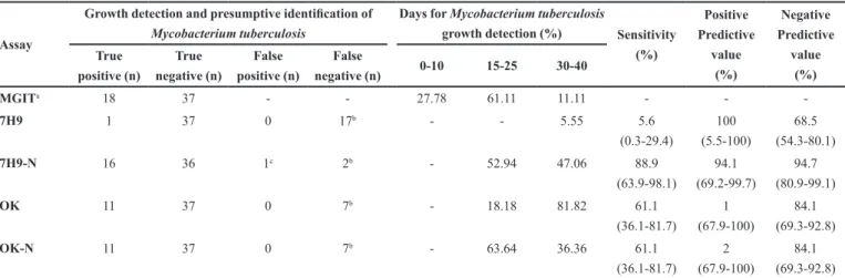
identification detected by 7H9-N and OK-N, respectively. From these positive cultures, 52.94% (9/17) and 63.64% (7/11) of growth, detection occurred from 15 to 25 days of incubation, respectively (Table I). No statistical difference between the performance of 7H9-N and MGIT has been observed (p>0.05). In contrast, there was a statistical difference (p≤0.05) when 7H9 and OK were compared to MGIT. Therefore, in low resource laboratories, these two assays could be auxiliary tools for a fast TB diagnosis.
The nitrate-supplemented Middlebrook broth (7H9-N) proved to be better for the detection of bacillus growth in clinical specimens. The 7H9-N assay revealed an easy
and fast evidence of the presumptive M. tuberculosis
growth by the strong reduction of nitrate into nitrite when compared to the solid media, OK and OK-N. Our results corroborate those by Rageade et al. (2014). The
difference between solid and broth media is well explained due to favorable growth in broth medium and the need to visualize colonies in solid form to perform nitrate reductase reaction. Nonetheless, OK-N or OK allows the visual observation of typical colony morphology and pigment production. The OK medium was chosen for sodium nitrate supplementation in current assay due to its buffered formulationand to the easy and fast sputum decontamination protocol, by which specimen cultures may be feasible in any clinical laboratory for implementing TB diagnosis.
Similar to the standardization process and rather than 7H9 with clinical specimens, 7H9-N detected bacillus
growth faster. The performance is statistically comparable to MGIT (p>0.05). Further, 7H9-N has an advantage over MGIT by allowing the preliminary identification of the bacillus simultaneously with growth detection. The addition of sodium nitrate in the medium caused fast bacillus phenotypic identification without impairing its growth, corroborating studies by (Meyer et al., 2003;
Goh, Rastogi, 2010). Macedo et al. (2012) reported that,
when sodium and potassium nitrate were compared, the two were metabolized as substrate.
The bacillus identification, based on the nitrate reductase reaction, requires attention due to clinical specimen contamination. Since nitrate reductase enzymes
occur in Klebsiella oxytoca, Pseudomonas aeruginosa,
Salmonella typhimurium and other bacteria, the strong
alkali decontamination process carried out in clinical specimens, as described by Kudoh (Kudoh, Kudoh, 1974), may minimize false positive results. Therefore, growth time, acid fastness and cord factor observed by Ziehl-Neelsen staining may determine the fast and presumptive presence of M. tuberculosis.
CONCLUSION
The culture medium sodium nitrate supplementation, similar to 7H9-N, was an inexpensive and fast alternative
method to detect M. tuberculosis growth through
simultaneous biochemical presumptive identification when automated culture systems are not available for TB
diagnosis. The 7H9-N performance in M. tuberculosis
TABLE I - Comparison of media without (7H9 and OK) and supplemented by sodium nitrate (7H9-N and OK-N) with the
Mycobacteria Growth Indicator Tube (MGIT) for Mycobacterium tuberculosis growth detection and fast presumptive identification in a 40-day culture
Assay
Growth detection and presumptive identification of Mycobacterium tuberculosis
Days for Mycobacterium tuberculosis
growth detection (%) Sensitivity (%) Positive Predictive value (%) Negative Predictive value (%) True positive (n) True negative (n) False positive (n) False
negative (n) 0-10 15-25 30-40
MGITa 18 37 - - 27.78 61.11 11.11 - -
-7H9 1 37 0 17b - - 5.55 5.6
(0.3-29.4)
100 (5.5-100)
68.5 (54.3-80.1)
7H9-N 16 36 1c 2b - 52.94 47.06 88.9
(63.9-98.1)
94.1 (69.2-99.7)
94.7 (80.9-99.1)
OK 11 37 0 7b - 18.18 81.82 61.1
(36.1-81.7)
1
(67.9-100)
84.1 (69.3-92.8)
OK-N 11 37 0 7b - 63.64 36.36 61.1
(36.1-81.7)
2 (67.9-100)
84.1 (69.3-92.8)
a: MGIT PANTA (Becton Dickinson Diagnostic Systems, Sparks, MD, USA) - assay used as reference; 7H9:Middlebrook 7H9 broth (Difco, Detroit, MI, USA);
growth detection from sputum samples was comparable to MGIT, except for growth detection time. Results show that the nitrate reductase assay may be an auxiliary tool for early TB diagnosis, particularly in low-income countries where laboratories have limited resources, providing independence of single-test commercial providers.
ACKNOWLEDGMENTS
The authors would like to thank the Medical Bacteriology Division of the Laboratório de Ensino e Pesquisa em Análises Clínicas (LEPAC), Universidade Estadual de Maringá, Maringá PR Brazil, for their collaboration in the study.
CONFLICT OF INTEREST
There is no conflict of interest.
ETHICAL APPROVAL
Formal consent is not required for this type of study.
REFERENCES
Adikaram CP, Perera J, Wijesundera SS. The manual mycobacteria growth indicator tube and the nitrate reductase assay for the rapid detection of rifampicin resistance of M. tuberculosis in low resource settings. BMC Infect Dis. 2012;12:326.
Coban AY, Cayci YT, Deveci A, Akgunes A, Uzun M, Durupinar B. A rapid detection of multidrug-resistant Mycobacterium tuberculosis by a nitrate reductase assay on blood agar. Mem Inst Oswaldo Cruz. 2011;106(3):378-80.
Coban AY, Deveci A, Sunter AT, Martin A. nitrate reductase assay for rapid detection of isoniazid, rifampin, ethambutol, and streptomycin resistance in Mycobacterium tuberculosis: a systematic review and meta-analysis. J Clin Microbiol. 2014;52(1):15-9.
Dixit P, Singh U, Sharma P, Jain A. Evaluation of nitrate reduction assay, resazurin microtiter assay and microscopic observation drug susceptibility assay for first line antitubercular drug susceptibility testing of clinical isolates of M. tuberculosis.
J Microbiol Methods. 2012;88(1):122-6.
Goh KS, Rastogi N. Simple and rapid method for detection of nitrate reductase activity of Mycobacterium tuberculosis and Mycobacterium canettii grown in the Bactec MGIT960 system. J Microbiol Methods. 2010;81(2):208-10.
Il YB, Orikiriza P, Rojas-Ponce G, Riera-Montes M, Atwine D, Nansumba M, et al. Use of colorimetric culture methods for detection of Mycobacterium tuberculosis complex isolates from sputum samples in resource-limited settings. J Clin Microbiol. 2013;51(7):2273-9.
Kent PT, Kubica GP. Public health mycobacteriology: a guide for the level III laboratory. Atlanta, Ga: Department of Health and Human Services; 1985. p. 96-103.
Kudoh S, Kudoh T. A simple technique for culturing tubercle bacilli. Bull World Heal Organ. 1974;51(1):71-82.
Kumar M, Khan IA, Verma V, Kalyan N, Qazi GN. Rapid, inexpensive MIC determination of Mycobacterium tuberculosis isolates by using microplate nitrate reductase assay. Diagn Microbiol Infect Dis. 2005;53(2):121-4.
Macedo MB, Von Groll A, Fissette K, Palomino JC, Almeida da Silva PE, Martin A. Nitrate reductase assay using sodium nitrate for rapid detection of multidrug resistant tuberculosis. Brazilian J Microbiol. 2012;43(3):981-3.
Martin A, Paasch F, Docx S, Fissette K, Imperiale B, Ribon W, et al. Multicentre laboratory validation of the colorimetric redox indicator (CRI) assay for the rapid detection of extensively drug-resistant (XDR) Mycobacterium tuberculosis. J Antimicrob Chemother. 2011;66(4):827-33.
Meyer A, Sedlacek L, Bange FC. Rapid testing for nitrate reductase activity of Mycobacterium tuberculosis grown in an automated culture system. Eur J Clin Microbiol Infect Dis. 2003;22(7):444-6.
Fast detection of Mycobacterium tuberculosis in culture-positive sputum samples by nitrate reductase activity
Rageade F, Picot N, Blanc-Michaud A, Chatellier S, Mirande C, Fortin E, et al. Performance of solid and liquid culture media for the detection of Mycobacterium tuberculosis in clinical materials: meta-analysis of recent studies. Eur J Clin Microbiol Infect Dis. 2014;33(6):867-70.
Richardson DJ, Berks BC, Russell DA, Spiro undefined S, Taylor CJ. Functional, biochemical and genetic diversity of prokaryotic nitrate reductases. Cell Mol Life Sci. 2001;58(2):165-78.
Rojas-Ponce G, Rachow A, Guerra H, Mapamba D, Joseph J, Mlundi R, et al. A continuously monitored colorimetric method for detection of Mycobacterium tuberculosis complex in sputum. Int J Tuberc Lung Dis. 2013;17(12):1607-12.
Siddiqi SH, Rüsch-Gerdes S. MGIT Procedure Manual for BACTECTM MGIT 960TM TB System. Geneva: FIND; 2006. 89 p.
Virtanen S. A study of nitrate reduction by mycobacteria. The use of the nitrate reduction test in the identification of mycobacteria. Acta Tuberc Scand Suppl. 1960;48:1-119.
World Health Organization. WHO. Strategic and Technical Advisory Group for Tuberculosis, Report of 13th meeting. Geneva, Switzerland: World Health Organization; 2013. 34 p.
World Health Organization. WHO. Noncommercial culture and drug-susceptibility testing methods for screening patients at risk for multidrug-resistant tuberculosis: policy statement. Geneva, Switzerland: World Health Organization; 2011. 12 p.
World Health Organization. WHO. Global Tuberculosis Report. Geneva, Switzerland: World Health Organization; 2016. 201 p.