Vol. 52, Special Number: pp. 69-76, November 2009
ISSN 1516-8913 Printed in Brazil BRAZILIAN ARCHIVES OF
BIOLOGY AND TECHNOLOGY
A N I N T E R N A T I O N A L J O U R N A L
Hematological and Cerebrospinal Fluid Changes in Cattle
Naturally and Experimentally Infected with the Bovine
Herpesvirus 5
Júlio Augusto Naylor Lisbôa
1*, Allan Jürgen Isernhagen
1, Alexandre Secorun Borges
2,
Rogério Martins Amorim
2, Mara Regina Stipp Balarin
3, Michele Lunardi
3and Amauri
Alcindo Alfieri
31
Departamento de Clínicas Veterinárias; Centro de Ciências Agrárias; Universidade Estadual de Londrina; Londrina - PR - Brasil. 2Departamento de Clínica Veterinária; Faculdade de Medicina Veterinária e Zootecnia; Universidade Estadual Paulista Júlio de Mesquita Filho; Botucatu - SP - Brasil. 3Laboratório de Virologia Animal; Departamento de Medicina Veterinária Preventiva; Universidade Estadual de Londrina; Londrina - PR - Brasil
ABSTRACT
Bovine herpesvirus 5 (BoHV-5) meningoencephalitis is one of the main causes of mortality by encephalopathy in Brazilian cattle herds. However, the neurological signs observed are common to several encephalopathies and do not contribute decisively to a diagnosis. In order to verify hematological and cerebrospinal fluid (CSF) changes, blood and CSF samples from naturally and experimentally infected bovines were evaluated. In natural cases (n=17), the samples were collected only once, and in experimental cases (n=7), the samples were sequentially obtained throughout disease progression. While routine methods were used to examine the samples, BoHV-5 infection was confirmed by a PCR assay. Blood analyses did not reveal any consistent hematological alterations and the leukogram results occasionally showed increases in leukocyte and segmented neutrophil. Hyperfibrinogenemia was noted in all experimentally infected calves and in half of the natural cases. Pleocytosis with mononuclear cells was a remarkable finding in CSF collected from both groups of animals and was present even in experimentally infected calves that remained asymptomatic. Therefore, CSF evaluation can be used as an auxiliary method in ante-mortem BoHV-5 diagnosis.
Key words: bovine, meningoencephalitis, herpesviruses, complete blood count, cerebrospinal fluid, diagnosis
*
Author for correspondence: janlisboa@uel.br
INTRODUCTION
Bovine herpesvirus 5 (BoHV-5) meningoencephalitis is one of the main causes of mortality by encephalopathy in Brazilian cattle herds. This infection is characterized by low morbidity and high lethality rates and mainly affects young cattle. In Brazil, the occurrence of
Nervous signs observed in this infection, during the evolution of clinical symptoms, can differ among individuals as well as in a single animal, during the course of the disease (Rissi et al., 2006). The neurological disturbances consist of a cerebral syndrome, depending on the extent and location of encephalic lesions; however, these signs are common to several encephalopathies and do not contribute decisively towards the diagnosis. Routine diagnosis has been only performed post-mortem by evaluating encephalic fragments using laboratory techniques such as histopathology (Rissi et al., 2008), virus isolation in cell culture, and/or BoHV-5 DNA detection by polymerase chain reaction (PCR) (Vogel et al., 2003; Claus et al., 2007).
Despite the high lethality rate, natural and experimental BoHV-5 infections have shown that the disease is not always lethal (Elias et al., 2004; Rissi et al., 2006; Spilki et al., 2006). Auxiliary methods for ante-mortem diagnosis of BoHV-5 infection, such as blood cell count and cerebrospinal fluid (CSF) analysis, are particularly important in animals presenting discrete and transitory dysfunctions. In addition, the results of these tests can lead to a preliminary differential diagnosis among other causes of encephalopathy in cattle.
Diverse aspects related to the pathogenesis of BoHV-5 meningoencephalitis were elucidated in studies involving experimental infection (Cascio et al., 1999; Vogel et al., 2003; 2004). However, data collected from hematological or CSF examinations in naturally or experimentally BoHV-5 infected cattle are not available.
The aim of this study was to present blood and CSF changes observed in both naturally and experimentally BoHV-5 infected cattle.
MATERIALS AND METHODS
Naturally affected cattle
Seventeen cattle naturally affected with BoHV-5 meningoencephalitis between 2001 and 2007 were evaluated. The cattle were from ten different herds from the Paraná (n=12) and São Paulo states (n=5). The animals were kept at Veterinary Teaching Hospitals (Londrina/PR and Botucatu/SP) where they were clinically evaluated until death, euthanasia, or discharge to return to the original farm. While alive, the animals were
evaluated daily using classical semiological methods.
Blood and CSF samples were collected by routine methods on the first or second day at the hospital (Mayhew, 1989). The only animal that survived the disease had four CSF samples collected at 5-day intervals. All blood and CSF samples were processed immediately after collection.
Experimentally infected cattle
BoHV-5 experimental infection was induced in seven clinically healthy male calves of the Holstein breed, ranging in age from 20 to 58 days. Only calves from eutocian births that had spontaneously fed on colostrum were selected. Calves that presented passive BoHV antibodies, detected by serum neutralization (Ferreira et al., 2005; Dias et al., 2008), were excluded from the experiment. The AA PAR BoHV-5 strain (Souza et al., 2002), isolated from a natural case of herpetic meningoencephalitis, was used for viral challenge. Experimental animals were inoculated through intranasal instilation of 0.5 mL of BoHV-5 inoculum (titer of 107.42 TCID50/mL) in each nostril (Isernhagen, 2005). Throughout the entire experimental period (30 days), routine physical examinations were performed twice daily.
The first blood and CSF collections were performed before viral challenge. Subsequent blood samples were collected at 48-hour intervals until the end of the experiment. In calves that presented neurological dysfunctions, CSF collections were performed at 48-hour intervals until death, starting on the day of onset of clinical signs. In asymptomatic calves, subsequent collections were performed on days 21, 23, 26, and 30 post-infection (p.i.). After death or euthanasia at 30 days p.i, all calves were submitted to necropsy.
Laboratorial parameters
coagulation), the global and differential cell counts, and the protein, glucose, pH, and specific gravity values were also evaluated (Coles, 1984).
PCR assay
In order to confirm the diagnosis of BoHV-5 infection in both naturally and experimentally BoHV-5 infected animals, CSF and fragments from diverse encephalic areas were collected and analyzed using a polymerase chain reaction (PCR) technique. For DNA extraction of encephalic fragments, a combination of the phenol/chloroform/isoamyl alcohol and silica/guanidine isothiocyanate methods was carried out according to Alfieri et al. (2006). CSF samples were submitted to the PCR assay without prior DNA extraction. The amplification of the BoHV-5 glycoprotein C gene by PCR was performed according Claus et al. (2005).
Statistical analysis
Dispersion and central tendency measurements were presented for the description of the results. In the experimental infections, the variation according to time was evaluated by means of analyzing the variation in the repeated measurements. An error probability of 5% was considered.
RESULTS AND DISCUSSION
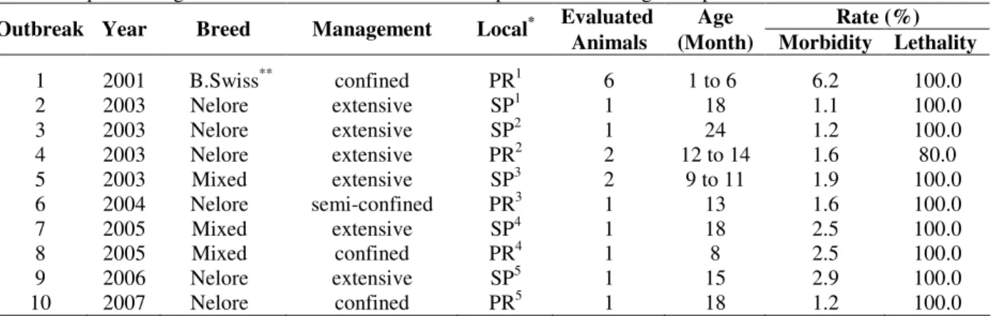
BoHV-5 infection was confirmed by PCR assay in encephalic samples from all 24 animals included in this study. As presented in Table 1, most natural BoHV-5 meningoencephalitis cases were observed in beef cattle (n=9) with extensive (n=6), semi-confined (n=1), and confined (n=2) management. In such herds, the infection was more frequent in young animals and was characterized by low morbidity rate.
The only BoHV-5 outbreak observed in dairy cattle involved younger calves and was characterized by a higher morbidity rate. The management in this farm was inadequate in several aspects, including environmental, nutritional, physiological, and sanitary aspects, which may have contributed to the higher morbidity and lethality rates.
In the natural cases of BoHV-5 infection included in this study, the lethality rate was not 100% because one of the calves survived. In all other evaluated outbreaks, no recovery of an affected animal was registered. This observation is in agreement with natural BoHV-5 outbreaks described in Brazilian cattle herds (Colodel et al., 2002; Rissi et al., 2006; Claus et al., 2007).
Table 1- Epidemiologic data from outbreaks of bovine herpesvirus 5 meningoencephalitis.
Evaluated Age Rate (%)
Outbreak Year Breed Management Local*
Animals (Month) Morbidity Lethality
1 2001 B.Swiss** confined PR1 6 1 to 6 6.2 100.0
2 2003 Nelore extensive SP1 1 18 1.1 100.0
3 2003 Nelore extensive SP2 1 24 1.2 100.0
4 2003 Nelore extensive PR2 2 12 to 14 1.6 80.0
5 2003 Mixed extensive SP3 2 9 to 11 1.9 100.0
6 2004 Nelore semi-confined PR3 1 13 1.6 100.0
7 2005 Mixed extensive SP4 1 18 2.5 100.0
8 2005 Mixed confined PR4 1 8 2.5 100.0
9 2006 Nelore extensive SP5 1 15 2.9 100.0
10 2007 Nelore confined PR5 1 18 1.2 100.0
*
PR (Paraná state), SP (São Paulo state)
PR1-Apucarana; SP1-Esperança do Sul; SP2-Avaré; PR2-Faxinal; SP3-Agudos; PR3-Congoinhas; SP4-Pardinho; PR4
-Londrina; SP5-Botucatu; PR5-Jaguapitã
**
Brown Swiss
Clinically, the neurological disturbances observed in the evaluated cattle and the intensity of those signs varied among individuals and during the disease progression. Cortical syndrome could be seen in most animals (n=15) through observation
amaurosis, paresis, proprioceptive deficits, and tongue paresis). These signs are similar to those previously described (Rissi et al., 2006; 2008). The presence of a concomitant cerebellar dysfunction could be observed in a few animals (n=4) and was characterized by ataxia and hypermetria. Similarly, only one calf had as the exclusive neurological sign a dysfunction of cranial nerves, suggesting a ponto-bulbar syndrome.
In most cases (n=14), a progressive worsening of the clinical signs preceded death or a critical clinical condition by up to one week. The only exception was a calf that survived after presenting mild and transitory clinical dysfunctions, including aggressivity, isolation from the herd, and anorexia. Unusually, the return of appetite and improvement in behavior were noticed in the same calf after it was hospitalized for 15 days. Such observations allowed the reintroduction of the animal to the herd, demonstrating the possibility of an occasional recovery in this disease (Spilki et al., 2006).
In this study, BoHV-5 experimental infection was not able to induce neurological signs in all infected calves. During the first 30 days p.i., three calves did not present any neurological dysfunction; however, BoHV-5 meningoencephalitis could be confirmed in the CNS of these calves using a PCR assay and histopathological examinations (Isernhagen, 2005). This observation demonstrates that the BoHV-5 infection may go unnoticed, with the virus maintained in a latent form (Cascio et al., 1999; Vogel et al., 2003; 2004).
Alterations in blood cells were investigated during the disease progression in experimentally infected calves, although no significant variation could be demonstrated for the erythron. In three out of four calves that presented neurological signs, the dysphagia observed was not accompanied by dehydration due to the rapid progression to death. In calves presenting encephalopathy, leukogram results were characterized by a trend of increase in the number of leukocytes accompanied by a significant elevation in the number of circulating segmented neutrophils. Such alterations were established from the day 10 p.i. onwards, coinciding with the beginning of signs. While the increase in the leukocytes did not characterize leukocytosis (maximum mean value observed: 12,900/mm3), neutrophils reached mean values similar to those of circulating lymphocytes (5,960
This feature may be a consequence of the elevation in the cortisol concentration due to stress that accompanies the disease (Coles, 1984; Jain, 1986). In asymptomatic calves, no variation in the leukogram could be observed.
In regard to the plasmatic fibrinogen concentration, hyperfibrinogenemia could be verified in all experimentally infected calves presenting encephalopathy. In these animals, individual values raging from 900 to 1,200 mg/dL were coincidentally reached with the onset of signs. The increase in the plasmatic fibrinogen level is known to accompany the early stages of inflammatory processes in cattle, with this increase observed earlier than leukocyte alterations (Jain, 1986).
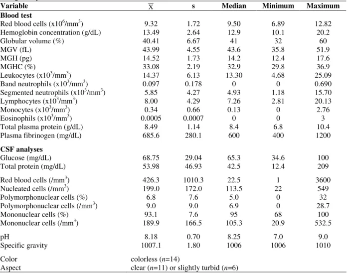
Hematological alterations were also not observed in animals affected by the natural form of BoHV-5 meningoencephalitis (Table 2). An increase in globular volume was observed in only one animal due to the hemoconcentration resulting from dysphagia. Leukocytosis was observed in four calves (ranging from 22,600 to 25,095 leukocytes/mm3), while neutrophilia was present in three of them (ranging from 10,288 to 15,704 segmented neutrophils/mm3). However, pneumonia could be observed simultaneously with meningoencephalitis in these animals. Hyperfibrinogenemia was observed only in half of naturally affected cattle, with the elevated values in a range from 800 to 1,200 mg/dL (Table 2), a result that differs from the results for the experimentally infected calves.
Most evaluated cattle presented 100 or more nucleated cells per mm3 of CSF (n=12), and mononuclear cells represented 90% or more of these cells (n=15). These findings are
characteristic of viral meningoencephalitis and differ from those of bacterial etiology (Mayhew, 1989; Scott, 2004).
Table 2 - Hematological and cerebrospinal fluid analyses of 17 natural cases of bovine herpesvirus 5 meningoencephalitis.
Variable X s Median Minimum Maximum
Blood test
Red blood cells (x106/mm3) 9.32 1.72 9.50 6.89 12.82
Hemoglobin concentration (g/dL) 13.49 2.64 12.9 10.1 20.2
Globular volume (%) 40.41 6.67 41 32 60
MGV (fL) 43.99 4.55 43.6 35.8 51.9
MGH (pg) 14.52 1.73 14.2 12.4 17.6
MGHC (%) 33.08 2.19 32.9 29.8 36.9
Leukocytes (x103/mm3) 14.37 6.13 13.30 4.68 25.09
Band neutrophils (x103/mm3) 0.097 0.178 0 0 0.690
Segmented neutrophils (x103/mm3) 5.85 4.27 4.93 1.18 15.70
Lymphocytes (x103/mm3) 8.00 4.29 7.26 2.81 20.13
Monocytes (x103/mm3) 0.34 0.66 0.13 0 2.76
Eosinophils (x103/mm3) 0.0005 0.0007 0 0 3
Total plasma protein (g/dL) 8.49 1.14 8.4 6.8 10.4
Plasma fibrinogen (mg/dL) 685.6 280.1 600 400 1200
CSF analyses
Glucose (mg/dL) 68.75 29.04 65.3 34.6 100
Total protein (mg/dL) 53.98 46.93 42.5 12.4 209
Red blood cells (/mm3) 426.3 1010.3 22.5 1 3600
Nucleated cells (/mm3) 199.0 172.0 113.5 22 549
Polymorphonuclear cells (%) 6.8 7.6 5.0 0 32
Polymorphonuclear cells (/mm3) 9.0 9.0 6.9 0 28.7
Mononuclear cells (%) 93.1 7.6 95 68 100
Mononuclear cells (/mm3) 189.9 166.5 105.3 20.9 532.5
pH 8.18 0.70 8.25 7.0 9.0
Specific gravity 1007.1 1.80 1006 1006 1010
Color colorless (n=14)
Aspect clear (n=11) or slightly turbid (n=6)
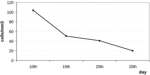
Asymptomatic experimentally infected calves presented the same cellular alterations as those observed in calves with encephalopathy (mean values of 9 cells/mm3 of CSF before challenge and approximately 46, 168, 67, and 20 cells/mm3 of CSF at days 21, 23, 26, and 30 p.i., respectively), indicating the occurrence of meningoencephalitis. It is important to note that the nucleated cell count decreased until the end of observation period. The same finding could be demonstrated in the calf that survived the natural disease, in which a mononuclear cell count that was slightly over
Figure 1 -Variation in cerebrospinal fluid mononuclear cells count during the disease progression of a calf that survived to natural form of bovine herpesvirus 5 meningoencephalitis.
Unlike pleocytosis, increase in CSF protein concentration was not characterized as a consistent alteration. Taking 40 mg/dL to be the maximum physiological limit (Scott, 2004), only a discrete increase in the CSF protein concentration could be observed in nine naturally infected calves (Table 2). According to Mayhew (1989), only four animals presented high concentrations (above 75 mg/dL). Among the experimentally infected calves, only one presented a high concentration at the end of the disease (108 mg/dL at day 19 p.i.). Nonetheless, this case must be considered as an exception due to the long duration of the disease. In fact, most CSF samples presented were colorless and clear and had pH and specific gravity values within physiological ranges (Scott, 2004).
BoHV-5 meningoencephalitis is not generally accompanied by consistent hematological alterations. Aspects of CSF cellularity can be considered of great value for ante-mortem diagnosis since they may indicate the possibility of BoHV-5 infection. Although they do not allow differentiation from other viral encephalitis, such as rabies and some cases of polioencephalomalacia, they allow exclusion of bacterial encephalitis, intoxication by lead, organophosphate and carbamate poisonings, hepatic encephalopathy, brain trauma, and
The protocol for the experimental induction of BoHV-5 infection was approved by the Ethics Committee in Animal Experimentation of Universidade Estadual de Londrina under registration number 25/05, process 9671/200, and was performed following the ethical principles instituted by COBEA.
ACKNOWLEDGEMENTS
The financial resources for the conduction of this study were supported by the project BioAgroPar financed by FINEP, SETI/PR, and Fundação Araucária/PR; and by CNPq/Brazil.
Part of the research activities of this study was carried out in the Agricultural Research Support Laboratory (Laboratório de Apoio à Pesquisa Agropecuária - LAPA) / PROPPG / UEL.
Alfieri A.A. is recipient of CNPq fellowship.
RESUMO
A meningoencefalite determinada pelo herpesvírus bovino 5 (BoHV-5) é considerada uma das principais causas de mortalidade por encefalopatia em bovinos no Brasil. O diagnóstico clínico da infecção é difícil de ser realizado pois os sinais neurológicos ocasionados pela infecção com o
0 20 40 60 80 100 120
10th 15th 20th 25th
day
c
e
ll
s
/m
m
BoHV-5 são comuns aos observados em encefalopatias bovinas de diferentes etiologias. Com o objetivo de determinar as alterações hematológicas e do líquido cefalorraquidiano, foram avaliadas amostras de sangue total e líquor colhidas de animais infectados tanto naturalmente quanto experimentalmente. Nos casos naturais da doença (n=17), as amostras foram coletadas apenas uma vez. Nos casos de infecção experimental (n=7), as amostras foram obtidas sequencialmente durante a evolução clínica da doença. Enquanto na análise das amostras clínicas foram empregadas metodologias rotineiras, a infecção pelo BoHV-5 foi confirmada em todos os animais incluídos nesse estudo por meio da técnica de PCR. As análises hematológicas não revelaram alterações consistentes e os resultados obtidos a partir do leucograma mostraram aumento ocasional de leucócitos e neutrófilos segmentados. A hiperfibrinogenemia pôde ser observada em todos os bezerros infectados experimentalmente e em metade dos casos naturais da doença. Diferentemente, a pleiocitose com predomínio de células mononucleares foi considerada um achado marcante nas amostras de líquor coletadas em ambos os grupos de animais, encontrando-se presente até mesmo nos bezerros infectados experimentalmente que permaneceram assintomáticos. Os resultados do presente estudo, obtidos tanto em condições de infecções naturais quanto experimentais, demonstraram que a avaliação do líquor cefalorraquidiano pode ser utilizada como método auxiliar para o diagnóstico ante-mortem
da infecção pelo BoHV-5.
REFERENCES
Alfieri, A. A., Parazzi, M. E., Takiuchi, E., Médici, K. C., Alfieri, A. F. (2006), Frequency of group A rotavirus in diarrhoeic calves in Brazilian cattle herds, 1998–2002. Trop Anim Health Prod, 38, 521-526
Cascio, K. E., Belknap, E. B., Schultheiss, P. C., Ames, A. D., Collins, J. K. (1999), Encephalitis induced by bovine herpesvirus 5 and protection by prior vaccination or infection with bovine herpesvirus 1. J Vet Diagn Invest, 11, 134-139
Claus, M. P., Alfieri, A. F., Folgueras-Flatschart, A. V., Wosiacki, S. R., Médici, K. C., Alfieri, A. A. (2005), Rapid detection and differentiation of bovine herpesvirus 1 and 5 glycoprotein C gene in clinical specimens by multiplex-PCR. J Virol Methods, 128, 183-188
Claus, M. P., Alfieri, A. F., Médici, K. C., Lunardi, M., Alfieri, A. A. (2007), Bovine herpesvirus 5 detection by virus isolation in cell culture and Multiplex-PCR in central nervous system from cattle with neurological disease in Brazilian herds. Braz J Microbiol, 38, 485-490
Coles, E. H. (1984), Patologia Clínica Veterinária. Manole, São Paulo
Colodel, E. M., Nakazato, L., Weiblen, R., Mello, R. M., Silva, R. R. P., Souza, M. A., Filho, J. A. O., Caron, L. (2002), Meningoencefalite necrosante em bovinos causada por herpesvírus bovino no estado de Mato Grosso, Brasil. Ciência Rural, 32, 293-298
Dias, J.A., Alfieri, A., Médici, K. C., Freitas, J. C., Neto, J. S. F., Müller, E. E. (2008), Fatores de risco associados à infecção pelo herpesvírus bovino 1 em rebanhos bovinos da região Oeste do Estado do Paraná. Pesq Vet Bras, 28, 161-168
Elias, F., Schild, A. L., Riet-Correa, F. (2004), Meningoencefalite e encefalomalacia por Herpesvírus bovino-5: distribuição das lesões no sistema nervoso central de bovinos naturalmente infectados. Pesq Vet Bras, 24, 123-131
Ferreira, M. C., Médici, K. C., Alfieri, A. F., Alfieri, A. A. (2005), Desenvolvimento e avaliação de um ensaio imunoenzimático para o diagnóstico sorológico da infecção pelo herpesvírus bovino. Semina Ci Agr, 26, 363-372
Gomes, L. I., Rocha, M. A., Costa, E. A., Lobato, Z. I. P., Mendes, L. C. N., Borges, A. S., Leite, R. C., Barbosa-Stancioli, E. F. (2002), Detecção de herpesvírus bovino 5 (BoHV-5) em bovinos do sudeste brasileiro. Arq Bras Med Vet Zootec, 54, 217-220
Isernhagen, A. J. (2005), Meningoencefalite herpética em bezerros: evolução clínica e diagnóstico. MSc Dissertation, Universidade Estadual de Londrina, Londrina, Brazil
Jain, N. C. (1986), Shalm’s Veterinary Hematology. Lea and Febiger, Philadelphia
Mayhew, I. G. (1989), Large Animal Neurology: a handbook for veterinarians clinicians. Lea and Febiger, Philadelphia
Rissi, D. R., Oliveira, F. N., Rech, R. R., Pierezan, F., Lemos, R. A. A., Barros, C. S. L. (2006), Epidemiologia, sinais clínicos e distribuição das lesões encefálicas em bovinos afetados por meningoencefalite por herpesvírus bovino-5. Pesq Vet Bras, 26, 123-132
Rissi, D. R., Pierezan, F., Silva, M. S., Flores, E. F., Barros, C. S. L. (2008), Neurological disease in cattle in southern Brazil associated with bovine herpesvirus infection. J Vet Diagn Invest, 20, 346-349
Salvador, S. C., Lemos, R. A. A., Riet-Correa, F., Roehe, P. M., Osório A. L. A. R. (1998), Meningoencefalite em bovinos causada por herpesvírus bovino-5 no Mato Grosso do Sul e São Paulo. Pesq Vet Bras, 18, 76-83
Scott, P. R. (2004), Diagnostic techniques and clinicopathologic findings in ruminant neurologic disease. Vet Clin Food Anim, 20, 215-230
Souza, V. F., Melo, S. V., Esteves, P. A., Schmidt, C. S., Gonçalves, D. A., Schaefer, R., Silva, T. C., Almeida, R. S., Vicentini, F., Franco, A. C., Oliveira, E. A., Spilki, F. R., Weiblen, R., Flores, E. F., Lemos, R. A., Alfieri, A. A., Pituco, E. M., Roehe, P. M. (2002), Caracterização de herpesvírus bovinos tipos 1 (BHV-1) e 5 (BHV-5) com anticorpos monoclonais. Pesq Vet Bras, 22, 13-18
Spilki, F. R., Silva, T. C., Esteves, P. A., Teixeira, M. B., Batista, H. B. C. R., Chiminazzo, C., Driemeier, D., Franco, A. C., Roehe, P. M. (2006), Co-infections with bovine herpesvirus type 5 and bovine viral diarrhoea virus. Arq Bras Med Vet Zootec, 58, 699-707
Vogel, F. S. F., Caron, L., Flores, E. F., Weiblen, R., Winkelmann, E. R., Mayer, S. V., Bastos, R. G. (2003), Distribution of bovine herpesvirus type 5 DNA in the central nervous systems of latently, experimentally infected calves. J Clin Microbiol, 41, 4512-4520 Vogel, F. S. F., Lima, M., Flores, E. F., Weiblen, R.,