CASE REPORT
Immunoglobulin-responsive chikungunya encephalitis:
two case reports
Stephanie Suzanne de O. Scott
1&Pedro Braga-Neto
2,3&Lícia Pacheco Pereira
1&Paulo Ribeiro Nóbrega
1&Francisco de Assis Aquino Gondim
2&Manoel Alves Sobreira-Neto
2,4&Claudia Carvalho Mendes Schiavon
1Received: 7 November 2016 / Revised: 23 April 2017 / Accepted: 16 May 2017 / Published online: 2 June 2017
#Journal of NeuroVirology, Inc. 2017
Abstract
Chikungunya virus is an alphavirus transmitted
by the mosquito
Aedes
, mainly
Aedes aegypti
and
Aedes
albopictus
, that can cause acute illness, mostly self-limited,
characterized by fever, maculopapular rash, and disabling
polyarthritis/arthralgia, with an incubation period of 1 to
12 days. Chikungunya was largely regarded as a non-fatal
and self-limited disease, but recently, serious cases have
been reported including some with severe involvement of
the nervous system, such as meningoencephalitis, myelitis,
polyradiculitis, and polyradiculoneuropathy. In this report,
we describe the clinical and laboratory findings of two
patients with encephalitis associated with chikungunya in
a northeastern city in Brazil, who exhibited a good
out-come, with improvement after treatment with i.v.
immuno-globulin (IVIg).
Keywords
Encephalitis . Chikungunya . Immunoglobulin .
Treatment
Introduction
Chikungunya, in the Makonde language, stands for
B
that
which bends
^
(Azevedo et al.
2015
), an expression that
de-notes the severity of joint pain in patients affected by the
disease. Chikungunya is a virus described primarily in the
1950s in East Africa and is classified as a RNA virus, from
the Togaviridae family and alphavirus genus (Azevedo et al.
2015
). The vector is the mosquito
Aedes
, mainly
Aedes
aegypti
and
Aedes albopictus
.
In 2004, there was a resurgence of the virus in the Indian
Ocean Islands with subsequent worldwide spread (Mohan
et al.
2010
), presenting as a febrile illness associated with
debilitating arthritis, skin rash, headache, maculopapular rash,
and myalgia. The clinical picture shows an improvement after
10 days, but the joint pain can last from months to years
(Gérardin et al.
2015
). Chikungunya was largely regarded as
a non-fatal and self-limited disease, but recently, serious cases
have been reported including some with severe involvement
of the nervous system, e.g., meningoencephalitis, myelitis,
polyradiculitis, and polyradiculoneuropathy (Donalisio and
Freitas
2015
; Crosby et al.
2016
; Mohan et al.
2010
).
Here, we report two patients in Fortaleza, a city in the
northeast of Brazil, with chikungunya encephalitis who
exhib-ited an excellent improvement after administration of i.v.
immunoglobulin.
Case 1
A 74-year-old man presented with fever, maculopapular rash,
and severe arthralgia. After 4 days, he developed confusion
and fluctuating level of consciousness and was taken to a local
hospital. Upon arrival, the patient was drowsy and disoriented
in space and time. After 12 days, he developed paraparesis
* Stephanie Suzanne de O. Scottstephaniesscott@hotmail.com
1 Hospital Universitário Walter Cantídio, Universidade Federal do
Ceará, Av Rogaciano Leite 900 apto 503, Torre Friburgo, Guararapes, Fortaleza, Ceará, Brazil
2 Department of Clinical Medicine, Universidade Federal do Ceará,
Fortaleza, Ceará, Brazil
3
Center of Health Sciences, Universidade Estadual do Ceará, Fortaleza, Ceará, Brazil
4 Universidade de Fortaleza, Fortaleza, Ceará, Brazil
with diffuse areflexia. Ancillary tests revealed the following:
cerebrospinal fluid (CSF) showing increased cell count with a
predominance of lymphocytes and increased protein
(Table
1
), serology for chikungunya was positive, and
electro-encephalogram showed disorganized electrical brain activity
with the presence of some waves with triphasic morphology.
Nerve conduction studies (NCS) and electromyogram (EMG)
revealed a sensorimotor axonal neuropathy (Tables
2
,
3
and
4
).
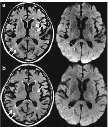
Magnetic resonance imaging (MRI) of the brain showed
scattered T2/flair hyperintense foci in the supratentorial white
matter in the semioval and subcortical centers, some with high
signal intensity on DWI sequence, with no contrast
enhance-ment (Fig.
1
). After this initial investigation, treatment with
intravenous human immunoglobulin (dose of 400 mg/kg/day
for 5 days) was initiated, with significant improvement, after
the third day, of his level of consciousness, becoming awake
and obeying verbal commands, also becoming oriented in
space and time, recognizing family members. On the other
hand, paraparesis only improved after 40 days, following
plas-mapheresis. A follow-up NCS (Tables
5
,
6
,
7
and
8
) showed
improvement in conduction velocities over time, and the
pa-tient was walking with unilateral assistance after 3 months.
Case 2
An 83-year-old man developed fever with chills, followed
after 3 days by important arthralgias. After 6 days, he
Table 1 Results of CSF studies in two patients with chikungunya
Patient 1 Patient 2
Cell count (cells/mm3) 90 42
Lymphocytes (%) 91 98
Neutrophils (%) 2 2
Plasma cells (%) 7 0
RBC 1 2
Glucose (mg/dl) 62 59
Protein (mg/dl) 179 100
Cultures Negative Negative
Table 2 Case 1: Electrophysiology studies. Motor nerve conduction study
Site Lat.
(ms) Dur. (ms)
Amp. (mV)
Area (mV ms)
Segment Distance
(mm)
Interval (ms)
NCB (m/s)
CCV N.D.
Ulnar Left
Wrist 2.6 18.0 4.4 31.5 Wrist 2.6
Below elbow 6.7 16.7 3.8 29.5 Wrist–below elbow 230 4.1 56.1 Above elbow 9.0 18.0 3.7 30.9 Above elbow–below elbow 105 2.3 45.7 Underarm 10.7 17.7 3.7 30.7 Above elbow–underarm 85 1.7 50.0
Ulnar Right
Wrist 3.0 16.3 4.6 32.8 Wrist 3.0
Below elbow 8.2 17.5 4.2 33.3 Wrist–below elbow 240 5.3 45.7 Above elbow 10.0 16.1 4.1 31. Below elbow–above elbow 90 1.8 50.0 Underarm 11.7 16.0 3.7 29.2 Above elbow–underarm 75 1.7 45.5
Tibial Left
Hallucis abductor 3.7 7.2 5.6 14.4 Hallucis abductor 2.3
Popliteal fossa 14.8 7.7 3.5 14.0 Hallucis abductor-popliteal fossa 400 12.6 31.9
Tibal Right
Hallucis abductor 3.7 7.2 5.6 14.4 Hallucis abductor 3.7
Popliteal fossa 13.1 8.8 2.3 9.6 Hallucis abductor–popliteal fossa 385 9.4 41.0 Fibular Left
Ext digitorum brevis
3.4 7.1 1.7 5.0 Ext digitorum brevis 3.4
Fibular head 11.1 13.0 1.0 7.9 Ext digitorum brevis–Fibular Head
300 7.7 39.0
Popliteal fossa 13.1 12.5 1.1 8.4 Fibular head-popliteal fossa 90 2.0 45.0 Fibular Right
Extens brev dig 3.1 4.5 3.5 1.2 Ext digitorum brevis 3.1 Fibular head 10.7 9.5 2.4 11.9 Ext digitorum brevis–fibular
head
305 7.7 39.9
developed lethargy and confusion with disorientation in
time and space. Neurological status evolved with
worsen-ing level of consciousness, leadworsen-ing to orotracheal
intuba-tion (OTI) for airway protecintuba-tion. Head CT scan was
per-formed and did not disclose any abnormalities. CSF
stud-ies revealed an increase in the cell count with a
predom-inance of lymphocytes and increased protein (Table
1
). At
this point, the patient was treated with acyclovir without
significant clinical improvement. Three days after
admis-sion, laboratory tests showed positive serology for
chikungunya, while serology for dengue (IgM and IgG)
was negative. Four days after initial neurological
symp-toms, treatment with i.v. immunoglobulin (IVIg) (at a
dose of 400 mg/kg/day for 5 days) was started with
sig-nificant neurological improvement on the fourth day of
infusion, with complete recovery of the encephalitis.
Table 3 Case 1: Electrophysiology studies. EMG findings summary
Muscle Side Ins. act. Fibs. Pos. wave Fasc. Myo. disch. Normal MUP Poly Low amp. High amp. Dur. Recruit Int. patt.
Pronator teres L Normal 0 0 0 0 0 N 0 0 Long Reduce Reduce
1st dorsal inter. L Incr. +2 +2 0 0 0 ++ 0 0 Long Reduce Reduce
Biceps brachii L Incr. +1 +1 0 0 0 ++ 0 0 Long Reduce PI 4
Triceps L Normal 0 0 0 0 0 N 0 0 Normal Full Full
Vastus lateralis L Incr. +2 +2 0 0 0 − − − − − No act
Vastus medialis R Incr. +2 +2 0 0 0 − − − − − No act
Gastroc. medial H L Incr. +2 +2 0 0 0 − − − − − No act
Gastroc. Medial H R Incr. +2 +2 0 0 0 − − − − − No act
Tibialis anterior L Incr. +2 +2 0 0 0 − − − − − No act
Tibialis anterior R Incr. +2 +2 0 0 0 − − − − − No act
Table 4 Case 1: Electrophysiology studies. Sensory nerve conduction study
Site Lat. 1 (ms)
Lat. 2 (ms)
Amp. (μV)
Area (μV ms)
Segment Distance (mm)
Interval (ms)
NCV (m/s)
CCV N.D. Temp.
Ulnar Left
Wrist–digito V Wrist–digito V
Elbow–digito V Ulnar Right
Wrist–digito V 2.4 3 5.7 0.2 Wrist– digito V
110 2.4 45.5
Elbow– digito V Radial Left
Wrist–radial 1.5 2.1 8 0.6 Wrist–radial 95 1.5 63.3 Sural Left
Retr exter–lat foot
2.5 3.3 4.6 0.6 Retr exter–lat foot
90 2.5 36
Sural Right Retr exter–lat
foot
2.5 2.9 1.5 0 Retr exter–lat foot
85 2.5 34
Fibular superf. Right Ankle–dorso
foot
Ankle–dorso foot Fibular superf. Left
Ankle–dorso foot
Ankle–dorso foot Median Left
Palm 2.2 3 7 0.4 Palm 105 2.2 47.7
Table 5 Case 1: Electrophysiology studies. Electrophysiology studies after treatment with plasmapheresis. Motor nerve conduction study
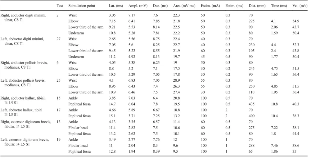
Test Stimulation point Lat. (ms) Ampl. (mV) Dur. (ms) Area (mV ms) Estim. (mA) Estim. (ms) Dist. (mm) Time (ms) Vel. (m/s)
Right, abductor digiti minimi, ulnar, C8 T1
2 Wrist 3.05 7.17 7.6 22.3 50 0.3 70
Elbow 7.15 6.41 7.85 21.8 50 0.3 225 4.1 54.9
Lower third of the arm 9.21 5.53 8.14 22.5 50 0.3 90 2.06 43.7
Underarm 10.8 5.28 7.81 22.2 50 0.3 80 1.59 50.4
Left, abductor digiti minimi, ulnar, C8 T1
27 Wrist 2.65 5.56 8.75 22.4 40 0.3 70
Elbow 7.05 5.6 8.25 22.7 40 0.3 230 4.4 52.3
Lower third of the arm 9.45 5.22 8.55 21.9 60 0.3 105 2.4 43.8
Underarm 11.2 4.92 8.13 19.7 45 0.5 90 1.77 50.4
Right, abductor pollicis brevis, medianus, C8 T1
6 Wrist 4.05 5.38 6.25 19 50 0.3 80
Elbow 8.8 5.2 7.1 17.5 30 0.2 245 4.75 51.5
Lower third of the arm 10.5 5.29 7.05 17.8 30 0.2 90 1.65 56.4
Left, abductor pollicis brevis, medianus, C8 T1
25 Wrist 4.1 6.83 7.05 28.9 55 0.3 80
Elbow 8.95 6.43 7.4 26.3 55 0.3 250 4.85 51.5
Lower third of the arm 10.9 6.46 7.5 27.4 30 0.2 110 1.95 56.4
Right, abductor hallux, tibial, I4 L5 S1
15 Ankle 3.85 7.03 6.4 20.8 100 0.5 70
Popliteal fossa 14.7 6.04 7.8 19.5 100 0.5 435 10.8 40.3
Left, abductor hallux, tibial I4 L5 S1
17 Ankle 4.66 5.89 6.67 18.8 100 2 70
Popliteal fossa 15.1 3.71 7.25 13.2 100 2 400 10.4 38.3
Right, extensor digitorum brevis, fibular, I4 L5 S1
13 Ankle 4.13 3.35 6.57 11.4 60 0.5 70
Fibular head 11.4 2.82 7.5 10.6 60 0.5 275 7.22 38.1
Popliteal fossa 13.2 2.62 7.5 10.1 60 0.5 80 1.8 44.4
Left, extensor digitorum brevis, fibular, I4 L5 S1
19 Ankle 3.49 2.77 7.76 12 100 1 70
Fibular head 11 2.04 8.3 9.6 100 1 288 7.46 38.6
Popliteal fossa 12.8 1.94 8.39 9.5 100 1 65 1.86 35
J.
N
eu
rov
ir
ol
.
(20
17
)
23
:6
25
–
63
Brain MRI performed almost 15 days after hospital
ad-mission was normal, when symptoms had resolved almost
completely.
Discussion
Chikungunya virus causes acute symptomatic illness. The
dis-ease is characterized by fever, rash, and disabling arthralgia,
with an incubation period that can range from 1 to 12 days
(Ganesan et al.
2008
). CNS involvement has been more
re-cently described. However, similar to those of the dengue
virus (Nelson et al.
2014
), it is uncertain whether the
neuro-logical manifestations are due to CNS viral invasion, immune
responses, or to both conditions (Das et al.
2010
).
There is some experimental evidence of CNS invasion. A
study in rats has demonstrated invasion of the choroid plexus
and meninges by the virus, however, not of the brain
paren-chyma (Ziegler et al.
2008
). Moreover, in vitro studies have
documented virus replication in astrocytes and
oligodendro-cytes (Das et al.
2015
). Additionally, the higher frequency of
symptoms related to CNS involvement in the elderly and
those with comorbidities, as seen in the present cases, suggests
that the virus can manipulate ineffective immune systems to
reach the brain and its surrounding structures since
immuno-logic response to antigens in elders weakens and infections
tend to be frequent and heavier with increased age
(Hjalmarsson et al.
2007
; Sansoni et al.
2008
).
Another experimental study with monkeys reported a
rela-tionship between high levels of cytokines and encephalopathy
(Labadie et al.
2010
), linking inappropriate immune response
Table 7 Case 1: Electrophysiology studies. Electrophysiology studies after treatment with plasmapheresis. F-wave
Test Fmin lat. (ms)
F ampl. (μV)
M lat. (ms)
Fmin-M lat. (ms)
F/M (%)
Max V (m/s)
V (m/s)
Right, abductor digit minimi, ulnar, C8 T1 5 31.3 316 2.75 28.5 2.8 Left, abductor digit minimi, ulnar, C8 T1 28 32.2 287 2.95 29.3 3 Right, abductor pollicis brevis, medianus, C8 T1 7 24.2 327 4.3 19.9 3.6 Left, abductor pollicis brevis, medianus, C8 T1 26 34.5 141 4.25 30.3 1.4 Right, abductor hallux, tibial, I4 L5 S1 16 57.5 435 4 53.5 3.8 Left, abductor hallux, tibial I4 L5 S1 18 58.6 377 4.2 54.5 3.9 Right, extensor digitorum brevis, fibular, I4 L5 S1 14 55.1 173 4.25 50.8 3.9 Left, extensor digitorum brevis, fibular, I4 L5 S1 20 54.7 102 3.9 50.9 3.1
Table 6 Case 1: Electrophysiology studies. Electrophysiology studies after treatment with plasmapheresis. Sensory nerve conduction study
Test Stimulation point
Lat. (ms)
Ampl. (μV)
Dur. (ms)
Area (nV s)
Estim. (mA)
Estim. (ms)
Dist. (mm)
Time (ms)
Vel. (m/s)
Right, superficial branch of radial nerve, C5 C6
8 1 1.22 24.1 2.73 17.8 20 0.2 78 1.22 64.1
Right, n. medianus 9 Palm 1.85 18.4 1.35 13.6 20 0.2 90 1.85 48.6 Right, n. medianus 11 Wirst 2.54 16.1 3.31 13.9 20 0.2 130 2.54 51.2
Left, medianus 30 Palm 2.2 15.6 1.1 9 20 0.2 95 2.2 43.2
Left, medianus 32 Wirst 3.07 11.6 2.98 11.2 15 0.2 140 3.07 45.6
Right, n. ulnar 12 Wirst 1.96 18 3.14 20.5 12 0.2 120 1.96 61.3
Right, n. ulnaris V dig. 31 Palm 1.59 10 2.46 6.4 15 0.2 95 1.59 59.8 Right, n. peroneus
superficialis, L4-S1
24 1 0 25 0.2
Left, n. peroneus superficialis, L4-S1
22 1 0 25 0.2
Right, n. suralis, S1–S2 23 1 1.95 7 1.6 6.6 25 0.2 80 1.95 41 Left, n. suralis, S1–S2 21 1 1.75 3.4 1.85 2.8 15 0.2 75 1.75 42.9
to neurologic manifestations. Taking this into account, a good
response to IVIg observed in our two patients suggests that an
immune mechanism could be the main cause of the
neurolog-ical symptoms, since this drug acts by neutralizing the
humor-al response elicited by the virus.
It is also possible that the involvement of the CNS may
occur through viral invasion, associated with an inappropriate
immune response (Courderc and Lecuit
2015
), justifying
higher frequency of these symptoms in the elderly and a good
response to immunoglobulin.
Our two patients were infected during a chikungunya
outbreak in the city of Fortaleza in 2016, had positive
IgM for chikungunya, and tested negative for dengue
fever. The CSF changes observed in our patients
sup-ported the diagnosis of encephalitis and therefore CNS
involvement.
As far as we know, this is the first description of an
IVIg-responsive encephalitis due to a chikungunya virus
infection. The mechanisms underlying the response to
IVIg in our patients are not clear. We can speculate that
blockade of a deleterious immune response by IVIg is a
possible mechanism. It is more difficult to understand
how IVIg could directly affect the viral activity or
replication.
However, given the lack of controlled studies about
this subject, it is also important to consider that the
improvement could be simply explained by natural
his-tory of the disease and that improvement after IVIg
could be due to chance.
In summary, it is possible that chikungunya encephalitis
can benefit from IVIg, at least in certain conditions.
However, controlled randomized trials are necessary to better
assess this therapeutic possibility.
Compliance with ethical standards
Conflict of interest The authors declare that they have no conflict of interest.
a
b
Fig. 1 Case 1: Brain MRI findings. a Multiple white matter hyperintensities are shown on FLAIR (left,arrows) and DWI (right, circle) predominantly in the periventricular and basal ganglia/capsular regions.bMRI of the same patient 4 months later, demonstrating partial regression of the hyperintense foci on FLAIR and disappearance of the alterations on DWI (circle). There was no abnormal gadolinium enhance-ment. Also, note mild diffuse brain atrophy
Table 8 Case 1: Electrophysiology studies. Electrophysiology studies after treatment with plasmapheresis. EMG findings summary
Test Spontaneous activity
Amplitude MUP
Duration MUP
Polyphasia MUP
Pattern
Right, biceps brachii, cutaneous muscle, C5 C6
34 N N N N
Left, biceps brachii, cutaneous muscle, C5 C6 43 N N N N
Right, triceps, radial, C6 C7 C8 T1 35 Increased Increased N Neurogenic
Right, interosseus, ulnar, C8 T1 36 N N N N
Left, interosseus, ulnar, C8 T1 44 N N N N
Right, vastus medialis, femoral, L2–L4a 37 Increased Increased N Neurogenic Left, vastus medialis, femoral, L2–L4 40 Increased Increased Increased Neurogenic Right, gastrocnemius, tibial, S1–S2 39 Increased Increased Increased Neurogenic Left, gastrocnemius, tibial, S1–S2 42 Increased Increased N Neurogenic Right, tibialis anterior fibular, L4 L5 s1 38 Increased Increased Increased 4
References
Azevedo RSS, Oliveira CS, Vasconcelos PFC (2015) Risco do Chikungunya para o Brasil. Rev Saude Publica 49
Courderc T, Lecuit M (2015) Chikungunya virus pathogenesis: from bedside to bench. Antivir Res 121:120–131
Crosby L, Perreau C, Madeux B, Cossis J, Armand C, Herrmann-Storke C, Najioullah F, Valentino R, Thiéry G (2016) Severe manifestations of chikungunya virus in critically ill patients during the 2013–2014 Caribbean outbreak. Intern J Infect Dis v 48:78–80
Das T, Jaffar-Bandjee M, Hoarau J, Trotot P, Denizot M, Lee-Pat-Yuen G, Sahoo R, Guiraud P, Ramful D, Robin S, Alessandri J, Gauzere B, Gasque P (2010) Chikungunya fever: CNS infection and patholo-gies of a re-emerging arbovirus. Prog Neurobiol 91:121–129 Das T, Horarau MC, Bandjee J, Gasque MMP (2015) Multifaceted immune
responses engaged by astrocytes, microglia and resident dendritic cells against chikungunya neuroinfection. J Gen Virol 96:294–310 Donalisio MR, Freitas AR (2015) Chikungunya in Brasil: an emerging
challenge. Rev Bras Epidemiol 18(1)
Ganesan K, Diwan A, Shankar SK, Desai SB, Saiani GS, Katrak SM (2008) Chikungunya encephalomyeloradiculitis: report of 2 cases with neuroimaging and 1 case with autopsy findings. AJNR 29
Gérardin P, Couderc T, Bintner M, Tournebize P, Renouil M, Lémant J, Boisson V, Borguerini G, Staikowsky F, Schramm F, Lecuit M, Michault A (2015) Chikungunya virus-associated encephalitis. Neurology 86
Hjalmarsson A, Blomqvist P, Skoldenberg B (2007) Herpes simplex en-cephalitis in Sweden, 1990–2001: incidence, morbidity, and mortal-ity. Clin Infect Dis 45(7):875–880
Labadie K, Larcher T, Joubert C, Mannioui A, Delache B, Brochard P et al (2010) Chikungunya disease in nonhuman primates involves long term viral persistence in macrophages. J Clin Invest 120:894–906 Mohan A, Kiran DHN, Manohar IC, Kumar DP (2010) Epidemiology,
clin-ical manifestations and diagnosis of chikungunya fever: lessons learned from the re-emerging epidemic. Indian J Dermatol 55(1):54–63 Nelson J, Waggoner J, Sahoo M, Grant P, Pinsky B (2014) Encephalitis
caused by chikungunya virus in a traveler from the Kingdom of Tonga. J Clin Microbiol 52(9):3459–3461
Sansoni P, Vescovini R, Fagnoni F, Biasini C, Zanni F, Zanlari L, Telera A, Lucchini C, Passeri G, Monti D, Franceschi C, Passeri M (2008) The immune system in extreme longevity. Exp Gerontol 43(2):61–65 Ziegler SA, Lu L, da Rosa AP, Xiao SY, Tesh RB (2008) An animal
model for studying the pathogenesis of chikungunya virus infection. AmJTrop Med Hyg 79:133–139