Vol.55, n. 3: pp. 375-379, May-June 2012
ISSN 1516-8913 Printed in Brazil BRAZILIAN ARCHIVES OF
BIOLOGY AND TECHNOLOGY
A N I N T E R N A T I O N A L J O U R N A L
Detection of Lsr2 Gene of
Mycobacterium leprae
in Nasal
Mucus
Luiz Antonio Custodio
1*, Alexandre Saito
2, Marla Karine Amarante
1, Thiago Cezar
Fujita
2, Aparecida de Lourdes Perim
1, Ivete Conchon Costa
2, Ionice Felipe
2and Shiduca
Itow Jankevicius
21Departamento de Patologia Aplicada, Análise Clínicas e Toxicológicas; Centro de Ciências da Saúde;
Universidade Estadual de Londrina – PR - Brasil. 2 Departamento de Patologia Geral; Centro de Ciências
Biológicas; Universidade Estadual de Londrina; Rodvia Celso Garcia Cid, 86051-990; Londrina - PR - Brasil
ABSTRACT
In the present study, nasal mucus from patients with leprosy were analyzed by PCR using specific primers for Lsr2
gene of Mycobacterium leprae. The presence of Lsr2 gene in the nasal mucus was detected in 25.80% of patients
with paucibacillari leprosy, and 23.07% of contacts. Despite the absence of clinical features in the contact individuals, it was possible to detect the presence of Lsr2 gene in the nasal mucus of these individuals. Therefore,
PCR detection of M. leprae targeting Lsr2 gene using nasal mucus samples could contribute to early diagnosis of
leprosy.
Key words:Mycobacterium leprae, leprosy, hanseniase, nasal mucus, PCR.
* Author for correspondence: harpialc@hotmail.com
INTRODUCTION
Although a long time has elapsed since the discovery of Mycobacterium leprae by Gerhard Armauer Hansen in 1873 (De Zubiria et al. 2003; Fernandes et al. 2004), the understanding of the biology of this microorganism is still incomplete. For instance, the reasons for the inability of M. leprae to grow in media and conditions commonly used for bacteria is still not known. Recent studies have presented interesting data about the relationship between M. leprae and its host (Zanazzi et al. 2000; Nurse 2003), but many relevant questions about it remain unanswered (Siddiqui et al. 2003). An important aspect of mycobacterial pathogenesis is the ability of the pathogen to establish long lasting latent infections in the host. Leprosy (or Hanses’s Disease) is a
to human genomes using BLAST method. It is known that B cell responses to specific sequences within the Lsr antigen have been shown to be associated with the immunopathological responses in leprosy patients with erythema nodosum leprosum. It has been observed that some B and T cell epitopes localized to the regions with amino acid substitutions may account for the putative differential responsiveness to this antigen in tuberculosis and leprosy (Oftung et al. 2000). (Misra et al. 1995) found that the patients who did not show acid-fast bacilli in the tissues by the conventional methods presented positive for M. leprae DNA. PCR was initially used for the detection this pathogen in 1989 (Hartskeerl et al. 1989; Woods et al. 1989) and since them it has been used in most different clinical specimens as skin (De Wit et al. 1991; Yoon et al. 1993), nasal swab and hair bulb (Santos et al. 1995) and oral swab (Goulart et al. 2001). A proteomic approach was undertaken to identify the proteins present in the soluble/cytosol and membrane subcellular fractions obtained from the armadillo derived from
M. leprae. Proteins from each fraction were separated by two-dimensional gel electrophoresis (2-DE) and identified by mass spectrometry. A total of 147 protein spots were identified from 2-DE patterns and shown to comprise the products of 44 different genes, 28 of them corresponding to new proteins (Marques et al. 2004).
Multidrug treatment has contributed to disease control, but new cases are being reported, showing that the development of early detection methods is imperative. Knowledge of the genomic sequence of M. leprae (Cole et al. 2001) is an important step for understanding this bacterium and its interactions with its host, as well as for developing the detection tests such as those based on PCR (Meima et al. 2004) or helicase-dependent amplification (HDA) (Young 2001; Vincente et al. 2004). Several PCR-based methods for the detection and evaluation of leprosy have been reported (Zanazzi et al. 2000; Santos et al. 2001; Guerrero et al. 2002; Torres et al. 2003; Sakamuri et al. 2009). The aim of this work was to evaluate the PCR detection of lsr2 gene of M. leprae in nasal mucus.
MATERIAL AND METHODS
With the approval at the Human Ethics Committee
of Londrina State University and Philantropic Society Humanitas, Parana, Brazil, 130 Brazilian patients were included in thisn study. DNA sequences of M. leprae were analyzed by BLAST (http://ncbi.nlm.nih.gov/BLAST) and the rate of repetition in the genome was checked searching for a unique sequence copy, as well as for similarity with the genome of other microorganisms. Lsr2 gene was analyzed in the PRIMER3 program (http://frodo.wi.mit.edu/ primer3/) to draw the primers and these were analyzed by the BLAST program, checking its specificity to M. leprae. The 130 patients enrolled in this study were grouped as following: 22 samples (~16,9%) from multibacillary leprosy, 31 samples (~23,8%) from paucibacillari leprosy, 52 samples (40%) from asymptomatic household contacts of patients with leprosy and 25 samples (~19,2 %) from people without history of contact with leprosy. Mycobacterium fortuitum (11 isolates), Mycobacterium scrofulaceum (1 isolate),
Mycobacterium tuberculosis (29 isolates),
Mycobacterium avium (4 isolates), which were kindly provided by Dra. Halha Ostrensky Saridakis, (Centro de Ciências Biológicas, Departamento de Microbiologia – Universidade Estadual de Londrina - Brasil), were used as controls.
Swab samples of nasal mucus were collected using sterile cotton swabs wet in phosphate buffer (pH 7.0), maintained at 4°C until transferred to Eppendorf® tubes containing 1.0 mL of ultra pure water and stored at –20°C until DNA extraction.
DNA extraction
Genomic DNA was isolated following the method proposed by (Torres et al. 2003) with modification. Briefly, 200µL of the sample was centrifuged 15 minutes at 12.000 g and the pellet was treated at 100°C for 1 minute and freezing in nitrogen for 3 minutes. It was resuspended in 100 µl of Tris- HCl 50 mM buffer (pH 8.0), containing lysozime 10 mg/mL, incubated at 37°C for30 minutes, added 10 µL of proteinase K 50 mg/mL, followed by the addition of 1.0 µl of Triton X-100 (10%). The mixture was kept at 55oC for two hours and after the addition of absolute ethanol centrifuged at 10.000 g for five minutes. The DNA was suspended in ultra pure water and stored at –20°C until use.
PCR products were analyzed by the electrophoresis on acrylamide gel (10%) and detected by a nonradioisotopic technique using a commercially available silver staining method (data not shown).
RESULTS AND DISCUSSION
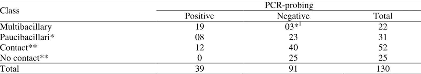
The Lsr2 gene of M. leprae codifies a protein of 15 kDa with capability of stimulating T lymphocytes. Table 1 shows the results of
amplifications by PCR from 130 samples of nasal mucus of leprosy patients, asymptomatic household contacts of patients with leprosy and people without contact history. Samples with negative PCR results were tested for the presence of inhibitors in the reaction, by adding standardized DNA to the extracted sample. To test the specificity of PCR-probing with the primers for M. leprae Lsr2 region to Lsr2, the gene of other mycobacterias as M. avium, M. fortuitum, M. scrofulaceum and M. tuberculosis were tested in the same region. In this context, all the strains were negative in PCR test.
Table 1 - Detection of Lsr2 gene of M. leprae in smear of nasal mucus from patients.
Class PCR-probing
Positive Negative Total
Multibacillary 19 03*1 22
Paucibacillari* 08 23 31
Contact** 12 40 52
No contact** 0 25 25
Total 39 91 130
1Three were discharged from treatment. *p<0,0001. **p<0,0227
In Brazil, despite actions for the treatment and control of leprosy, the number of new cases is highly threatening, and considering the existence of infected and undiagnosed people as a true reservoir of M. leprae and their contacts (Braber 2004) as infection sources, the development of methods for early diagnosis become imperative. PCR is a useful tool for the diagnosis of leprosy and its recent developments such as helicase dependent amplification point to the possibility of developing methods of detection with better sensitivity and specificity. (Bang et al. 2009) evaluated the sensitivity and utility of polymerase chain reaction (PCR) to detect M. leprae in comparison with other conventional methods for the diagnosis such as split skin smears, histopathology and serodiagnosis. They observed that the PCR detection of M. leprae targeting 16S ribosomal RNA was specific and more sensitive than the conventional methods, and could contribute to early and accurate diagnosis of leprosy. The present work standardized a PCR technique to amplify the single copy gene Lsr2 of
M. leprae. This technique presented 100% of specificity for M. leprae relative to other mycobacteria, in agreement with previous reports (Santos et al. 2001; Guerrero et al. 2002; Torres et al. 2003; Willams et al. 2003; Braber 2004; Cortes-Herrera et al. 2008). Therefore, it seemed
possible to use this gene as a target for large-scale screening of human reservoirs of M. leprae,
of patients with leprosy with a high risk for developing or transmitting the disease would be of clinical and predictive value. While the prevalence of leprosy has declined around the world, there has not been a corresponding decrease in its incidence, thus indicating that it has not been possible to prevent the transmission of the disease.
ACKNOWLEDGEMENT
We thank Sociedade Filantrópica Humanitas for their colaboration, Dr. John Spencer and Dr. Patric Brennan for their inestimable attention, and also Dr. Halha Ostrensky Saridakis for samples supply.
REFERENCES
Bang PD, Suzuki K, Phuong le T, Chu TM, Ishii N, Khang TH, Evaluation of polymerase chain reactionbased detection of Mycobacterium leprae for the diagnosis of leprosy. J. Dermatol, 2009, 5: 269-276.
Braber KL, Na Evaluation of GAEL, the Global Alliance for the Elimination of Leprosy. Leprosy Rev, 2004, 75:208-213.
Braga FJHN, Nuclear medicine in tropical diseases.
Braz Arch Biol Technol, 2002, 45: 1-7.
Chae GT, Lee SB, Kang TJ, Shin HK, Kim JP, Ko YH, Kim SH, Kim NH, Typing of clinical isolates of
Mycobacterium leprae and their distribution in
Korea. Lepr Rev, 2002, 73: 41-46.
Cole ST, Eiglmeier K, Parkhill J, James K.D, Thomson NR, Wheeler PR, Honoré N, Garnier T, Churcher C, Harris D, Mungall K, Basham D, Brown D, Chillingworth T, Connor R, Davies RM, Devlin K, Duthoy S, Feltwell T, Fraser A, Hamlin N, Holroyd S, Hornsby T, Jagels K, Lacroix C, Maclean J, Moule S, Murphy L, Oliver K, Quail MA, Rajandream MA, Rutherford KM, Rutter S, Seeger K, Simon S, Simmonds M, Skelton J, Squares R, Squares S, Stevens K, Taylor K, Whitehead S, Woodward JR, Barrell BG, Massive gene decay in the leprosy bacillus. Nature, 2001, 409: 1007-1011.
Cortez-Herrera E, Sperhacke RD, Becker D, Kritski A, Zaha A, Rossetti MLR, Internal control in PCR for
Mycobacterium tuberculosis: usefulness and
improvement of the diagnosis. Braz Arch Biol
Technol, 2008, 4: 685-691.
De Wit MYL, Faber WR, Krieg SR, Douglas JT, Lucas SB, Montreewasuwar N, Application of a polymerase chain reaction for the detection of Mycobacterium
leprae in skin tissues. J Clin Microbiol, 1991, 29:
906-910.
De Zubiria R, Rodriguez G, Historia de la lepra: ayer, hoy y mañana. Medicina (Bogotá), 2003, 61: 33-46. Fernández JL, Rangel Mayoral JF, Rubio FJ, A review
on hansen’s disease. Farm Hosp, 2004, 2: 123-129. Goulart IMB, Ferreira FR, Goulart LR, Pinheiro CA,
Borges DS, Cunha G, Detection of Mycobacterium
leprae by PCR in nasal and buccal mucosae in
leprosy patients and household contacts. Int J Lepr
other Mycobact Dis, 2001, 69: S230.
Guerrero MI, Arias MT, Garcés MT, León CI, Developing and using a PCR test to detect subclinical
Mycobacterium leprae infection. Rev Panam Salud
publica, 2002, 4: 228-34.
Hartskeerl RA, De Wit MYL, Klatser PR, Polymerase chain reaction for the detection of Mycobacterium
leprae. J Gen Microbiol, 1989, 135: 2357-2364.
Jianping S, Wenzhong L, Meiwen Y, Jun Y, Longchao Z, Rongmao W, Lufang H, Hongjiang M, Fuchang Y, Xinguo H and Liangde P, Analysis on the detection of new leprosy cases before, during and after the year of leprosy elimination campaings. Lepr Rev, 2004, 2: 157-163.
Marques MA, Espinosa BJ, Xavier da Silveira EK, Pessolani MC, Chapeaurouge A, Perales J, Dobos KM, Belisle JT, Spencer JS, Brennan PJ, Continued proteomic analysis of Mycobacterium leprae
subcellular fractions. Proteomics, 2004, 10: 2942-2953.
Matsuoka M, Recent advances in the molecular epidemiology of leprosy. Nihon Hansenbyo Gakkai
Zasshi, 2009, 1: 67-73.
Meima A, Smith WCS, Oortmarssen GJV, Richardus JH, Habbema JDF, The future incidence of leprosy: a scenario analysis. Bull World Health Organ, 2004, 5: 373-386.
Misra N, Ramesh V, Misra RS, Narayan NP, Colston MJ, Nath I, Clinical utility of LSR/A15 gene for Mycobacterium leprae detection in leprosy tissues using the polymerase chain reaction. Int J Lepr other
Mycobact Dis, 1995, 1: 35-41.
Nurse P, Systems biology: understanding cells. Nature,
2003, 424: 883.
Oftung F, Mustafa AS, Wiker HG, Extensive sequence homology between the Mycobacterium leprae LSR (12 kDa) antigen and its Mycobacterium tuberculosis
counterpart. FEMS Immunol Med Microbiol, 2000, 1: 87-89.
in Cebu, Philippines. J Clin Microbiol, 2009, 9: 2844-2854.
Santos AR, Balassiano V, Oliveira MLW, Pereira MAS, Santos PB, Degrave WM, Suffys PN, Detection of Mycobacterium leprae DNA by polimerase chain reaction in the blood of individuals, eight years after completion of anti-leprosy therapy.
Mem Inst Oswaldo Cruz, 2001, 8: 1129-1133.
Santos AR, Filho JT, Nery JC, Duppre NC, Gallo ME, Suffys PN, Degrave WM, Goes evaluation of PCR mediated DNA amplification in noninvasive biological specimens for subclinical detection of
Mycobacterium leprae. FEMS Immunol Med
Microbiol, 1995, 11: 113-120.
Siddiqui MR, Meisner S, Tosh K, Balakrishnan K, Ghei S, Fisher SE, Golding M, Narayan NPS, Sitaraman T, Sengupta U, Pitchappan R, Hill AVS, A major susceptibility locus for leprosy in India maps to chromosome 10p13. Nature, 2001, 4: 439-441. Torres P, Camarena JJ, Gomez JR, Nogueira JM,
Gimeno V, Navarro JC, Olmos A, Comparison of PCR mediated amplification of DNA and the classical methods for detection of Mycobacterium
leprae in different types of clinical samples in leprosy
patients and contacts. Lepr Rev, 2003, 74: 18-30. Vincent M, Xu Y, Kong H, Helicase-dependent
isothermal DNA amplification. EMBO Reports, 2004; 5: 795-800.
Willams DL, Scollard DM, Gillis TP, PCR-based diagnosis of leprosy in the United States. Clin
Microbiol Newsletter, 2003, 8: 57-61.
Wiwanitkit V, Analysis of Mycobacterium leprae
genome: in silico searching for drug targets.
Southeast Asian J Trop Med Public Health, 2005, 4:
225-227.
Woods SA, Coloe ST, A rapid method for the detection of potentially viable Mycobacterium leprae in human biopsies: a novel application of PCR. FEMS
Microbiol Letter, 1989, 65: 305-310.
Yoon K, Cho S, Lee M, Hablaos RM, Cellona RV, Fajardo Jr TT, Evaluation of polymerase chain reaction amplification of Mycobacterium leprae: specific repetitive sequence in biopsy specimens from leprosy patients. J Clin Microbiol, 1993, 31: 895-899. Young D, Leprosy and genome –not yet a burntout
case. The Lancet, 2001, 357: 1639-1640.
Zanazzi NVG, Timpl R, Talts JF, Salzer JL, Brennan PB, Rambukkana A, Role of the cell wall phenolic lycolipid-1 in the peripheral nerve predilection of
Mycobacterium leprae. Cell, 2000, 103: 511-524.