Bianca Leopoldo Gonçalves
Licenciada em Bioquímica
Porous Structures for the Purification
of Biopharmaceuticals
Dissertação para obtenção do Grau de Mestre em Biotecnologia
Orientador: Prof.ª Doutora Ana Cecília Afonso Roque,
Secção de Engenharia Química e Bioquímica, Faculdade
de Ciência e Tecnologias (FCT) da Universidade Nova
de Lisboa (UNL)
Co-orientador: Prof.ª Doutora Ana Isabel Nobre Martins
Aguiar de Oliveira Ricardo
,Secção de Engenharia
Química e Bioquímica, Faculdade de Ciência e
Tecnologias (FCT) da Universidade Nova de Lisboa
(UNL)
Júri:
Presidente: Prof. Doutor Carlos Alberto Gomes Salgueiro
Arguente: Doutora Ana Margarida Nunes da Mata Pires de Azevedo
Abstract
Smart Macroporous Structures for the Purification of
Biopharmaceuticals
“Copyright”
Bianca Leopoldo Gonçalves
Faculdade de Ciências e Tecnologia Universidade Nova de Lisboa
Acknowledgments
i
Acknowledgments
This past year has been a real challenge to me. I had the amazing opportunity to work in totally different areas: materials engineering and virology; and so, expand my knowledge and open my scientific horizons. I feel I am stronger, more autonomous, more confident, more knowledgeable. I feel I have grown a lot, and not only as a professional but also as a person. But one thing is for sure, I could not have done it without all the support and encouragement of countless people that I will never forget.
First and foremost I would like to thank my advisors Prof. Ana Cecília Roque and Prof. Ana Aguiar Ricardo, for the opportunity to work with them, in this project and in their labs. It meant a lot, and it was a real honour. Thank you for all the help and thoughtful input that allowed the development of this work, despite the misfortunes faced. Dear Prof. Cecília Roque I want to thank you for all your support, guidance, patience, understanding, exigency, positive mind, constructive criticism, and mostly for your trust and help on making me a more mature person at professional level. Dear Prof. Ana Aguiar Ricardo, I am very thankful for your enlightening and pertinent suggestions that gave me the motivation and guidance to improve this work, and also for your support and exigency. For all stated reasons, I do not have enough words to express my gratefulness.
Then I would like to specially acknowledge Dr. Telma Barroso and Dr. Ana Pina for their fundamental help in the fulfilment of this important stage of my academic and personal life. Dr. Telma thanks for the encouragement, advices, pertinent suggestions, the teachings about materials science, and exigency that contributed for the more confident and autonomous person I have become. Of course I will always remember the awesome periods of unstoppable laughing and relaxation that were crucial for my motivation! Dr. Ana Pina, I want to thank you deeply for all advises and suggestions, cheering and supportive talks, the company in the lab until late, the preoccupation and the rides home. You were very important in this final period of this hard journey! Foremost, thank you both for your availability and meaningful friendship. Working with you was a real pleasure.
Then I would like to acknowledge Claudia for her incomparable company, for the patience and hearing, as well as the advices and encouragement. Thank you
Acknowledgments
ii From lab 510 I would like to acknowledge all committee for the so pleasant working environment, for the suggestions and for the cake afternoons! Vanessa Correia and Rita Restani a special thanks for your availability on helping me, for the supportive and encouraging talks, for the brainstormings and the funny moments. Vanessa Almeida thank you for the interesting debates, undoubtedly good laughing times and the Ben&Jerry’s afternoons!
Thank you Dr. João Canejo for the help with the traction equipment, for the sympathy and willingness to help, and for the enlightening conversations about biomaterials engineering.
I would like to acknowledge also Dr. Cristina Peixoto from IBET and the project PTDC/EBB-BIO/118317/2010.
Thank you my forever friends Catarina Alves, Patrícia Ramos and Joana Gonçalves for the companionship, encouraging supportive talks, for the moments of outburst in times of stress, and foremost for always believing in me. Sofia Pinto your company in the shallow underground where we listened to each other’s lab day problems and we talked about genetics and materials engineering all the time, was really important, after the long days of work. Thank you! Thank you also for listening to me and supporting me unreservedly. Thank you to all my friends!
Finally, I would like to acknowledge my family that I love more than anything. Specially my mother, father and sister thank you for everything, thank you for being there all the time. Is in you that I find my inside strength to fight and continue this journey that is life. You are the reason I am here. I do not have enough words to express my deep gratefulness.
Abstract
iii
Abstract
This work aimed at the development of a (bio)polymeric monolithic support for biopharmaceuticals purification and/or capture. For that, it was assured that functional groups on its surface were ready to be involved in a plethora of chemical reactions for incorporation of the desired and most suitable ligand. Using cryogelation as preparation method a screening on multiple combinations of materials was performed in order to create a potentially efficient support with the minimal footprint, i.e. a monolithic support with reasonable mechanical properties, highly permeable, biocompatible, ready to use, with gravitational performance and minimal unspecific interactions towards the target molecules, but also biodegradable and produced from renewable materials. For the pre-selection all monoliths were characterized physico-chemically and morphologically; one agarose-based and two chitosan-based monoliths were then subjected to further characterizations before and after their modification with magnetic nanoparticles. These three specimens were finally tested towards adenovirus and the recovery reached 84% for the chitosan-GMA plain monolith prepared at -80°C.
Monoliths based on chitosan and PVA were prepared in the presence and absence of magnetic particles, and tested for the isolation of GFP directly from crude cellular extracts. The affinity ligand A4C7 previously selected for GFP purification was synthesized on the monolith. The results indicated that the solid-phase synthesis of the ligand directly onto the monolith might require optimization and that the large pores of the monoliths are unsuitable for the purification of small proteins, such as GFP.
Resumo
v
Resumo
Este trabalho teve como objetivo desenvolver um suporte monolítico (bio)polimérico para purificação/captura de biofármacos. Para isso, a presença de grupos funcionais na superfície, prontos para intervir em múltiplas reacções químicas como a incorporação do ligando desejado, foi assegurada. Usando a criogelação como método de preparação, foi realizada uma selecção preliminar a partir de múltiplas combinações de materiais, para assim se obter um suporte monolítico potencialmente eficiente com impacto ambiental mínimo, ou seja, um suporte com propriedades mecânicas razoáveis, altamente permeável, biocompatível, com desempenho gravitacional e interacções inespecíficas mínimas entre o alvo e o suporte, mas que seja também biodegradável e produzido a partir de materiais renováveis. Para a pré-seleção todos os monolitos foram caracterizados físico-química e morfologicamente. Em seguida, os três monolitos pré-selecionados - um monolito tendo como biopolímero base a agarose e dois monolitos tendo como biopolímero base o quitosano - foram submetidos a outras caracterizações, antes e depois da sua modificação com nanopartículas magnéticas. Por fim, as três espécies mencionadas, modificadas ou não com nanopartículas magnéticas, foram testadas com uma solução previamente purificada de adenovírus. O valor máximo de recuperação foi de 84% para o monólito quitosano-GMA nativo preparado a -80°C.
Prepararam-se monolitos de quitosano e PVA na presença e ausência de nanopartículas magnéticas. Estes foram testados na isolação de GFP directamente a partir de estratos celulares brutos. O ligando de afinidade A4C7, previamente seleccionado para a purificação de GFP, foi sintetizado na superfície do monólito. Os resultados indicaram que a síntese em fase sólida do ligando directamente no monolito requer optimizações e que os grandes poros dos monolitos preparados não são adequados para a purificação de pequenas proteínas como a GFP.
Table of Contents
vii
Table of Contents
Acknowledgments ... i
Abstract ...iii
Resumo ... v
Table of Contents ... vii
Index of Figures ... ix
Index of Tables ... xv
List of Abbreviations ... xvii
1 LITERATURE REVIEW ... 1
1.1. Monoliths in Bioseparation... 3
1.1.1. Methods to Produce Monoliths ... 8
1.1.2. Surface Modification in Monoliths ... 13
1.2. Motivation and Aim of the Work ... 14
2 EXPERIMENTAL ... 17
2.1. Materials ... 19
2.1.1. Chemical Compounds ... 19
2.1.3. Equipment ... 20
2.2. Methods ... 21
2.2.1. Monolith Preparation ... 21
2.2.1.1. Smart Monolith Preparation ... 23
2.2.2. Monoliths Characterization – Chemical Properties ... 25
2.2.3. Monolith Characterization – Morphological and Mechanical Properties ... 26
2.2.4. Screening of Non-Functionalized Monoliths with Ad5 Virus ... 28
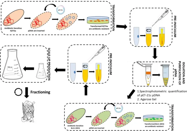
2.2.5. Production of GFP Containing-Crude Extracts ... 29
2.2.5.1. Preparation of LB Medium and LB Agar Plates with Ampicillin ... 29
2.2.5.2. Transformation of pET-21c Plasmid in NZY5α Competent Cells ... 30
2.2.5.3. Isolation and Purification of pET-21c pDNA ... 30
Table of Contents
viii
2.2.5.5. Agarose Gel ... 32
2.2.5.6. Large Scale Expression of GFP ... 32
2.2.6. Chitosan-based Monoliths Fuctionalization Towards GFP Protein ... 35
2.2.6.1. Monoliths Amination by Plasma Technology ... 35
2.2.6.2. Aldehyde Groups on the Surface of Previously Aminated Monoliths ... 37
2.2.6.3. A4C7 ligand Solid-Phase Synthesis on Monolith Platform ... 38
2.2.7. Screening Assays with GFP and Ligand Leaching Tests ... 40
2.2.7.1. SDS-PAGE Analysis ... 40
3 DEVELOPMENT OF MONOLITHS FOR VIRAL PARTICLES PURIFICATION ... 43
3.1. Introduction ... 45
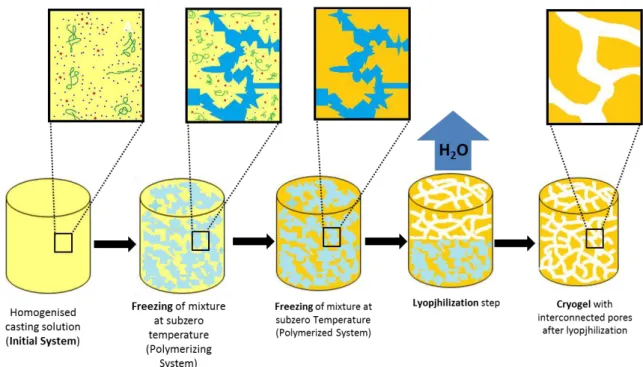
3.2. Preparation of Monoliths by Freeze-Drying ... 51
3.3. Monoliths Architecture and Analysis of its Properties through Characterization54 3.3.1. Materials Employed: an Overview ... 54
3.3.2. Monoliths Characterization ... 59
3.3.3. Magnetic Field Responsive Monoliths ... 70
3.4. Testing for Non-Specific Binding of Ad5 ... 78
3.5. Concluding Remarks ... 79
4 AFFINITY MONOLITHS FOR GFP PURIFICATION ... 83
4.1. Introduction ... 85
4.2. Results and Discussion ... 87
4.2.1. GFP Expression and Production ... 87
4.2.2. Preparation of Affinity Monolith towards GFP Purification ... 90
4.2.2.2. Evaluation of Affinity Monoliths for GFP Purification ... 97
4.4. Concluding Remarks ... 102
Index of figures
ix
Index of Figures
Figure 1.1.- Classification of chromatography stationary phases according to their morphology4,5. Micropores size correspond to values below 2 nm, mesopores size to
values between 2-50 nm, macropores size to values between 50-5000 nm, and super-macropores to values between 5000-105 nm6–9. ... 3
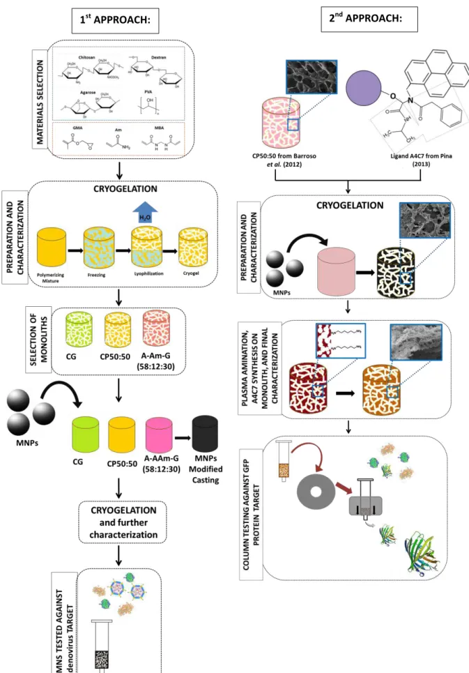
Figure 1.2.- Schematic depiction of research approaches followed in present work. ... 15
Figure 2.1. – Reaction mechanism in the base of Magnetite Method. ... 24
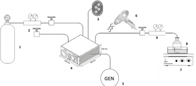
Figure 2.3. – Layout of Amination apparatus used for monolithic samples: argon gas bottle (1), gas 2 manometer (2), vacuum pumb (3), plasma chamber (4), high frequency generator (5), heat gun (6), stirrer hot plate (7), 1,6-diaminohexane vessel (8), gas 1 manometer (9). ... 36
Figure 2.4. – Kaiser test reaction. Compound 1 absorbs at 570 nm. ... 37
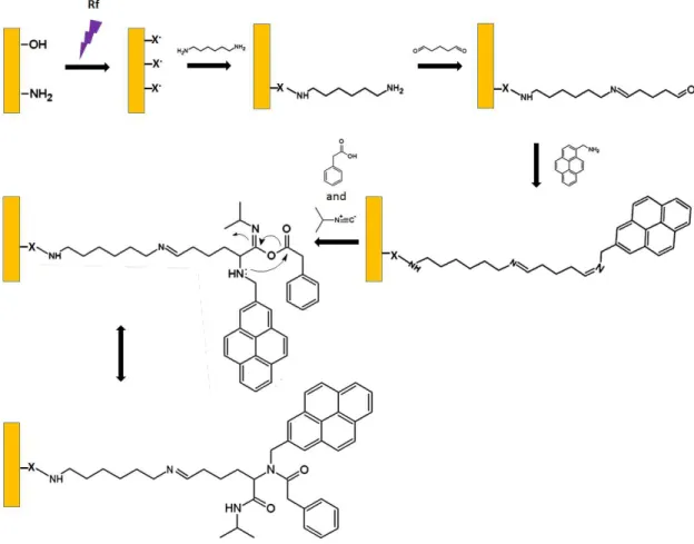
Figure 2.5. – Plasma amination104 followed by Ugi reaction onto monolith. “X” denotes
oxygen, nitrogen or carbon atoms. ... 39
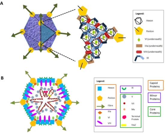
Figure 3.1. – Adenoviral particle external (A) and internal (B) structure. Structures based on Martín118 and Russel116 works respectively. ... 46
Figure 3.2. – General scheme for Ad downstream purification. Black spheres represent possible applicable unit operations (most common ones); numbering represents sequential steps (most common ones. The diagram was based on Prazeres work114. On
capture step AEC is the only method present once it is the most commonly applied one, however Ad can also be separated based on size, hydrophobicity, and metal affinity. AEC: anion-exchange chromatography; SEC: size exclusion chromatography; IPRPC: ion-pair reversed phase chromatography; IMAC: immobilized-metal affinity chromatography. .... 48
Index of Figures
x sublimated; the surface tension between solvent and gel phase guarantees the round smooth shape of pores. Green ribbons represent polymers, blue dots represent solvent molecules and the red ones represent the low-molecular weight solutes (e.g. monomers, initiators). Schem based on138,139. ... 52
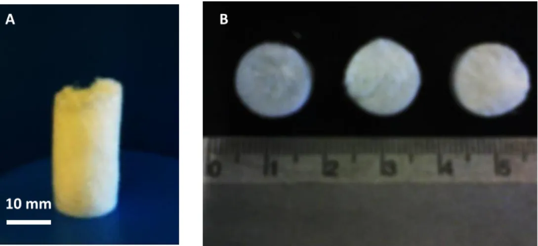
Figure 3.4. - Whole dextran-based monolith (A) and the three samples in which it was sliced (B). ... 53
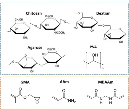
Figure 3.5. – Polymers (blue) and monomers (orange) used in monoliths preparation towards a novel, green and virus purifying support. ... 54
Figure 3.6. – Polymer scale arrangement of composites into monoliths. Structure of chitosan blended with PVA monolith (C/P) (A). Hydrogen bonds are established between polymer chains; and MBAAm polymerizes and imprisons the H-bonding stabilized chains improving. Structure of semi-IPN C-G monolith (B). Here MBAAm crosslinks poly-GMA imprisoning chitosan at some regions. Structure of agarose and dextran-based monoliths (C) where the closed (no loose ends) AAm-GMA copolymer entangles and imprisons agarose/dextran. Orange ribbon represents chitosan; blue ribbon represents PVA molecule; black piece represents MBAAm monomer; black ribbon represents polymerized MBAAm imprisoning bonding stabilized C/P chains; green shadow highlights the H-bonding. Purple chains represent poly-GMA; brown sticks represents intra-chain covalent bonds. Pink chain represents agarose/dextran; green chain represents poly(AAm-GMA) chains. ... 57
Figure 3.7. – Cyclical swelling analysis: variation of percent swelling degree (W) with time (t). Each monolith (frozen at -20°C) is alternately plunged into two different pH buffers (pH7 and 5) over time. C2.9% (a); C2% (b); C-G (c) C/P(50:50) (d); C/P(33:67) (e). All samples are presented in duplicate. ... 62
Figure 3.8. – Variation of percent swelling degree (W) with time (t). Each monolith (frozen at -80°C) is alternately plunged into two different pH (pH 7 and 5) solutions over time (t). C2.9% (a); C2% (b); C/P(50:50) (c); C/P(33:67) (d). All samples are presented in duplicate. ... 64
Figure 3.9- Swelling kinetics of A-AAm-G(58:12:30), C-G and C/P(50:50). ... 70
Index of Figures
xi Figure 3.11. –Permanent magnets used for field response testing. ... 74
Figure 3.12.– Magnetic-field response of Agarose (A-AAm-G (58:12:30)) and chitosan-based monoliths (C/P (50:50), C-G) to different magnetic-flux densities: 0.25T, 0.50T, 0.53T and 1.5T. The first five points plotted no each graph corresponds to deformation under external magnetic-field, the following five corresponds to matrix behaviour after external field removal (when reached the initial length no more points were plotted). Data was obtained from duplicated measurements. ... 75
Figure 3.13. – SEM micrographs of non-magnetic (A,C,E) and magnetic (B,D,F) chitosan and agarose-based monoliths at x300 magnification: Ag-Am-G(58:12:30) corresponds to A and B; C/P(50:50) to C and D; and C-G to E and F. For C-G monoliths a micrograph with lower magnification (x100 (left) x150 (right)) is shown. ... 77
Figure 3.14. – SEM micrograph of C/P(50:50) native monolith at a magnification of x500. Notice the peculiar pendant polymer strings. ... 78
Figure 4.2. – pET-21c isolation and purification was successfully achieved as agarose gel electrophoresis (0.8%(w/v) agarose, stained after running) can prove (A): marker, 1st
elution, 2nd elution (lanes 1, 2, 3 from left to right). First and second elutions recovered
through NZYminiprep kit for DNA purification were quantified and analysed by NanoDrop spectrophotometer (B). ... 87
Figure 4.3. – Monitoring of Cells growth and GFP expression through OD600nm (A) and
fluorescence intensity measurements (B), respectively. A correlation between phenomena can be seen (conversion of nearly all cell’s resources towards GFP gene expression209). .... 88
Figure 4.4. – Time course SDS-PAGE gel (12.5% acrylamide gel stained with Coomassie Blue R-250). GFP mass production can be visualized. M represents protein marker; PRE corresponds to sample collected at tinduction=0; all following lanes matches the GFP profile at
different times of induction (2h, 4h, 5h and 18h). The band of GFP is expected to be placed at ~29 kDa101,201. The loading volume of each sample was normalized to a constant specific
optical density value (1.2). ... 89
Index of Figures
xii Figure 4.6. – Silver mirror test on aldehyde functionalized monoliths: non-magnetic and non-functionalized monolith (NC, negative control); non-magnetic and functionalized monolith (NL); magnetic and functionalized monolith (ML); Glutaraldehyde as positive control (C+); and magnetic and non-functionalized monolith (MC, negative control) (from left to right). ... 92
Figure 4.7. – Pyrene presence at the surface of NL and ML monoliths: non-magnetic monolith functionalized with A4C7 (NL) (A,E); non-magnetic and non-functionalized monolith (NC) (B,F), magnetic monolith functionalized with A4C7 (ML) (C,G), magnetic non-functionalized monolith (MC) (D,H) (from left to right). Pictures were taken on the fluorescence microscope under bright field filter (A,B,C,D) and fluorescence filter (E,F,G,H) at x40 magnification. All supports were regenerated before analysis. ... 93
Figure 4.8. – SEM micrographs of NC monolith with x300 magnification kindly provided by Barroso et al.27 (A), MC monolith with x300 magnification (B), NL monolith with x1000
magnification (C), and ML monolith with several magnifications: x30 (D), x500 (E) and x1000 (F). ... 95
Figure 4.9. – Visual comparison between stages of monolith surface modification. C/P(50:50)80 native just lyophilized monolith (A), after aldehyde functionalization (B), after A4C7 solid-phase synthesis (C) (from left to right). ... 95
Figure 4.10. – Magnetic-field response of magnetic C/P (50:50)80 monolith at various modification stages at its dry (square) and wet states (diamond): without any modification (A); after aldehyde functionalization (B); after stability test in 100%(v/v) MeOH during 48h (C); C/P(50:50)80 after A4C7 functionalization. First five points plotted no each graph corresponds to deformation under external magnetic-field (1.5T), the following five corresponds to matrix behaviour after external-field removal (when reached the initial length no more points were plotted). Data obtained from duplicated measurements. ... 96
Figure 4.11. – Ligand Leaching assays. A4C7 leaching for NL and ML monoliths at 0.1M glycine-NaOH pH 9 (E1) and 0.1M glycine-NaOH pH 9, 50%ethylene glycol (E2 ... 97
Figure 4.12. – Selectivity of ligand A4C7 towards GFP at different pH values: pH7.4 (A) and pH9 (B). Results refer to a batch system where the protein is incubated with the support for 15 minutes. TP denotes for total protein. ... 98
Index of Figures
xiii Figure 4.14. – SDS-PAGE analysis of GFP screening on magnetic functionalized (ML) monoliths: Continuum assay (A); 15 minutes batch assay (B); 60 minutes batch assay. M corresponds to protein marker and lanes 1, 2, 3, 4, 5, 6, 7, 8, 9 denotes for: loading, flow-through, 1st wash, 2nd wash and 1st elution for ML and loading, flow-through, 1st wash, 2nd
wash and 1st elution for MC, respectively. GFP bands position (~29kDa) is highlighted. 101
Index of Tables
xv
Index of Tables
Table 1.1.- Benefits and limitations of each chromatographic media type used in bioseparation. The present comparison of generalized structures of porous beads, stacked membranes, and monoliths is not drawn to scale. Black arrows show the bulk convective flow, and shaded orange areas the diffusion regions. Green curling arrows show turbulent flow (eddies) with consequent counter-current between laminar flow and eddy flow (shear). In case of beads media green arrows represent inter-particles eddies20,21,5,2. ... 6
Table 1.2.- Examples of the application of monoliths in bioseparation. ... 7
Table 1.3.- Methods for preparation of monoliths to be applied in separation science12,54,48.9
Table 1.4.- Benefits and limitations associated with each type of monolith structure.80,81,79,5412
Table 2.1.- Casting solutions processed for monoliths preparation. All casting solutions were formulated with 3 mL of distilled water per monolith. Polymers and/or monomers content in every 2.9%(w/w) casting solution is 90mg. Conversely in PVA:GMA 79:21%(w/w) casting is 101mg), AAm:MBAAm:GMA 95mg/210mg, agarose:AAm:GMA and dextran:AAm:GMA 142 mg/172 mg. ... 22
Table 2.2. – Required volumes to prepare one 12.5% Acrylamide gel. ... 34
Table 3.1. – Traditional methods used in Ad purification. ... 47
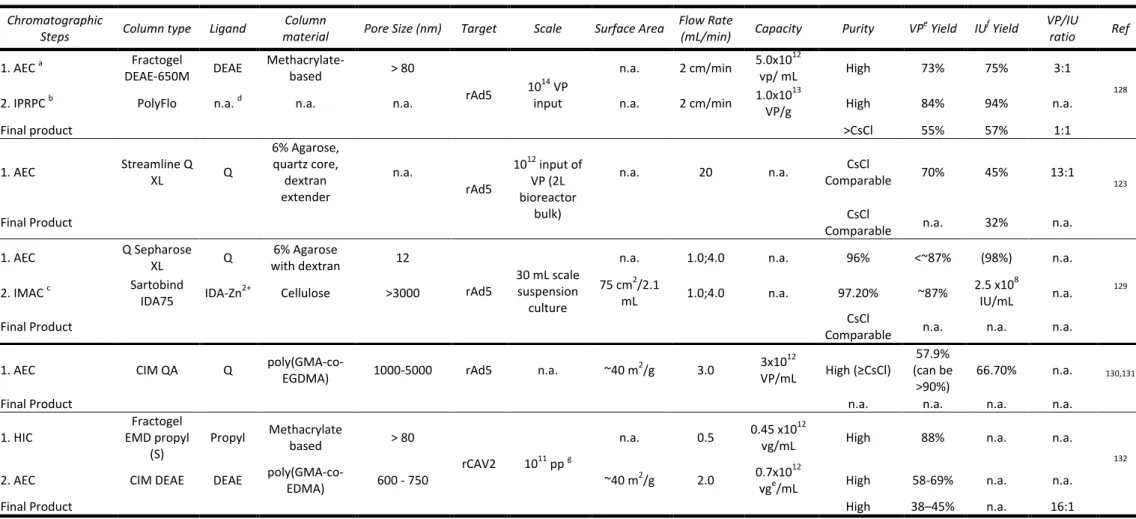
Table 3.2.- Summary of possible combinations of chromatographic steps in Ad purification steps. ... 50
Table 3.3. – Monoliths prepared for screening tests accompanied by the respective monomeric/polymeric ratios. ... 56
Table 3.4.– Stability Tests performed at pH3, 7 and 11 with different monoliths prepared at -20°C/-80°C. Monoliths were macroscopically analysed during fourteen days. ... 59
Index of Tables
xvi Table 3.6. – Morphological and mechanical properties of all monoliths prepared at -20°C. All data was obtained from duplicated measurements (in case of water flux measurements each one of the two samples was measured three times). ... 65
Table 3.7. – Morphological and mechanical properties of all monoliths prepared at -80°C. All data was obtained from duplicated measurements (in case of water flux measurements each one of the two samples was measured three times). ... 68
Table 3.8. – Morphological and mechanical comparison between non-magnetic and magnetic monoliths with MNPs at two different concentrations for each specimen. All data was obtained from duplicated measurements (in case of water flux measurements each one of the two samples was measured three times). M_C/P(50:50) denotes magnetic C/P(50:50), the same is true for the others. ... 72
Table 3.9. – Pore volume and density values for magnetic and non-magnetic (native) monoliths. All data was obtained from duplicated measurements. M_C/P(50:50) denotes magnetic C/P(50:50), the same is true for the others. ... 73
Table 3.10. – Comparative analysis of different monolithic supports for recovery of adenovirus vectors. ... 79
Table 4.1. – Comparative analysis on the efficiency of amination through three different approaches: traditional preliminary epoxyactivation, non-thermal plasma treatment followed by amination out-of-chamber, and non-thermal plasma with direct amination inside chamber. MC denoted for magnetic functionalized monolith and NC for non-magnetic non-functionalized monolith. ... 91
Table 4.2. – Morphological and mechanical properties of functionalized and non-functionaliized monoliths. All data was obtained from duplicated measurements (in case of water flux each one of the two samples was measured three times). ... 94
List of Abbreviations
xvii
List of Abbreviations
AAm Acrylamide
Ad Adenovirus
AEC Anion Exchange Chromatography
Ad5 Adenovirus type 5 APS Ammonium Persulphate
CEC
Capillary electrochromatography
DLS Dynamic light scattering
E1 Elution condition 1: 0.1M glycine-NaOH pH9
E2 Elution condition 2: 0.1M glycine-NaOH pH 9, 50%(v/v) ethylene glycol
EMA
European Medicines Agency
GFP Green Fluorescent Protein GCE GFP crude extract
GMA Glycidyl Methacrylate
HPLC
High performance liquid chromatography
IPTG Isopropyl β-D-1-thiogalactopyranoside NC Plain C/P(50:50) monolith
NL C/P(50:50) monolith non-modified with MNPs, functionalized with A4C7 MAbs Monoclonal antibodies
MBAAm N,N'-methylene-bis-acrylamide
MC C/P(50:50) monolith modified with MNPs, non-functionalized in any way MCR Multicomponent reactions
ML C/P(50:50) monolith modified with MNPs, functionalized with A4C7
Mw
Molecular weight
PBS Phosphate Buffer Saline PDB Protein Data bank
pDNA Plasmid DNA
PVA poly(vinyl alcohol)
rAd Recombinant Adenovirus SEM Scanning Electron Microscopy SDS Sodium Dodecyl Sulphate SPOS Solid Phase Organic Synthesis
Literature Review
1
Literature Review
Literature Review
3
1.1. Monoliths in Bioseparation
Adsorption chromatography can be performed using distinct solid phase media, namely porous beads, membranes, and monoliths (Figure 1.1.). Porous particle-based supports are currently the most widely employed stationary phases for purification of biomolecules, particularly proteins1. However, due to present research
and market evolution towards large biomolecules (virus, DNA, intact cells, complex proteins) in (bio)pharmaceutical industry, and particle-based media inadequacy to purify this types of molecules, monoliths arise as a promising alternative1–3.
Figure 1.1.- Classification of chromatography stationary phases according to their morphology4,5. Micropores size correspond to values below 2 nm, mesopores size to values
between 2-50 nm, macropores size to values between 50-5x10⁵ nm, and super-macropores to
values between 5x10³-1x10⁵ nm6–9.
Monolith is defined as a continuous and porous stationary phase moulded as a column and inserted in a chromatography housing2,5. Their “format can be compared to a single large particle”10, and include “compressed hydrophilic gels, macroporous polymer discs, columns, tubes and silica rods”10.
Beyond the possibility of being prepared through several procedures and distinct chemistries, monoliths can also be tailored to present differences at microstructural level (e.g. pore size and geometry)11,12. However, they are all
Literature Review
4 network of channels5. This single structure allows mobile phase to flow through these
channels by convection, minimizing mass transport resistance (low backpressure) and increasing separation speed, independently of molecular size or diffusion coefficient5.
These flow-through pores characterized by convective transportation of mass are thus responsible for the flow independent chromatographic properties of monoliths, such as dynamic binding capacity and resolution, and consequently for the efficient high speed assays13,14. In fact, monoliths exhibit plate efficiencies that compete with finest
bead-packings14.
This constitutes a totally different picture compared to diffusion, the representative driving force of mass transfer between solid surface and bulk liquid phases on porous packed-beads separation media, a slow phenomenon dependent on molecular weight5,2. This phenomenon takes place due to the adsorption surface
shallow dead-end pores with 10–100 nm large, where neither convective transport can be achieved nor big molecules like virus, DNA and cells can have access5,15. The mass
transport dependence on molecular weight of analytes comprises the speed of the assays and can only be overcome with resolution and binding capacity commitment. The independence of dynamic binding capacity and resolution from flow-rate can be achieved with non-porous micrometerized small beads (<5µm) made of silica or synthetic polymers. However due to its low porosity compared to monoliths, only short column lengths can be used to avert high backpressures and achieve attractive assay speeds13. Moreover in particulate-bead packings there are preferential paths for
the solution – interparticle void volumes (~40% of total bed volume1) – where flow
vortices (eddies) are created due to differential friction between particle surfaces and inter-particle void areas. This eddies origin turbulent mixing that reduce resolution, broaden peaks, and may cause shear forces that can harm sensitive/unstable molecules, lowering yields. Perfusive particles, with channels transecting them, allow a little increase on convective mass transport together with an increment on bio-nanoparticles accessible area, however this type of beads are not free from the undesired void volumes, where fluid flows preferentially and eddies occur1.
Conversely to packed beads, and equally to monoliths, membranes are designed particularly to take the maximum advantage of convective transport. In fact, a membrane can even be almost equalized to a monolith, once it is cast as a single continuous unit provided with large channels rather than pores. Nevertheless, their exceedingly flat bed height, their usually smaller channels width (generally not surpassing 1µm1 against ~100µm for monolithic cryogels9,16), together with the different
physical arrangements in which they are applied (e.g. stacked membranes, pleated cartridge) make them distinct from monoliths in terms of operating features17,1. In fact
Literature Review
5 between membrane layers forming the dead or void volumes; all this is due to the discontinuity of channels, consequence of the discontinuous character of the whole format (discontinuous pore distribution) and the characteristic inlet arrangement itself5. As the turbulent mixing occurring in these void volumes can be compared to
eddy dispersion in porous bead supports1, the threat of shear forces presence as
consequence of eddies formation is a possibility, and so it is product integrity commitment. The flow aberrations located on the outlet side of the membrane generate dispersion decreasing process performance5. So the flow uneven distributions and
undesired behaviours are thus responsible for a slight decrease on capture efficiency and strong reduction on elution efficiency (unwell resolved peaks).
In turn, monoliths not only exhibit a binding capacity three times wider than membranes14 but also lack the dead volumes. In fact the laminar flow through all
monolith avoid eddies, decrease shear, and guarantees immediate response to variations in buffer composition, maximizing elution kinetics and contributing to sharper, better resolved and more concentrated elution peaks, and high functional recoveries. They offer“the selectivity of particulate resins and the throughput of membrane absorbers”18, suggesting that monoliths should be more efficient, especially when it
comes to larger biomolecules purification14,2.
Table 1.1. presents a comparison between the three main chromatographic media available.
Monoliths that can be prepared in multiple ways and also find diverse applications, are usually easy to prepare in various sizes and shapes from different materials and through different methods. Monoliths surface can then be or not chemically modified with multiple molecules and applied in the capture/purification of large biomolecules, as they present fast performance at low pressures and room temperature, and a high productivity due to their flow-independent properties2,13.
Literature Review
6 Table 1.1.- Benefits and limitations of each chromatographic media type used in bioseparation. The present comparison of generalized structures of porous beads, stacked
membranes, and monoliths is not drawn to scale. Black arrows show the bulk convective flow, and shaded orange areas the diffusion regions. Green curling arrows show turbulent flow (eddies) with consequent counter-current between laminar flow and eddy flow (creating shear). In case of beads media green arrows represent inter-particles eddies. Table based on 20,21,5,2.
Chr
omatogr
aphi
c Medi
a
Beads Membranes Monoliths
Advantag
es
High product purity (SM); Consistency and safety; Industrial scale well known; High adsorption and elution
efficiency (SM);
High surface area and high binding capacity (SM);
Multiple column chemistries and HPLC column dimensions are commercially available;
Validated applications/assays.
Low pressure drop; high flow rates;
Small footprint; Inexpensive; Disposable;
Moderate resolution; High hydraulic permeability; Rapid mass transfer
(convection), allowing high flow rates with high productivity and major decrease in separation times.
High porosity;
Large interconnected channels with rapid mass transfer (convection), allowing high flow rates with major increase crease in separation times; High productivity;
No void volumes and laminar flow, so no eddies are formed, neither shear;
High purity products;
Ease of preparation and processing in various volumes and shapes (PM);
Flexible surface chemistry for ligand attachment, due to plethora possible usable materials;
High hydraulic permeability; Inexpensive;
Moderate-high resolution; Low-moderate pressure drop; High binding capacity (BM); Mechanically robust;
Li
mi
ta
ti
ons
High pressure drop;
Weak mechanical properties; Eddy dispersion and shear forces
due to voids compromising productivity;
Extensive footprint; Moderately expensive; Low binding capacity (BM); Peak broadening, resolution and
recovery worsens with flow rate (BM);
Low mass transfer (diffusion) with consequent low flow-rates High backpressure;
Extra machinery required for sample solution to cross media; Air incursion into column
destroys bed integrity.
Low binding capacity; Limited available surface
area;
Shear forces due to eddies; can compromise productivity Flow aberrations compromise
performance; Fouling;
Broad peaks due to eddies (desirable in situations where neither high purity nor high eluted product concentration is required);
Accumulated bubbles difficult to displace without breaking system sterility.
Scale-up difficult;
Low specific surface area per unit volume, and so low binding capacity for SM and medium large proteins as monoclonal antibodies; Extensive footprint;
Low efficiency (SM) and HPLC column to column reproducibility;
Limited column chemistries and dimensions commercially accessible;
Constrained use in routine analysis due to few commercial suppliers available.
Literature Review
7 Currently, this new category of porous media has been extensively applied in analytical chemistry, mainly in separation science areas. Through liquid chromatography, namely high performance liquid chromatography (HPLC) and capillary electrochromatography (CEC)22, monolith stationary phase has been used to
capture, purify, enrich and analyse diverse bio-nanoparticles, from plasmidic and genomic nucleic acids to organelles, inclusion bodies, virus and other macromolecular assemblies2,23. The specific interactions between the target macro-biomolecule and the
adsorptive matrix allow their isolation from related small molecules24. Table 1.2. shows
some applications of monoliths as sorbents for isolation of macro-biomolecules.
Table 1.2.- Examples of the application of monoliths in bioseparation.
Monolith
Material Mode Ligand Application/Target(s)
Pore Size (µm)
Surface Area (m2/g)
Capacity Recovery
(%) Purity Ref
GMA-EDMA CEC Acetic Acid
Purification and simultaneous renaturation
of rhIFN-γ
0.4-2.0 (>80%) n.a.
a
n.a. n.a. 93% 25 (Hybrid
silica) TEOS-AEAPMDMS
AEC Amine groups
Extraction of genomic
DNA from blood ≤6.0 n.a.
9.3
ng/cm 52.1 n.a. 26
Chitosan-PVA cryogel Affinity
(Artificial protein A) ligand 22/8
Capture of pure IgG, and direct capture and recovery of mAb from a non-clarified homogenate
~45 2.3 150
mg/g 90, 48 98% 27
CIM AEC Q
Isolation of bacterial ribosomes from crude cell
lysates
0.6-5.0 ~40 n.a. n.a.
< sucrose gradient centrifugati on 28 Concentration and purification of rubella virus from a complex biological suspension
0.6-5.0 ~40 n.a. ~100 High 29
Aam-AGE-MBA IMAC IDA-Cu 2+
Direct capture of enzyme (His)6-LDH from
non-clarified crude cell homogenate
0.01-100 n.a.
0.13
mg/ml 70-90
Need to be improved
9 Chromatography of E. coli
cells
0.01-100 n.a. n.a. 80 Reasonable 30
Aam- DMAEMA-MBAAm
AEC DEAE Chromatography of E. coli cells
0.01-100 n.a. n.a 70–80
Need to be improved 30 LMA-EDMA-VPBA CEC and HIC Boronic acid and C12 chain
Analysis and identification of cis-diol biomolecules/TRF
5x10⁻³– 50x10-3
43.5-54.8 n.a. n.a.
Need to be improved
31
Aam- MBAAm-GMA
Affinity Streptavidin
Single-step capture of chemically biotinylated
MoMuLV
0.01-100 n.a.
2x10⁵
cfu/mL <8 High 32,9
PHEMAH cryogel
Pseudo
Affinity MAH purification of pDNA 10–100 n.a.
13,350
µg/g 90 Reasonable 33
CEC: Cation-exchange chromatography; AEC: Anion-exchange chromatography; IMAC: Immobilized methal affinity chromatography;
HIC: Hydrophobic interaction chromatography; rhIFN-γ: Recombinant human interferon gamma (growth factor); TRF: Transferrin;
MAH: N-methacryloyl-(l)-histidine methyl ester; PHEMAH: Poly(hydroxyethyl methacrylate-Nmethacryloyl-(L)-histidine methyl ester).
Literature Review
8 Besides their wide applicability in bioseparation (liquid chromatography, capillary separations, capillary electrochromatography (CEC), thin-layer chromatography, gas chromatography)9,29,32,27,25,34,22 as adsorbent matrices, monoliths
have also found usefulness in sample pre-treatment21,23, catalysis35,36 (mainly towards
micro-scale protein mapping or proteomic analysis36,37), solid phase and combinatorial
chemistry38,39, scavenging40, as static mixers41, drug delivery, in vitro cell cultivation,
and tissue engineering42,43.
1.1.1. Methods to Produce Monoliths
Despite the possibility of monoliths to be miniaturized into capillaries, microfluidic devices or microarrays, they are usually prepared on an analytical scale: in a conventional large column/rod, tube or disk format, in a multi-well plates format for screenings assays44,2, in a thin-layer format for planar chromatography34,2, or with a tip
geometry45,2.
Monoliths can be divided into organic polymer monoliths, inorganic silica monoliths and hybrid organic-silica monoliths. Inorganic silica monoliths can be fabricated by (i) fusion of porous silica beads through thermal sintering, (ii) cementing/immobilizing silica beads in a packed bed by cross-linking/entrapping them through sol-gel process, or (iii) polymerization of sol-gel precursors (silicon alkoxide). The latter, a waste-free method, is the most commonly used46.
Recently a review on what authors called ‘exotic monoliths’, shows that beyond the famous silica gel-based monoliths inorganic monoliths can be prepared from both a ‘pure’ metal or a metal-oxide, and be applied in separation science47.
In turn organic polymer monoliths can be prepared from i) solely a polymer, ii) a blend of polymers, iii) a polymerization of monomers in presence or not of one or more polymers, or (iv) co-polymerization of monomers in presence or not of one or more polymers; using a variety of possible methods12,48,49 (Table 1.3.). Generally they
are produced by in situ polymerization of a mixture containing monomer(s) (commonly acrylamides, methacrylates, or styrene50,6), crosslinker, porogenic solvent(s)
and an initiator, using a simple moulding procedure executed inside a mould such as a chromatographic column, capillary or micro-channel (see figure 2 on Nordborg et al.
work51). The most employed method is the free radical polymerization, more precisely
the thermally and UV irradiation photo-initiated approaches48. Other approaches have
been explored, such as microwave or γ radiation initiated polymerizations51,12,52.
Literature Review
9 Table 1.3.- Methods for preparation of organic monoliths to be applied in separation science12,54,48.
Preparation
Method Materials
Initiator/ Porogen/ Other Pore Size (µm)
Application Obs. Ref
Thermally initiated free radical polymerization (TIFRP) PA:PDA (50:50 %w/w) AIBN / 2-propanol, THF 0.22 Separation of proteins and
oligo-deoxynucleotides
1st method used for preparation of rigid polymer-based monoliths;
Very simple;
Process origin can be traced down to techniques generally applied in preparation of porous beads by suspension polymerization;
High reproducibility;
Assembled by irregular micro-globules forming aggregated clusters, leading to some limitations (e.g. permeability) (all FRP). 54,55, 12,56 Photo-initiated free radical polymerization (UV rays) GMA: EGDMA: BMA (51:40:5 %v/v) AIBN / 1-dodecanol, cyclohexanol 0.5-3 High throughput sample clean-up throughput. Roscovitine and lidocaine in plasma
samples used as model substances.
Faster than TIFRP;
Can lead to columns with lower backpressure, and better chromatographic performance than TIFRP (comparing columns of same pore size);
Reaction can be stopped when irradiation source is removed and column is flushed;
Limited by use of UV transparent molds with a small size in one dimension and UV transparent monomers, exclusion of aromatic monomers, and wavelength of maximum absorbance of initiator;
57,54, 12,58, 59 Photo-initiated free radical polymerization (visible light) St:DVB (50:50 %v/v)
mixture of CQ, EDAB, MPPB /
ACN, 1-propanol,
1-decanol
n.a.a
Separation of mixture of standard
proteins: ribonuclease A, cytochrome c,
myoglobin and ovalbumin
Performed at room temperature, allows less common porogens usage, including those with low boiling point.
58
Radiation initiated free radical polymerization (ƴ-rays)
DEGDMA: GMA
No initiator / t-butanol or methanol or
ethanol or propanol or acetone or THF
or ethylpropionat
e
~3 Diagnostics and purification
Faster than TIFRP;
Greater penetration depth of radiation than UV-initiated polymerization, allowing preparation of any volume monoliths;
No initiator needed;
Pore volume and pore size distribution tuning in a broad range through process variables as irradiation dose and dose rate, non-available in other polymerization processes. 60,61 Radiation initiated free radical polymerization (microwaves) St:DVB: MAA (33.3: 33.3:33.3 %v/v)
AIBN / toluene, isooctane
0.28 -8.88
pCEC, CEC, LPLC of neutral compounds (thiourea, benzene, toluene, ethyl benzene, biphenyl, naphthalene)
Polimerization time shortened from 24h (TIFRP) to 15min;
Lower expense than TIFRP and UV-ligth initiated FRP.
62
Literature Review
10 Table 1.3. (Continued)
Preparation
Method Materials
Initiator/ Porogen/ Other Pore Size (µm)
Application Obs. Ref
Radiation initiated free radical polymerization (electron beam) EMA: TMPTA (50:50 %w/w)
No initiator / 2-propanol, 1-dodecanol 0.08-0.11 Separation of proteins: lysozyme, ribonuclease, insulin, cytochrome c, andalbumin; heterogeneous catalysis
No initiator needed;
Successful column scale up reported; Fastest separation with sufficient peak
resolution, in comparison to ROMP prepared monoliths. 52 Polymerization by high internal phase emulsions (polyHIPE) GMA: EGDMA (77:23 %w/w) Potassium persulfate / Emulsified water droplets / calcium chloride hexahydrate (el ectrolyte) and Synperonic PEL 121 (surfactant) ~0.1 (holes size 1 -10) Separation of standard protein mixture of myoglobin, conalbumine and trypsin inhibitor
Good separation in a very short time, comparable to separation achieved by commercial methacrylate monoliths (FRP);
Monoliths characterised by high porosity (>70%) and large spherical
hollows interconnected by ‘‘windows’’;
Possible drawback: monoliths present low specific surface area, restraining its use in separation science.
63,12, 64 Cryogelation HEMA: MAH (PHEMAH) APS / Water crystals /
TEMED (catalyst) 10– 100 Purification of pDNA
Freezing temperature define pore size; Due to large produced pores (1–100µm)
and high porosity (≤90%),
hydrodynamic cryogels properties are exceptional. 12,65, 33 Living Polymerization Nitroxide Mediated (SFRP) St:DVB (50:50 %w/w) Benzoyl peroxide / PEG 400, 1-decanol / 3-carboxy-PROXYL or 4- carboxy-TEMPO (promoter) ≤0.01 -1 separation of mixture of myoglobin, cytochrome c, and
lysozyme
Slower kinetics characterizing TEMPO-mediated polymerizations avoids significant shifts in pore size distribution;
TEMPO-capped dormant radicals usable for grafting pore surface and tailoring its chemistry;
Initiator remains on or within the material, enabling post-polymerization modifications.
Least versatile (against ATRP, and RAFT) 12,66
Living
Polymerization (TERP)
MBAAm
AIBN / PEO (phase- separator) / BTEE (promoter) 0.5 -2 Aqueous phase applications (bioseparation, support for catalysis)
A recent strategy lacking preparation of columns and chromatographic evaluation of their performance; High surface areas attained may ease
separation of small molecules in isocratic mode;
High temperatures employed.
67 Living Polymerization (ATRP) VC:EDMA (50:50 %v/v)
CCl4 / dodecyl alcohol / FeCl2
(catalyst)
0.85
Separation of: IgG from human plasma, lysozyme from egg white, and
mixture of papain, snailase, IgG.
Control over rate of monomer combination with growing polymer chain (chains similar in length) (all LP); Highly homogeneous crosslinking due
to isotropic spinodal decomposition promotion possibility (ATRP, TERP); Popular in general polymer chemistry,
but poorly explored in monoliths preparation. 68,12 Living Polymerization (RAFT) MAA: EDMA
AIBN / Toluene, dodecanol / DBTTC (chain
transfer)
n.a.
Extraction of clenbuterol from biological samples
Surface functionalization eased (all LP); Control over polymerization kinetics,
structure morphology and surface functionality (all LP).
69,70
Literature Review
11 Table 1.3. (Continued)
Preparation
Method Materials
Initiator/ Porogen/ Other Pore Size (µm)
Application Obs.
Living Polymerization (ROMP) NBE: DMN-H6 or COE:CL (50:50 %w/w) [RuCl2 (PCy3)2(CHPh)]
or [RuCl2(Py)2(IMe
sH2)CHPh] / 2-Propanol, toluene 0.006 -~0.04 Separation of Ribonuclease A , carbonic Anhydrase, insulin,
cyctochrome C, albumin
Restricted range of possible monomers; Noticeable irregularities in the porous structure with increasing ratio of pore size to the capillary diameter.
71,12 Poly-condensation polyglycer ol-3-glycidyl ether BF3·Et2O in dioxane / Toluene, t-butyl methyl ether
22 Capture of Gram-negative bacteria
Oxygen insensitive reaction, rendering unnecessary the careful de-aeration required for FRP;
Produces attractive morphological structures for separation;
Mild reaction conditions and possibility of room temperature employment avoids pore structure heterogeneities in contrast to FRP.
72 Thermally induced phase separation Polyamide No initiator needed / Benzyl alcohol
~0.01-~0.02 n.a.
Structures produced present uniform architecture. Exceptionally simple method (thermally controlled dissolution and phase segregation process) for preparing monoliths with attractive chemical, physical and porous properties. 73 Non-solvent induced phase separation Polycarbo nate No initiator needed / Cyclohexane 0.45-3.2 Adsorption ofmetal ions and purification of proteins
Easy and clean process, so morphology tailoring is easy.
74
Another attractive method for the preparation of monoliths is cryogelation. This versatile technique allows the preparation of elastic and sponge-like structures with a broad range of porosities, and gives rise to highly interconnected supermacroporous matrices with 100µm sized pores. Moreover its green character does not go unnoticed75,12,65,33.
A 2010 review from Svec12 gathers all different polymerization methods that
could be used to prepare polymeric monolith structures, so far. However since that comprehensive publication, several developments in this area have been made, with some breakthrough approaches reported48, namely, the growing incorporation of
nanostructures into monoliths like nanoparticles of silica, gold, silver, metal oxides, hydroxiapatite, and polymers, or carbon nanotubes76. This strategy aims to tailor
surface characteristics, incorporating nanostructures features into monoliths, what increases surface area-to-volume ratio, and consequently offers an extended surface for biomolecules adsorption, possibly facilitating mass transfer and improving separation
THF: Tetrahydrofuran; PA: Phenyl Acrylate; PDA: 1,4-Phenyl Diacrylate; BMA: Butyl Methacrylate; EDMA: Ethylene Glycol Dimethacrylate;
St: Styrene; DVB: Divinyl Benzene; MAA: Methacrylic Acid; PEO: Poly(ethylene oxide); TEMPO: 2,2,6,6-Tetramethyl-1-piperidyloxy VC: Vinyl Carboxylate; SFRP: Stable Free Radical Polymerization TERP: Organotellurium-mediated living Radical Polymerization; ATRP: Atom Transfer Radical Polymerization; NMP: Nitroxide-Mediated Polymerization; RAFT: Reversible Addition-Fragmentation Chain Transfer; ROMP: Ring-Opening Metathesis Polymerization; polyHIPE: Polymerization by High Internal Phase Emulsion; LP: Living Polymerization; FRP: Free radical Polymerization.
Literature Review
12 efficiency76,77. The incorporation of particles can be performed by embedding them into
the matrix, which includes simply its dispersion (entrapment) or polymerization of their dispersions into polymerizing mixture (co-/polymerizing monomers attached by functionalized nanoparticles), or by immobilizing them on surface of manufactured monoliths through surface coating76,78. To our knowledge, up to now, just a recent
unpublished work accomplished the embedding of iron oxide MNPs into monoliths to be used in analytes separation (IgG), more specifically an external magnetic field aided separation79.
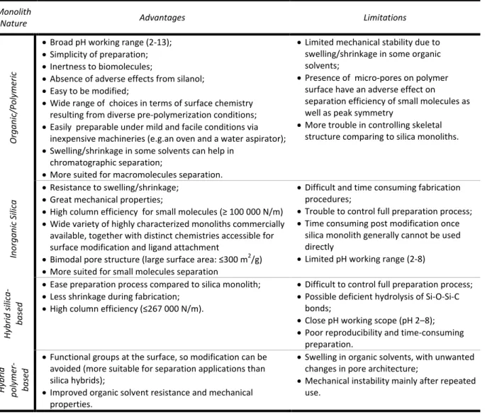
Table 1.4.- Benefits and limitations associated with each type of monolith structure.80–82,54
Monolith
Nature Advantages Limitations
Or gani c/Pol yme ri c
Broad pH working range (2-13); Simplicity of preparation; Inertness to biomolecules;
Absence of adverse effects from silanol; Easy to be modified;
Wide range of choices in terms of surface chemistry resulting from diverse pre-polymerization conditions; Easily preparable under mild and facile conditions via
inexpensive machineries (e.g.an oven and a water aspirator); Swelling/shrinkage in some solvents can help in
chromatographic separation;
More suited for macromolecules separation.
Limited mechanical stability due to swelling/shrinkage in some organic solvents;
Presence of micro-pores on polymer surface have an adverse effect on separation efficiency of small molecules as well as peak symmetry
More trouble in controlling skeletal structure comparing to silica monoliths.
Inorga
ni
c S
il
ica
Resistance to swelling/shrinkage; Great mechanical properties;
High column efficiency for small molecules (≥ 100 000 N/m)
Wide variety of highly characterized monoliths commercially available, together with distinct chemistries accessible for surface modification and ligand attachment
Bimodal pore structure (large surface area: ≤300 m2 /g) More suited for small molecules separation
Difficult and time consuming fabrication procedures;
Trouble to control full preparation process; Time consuming post modification once
silica monolith generally cannot be used directly
Limited pH working range (2-8)
H ybr id si li ca -bas ed
Ease preparation process compared to silica monolith; Less shrinkage during fabrication;
High column efficiency (≤267 000 N/m).
Difficult to control full preparation process; Possible deficient hydrolysis of Si-O-Si-C
bonds;
Close pH working scope (pH 2–8); Poor reproducibility and time-consuming
preparation. H ybr id pol yme r-bas ed
Functional groups at the surface, so modification can be avoided (more suitable for separation applications than silica hybrids);
Improved organic solvent resistance and mechanical properties.
Swelling in organic solvents, with unwanted changes in pore architecture;
Mechanical instability mainly after repeated use.
Literature Review
13 hybrid monolith flexibility, service longevity, exceptional biocompatibility, mechanical stability, ease of preparation and design at molecular level, the limitations they entail (Table 1.4.) have slowed its preferential use process82,80.
1.1.2. Surface Modification in Monoliths
Multiple approaches have been developed over the years, and are now accessible for the preparation of various functionalised monoliths48,83,12,84. The simplest,
and possibly the most straightforward methodology to tailor monoliths’ surface chemistry is just by choosing the suitable monomers that possess the desired functional groups (ionic, polar, non-polar, zwitterionic, etc.), once these groups are going to be exposed on the surface of the monolith after its preparation48,83. Nevertheless, every
time a new monomer system is employed there is a need of polymerization process de novo optimization so that a monolith with the desired properties is attained; however
this could end in a dull experimental procedure. Moreover as both monomers and crosslinkers become part of the final structure part of functional monomers added will be buried on the polymer bulk and not exposing its functional groups on the surface of the monolith for reaction48,12. Additionally proteins attachment on surface is virtually
impossible through this strategy due to proteins denaturation into casting solution. Thus besides this simple but limited approach, plethora strategies were developed to make possible the tailoring of monolith surface functionality as user pleases48,83,12,84. One
approach, less direct but perhaps more convenient, comprises the functionalization of a pre-formed monolith by post-preparation modification of reactive groups protruding from its surface. This type of modification, where each single reactive site provides one new functionality, allows the non-dependent optimization of bulk monolith properties and surface chemistry, enabling a onetime optimization of the monolith in question. The post-preparation modifications comprise the reaction of functional reagents with material surface groups (in case of silica-based monoliths it is first required the introduction of reactive sites or anchor groups for further incorporation of functionality to be accomplished); monomer/polymer chains grafting to or from monolith surface; dynamic or static coating in case of polymeric monoliths, and permanent or semi-permanent coating in case of silica-based monoliths. It is noteworthy that grafting strategy is frequently used to increment the ligand density (thus binding capacity) and also to improve hydrophilicity of column surface, to minimize non-specific interactions between analyte and monolithic media48. However
beyond copolymerization and post-preparation modifications (covalent immobilization) monoliths can also be modified by entrapment or bio-specific adsorption of ligand85. These methodologies are reviewed in some comprehensive
Literature Review
14 To these traditional methodologies used to tailor monolith surface chemistry, new surface functionalities can be afforded via attaching nanoparticles with a broad range of properties on monolith surface. These nanostructures (silica, silver, gold, metal oxides or polymers-based particles, or even carbon nanotubes) have been employed to enhance parameters as selectivity, chemical stability, and efficiency of 3D monolithic structure in gas and liquid chromatography, electrophoresis, and solid-phase extraction77,76.
1.2. Motivation and Aim of the Work
Monoliths show an attractive potential towards separation, especially of biomolecules. Moreover the astonishing growth of biopharmaceutical industry over the last decade denounce the urge for the development of novel, productive and efficient methods of purification, namely chromatographic matrices, once chromatography is the most widespread used and efficient purification approach nowadays.
The present work can be divided in two main parts. In a first approach a screening of materials processed by cryogelation was made in order to develop suitable monolithic structures for adenovirus purification. All structures were characterized physically and chemically. In the end three monolithic materials were elected, analysed morphologically and tested for binding adenovirus type 5 (Ad5).
In a second approach a monolithic structure was used for the first time as solid-phase platform for the synthesis of a small synthetic ligand specific for Green Fluorescent Protein (GFP). For this it was used an existing monolith previously developed in our lab by cryogelation. The matrix was characterized physically, chemically and morphologically before and after each step of the synthesis protocol to evaluate the presence or not of significant changes on the support during the process. Finally the functionalized affinity support was tested towards the target.
In the two approaches iron oxide magnetic nanoparticles (MNPs) were synthesized and embedded in the selected monoliths. The respective physical and morphological characterization was performed and compared with the respective plain supports in order to analyse the changes triggered. The MNPs modified monoliths were analysed regarding its performance on bioseparation of the protein in study upon external field exposure or not.
Literature Review
15
Figure 1.2.- Schematic depiction of research approaches followed in present work.
Literature Review
Experimental
17
Experimental
Experimental
19
2.1. Materials
2.1.1. Chemical Compounds
Chitosan (75-85% deacylated, medium Mw), dextran (from Leuconostoc mesenteroides, Mw ≈150,000), acrylamide (AAm, for electrophoresis, purity ≥99%),
poly(vinyl alcohol) (PVA, 99.0-99.8%(mol) hydrolysed, Mw 89,000-98,000), glycidyl methacrylate (GMA, 97%), 1,6-hexanodiamine 98%, N,N-dimethyl formamide (DMF, purity ≥99.8%) , phenylacetic acid 99%, iron (III) chloride hexahydrate (purity ≈ 98.0-102%), iron (II) chloride tetrahydrate (purity ≥99.0%), β-mercaptoethanol (purity ≥99.0%), phenol (unstabilized, purity ≥99%), potassium cyanide (purity ≥96.0%), pyridine (purity ≥99%), glutaric dialdehyde solution 50 wt% in water, silver nitrate (purity ≥99.0%), 1-pyrenemethylamine hydrochloride (purity 95%), isopropyl isocyanide (purity ≈97%), glycerol (purity ≥99.5%), ethylenediaminetetraacetic acid (EDTA) (purity ≥98.5%), bichinchoninic acid (BCA) kit and phosphate buffered saline tablet (PBS) were supplied by Sigma-Aldrich.
Ammonium persulphate (APS, purity ≥98%) and N,N,N’,N’ -tetramethylethylenediamine (TEMED, purity ≈99%), methanol (purity ≥99%), bromophenol blue sodium salt and 2-propanol were purchased from Roth.
N,N’-Methylenebisacrylamide (MBAAm, purity ≥98%), ninhydrin (purity ≥99%), ammonium hydroxide (purity ≈25%), maleic acid (purity ≥98%) and 1,10-phenanthroline 1-hydrate (purity ≥99.0%) were acquired from Fluka.
Glacial acetic acid (purity ≥99,7%) was purchased from Fisher Chemical. Bacteriological Agar powder was acquired from HIMEDIA.
β-D-1-Thiogalactopyranoside (IPTG), luria broth (LB), agarose (electrophoresis grade), ampicillin, glycine ultrapure for molecular biology, NZYMiniprep kit, tris(hydroxymethyl)aminomethane (Tris) Base ultrapure for molecular biology, and Greensafe were acquired from NZYTech.
Sodium citrate dihydrate (purity ≥99%) was supplied by Merk. Absolute ethanol (purity ≥99.9%) was purchased from Scharlau.
Glycine (98% purity) and hydroxylamine hydrochloride (97% purity) were obtained from Acros.
Sodium hydroxide, hydrochloric acid 37%, ethylene glycol and sodium chloride were supplied by Panreac.
Experimental
20 development reagent and development accelerator reagent) were purchased from BIO-RAD.
The Coomassie Plus (Bradford) assay kit was supplied by Thermo Scientific. Nitrogen and argon were provided by Air Liquide.
2.1.2. Biochemical Reagents
Albumin from bovine serum (BSA, purity ≥98%) was purchased from Sigma. Recombinant green fluorescent protein rTurboGFP (FP552-Evrogen) was acquired from Biocat GmbH. The plasmid pET-21c containing the DNA fragment encoding for GFP was synthesized and subcloned by GeneartTM (Germany). Competent cells NZY5α
and BL21(DE3), DNA marker ladder III and low molecular weight protein marker were purchased from NZYTech.
DNaseI was aquired from Roche.
Ad5 virions 10 times concentrated and 5 times diafiltrated (21st February
DM/CP) was kindly produced and manipulated by Dr Cristina Peixoto’s laboratory on ITQB-UNL/IBET, Portugal.
2.1.3. Equipment
Stirring of the casting solutions was performed using Dragon LAB MS-H-Pro stirring plates.
The lyophiliser used was a Telstar cryodos-50.
For the swelling tests, growing assays, GFP expression, A4C7 ligand synthesis, and BCA assays it was used an IKA KS 4000 i control incubator shaker.
The uniaxial compression measurements were attained using the compressive mode of tensile testing equipment (MINIMAT firmware v.3.1).
An Hitachi S 2400 equipment was used for SEM micrographs acquisition. The amination of monoliths was performed in a Plasma system FEMTO, version 3, Diener Electronics.
Hydrodynamic diameter and Zeta Potential measurements of MNPs samples were accomplished in a Malvern Dynamic Light Scattering (DLS) Zetasizer Nano ZS.
For magnetite assays the pH of solutions was adjusted in a Hanna Instruments microprocessor-based pH/mV/°C bench meter.