Comparisons and correlations of pain intensity and
respiratory and peripheral muscle strength in the
pre- and postoperative periods of cardiac surgery
INTRODUCTION
Cardiac surgeries are still considered the procedures of choice for reducing symptoms and mortality.(1-3) The main cardiac surgeries are myocardial revascu-larization surgery (MRS), surgery for valvulopathies, correction of aortic disea-ses, and cardiac transplantation.(1,4)
In this context, surgical stress induces loss of muscle mass due to
dysregu-lation in protein metabolism.(5,6) This condition culminates in the reduction
of muscle strength, causing long-term deficiencies such as persistent muscle
weakness.(7) Therefore, prevention of muscle proteolysis induced by surgical
Thayse Campos de Menezes1, Daniela Bassi2, Ricardo César Cavalcanti3, Juliana Emanuelle Santos Luz Barros1, Karolyne Soares Barbosa Granja1, Ana Carolina do Nascimento Calles1, Ana Luiza Exel1
1. Department of Physical Therapy, Centro Universitário Tiradentes - Maceió (AL), Brazil. 2. Department of Physical Therapy, Universidade Ceuma - São Luís (MA), Brazil.
3. Center of Clinical Research in Cardiology, Hospital do Coração - Maceió (AL), Brazil.
Objective: To evaluate respiratory and peripheral muscle strength after car-diac surgery. Additionally, we compared the changes in these variables on the third and sixth postoperative days.
Methods: Forty-six patients were
recruited, including 17 women and 29 men, with a mean age of 60.50 ye-ars (SD = 9.20). Myocardial revasculari-zation surgery was performed in 36 pa-tients, replacement of the aortic valve in 5 patients, and replacement of the mitral valve in 5 patients.
Results: A significant reduction in respiratory and peripheral muscle streng-th and a significant increase in pain in-tensity were observed on the third and sixth postoperative days (p < 0.05), ex-cept for the variable maximal inspira-tory pressure; on the sixth postoperative day, maximal inspiratory pressure values were already similar to the preoperative and predicted values (p > 0.05). There was an association between peripheral
Conflicts of interest: None.
Submitted on February 7, 2018 Accepted on July 26, 2018
Corresponding author:
Daniela Bassi
Departamento de Fisioterapia Centro de Ciências da Saúde Universidade Ceuma
Rua Josué Montello, 1 - Jardim Renascença Zip code: 65075-120 - São Luís (MA), Brazil E-mail: [email protected]
Responsible editor: Alexandre Biasi Cavalcanti
Comparações e correlações da intensidade da dor e da força
muscular periférica e respiratória no pré e pós-operatório de
cirurgia cardíaca
ABSTRACT
Keywords: Rehabilitation; Pain; Cardiac surgery; Respiratory muscles; Muscle strength; Postoperative period muscle strength, specifically between maximal expiratory pressure preope-ratively (rs = 0.383; p = 0.009), on the third postoperative day (rs = 0.468; p = 0.001) and on the sixth postoperative day (rs = 0.311; p = 0.037). The effect sizes were consistently moderate-to-large for respiratory muscle strength, the Me-dical Research Council scale and the vi-sual analog scale, in particular between preoperative assessment and the sixth postoperative day.
Conclusion: There is a decrease in respiratory and peripheral muscle streng-th after cardiac surgery. In addition, ma-ximal expiratory pressure is the variable that is most associated with peripheral muscle strength. These variables, espe-cially respiratory and peripheral muscle strength, should be considered by pro-fessionals working in the intensive care setting.
in the postoperative (PO) period and is described as a de-terminant of decreased functional capacity.(9)
In cardiac surgery patients, decreased RMS has been associated with decreased functional capacity and has con-tributed to a prolonged period of recovery of lung func-tion and the occurrence of physical decondifunc-tioning, whi-ch can last for several weeks.(10) Respiratory repercussions also generate changes in RMS, as well as changes in lung volumes and capacities, alveolar dysfunction, depression of central respiratory stimulation, and mechanical disor-ders of thoracic function.(3,4,11) In addition, it is known that most cardiac surgery patients present with episodes of muscle weakness in the preoperative period, which is accentuated after the surgical procedure.(12) However, this muscle weakness is more noticeable in the respiratory muscles than in the peripheral muscles, although the latter muscles are also inactive.(12,13)
Another important factor in this context is the role of postoperative pain in the functional recovery of the patient, which is an important indicator to estimate the physical and psychological tolls because prolonged painful stimuli cause suffering and complications in the PO pe-riod, which correlate with increased morbidity and mor-tality by affecting the ability to cough, breathe, and move properly.(14,15)
In view of the above, the objectives of this study were to evaluate RMS and peripheral muscle strength (PMS) after cardiac surgery and to analyze the changes in these variables on the third and sixth PO days, observing ble alterations in maximal respiratory pressures and possi-ble correlations with PMS and pain.
METHODS
This was a longitudinal observational study. The data collection was carried out from March to October 2016 after approval of the project by the Research Ethics
Com-mittee of the Centro Universitário Tiradentes (protocol
number 40004314.6.0000.5641). The nonprobabilistic convenience sample was composed of patients in the pre- and PO periods for cardiac surgery who were admitted to
ment of RMS, PMS, and pain intensity. Patients who had hospitalization-related complications that prevented their reevaluation were also excluded.
The evaluations were carried out at three time points. In the preoperative period (on the day before cardiac surgery), an evaluation form with identification, disease, treatment, and anthropometric data was used; RMS, PMS, and pain intensity were measured.
On the third and sixth PO day, the same evaluations performed in the preoperative period were performed again by the same evaluator as shown in the figure 1. The patients were in the intensive care unit (ICU) on the third PO day, while they were in the ward on the sixth PO day; thus, the time interval between evaluations is justified be-cause the clinical presentation of the patients in the two postoperative periods is different with regard to functional performance.
For the evaluation of RMS, the analog manovacuom-eter M120 (Porto Alegre, RS, Brazil) was used, with a scale
of ± 120cmH2O. Respiratory muscle strength tests were
performed with patients seated, lower limbs hanging, and feet supported. The nostrils were occluded with a nasal clip and the mouthpiece of the equipment was coupled to the mouth. To evaluate the maximal expiratory pres-sure (EPmax), the patient took a deep inspiration to total lung capacity (TLC), during which the occlusion of the buccal orifice was performed, followed by a maximal expi-ration until the residual volume was sustained for at least 2 seconds. To assess maximal inspiratory pressure (IPmax), an expiration to residual volume was taken, followed by occlusion of the orifice and maximal inspiration to TLC; the patient then maintained sustained strength for at least
2 seconds.Both maneuvers were repeated three times with
a one-minute interval, and the best measure was recorded for statistical analysis. Differences of 10% or less between values were accepted for each repetition. For the calcula-tion of the predicted pressures, the equacalcula-tions proposed by Neder et al.(16) were used.
individu-als remained seated in a chair with the hip joint at 90º of flexion, the knee at 60º of flexion, and the trunk erect. The MRC test measures muscle strength capable of joint displacement against manual resistance applied by the evaluator during the following joint movements: shoul-der abduction, elbow flexion, wrist extension, hip flexion, knee extension, and ankle dorsiflexion. When the scores for each evaluated movement are summed, the final MRC score ranges from 0 (tetraplegia) to 60 (normal muscle strength); patients with scores lower than 48 are
conside-red to have muscle weakness.(17) Measurements were
repe-ated three times, with a one-minute interval, and the best measure was recorded for statistical analysis.
Pain intensity was obtained by the visual analog scale (VAS), which consists of a horizontal line 10cm in length that shows a range of pain levels from absence of pain to the most intense pain and (provides a simple and efficient pain intensity measurement.(18)
Statistical analysis
The data were entered into and stored in a database created in Microsoft Excel 2010 software (Redmond, WA, USA). Continuous variables were presented as the mean and standard deviation; categorical variables were presen-ted as relative and absolute frequencies. Normality was tested using the Shapiro-Wilk test. Changes over time in respiratory and peripheral muscle forces were compared using the Kruskal-Wallis test. Correlations were evalua-ted using the Spearman correlation coefficient. An alpha value of 5% was adopted, and the Statistical Package for Social Science (SPSS) version 20.0 (IBM Inc., Chicago, IL, USA) and BioStat® 5.3 (Belém, PA, Brazil) were used.
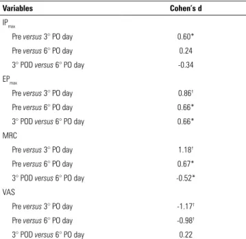
Cohen’s d was used to determine the clinical effect size of the proposed physiotherapeutic interventions, with the interpretation based on the classification established by Cohen(19) and Fernández-Lao et al.:(20) less than 0.20,
negligible effect; 0.20 to 0.50, small effect; 0.50 to 0.80, moderate effect; and > 0.80, large effect.
RESULTS
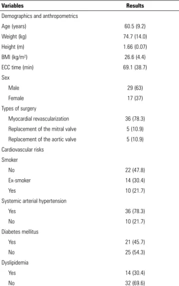
Forty-six patients were enrolled and evaluated in the study, with no losses. Thus, 17 (36.96%) women and 29 men (63.04%) with a mean age of 60.50 years (stan-dard deviation [SD] = 9.20) and a mean body mass index
(BMI) of 26.60kg/m2 (SD = 4.40) were included.
Thirty--six (78.26%) patients had systemic arterial hypertension, and 21 (45.65%) had type 2 diabetes mellitus, whereas 22 (47.83%) patients never smoked, 14 (30.43%) had not smoked for at least 6 months, and 10 (21.74%) were smokers.
Regarding the surgeries, MRS was performed in 36 (78.26%) patients, replacement of the aortic valve in 5 (10.87%) patients, and replacement of the mitral valve in 5 (10.87%) patients. Of these, 41 (89.13%) surgeries were performed with extracorporeal circulation, with a mean time of 69.10 minutes (SD = 38.70). The mean length of hospital stay was 7.10 days (SD = 2.1) (Table 1). Regarding the frequency of postoperative complications, only one patient had a pleural effusion, and two patients had pulmonary hypersecretion.
As shown in table 2, there was a significant reduction of RMS and PMS and a significant increase in pain intensity on the third and sixth PO day(p < 0.05), except for the va-riable IPmax, which on the sixth PO day already had values similar to the presurgical value and predicted (p > 0.05).
Curiously, there was a positive association between EPmax and PMS at different time points. Pain intensity was not correlated with RMS or PMS. Further details are described in table 3.
Table 4 shows the consistently moderate-to-large ef-fect sizes for RMS, MRC and VAS, particularly between the preoperative assessment and the sixth PO day.
RMS seen on the third PO day; however, it had returned at the sixth PO day. Corroborating in part the results of the current study, Roncada et al.(23) concluded that after coronary artery bypass grafting surgery, there was a ma-jor reduction in pulmonary function, which the author attributed to changes in circulatory factors that affect the synthesis of muscle proteins. In contrast, Urell et al.(9) ob-served that RMS was not reduced at two months after cardiac surgery. However, the authors did not evaluate pa-tients in the immediate postoperative period; this may be particularly relevant because pain would have needed to be considered.
In an evaluation of the clinical efficacy and feasibility of a respiratory muscular training device applied to pa-tients after cardiothoracic surgery, Crisafulli et al.(24) ob-served improvement in both IPmax and EPmax 14 days after surgical procedures associated with the use of the device.
While IPmax refers mainly to the force of the diaphragm,
EPmax mainly reflects the strength of the abdominal and
intercostal muscles.(25) In the present study, the reduction in IPmax was less than the reduction in EPmax. This could be explained by the fact that peak postoperative
diaphragma-tic dysfunction (decreased IPmax) occurs between two and
eight hours after surgery, whereas the muscles associated with EPmax suffer greater damage from incision and surgi-cal manipulation.(26)
Saglam et al.(27) , Faustini Pereira et al.(28) and Santos et al.(7) reported that the loss of RMS was related to the de-cline in PMS, corroborating the results of the present stu-dy, which demonstrated a positive and significant correla-tion between EPmax and PMS evaluated by MRC both
Male 29 (63)
Female 17 (37)
Types of surgery
Myocardial revascularization 36 (78.3) Replacement of the mitral valve 5 (10.9) Replacement of the aortic valve 5 (10.9) Cardiovascular risks
Smoker
No 22 (47.8)
Ex-smoker 14 (30.4)
Yes 10 (21.7)
Systemic arterial hypertension
Yes 36 (78.3)
No 10 (21.7)
Diabetes mellitus
Yes 21 (45.7)
No 25 (54.3)
Dyslipidemia
Yes 14 (30.4)
No 32 (69.6)
BMI - body mass index; ECC - extracorporeal circulation. The results are presented as mean (SD) or n (%).
Table 2 - Comparison of respiratory and peripheral muscle strength and pain intensity over time
Variables Predicted Presurgery 3rd PO day 6th PO day
IPmax (cmH2O) -102.50 (-82.90 - -109.30) -120.00 (-85.00 - -120.00) -80.00 (-40.00 - -120.00)*.† -120.00 (-55.00 - -120.00) EPmax (cmH2O) 111.84 (82.60 - 119.13) 90.00 (60.00 - 115.00)* 60.00 (40.00 - 80.00)*.† 50.00 (47.50 - 85.00)*.†
MRC (score) 60.00 (54.50 - 60.00) 51.00 (46.50 - 56.00)† 55.00 (48.00 - 58.00)†
VAS (score) 0 (0 - 0) 2.00 (0 - 6)† 2.00 (0 - 3.50)†
PO - postoperative; IPmax - maximum inspiratory pressure; EPmax - maximum expiratory pressure; MRC - Medical Research Council scale; VAS - Visual Analog Scale. *Differs from the predicted (p < 0.05, Kruskal-Wallis Test post hoc Dunn); † Differs from presurgery (p < 0.05, Kruskal-Wallis Test post hoc Dunn).
DISCUSSION
Table 3 - Correlation between respiratory and peripheral muscle strength and pain intensity
Correlation Presurgery 3rd PO day 6th PO day
IPmaxversus EP
max rs = 0.397. p = 0.006* rs = 0.675. p = 0.000* rs = 0.598. p = 0.000*
IPmaxversus MRC rs = 0.115. p = 0.447 rs = -0.125. p =0.406 rs = 0.289. p = 0.055 IPmaxversus VAS r
s = -0.275. p = 0.064 rs = -0.274. p = 0.066 rs = -0.244. p = 0.106 EPmaxversus MRC r
s = 0.383. p = 0.009* rs = 0.468. p = 0.001* rs = 0.311. p = 0.037* EPmaxversus VAS rs = -0.174. p = 0.246 rs = -0.086. p = 0.571 rs = -0.190. p = 0.211 MRC versus VAS r
s = 0.024. p = 0.872 rs = -0.219. p = 0.143 rs = -0.183. p = 0.223
POD - postoperative; IPmax - maximum inspiratory pressure; EPmax - maximum expiratory pressure; MRC - Medical Research Council scale; VAS - Visual Analog Scale. *Significant correlation (p ≤ 0.05, Spearman correlation coefficient).
Table 4 - Clinical effect sizes for measurements at pre- and postoperative days
Variables Cohen’s d
IPmax
Pre versus 3° PO day 0.60*
Pre versus 6° PO day 0.24
3° POD versus 6° PO day -0.34
EPmax
Pre versus 3° PO day 0.86†
Pre versus 6° PO day 0.66*
3° POD versus 6° PO day 0.66*
MRC
Pre versus 3° PO day 1.18†
Pre versus 6° PO day 0.67*
3° POD versus 6° PO day -0.52*
VAS
Pre versus 3° PO day -1.17†
Pre versus 6° PO day -0.98†
3° POD versus 6° PO day 0.22
IPmax - maximum inspiratory pressure; PO - postoperative day; EPmax - maximum expiratory
pressure; MRC - Medical Research Council scale; VAS - Visual Analog Scale. *Moderate effect size; † large effect size.
in the preoperative period and at both POD time points, that is, the third and sixth PO day. Santos et al.,(7) Saglam et al.,(27) Faustini Pereira et al.,(28) also reported that peri-pheral muscle strength showed an initial postsurgical loss with partial recovery during the PO period. This result is inconsistent with the results of the present study. Howe-ver, it should be noted that the postsurgical limitations may last for a period of six weeks to six months.(7) In addi-tion, it is already shown in the literature that worsening of RMS and PMS, as well as cognitive and motor disorders, can be caused by neuromuscular lesions from factors such as mechanical ventilation,(29,30) anesthesia, extracorporeal circulation time,(31) medicines,(26) malnutrition,(32) and bed immobility. Additionally, a study conducted by Santos et al., which evaluated the PMS of patients undergoing elective cardiac surgery, concluded that PMS values were
remarkably reduced after surgery and returned to near ba-seline by the time of discharge,(7) corroborating the results of the present study.
In recent years, protocols for the early withdrawal of sedation and for early mobilization have been used in se-veral intensive centers.(11,33,34) Such protocols highlight the importance physiotherapeutic intervention, which faci-litates important gains in both RMS and PMS and is a viable and safe strategy that prevents complications, redu-ces the deleterious effects of immobility, preserves muscle strength, and thus results in higher functional performan-ce.(12,35,36)
The decrease in RMS and PMS in the postoperative period seems to maintain a direct relationship with pain. (37) In the current study, there was no correlation between
VAS and RMS. The study of Sasseron et al.,(38) which
ai-med to evaluate the intensity and location of pain during hospitalization and its effects on the RMS of cardiac sur-gery patients, showed a correlation between pain in the first PO day and the decrease in the IPmax. Decreased PMS in individuals with heart disease has been reported in the literature. In addition, peripheral muscle weakness is asso-ciated with reduced muscle strength and loss of physical function.(39) Another study with the objective of evalua-ting the interaction between handgrip strength (HGS)
and myocardial oxygen consumption index (MVO2)
before and after cardiac surgery showed that hand grip strength had different effects on MVO2 prior to and after myocardial revascularization; HGS might be used as a pre-dictor to assess oxygen consumption in cardiac patients.(40) In our study, there was a significant increase in pain on the third PO day, with maintenance of the pain inten-sity until the sixth PO day, corroborating the findings of
Andrade et al.(41) who observed increased pain until the
sis of severity, assessment of functional implications, and determination of the risks of pulmonary and neuromus-cular dysfunction, thus providing information supporting the need for adequate muscular training to improve the strength of the respiratory and peripheral muscles and thus improving functional capacity.
The present study has some limitations. First, the number of patients was relatively small. Second, there is no gold standard for the assessment of PMS; however, dy-namometry has been demonstrated in the literature to be
We conclude that there is a decrease in respiratory and peripheral muscle strength associated with cardiac surgery. In addition, maximal expiratory pressure is the variable that is most associated with peripheral muscle strength. Thus, to improve both respiratory and peripheral muscle strength, professionals working in intensive care settings should consider these variables in relation to preoperative and postoperative interventions.
Objetivo: Avaliar a força da musculatura respiratória e peri-férica após cirurgia cardíaca, e comparar as modificações nestas variáveis no terceiro e no sexto dias pós-operatórios.
Métodos: Recrutaram-se 46 pacientes, dos quais 29 eram homens, com média de idade de 60,50 anos (DP = 9,20). Fo-ram submetidos à cirurgia de revascularização do miocárdio 36 pacientes, cinco pacientes foram submetidos à substituição de válvula aórtica, e outros cinco à substituição da válvula mitral.
Resultados: Observaram-se redução significante da força da musculatura respiratória e periférica, e significante aumento da intensidade da dor no terceiro e no sexto dias pós-operatórios (p < 0,05), exceto para a variável pressão inspiratória máxima. No sexto dia pós-operatório, os valores da pressão inspiratória máxima já tinham nível similar aos do período pré-operatório e aos valores previstos (p > 0,05). Ocorreu associação entre a
força da musculatura periférica, especificamente entre a pressão expiratória máxima no pré-operatório (rs = 0,383; p = 0,009), no terceiro dia pós-operatório (rs = 0,468; p = 0,001) e no sex-to dia pós-operatório (rs = 0,311; p = 0,037). Os tamanhos de efeitos foram coerentes em nível moderado à grande para força muscular respiratória, escores segundo a escala Medical Research Council e a Escala Visual Analógica, em particular entre a avalia-ção pré-operatória e a do sexto dia pós-operatório.
Conclusão: Após cirurgia cardíaca, ocorre diminuição da for-ça muscular respiratória e periférica. Além disto, a pressão expi-ratória máxima é a variável mais associada com a força muscular periférica. Essas variáveis, especialmente a força muscular respira-tória e periférica, devem ser consideradas pelos profissionais que atuam no ambiente de terapia intensiva.
RESUMO
REFERENCES
1. Peric V, Stolic R, Jovanovic A, Grbic R, Lazic B, Sovtic S, et al. Predictors of quality of life improvement after 2 years of coronary artery bypass surgery. Ann Thorac Cardiovasc Surg. 2017;23(5):233-8.
2. Hawkes AL, Nowak M, Bidstrup B, Speare R. Outcomes of coronary artery bypass graft surgery. Vasc Health Risk Manag. 2006;2(4):477-84. 3. Siregar S, Groenwold RH, de Mol BA, Speekenbrink RG, Versteegh MI,
Brandon Bravo Bruinsma GJ, et al. Evaluation of cardiac surgery mortality rates: 30-day mortality or longer follow-up? Eur J Cardiothorac Surg. 2013;44(5):875-83.
4. Scherner M, Madershahian N, Kuhr K, Rosenkranz S, Stöger E, Rahmanian P, et al. Aortic valve replacement after previous heart surgery in high-risk patients: transapical aortic valve implantation versus conventional aortic valve replacement-a risk-adjusted and propensity score-based analysis. J Thorac Cardiovasc Surg. 2014;148(1):90-7.
5. van Venrooij LM, Verberne HJ, de Vos R, Borgmeijer-Hoelen MM, van Leeuwen PA, de Mol BA. Postoperative loss of skeletal muscle mass, complications and quality of life in patients undergoing cardiac surgery. Nutrition. 2012;28(1):40-5.
6. Şimşek T, Şimşek HU, Cantürk NZ. Response to trauma and metabolic changes: posttraumatic metabolism. Ulus Cerrahi Derg. 2014;30(3):153-9.
7. Santos KM, Cerqueira Neto ML, Carvalho VO, Santana Filho VJ, Silva Junior WM, Araújo Filho AA, et al. Evaluation of peripheral muscle strength of patients undergoing elective cardiac surgery: a longitudinal study. Rev Bras Cir Cardiovasc. 2014;29(3):355-9.
8. Iida Y, Yamazaki T, Kawabe T, Usui A, Yamada S. Postoperative muscle proteolysis affects systemic muscle weakness in patients undergoing cardiac surgery. Int J Cardiol. 2014;172(3):5957.
9. Urell C, Emtner M, Hedenstrom H, Westerdahl E. Respiratory muscle strength is not decreased in patients undergoing cardiac surgery. J Cardiothorac Surg. 2016;11:41.
10. Hermes BM, Cardoso DM, Gomes TJ, Santos TD, Vicente MS, Pereira SN, et al. Short-term inspiratory muscle training potentiates the benefits of aerobic and resistance training in patients undergoing CABG in phase II cardiac rehabilitation program. Rev Bras Cir Cardiovasc. 2015;30(4):474-81.
11. da Costa Torres D, Dos Santos PR, Reis HJ, Paisani DM, Chiavegato LD. Effectiveness of an early mobilization program on functional capacity after coronary artery bypass surgery: A randomized controlled trial protocol. SAGE Open Med. 2016;4:2050312116682256.
12. Cordeiro AL, de Melo TA, Neves D, Luna J, Esquivel MS, Guimarães AR, et al. Inspiratory muscle training and functional capacity in patients undergoing cardiac surgery. Braz J Cardiovasc Surg. 2016;31(2):140-4. 13. Caruso FR, Arena R, Phillips SA, Bonjorno JC Jr, Mendes RG, Arakelian
VM, et al. Resistance exercise training improves heart rate variability and muscle performance: a randomized controlled trial in coronary artery disease patients. Eur J Phys Rehabil Med. 2015;51(3):281-9.
14. Sattari M, Baghdadchi ME, Kheyri M, Khakzadi H, Ozar Mashayekhi S. Study of patient pain management after heart surgery. Adv Pharm Bull. 2013;3(2):373-7.
15. Mello LC, Rosatti SF, Hortense P. Assessment of pain during rest and during activities in the postoperative period of cardiac surgery. Rev Lat Am Enfermagem. 2014;22(1):136-43.
16. Neder JA, Andreoni S, Lerario MC, Nery LE. Reference values for lung function tests. II. Maximal respiratory pressures and voluntary ventilation. Braz J Med Biol Res. 1999;32(6):719-27.
17. Paternostro-Sluga T, Grim-Stieger M, Posch M, Schuhfried O, Vacariu G, Mittermaier C, et al. Reliability and validity of the Medical Research Council (MRC) scale and a modified scale for testing muscle strength in patients with radial palsy. J Rehabil Med. 2008;40(8):665-71.
18. Ferreira-Valente MA, Pais-Ribeiro JL, Jensen MP. Validity of four pain intensity rating scales. Pain. 2011;152(10):2399-404.
19. Cohen J. Statistical power analysis for the behavioral sciences. New York: Academic Press; 1977. 474 p.
20. Fernández-Lao C, Cantarero-Villanueva I, Fernández-de-las-Peñas C, del Moral-Ávila R, Castro-Sánchez AM, Arroyo-Morales M. Effectiveness of a multidimensional physical therapy program on pain, pressure hypersensitivity, and trigger points in breast cancer survivors: a randomized controlled clinical trial. Clin J Pain. 28(2):113-21.
21. Platz JJ, Fabricant L, Norotsky M. Thoracic trauma: onjuries, evaluation, and treatment. Surg Clin North Am. 2017;97(4):783-99.
22. Stoliński J, Plicner D, Fijorek K, Grudzień G, Kruszec P, Andres J, et al. Respiratory system function in patients after aortic valve replacement through right anterior minithoracotomy. Thorac Cardiovasc Surg. 2017;65(3):182-90.
23. Roncada G, Dendale P, Linsen L, Hendrikx M, Hansen D. Reduction in pulmonary function after CABG surgery is related to postoperative inflammation and hypercortisolemia. Int J Clin Exp Med. 2015;8(7):10938-46.
24. Crisafulli E, Venturelli E, Siscaro G, Florini F, Papetti A, Lugli D, et al. Respiratory muscle training in patients recovering recent open cardiothoracic surgery: a randomized-controlled trial. Biomed Res Int. 2013;2013:354276.
25. Sachs MC, Enright PL, Hinckley Stukovsky KD, Jiang R, Barr RG; Multi-Ethnic Study of Atherosclerosis Lung Study the M-ES of A (MESA) L. Performance of maximum inspiratory pressure tests and maximum inspiratory pressure reference equations for 4 race/ethnic groups. Respir Care. 2009;54(10):1321-8.
26. Sasaki N, Meyer MJ, Eikermann M. Postoperative respiratory muscle dysfunction: pathophysiology and preventive strategies. Anesthesiology. 2013;118(4):961-78.
27. Saglam M, Vardar-Yagli N, Calik-Kutukcu E, Arikan H, Savci S, Inal-Ince D, et al. Functional exercise capacity, physical activity, and respiratory and peripheral muscle strength in pulmonary hypertension according to disease severity. J Phys Ther Sci. 2015;27(5):1309-12.
28. Faustini Pereira JL, Galant LH, Rossi D, Telles da Rosa LH, Garcia E, de Mello Brandão AB, et al. Functional capacity, respiratory muscle strength, and oxygen consumption predict mortality in patients with cirrhosis. Can J Gastroenterol Hepatol. 2016;2016:6940374.
29. Tobin MJ, Laghi F, Jubran A. Narrative review: ventilator-induced respiratory muscle weakness. Ann Intern Med. 2010;153(4):240-5. 30. Baldwin CE, Bersten AD. Alterations in respiratory and limb muscle
strength and size in patients with sepsis who are mechanically ventilated. Phys Ther. 2014;94(1):68-82.
31. Calles AC, Lira JL, Granja KS, Medeiro JD, Farias AR, Cavalcanti RC. Pulmonary complications in patients undergoing coronary artery bypass grafting at a hospital in Maceio, Brazil. Fisioter Mov. 2016;29(4):661-7. 32. Dassios T, Katelari A, Doudounakis S, Dimitriou G. Aerobic exercise and
respiratory muscle strength in patients with cystic fibrosis. Respir Med. 2013;107(5):684-90.
33. Cassina T, Putzu A, Santambrogio L, Villa M, Licker MJ. Hemodynamic challenge to early mobilization after cardiac surgery: a pilot study. Ann Card Anaesth. 2016;19(3):425-32.
34. Ramos dos Santos PM, Aquaroni Ricci N, Aparecida Bordignon Suster É, de Moraes Paisani D, Dias Chiavegato L. Effects of early mobilisation in patients after cardiac surgery: a systematic review. Physiotherapy. 2017;103(1):1-12.
35. Urell C, Westerdahl E, Hedenström H, Janson C, Emtner M. Lung function before and two days after open-heart surgery. Crit Care Res Pract. 2012;2012:291628.
36. Urell C. Lung function, respiratory muscle strength and effects of breathing exercises in cardiac surgery patients [dissertation]. Acta Universitatis Upsaliensis; 2013. 58p. [ Digital Comprehensive Summaries of Uppsala Dissertations from the Faculty of Medicine, 857].