Hélder Filipe Badim Costa
Chemical and biological characterization
of an aqueous
Sambucus nigra
L. flower
extract
Dissertação de Mestrado
Mestrado em Biotecnologia
Trabalho efetuado sob a orientação de
Doutora Cláudia Botelho
Professor José António Teixeira
Nome: Hélder Filipe Badim Costa
Endereço eletrónico: helderbadim@hotmail.com Telefone: +351 916257199
Cartão do Cidadão: 14402402
Título da dissertação: Chemical and biological characterization of an aqueous Sambucus
nigra L. flower extract
Orientadores:
Doutora Cláudia Botelho Professor José António Teixeira
Ano de conclusão: 2019
Mestrado em Biotecnologia
É AUTORIZADA A REPRODUÇÃO INTEGRAL DESTA DISSERTAÇÃO APENAS PARA EFEITOS DE INVESTIGAÇÃO, MEDIANTE DECLARAÇÃO ESCRITA DO INTERESSADO, QUE A TAL SE COMPROMETE.
Universidade do Minho, _____/_____/_____
Assinatura:
A
CKNOWLEDGMENTS
Inicialmente gostaria de agradecer à Doutora Cláudia Botelho pela orientação, motivação, palavras de encorajamento, disponibilidade e amizade. Foi uma tese longa, com muitos obstáculos, alguns até pareciam impossíveis de ultrapassar, mas com a sua persistência foram contornados sempre da melhor maneira, e por isso um obrigado gigante por todo o seu esforço e dedicação.
Gostava também de agradecer ao Professor José António Teixeira, por me ter dado as condições e a confiança necessárias para o desenvolvimento desta tese.
À professora Cristina Wilson, que desde o momento em que cheguei ao departamento de biologia foi incansável no acompanhamento. Desde os ensinamentos às reuniões realizadas, teve um papel fundamental no meu desenvolvimento enquanto investigador e no desenrolar desta tese. Um agradecimento especial para a equipa da Universidade de Aveiro, nomeadamente, ao Doutor Ângelo Salvador e à Doutora Sónia Santos pelo acompanhamento irrepreensível. Estas duas pessoas foram fundamentais numa fase inicial do trabalho, e com eles desenvolvi novas competências que foram fundamentais para a realização desta tese.
À professora Paula Sampaio e à professora Andreia Gomes, uma palavra de agradecimento pela preocupação demonstrada ao longo do desenrolar deste trabalho.
Aos membros do LBA, Diogo, Mário, Meirielly, Catarina, Anabela, Teresa, Cristina, Jorge e Rita. Acolheram-me desde o início da minha estadia e com eles passei a perceber o que realmente significa trabalhar num laboratório.
Aos membros do Tea Time, em especial, ao Mendanha, à Andreia e ao Mile (aka Bruno). Depois de uma semana dura de trabalho, este grupinho foi sempre a receita ideal para voltar a colocar o ânimo bem lá no alto.
À minha família, em especial ao meu pai e à minha mãe, que sempre me apoiaram durante toda a minha jornada académica. Pela paciência inesgotável e por sempre acreditaram em mim, um obrigado gigante.
À minha princesa Angélica por me animar, reconfortar e compreender. Como minha namorada e melhor amiga fez-me ficar cada vez melhor dia após dia. Há coisas que se levam desta tese e outras que se deixam ficar, ela com certeza é uma que eu quero levar para a vida toda.
R
ESUMO
Atualmente, há um crescente interesse no uso de recursos naturais como fonte de compostos bioativos, com potenciais benefícios para a saúde. Desde a antiguidade que é recorrente o uso plantas ao nível da medicina tradicional, nomeadamente a flor de sabugueiro (Sambucus nigra L.). Recentemente, esta planta tem sido a base de vários produtos nutracêuticos, contudo, ainda não existem evidências científicas claras que fundamentem a sua bioatividade, bem como os mecanismos celulares associados. De acordo com a literatura, os extratos de sabugueiro são constituídos sobretudo por compostos terpénicos voláteis, triterpénicos, esteróis, e compostos fenólicos. É, contudo, importante salientar que a composição de cada uma destas famílias é dependente do método de extração (solvente, temperatura e tempo).
O principal objetivo deste estudo foi a caracterização química de um extrato aquoso de flor de sabugueiro e a sua validação como agente anticancerígeno.
A extração aquosa da flor de Sabugueiro foi realizada a diferentes temperaturas (50, 70 e 90 ºC). O extrato aquoso obtido a 90 ºC apresentou a atividade anti-radicalar mais elevada (0.157 ± 0.001 mmol TE g-1) e uma concentração de compostos fenólicos mais elevada (45.32 ± 2.20 mg CAE g-1).
As análises qualitativas e quantitativas de GC-MS e HPLC-MS do extrato aquoso de flor de sabugueiro permitiram identificar 46 compostos, sendo a quercetina e o ácido clorogénico representativos de 86 % da totalidade dos compostos fenólicos identificados na fração hidrofílica, e a naringenina (27.16 %) o composto maioritário na fração lipofílica.
A atividade epigenética do extrato da flor de sabugueiro na desmetilação do MLH1 foi testada em combinação com o 5-fluorouracil em células do cancro colorretal (RKO), usando a 5-azacitidina como controlo positivo. Os resultados sugerem que a combinação do extrato de sabugueiro com o 5-FU não resulta num efeito sinergético, o que indica que o extrato não tem influência na desmetilação do MLH1. Apesar do extrato da flor sabugueiro ter diversas moléculas com capacidade antioxidante, o extrato não demonstrou proteção ou indução de reparação de danos do DNA após um insulto oxidativo (H2O2).
Ao nível da atividade antimicrobiana, verificou-se que bactérias gram-positivas são mais suscetíveis à presença deste extrato, nomeadamente, Staphylococcus aureus clinicamente isolado e Staphylococcus epidermidis.
Em conclusão, verificou-se que o extrato de flor de sabugueiro não possui capacidade de modular atividade epigénica e estimular a proteção/reparação de danos oxidativos. Ao invés, verificou-se que o extrato possui atividade microbiana, nomeadamente contra bactérias gram-positivas.
A
BSTRACT
Nowadays, there is a growing interest on the use of natural resources as sources of bioactive compounds with potential health benefits. Since the antiquity, that is recurrent the use of plants in folk medicine, such as the elderflower (Sambucus nigra L.). Recently, this plant has been the basis of several nutraceutical products. Thus, there is not a clear scientific explanation for its bioactivity, as well as the responsible cellular mechanisms. According to the literature, elderflower extracts are mainly composed by volatile terpenic, triterpenic, sterols and phenolic compounds. However, the composition of each one of this families will be dependent of extraction method (solvent, temperature, time).
The main gold of this study was the chemical characterization of an aqueous elderflower extract and its validation as an anticarcinogenic agent.
The aqueous extraction of elderflower was performed at different temperatures (50,70 and 90 ºC). The aqueous extract obtained at 90 ºC exhibited the highest antiradical activity (0.157 ± 0.001 mmol TE g-1) and the highest concentration of phenolic compounds (45.32 ± 2.20 mg CAE g-1).
The qualitative and quantitative analysis of GC-MS and HPLC-MS of elderflower aqueous extract allowed the identification of 46 compounds, being quercetin and chlorogenic acid representative of 86 % of the total of phenolic compounds identified in hydrophilic fraction, and naringenin (27.16 %), the major compound in lipophilic fraction.
The epigenetic activity of elderflower aqueous extract on demethylation of MLH1 was tested in combination with 5-fluorouracil (5-FU) in colorectal cancer cells (RKO), using 5-azacytidine (5-azaC) as a positive control. The results suggest that the combination of elderflower extract with 5-FU did not result in a synergetic effect, which indicates that elderflower extract did not have an influence on MLH1 demethylation.
Even though, the elderflower extract has several molecules with antioxidant potential, the extract did not exhibit protection or induction of repair of DNA damage after exposure to oxidative stress (H2O2).
The antimicrobial activity of elderflower extract was evaluated, and it was verified that gram-positive bacteria were the most susceptible to the presence of this extract, namely, Staphylococcus aureus clinically isolated and Staphylococcus epidermidis.
In conclusion, it was verified that S. nigra extract does not have the ability to modulate epigenetic activity and stimulate DNA protection/repair against oxidative stress. Although, this extract exhibits antimicrobial activity, particularly on gram-positive bacteria.
T
ABLE OF
C
ONTENTS
Acknowledgments _____________________________________________________ III Resumo ______________________________________________________________ V Abstract _____________________________________________________________VII Table of Contents _____________________________________________________ IX List of Figures _______________________________________________________ XIII List of Tables _______________________________________________________ XVII List of Abbreviations and Acronyms _____________________________________ XIX 1. Introduction _______________________________________________________ 3
1.1. Phytochemicals ________________________________________________ 3 1.1.1. Dietary polyphenols _____________________________________________ 4 1.1.2. The concept of oxidative stress _____________________________________ 5 1.1.3. Antioxidant Activity of Polyphenols ___________________________________ 7 1.1.4. Anti-inflammatory Activity of Polyphenols ______________________________ 9 1.2. Oxidative DNA damage and Mismatch Repair Pathway ___________________ 10 1.2.1. 5-Azacytidine/5-Azacitidine (5-azaC) on Cancer Therapy __________________ 11 1.2.2. 5-Fluorouracil (5-FU) on Cancer Therapy _____________________________ 13 1.3. Sambucus nigra L. _____________________________________________ 14 1.3.1. General Considerations __________________________________________ 14 1.3.2. Chemical composition and nutritional value ___________________________ 15 1.3.3. Harmful compounds ____________________________________________ 15 1.3.4. Overview of the S. nigra potential health benefits ________________________ 16
1.3.4.1. Antioxidant Activity _____________________________________________ 16
1.3.4.2. Anti-inflammatory Activity ________________________________________ 18
1.3.4.3. Anti-infective activity ____________________________________________ 19
1.3.4.4. Activity on colorectal cancer modulation ______________________________ 20
1.3.4.5. Diabetes mellitus ______________________________________________ 20 1.4. Solid-liquid extraction ___________________________________________ 21 1.5. Chromatographic-based technologies ________________________________ 21 1.5.1. One dimensional gas chromatography _______________________________ 22 1.5.2. High-performance liquid chromatography (HPLC) _______________________ 23
X
1.5.3. Data processing and interpretation __________________________________ 25 1.6. Context and objectives of the thesis _________________________________ 28
2. Material and Methods _____________________________________________ 31
2.1. Extract isolation and characterization ________________________________ 31 2.1.1. Plant material and extraction ______________________________________ 31 2.1.2. DPPH scavenging activity and trolox equivalent antioxidant capacity (TEAC)_____ 31 2.1.3. Phenolic content of elderflower aqueous extract ________________________ 31 2.1.4. GC-MS spectrometry analysis _____________________________________ 32 2.1.5. Characterization of HPLC-MS ______________________________________ 32 2.2. In vitro studies ________________________________________________ 33 2.2.1. Cell lines and culture conditions____________________________________ 33 2.2.2. Antiproliferative assay in human cells ________________________________ 34 2.2.3. Epigenetic activity of elderflower extract on MLH1 demethylation ____________ 34 2.2.4. Effect of S. nigra extract on oxidative DNA damage ______________________ 35 2.2.5. Comet Assay _________________________________________________ 35 2.2.6. Cellular Repair Assay ___________________________________________ 35 2.2.7. Susceptibility Testing ___________________________________________ 36 2.3. Statistical Analysis _____________________________________________ 37
3. Results __________________________________________________________ 41
3.1. Antioxidant activity and Phenolic Content _____________________________ 41 3.2. Identification and characterization of the compounds on the elderflower extract __ 42 3.2.1. GC-MS Analysis _______________________________________________ 43 3.2.2. HPLC-MS Analysis _____________________________________________ 48 3.3. In vitro analysis _______________________________________________ 54 3.3.1. In vitro proliferative activity _______________________________________ 54 3.3.2. Epigenetic activity of elderflower extract on MLH1 demethylation ____________ 54 3.3.3. Effects of elderflower extract on oxidatively induced-DNA damage ____________ 56 3.3.4. Effects of elderflower extract on repair ability __________________________ 57 3.3.5. Antimicrobial activity of elderflower aqueous extract _____________________ 59
4. Discussion _______________________________________________________ 63
4.2. Identification and characterization of elderflower extract ___________________ 64 4.3. In vitro analysis _______________________________________________ 66
5. Conclusions and Future Perspectives _________________________________ 73 6. References ______________________________________________________ 77 Supplementary Data __________________________________________________ 97
Supplementary Data I – GC calibration curve __________________________________ 99 Supplementary Data II – HPLC calibration curve of rutin ________________________ 100 Supplementary Data III – HPLC calibration curve of chlorogenic acid ________________ 101 Supplementary Data IV – Antiproliferative activity by MTT assay ___________________ 102 Supplementary Data V - Kinetics of attenuation of H2O2-induced DNA damage on RKO cells 102
Supplementary Data VI - Kinetics of SBs rejoining using H2O2 _____________________ 104
Supplementary Data VII – Yield of elderflower aqueous extract ____________________ 105 Supplementary data VIII – Antimicrobial activity of flavonoids _____________________ 106 Supplementary Data IX – Absorption of rutin and quercetin ______________________ 107 Supplementary Data X – List of material ____________________________________ 108
L
IST OF
F
IGURES
Figure 1. Phytochemicals classes and some examples of most common compounds of each class.
Adapted from Pandey and Rizvi (2009). _________________________________________ 4
Figure 2. Chemical structure of some common phenolic acids: gallic acid (A) and ferulic acid (B). __ 5 Figure 3. Chemical structure of some common flavonoids: kaempferol (A) and naringenin (B). _____ 5 Figure 4. Flavonoid scavenging a radical, leading to the formation of a flavonoid radical or a prooxidant.
______________________________________________________________________ 7
Figure 5. Molecular signaling transductions controlled by ROS molecules. ROS diffused into the cell
mitochondria stimulate activations of NF-κB pathway and Nrf2 related pathways. Initiation of NF-κB provokes stimulation of inflammatory transcription factors to upregulate pro-inflammatory molecules (e.g. TNF-a, IL-6, IL-8 and IL-1β). Activation of Nrf2 results in the transcription of antioxidant enzymes (GPx, SOD, CAT, etc). Both endogenous and exogenous antioxidants can scavenge ROS or suppressing NF-kB activated pro-inflammatory signal transduction, thus attenuating oxidative stress. Arrows indicate activation (→), whereas perpendicular lines show inhibition (Ⱶ). ARE, antioxidant response element; BAX, Bcl-2-associated X protein; Maf, transcription factor Maf; NF-κB, nuclear factor kappa-light-chain-enhancer of activated B cell; Nfr2, nuclear factor (erythroid-derived 2)-like 2; O2•–, superoxide anion;P53, tumor protein p53; TNFα, tumor necrosis factor α;
TNFR, tumor necrosis factor receptor. Adapted from Zhang and Tsao (2016). ______________ 8
Figure 6. Schematic representation of action of polyphenols as anti-inflammatory agents. COX1,
cyclooxygenase-1; COX2, cyclooxygenase-2; iNOS, inducible nitric oxide synthase; LPS, lipopolysaccharide; NADPHox, NADPH oxidase; NF-κB, nuclear factor kappa-light-chain-enhancer of activated B cells; pNF-κB, nuclear factor kappa-light-chain-enhancer of activated B cells promoter; ROS, reactive oxygen species; TLR, toll-like receptor. Adapted from Ambriz-Pérez et al. (2016). 10
Figure 7. Schematic illustration of the mammalian MMR pathway, indicating protein complexes and
processes involved in recognition of the mismatch to DNA resynthesis and DNA ligation (Jiricny, 2006). EXO1, exonuclease-1; MutSα, MutSα complex; MutLα, MutLα complex; Pol δ, DNA polymerase δ; PCNA, proliferating cell nuclear antigen; RFC, replication factor C; RPA, replication protein A. Adapted from Jiricny (2006). ________________________________________ 11
XIV
Figure 8. Schematic representation of reactivation of a silent tumor suppressor gene using 5-azaC. ↑ -
overexpression. Adapted from Momparler (2005). _________________________________ 12
Figure 9. 5-Fluorouracil (5-FU) metabolism. FUTP metabolite is extensively incorporated into RNA,
disrupting normal RNA processing and function. FdUTP can be misincorporated into DNA lead to strand breaks and consequently cell death. Adapted from Longley et al. (2003). DHFU, dihydrofluorouracil; DPD, dihydropyrimidine dehydrogenase; FdUDP, 5-fluorodeoxyuridine diphosphate; FdUMP, fluorodeoxyuridine monophosphate; FdUTP, fluorodeoxyuridine triphosphate; FUMP, fluorouridine monophosphate; FUDP, fluorouridine diphosphate; FUTP, fluorouridine triphosphate. ___________________________________________________________ 13
Figure 10. Sambucus nigra L. tree (A), flower (B) and berries (C). ________________________ 14
Figure 11. One-dimensional gas chromatographic system. _____________________________ 23 Figure 12. High-performance liquid chromatography system. ____________________________ 25
Figure 13. Radical scavenging activity of Sambucus nigra L. flower extracts at different isolation
temperatures. (A) Radical scavenging activity determined by DPPH assay. (B) Trolox equivalent antioxidant capacity (TEAC) in millimole of trolox equivalents per gram of flower (mmol TE g-1). Values
are the mean of three independent extraction flasks ± SD. Asterisks denote significant differences (*p < 0.05). ____________________________________________________________ 41
Figure 14. Phenolic content of Sambucus nigra L. flower extract at different isolation temperatures
expressed in caffeic acid equivalents (CAE). Values are the mean of three independent extraction flasks ± SD. Asterisks denote significant (**p < 0.01). ______________________________ 42
Figure 15. HPLC-PDA profile chromatogram of aqueous extract of S. nigra flowers. Time of retention is
expressed in minutes. Compounds are signed by numbers as stated in Table 6 and Table 7. __ 48
Figure 16. Effect on cellular viability of elderflower extract, 5-azaC and 5-FU. The cells were
pre-incubated during 48h with 5-azaC (2 µM) or with aqueous elderflower extract (10 µg mL-1 and 20
µg mL-1) before 5-FU (10 µM) treatment for 48h. The percentage of cell viability was calculated by
the absorbance relative to the value detected for the control cells (untreated cells), that was defined as 100% of cell viability. For each condition were performed three independent experiments and
data are expressed as mean ± SD. Asterisks represent significant difference (**p < 0.01; ***p < 0.001). _______________________________________________________________ 55
Figure 17. Effects of 24 and 48h of pre-treatment with luteolin (20 µM) and elderflower extract (200
and 400 µg mL-1) on oxidative DNA damage induced by 75 µM H2O2 (5 min, on ice) in RKO cells.
[+] - compound present; [-] - compound absent; () – percentage of protection regarding to the respective control. Results are expressed as mean ± SD, of at least three independent experiments. Asterisks represent significant difference (**p < 0.01). _____________________________ 57
Figure 18. Extent of repair of 75 µM H2O2-induced damage in RKO cells after pre-incubation during 24
and 48 hours with elderflower aqueous extract (200 and 400 µg mL-1) and 20 µM L-7-G followed
by 5 minutes (A) and 10 minutes (B) of recovering time. [+] - compound present; [-] - compound absent. Results are expressed as mean ± SD, of at least three independent experiments. Asterisks represent significant difference (***p < 0.001). ___________________________________ 58
L
IST OF
T
ABLES
Table 1. Relevant finding using in vitro and in vivo assays regarding to antioxidant activity using
elderflower and elderberries extracts. __________________________________________ 17
Table 2. Inflammation process involving the activation of monocytes and/or macrophages caused by
pathogens or harmful stimulus when exposed to elderflower or elderberries formulations. ↓ - downregulation; ↑ - activation/stimulation. (Harokopakis et al., 2006; Ho et al., 2017; Olejnik et al., 2015). ________________________________________________________________ 19
Table 3. Data pre-processing, pre-treatment, identification and quantification, processing,
post-processing, validation and interpretation (Goodacre et al., 2007). ______________________ 26
Table 4. The compounds identified in elderflower dichloromethane extract by GC/MS analysis, and their
identified biological activities in literature. _______________________________________ 44
Table 5. Composition of elderflower dichloromethane extract (µg g-1 of flower) with the respective relative
standard deviation (RSD). __________________________________________________ 47
Table 6. Phenolic compounds identified in S. nigra flower hydrophilic extract and corresponding MSn
fragmentation profiles, and their identified biological activities in literature. _______________ 50
Table 7. Concentration of the polyphenolic compounds in elderflower aqueous extract (µg g-1 of flower)
determined by HPLC. _____________________________________________________ 53
Table 8. IC50 values for elderflower extract on different cell lines expressed in mg mL-1. _________ 54
Table 9. MBC and MFC performance of elderflower aqueous extract against pathogenic organisms. MBC
and MFC values are presented in mg mL-1. Results are expressed as mean of at least three
L
IST OF
A
BBREVIATIONS AND
A
CRONYMS
1D-GC One-dimensional chromatography
5-azaC 5-Azacytidine/5-Azacitidine
5-FU 5-Fluorouracil
ANOVA Analysis of variance
Caco-2 Human colon adenocarcinoma cell line
CAE Caffeic acid equivalents
CAT Catalase
CO2 Carbon dioxide
COX2 Cyclogenease-2
DSBs Double stranded DNA breaks
EI Electron ionization
ESI Electrospray ionization
EXO1 DNA endonuclease
FdUMP Fluorodeoxyuridine monophosphate
FdUTP Fluorodeoxyuridine triphosphate
FUTP Fluorouridine triphosphate
GC-MS Gas chromatography-mass spectrometry
GPx Glutathione peroxidase
GSH Glutathione
H2O2 Hydrogen peroxide
HCT-116 Human colon cancer cell line
HIV-1 Human immunodeficiency virus
HO• Hydroxyl radicals
HPLC-MS High pressure liquid chromatography-mass spectrometry
HT29 Human colorectal adenocarcinoma
IC50 The half maximal inhibitory concentration
IL Interleukins
L-7-G Luteolin-7-glucoside
LCFAs Long chain fatty acids
XX
MCFAs Medium chain fatty acids
MFC Minimum fungicidal concentration
MMR Mismatch repair pathway
MS Mass spectrometry
NCTC 2544 Human keratinocyte cell line
NMR Nuclear magnetic resonance
NO Nitric oxide
Nrf2 Nuclear factor erythroid-related factor
O2⁻ Superoxide anion
PCNA Proliferating cell nuclear antigen
PI3K Phosphatidylinositol-3-kinase
PPARγ Peroxisome proliferator-activated receptor
QR Quinone reductase
RAW 264.7 Murine macrophage cell line
RKO Human colon carcinoma cell line
RNS Reactive nitrogen species
ROS Reactive species of oxygen
S. nigra Sambucus nigra L.
SBs Stranded DNA breaks
SD Standard deviation
SDA Sabouraud dextrose agar
SDB Sabouraud dextrose broth
SOD Superoxide dismutase
TE Trolox equivalents
TEAC Trolox equivalent antioxidant capacity
TMS Trimethylsilyl
TNF-α Tumor necrosis factor
TS Thymidylate synthase
TSA Trypic soy agar
TSB Trypic soy broth
1.
Introduction
Historically, natural products have provided an endless source of medicine. Plant products – as parts of foods or botanical portions and powders – have been used with variable success to cure and prevent diseases. Recently, there is a growing interest in so-called functional foods. In this context, phytochemical compounds stand out due to their health-promoting and/or medical properties (Wildman, 2016).
There is abundant evidence that a diet rich in plant derivatives is associated with lower risk of disease development, such as cardiovascular and inflammatory diseases (Guimarães et al., 2013; Sá et al., 2013). This may be attributed to the numerous bioactive substances present in plants, both of low molecular weight, like phenolic compounds, and high molecular weight, as is the case of polysaccharides. From a vast array of medicinal plants, Sambucus nigra L. (S. nigra) has been the subject of considerable interest due to their heterogenous mixture of phytochemicals, specially from the family of polyphenols. The study of its properties is of extreme importance to society since they can be integrated in many fields, including cosmetics and pharmaceutical industries.
1.1.
Phytochemicals
Phytochemicals are bioactive compounds present in plants, generally produced as secondary metabolites. These molecules play a crucial function in protecting plants against some environmental stresses (hordes of bacteria, fungi, insects, and other predators) (Dixon, 2001). The most bioactive phytochemicals on humans are phenolics, carotenoids, alkaloids, saponins, glucosinolates, cyanogenic glycosides and terpenes (Figure 1) (Mann & Truswell, 2017). Many of these natural products are assumed to have health promoting properties (Pandey & Rizvi, 2009).
4
Figure 1. Phytochemicals classes and some examples of most common compounds of each class. Adapted from
Pandey and Rizvi (2009).
1.1.1. Dietary polyphenols
Dietary polyphenols, found in fruits, vegetables, grains, tea and essential oils, are one of the most important class of natural antioxidants and chemopreventive agents found in human diet. There is currently evidence that dietary phenolic compounds can improve human health by lowering risk an preventing the onset of degenerative diseases including cancers, cardiovascular diseases and metabolic disorders (Scalbert et al., 2005). Phenolic compounds can be categorized into several sub-groups according their chemical structure, although, those usually found in plant derivatives can be separated into three main sub-groups: phenolic acids, flavonoids and non-flavonoids (Tsao, 2010). Phenolic acids are hydroxyl derivates of aromatic carboxylic acids which have a single phenolic ring (Figure 2).
Figure 2. Chemical structure of some common phenolic acids: gallic acid (A) and ferulic acid (B).
Flavonoids contain two phenolic rings (Ring A and B) linked by three carbon bridges to an oxygenated heterocycle (Ring C) as shown in Figure 3.
Figure 3. Chemical structure of some common flavonoids: kaempferol (A) and naringenin (B).
The antioxidant and anti-inflammatory properties and also other biological functions of polyphenols have been principally attributed to their particular chemical structure. The aromatic ring feature and the highly conjugated system with several hydroxyl groups make them good electron or hydrogen atom donors, capable of neutralize free radicals and other reactive oxygen species (ROS) (Zhang & Tsao, 2016).
1.1.2. The concept of oxidative stress
Oxidative stress is described as the loss of balance between the production/exposure to reactive oxygen/nitrogen species (ROS/RNS) and the organisms ability to counteract their action by the antioxidative protection systems (Koopman et al., 2010). It occurs due to an enhanced ROS/RNS formation or due to decay on the antioxidant protective capacity of an organism.
6
ROS can have opposite effects in the human body, operating as signaling molecules or harmful agents. They are generated as a result of aerobic metabolism (e.g. oxidative metabolism, β-oxidation of fatty acids me, induction of cytochrome P450 and respiratory burst of neutrophils during inflammatory response) or due to exposure to environmental elements (e.g. UV light, ionization radiation, chemical compounds, heavy metals) (Ames et al., 1993; Cooke et al., 2003; Halliwell, 1996). In homeostatic conditions, ROS are originated endogenously in controlled levels, being crucial for cell survival, differentiation and apoptosis (Bartosz, 2009). In contrast, an disproportion between ROS production and the activity of antioxidant mechanisms, in favor of the former, can lead to oxidative damage of lipids, proteins and DNA (Balaban et al., 2005). Free radical-induced damage induced oxidative stress has been confirmed as a contributor to the pathogenesis and pathophysiology of many chronic conditions such as neurodegenerative diseases (Parkinson, Alzheimer, Huntington’s disease and amyotrophic lateral sclerosis), emphysema, cardiovascular and inflammatory diseases, cataracts and cancer (López-Alarcón & Denicola, 2013; Maulik et al., 2013; Sies, 1985).
The most important ROS include superoxide anion (O2⁻) and hydroxyl radicals (HO•), and
non-radical molecules such hydrogen peroxide (H2O2) generated by endogenous and exogenous sources
(Slupphaug et al., 2003). The radical HO• is defined as the main ROS responsible for damaging DNA
once it is highly reactive and can directly damage DNA (Kryston et al., 2011). O2⁻ and H2O2 do not have
the ability to damage DNA directly, but the former can be transformed by the action superoxide dismutase (SOD) into H2O2, that, in the presence of divalent ions (e.g. Fe2+ and Cu2+), can originate HO• via Fenton
reaction (Equation 1) (Cabiscol et al., 2000).
𝐹𝑒2++ 𝐻2𝑂2 → 𝐹𝑒3++ • 𝑂𝐻 + 𝑂𝐻− (Equation 1)
Therefore, the DNA-damaging outcome of non-reactive ROS, like H2O2, is mediated by HO• which
can lead to base oxidation and strand breaks (Iyama & Wilson, 2013). The most frequent DNA damage product of oxidative stress responsible for induction of carcinogenesis mutations is the modified guanine base 8-oxoG, being result of the addition of HO• to the C8 position of the guanine ring, which is after
incorporated by the action of DNA polymerase during DNA synthesis (Barzilai & Yamamoto, 2004). During evolutionary course, organisms have developed several antioxidant defense systems that are utilized to counterbalance the deleterious effects produced by oxidative stress. Presently, it is well-known that cells exposed to oxidants express stress-induced genes or genes encoding antioxidant defenses (Evans et al., 2004). The counteracting mechanisms can be categorized into enzymatic and
non-enzymatic antioxidants. The antioxidant enzymes are located in the cell and include catalase, SOD, glutathione transferase, glutathione peroxidase, among others (Birben et al., 2012). The enzymatic reactions are capable to eliminate oxygen radicals and their products, lowering the damage caused by oxidative stress. The non-enzymatic antioxidants are mainly of dietary source, involving tocopherol (vitamin E), ascorbic acid (vitamin C), carotenoids and polyphenols (Brieger et al., 2012). Consequently, diets rich in antioxidant compounds provide an increased antioxidant defense/response, defending cellular machineries from damaging effect of excessive levels of ROS, and are linked with an inferior risk to develop degenerative diseases (Ames et al., 1993).
1.1.3. Antioxidant Activity of Polyphenols
A progressive and irreversible escalation of oxidative damage by ROS exerts severe influence on critical aspects of the biology (Maulik et al., 2013). Dietary phenolics possesses a good scavenging activity, having the ability to neutralize free radicals by donating an electron or a hydrogen atom. Namely, phenolic acids and flavonoids have an effective radical scavenging activity, although, the metal chelating ability and reducing power can differ depending on their structural properties (Perron & Brumaghim, 2009). However, when a phenolic molecule loses an electron or when it acts as a reducing agent, the molecule itself becomes a radical, but a relatively stable one, becoming a prooxidant (Bouayed & Bohn, 2010) (Figure 4). Consequently, polyphenols can be a double-edge sword: on one hand, when consumed correctly in the form of food or functional food, they are strong antioxidants, counteracting the excessive oxidative stress; on the other hand, they can exhibit prooxidant activity when used in high doses such as by administration of supplements (Bouayed & Bohn, 2010). The polyphenols ingested are digested by colonic microflora producing bioactive molecules that may interfere in the regulation of gut microbiota or be uptake by colonic epithelia leading to systemic antioxidant and anti-inflammatory effects (Nicholson et al., 2012).
8
The ability of dietary polyphenols to restore redox homeostasis and prevent systemic or localized inflammation by enhancing the activity of SOD, catalase (CAT) and glutathione peroxidase (GPx). It has been demonstrated that the expression of these antioxidant enzymes is controlled by a key transcription factor nuclear factor erythroid-related factor (Nrf2). Nfr2 is triggered by the presence of ROS at cellular level, being translocated into the nucleus. There, it regulates the expression of antioxidant-responsive elements (ARE)-mediated of numerous genes encoding the above cited antioxidant enzymes (Figure 5) (Kansanen et al., 2013).
Figure 5. Molecular signaling transductions controlled by ROS molecules. ROS diffused into the cell
mitochondria stimulate activations of NF-κB pathway and Nrf2 related pathways. Initiation of NF-κB provokes stimulation of inflammatory transcription factors to upregulate pro-inflammatory molecules (e.g. TNF-a, IL-6, IL-8 and IL-1β). Activation of Nrf2 results in the transcription of antioxidant enzymes (GPx, SOD, CAT, etc). Both endogenous and exogenous antioxidants can scavenge ROS or suppress NF-kB activated pro-inflammatory signal transduction, thus attenuating oxidative stress. Arrows indicate activation (→), whereas perpendicular lines show inhibition (Ⱶ). ARE, antioxidant response element; BAX, Bcl-2-associated X protein; Maf, transcription factor Maf; NF-κB, nuclear factor kappa-light-chain-enhancer of activated B cell; Nfr2, nuclear factor (erythroid-derived 2)-like 2; O2•–, superoxide anion;P53, tumor protein p53; TNFα, tumor necrosis factor α; TNFR, tumor necrosis factor
1.1.4. Anti-inflammatory Activity of Polyphenols
In the course of inflammation, macrophages can be activated through the detection of a pathogen endotoxin, such as lipopolysaccharide (LPS), via macrophages toll-like receptor (TLR). This action activates a signaling pathway that promotes the release of NF-κB (Nuclear factor kappa-light-chain-enhancer of activated B cells), a transcription factor responsible for the activation of transcription of inflammatory mediator genes, such as interleukins, TNF-α and prostaglandins (PGs); inflammatory enzymes such as inducible nitric oxide synthase (iNOS) responsible for the synthesis of NO; and cyclooxygenases (COXs). TLR signaling also triggers the generation of reactive oxygen species (ROS) (Friedman & Hughes, 2002; Kumar et al., 2017). Overexpression of these mediators, as happens in chronic inflammation, might lead to the occurrence of several chronic diseases.
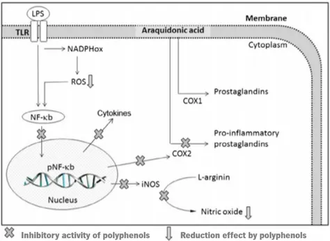
Polyphenols might exert anti-inflammatory properties especially through radical scavenging activities, regulation of cellular events, and modulation of enzyme action of arachidonic acid metabolism (phospholipase A2, COX) and arginine metabolism (NOS), as well as modulation of proinflammatory molecules production (Figure 6) (Ambriz-Pérez et al., 2016).
Molecular mechanisms of polyphenol anti-inflammatory activities comprise inhibition of enzymes related with proinflammatory properties such as COX2, LOX, and iNOS, inhibition of NF-κB and the activating protein-1 (AP-1), activation of phase-II antioxidant detoxifying enzymes, and activation of mitogen activated protein kinase (MAPK), protein kinase-C, and nuclear fact erythroid 2-related factor (Santangelo et al., 2007).
10
Figure 6. Schematic representation of action of polyphenols as anti-inflammatory agents. COX1,
cyclooxygenase-1; COX2, cyclooxygenase-2; iNOS, inducible nitric oxide synthase; LPS, lipopolysaccharide; NADPHox, NADPH oxidase; NF-κB, nuclear factor kappa-light-chain-enhancer of activated B cells; pNF-κB, nuclear factor kappa-light-chain-enhancer of activated B cells promoter; ROS, reactive oxygen species; TLR, toll-like receptor. Adapted from Ambriz-Pérez et al. (2016).
1.2.
Oxidative DNA damage and Mismatch Repair Pathway
A significant consequence of oxidative stress is DNA damage, leading to genomic instability. DNA damage induced by ROS involves structural changes due to single- or double- stranded DNA breaks (SBs), alteration of purine, pyrimidine or deoxyribose; and development of DNA crosslinks through oxidation, depurination, methylation and deamination reactions.
To respond to these lesions, cells have DNA repair mechanisms such as the mismatch repair pathway (MMR) (Figure 7). MMR pathway starts when MutSα including Msh2 and Msh6 binds to a mismatch, followed by binding of MutLα [Mlh1 and Pms2]. Later, a proliferating cell nuclear antigen (PCNA) along with an accessory protein (RFC), triggers DNA endonuclease (EXO1), which guided by the MutS/MutL travels to the clamp. The elimination of daughter-strand DNA starts in the direction of the mismatch and then beyond it. Once the incongruity is removed, the activity of EXO1 is blocked by MutL, stopping DNA excision. After this procedure, repair is completed by correct DNA synthesis and ligation by the action of DNA polymerase and a DNA ligase, respectively.
Figure 7. Schematic illustration of the mammalian MMR pathway, indicating protein complexes and processes
involved in recognition of the mismatch to DNA resynthesis and DNA ligation (Jiricny, 2006). EXO1, exonuclease-1; MutSα, MutSα complex; MutLα, MutLα complex; Pol δ, DNA polymerase δ; PCNA, proliferating cell nuclear antigen; RFC, replication factor C; RPA, replication protein A. Adapted from Jiricny (2006).
1.2.1. 5-Azacytidine/5-Azacitidine (5-azaC) on Cancer Therapy
DNA methylation is a heritable epigenetic mark which involves the covalent transfer of a methyl group to the C-5 position of the cytosine ring of DNA by the action of DNA methyltransferases (DNMTs). Most DNA methylation is essential for normal development, and it plays an important role in a number of key processes including genomic imprinting, X-chromosome inactivation, and suppression of repetitive
12
element transcription and transposition. When dysregulated, this process contributes to diseases like cancer by the inactivation tumor suppressor genes such as MLH1 (Jones & Laird, 1999). The molecular mechanism of silencing gene expression appears to be due to the binding of 5-methylcytosine proteins to the methylated promoter, which blocks the action of transcription factors (Jones et al., 1998).
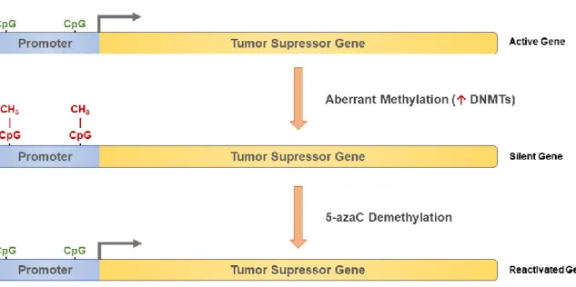
5-azaC (also known as Dacogen or Vidaza) is an analogue of cytidine ribose nucleoside. It is considered a prodrug and it has two main mechanisms of antineoplastic action. First, its ability to incorporate 5-azacetidine triphosphate into RNA which causes a disruption of nuclear and cytoplasmatic RNA metabolism with subsequent inhibition of protein synthesis (Li et al., 1970). Second, its ability to inhibit DNA methylation, by trapping DNA methyltransferases (DNMTs), leading to global demethylation as cells divide. DNA methylation refers to the addition of a methyl group to the cytosine residue with a high frequency of CG dinucleotides that are typically located in proximity of gene promoters. The degree of methylation of CpG islands plays a role in the control of gene transcription. Usually, fully methylated sites are associated with suppression of gene expression, while hypomethylated or unmethylated CpG islands are linked to active transcription. Forming a tight-binding complex 5-azaC irreversibly binds to DNA methyltransferases, which inhibits its progression along the DNA duplex, resulting in intracellular depletion of the enzyme. Consequently, unmethylated DNA can lead to the transcription of previously quiescent genes (Jones & Taylor, 1981; Taylor & Jones, 1982). 5-azaC demethylation mechanism is shown in Figure 8.
Figure 8. Schematic representation of reactivation of a silent tumor suppressor gene using 5-azaC. ↑ -
1.2.2. 5-Fluorouracil (5-FU) on Cancer Therapy
The action of anticancer agent classified as antimetabolite drugs, happens through the inhibition of essential biosynthetic processes and/or by being incorporated into macromolecules, such as DNA and RNA, inhibiting the cells normal function. The fluoropyrimidine 5-FU does both (Grem, 2000).
For the last 70 years, the fluoropyrimidine 5-fluorouracil (5-FU) has been positioned in the first line as chemotherapy agent of various cancers, including colorectal, head, neck and breast cancer (Grem, 2000; Toloudi et al., 2015). 5-FU is an analogue of uracil with a fluorine atom at the C-5 position in place of hydrogen. It rapidly enters the cell using the same facilitated transport mechanism as uracil (Wohlhueter et al., 1980). 5-FU is converted intracellularly to several active metabolites: fluorodeoxyuridine monophosphate (FdUMP), fluorodeoxyuridine triphosphate (FdUTP) and fluorouridine triphosphate (FUTP) — these active metabolites disrupt RNA synthesis and increase DNA damage. It results in cell growth arrest and apoptosis (Figure 9) (Longley et al., 2003).
Figure 9. 5-Fluorouracil (5-FU) metabolism. FUTP metabolite is extensively incorporated into RNA, disrupting
normal RNA processing and function. FdUTP can be misincorporated into DNA lead to strand breaks and consequently cell death. Adapted from Longley et al. (2003). DHFU, dihydrofluorouracil; DPD, dihydropyrimidine dehydrogenase; FdUDP, 5-fluorodeoxyuridine diphosphate; FdUMP, fluorodeoxyuridine monophosphate; FdUTP,
14
fluorodeoxyuridine triphosphate; FUMP, fluorouridine monophosphate; FUDP, fluorouridine diphosphate; FUTP, fluorouridine triphosphate.
However, the response rates of 5-FU for advanced colorectal cancers is less about 10%, when given as a single agent, and its bioavailability is also limited (rapid degradation to DHFU by DPD) (Diasio & Harris, 1989; Giacchetti et al., 2000). Furthermore, 5-FU induces severe adverse reactions at different levels, namely, hematological, neural, cardiac and dermatological reactions, and at gastrointestinal tract.
1.3.
Sambucus nigra
L.
1.3.1. General Considerations
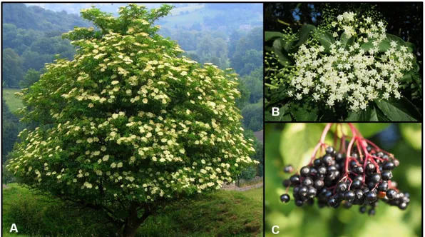
S. nigra or black elder (Figure 10) is widespread native plant from the British Isles and continental Europe. In Portugal, it can be found in the Northern Region (Tarouca, Lamego, Moimenta da Beira) (Cunha et al., 2016).
Sambucus belongs to the Adoxaceae family, a major group of Angiosperms (flowering plants), however some studies refer to Sambucus as a Caprifoliceae family member (Charlebois et al., 2010; Donoghue et al., 2003). S. nigra is a deciduous shrub or small tree that can grow up to 10 meters. There are two important subspecies of S. nigra: the black elder, a common plant in Europe, and American elder, also known as elder or elderberry (Charlebois, 2007).
S. nigra berries and flowers have a long history of usage in traditional European medicine, being used on diverse formulations that range from food products to medical formulations. Recently, S. nigra has been used on the development of supplements and nutraceuticals (Wildman, 2016). It has been extensively used in phytotherapy for the treatment of disorders associated with the respiratory and gastrointestinal tract, rheumatism, inflammation, diabetes, as well as viral infections and fevers and is currently one of the most-used medicinal plants worldwide (Jarić et al., 2007; Sidor & Gramza-Michalowska, 2015).
The agroindustrial market pursues the consumer desire for added value-added products which are effective in disease prevention, as well as, in promoting a healthy aging (Silva, 2009). In the last decades, S. nigra flowers and berries secondary metabolites attracted attention due to their pharmacological activities, mainly the monoterpenic, sesquiterpenic and triterpenic compounds, sterols and phenolic compounds. The chemical composition of the berries and flowers depends on a series of factors, such as habitat/location, fertilization, maturation and harvest period.
1.3.2. Chemical composition and nutritional value
Elderflowers are a rich source of bioactive flavonoids and phenolic acids (Christensen et al., 2008). Among flavonoids, flavonol glycosides, quercetin-3-rutinoside (rutin), kaempferol-3-rutinoside and isoharmnetin-3-rutinoside are the major flavonoids in elderflowers contributing as much as 90% of the total flavonoids content. The concentration of flavonoids is higher in elderberry flowers in comparison with berries and leaves (Dawidowicz et al., 2006). The 5-caffeoylquinnic acid and 1,5-di-caffeoylquinic acid (chlorogenic acids) comprise over of 70% of the total phenolic acid content present in the S. nigra flowers. Opposed to the chemical composition of the elderberry fruit, which is especially rich in anthocyanins, flowers do not contain any pigments from this group.
1.3.3. Harmful compounds
Not all constituents of the S. nigra plant are safe for consumption. In fact, only S. canadensis and S. nigra flowers have been approved by the United States Food and Drug Administration (FDA) as Generally Recognized as Safe (GRAS) for use as a flavoring ingredient (Food). All elderberry parts contain cyanogenic glycosides, being the most abundant sambunigrin and prunasin. Furthermore, elderberry contains m-hydroxysubstituted glycosides, such as zierin and holocalin (Dellagreca et al., 2000). These compounds are potentially toxic and life-threatening, because they can hydrolyzed resulting in the release
16
of cyanide (Bromley et al., 2005). However, they occur primarily in unripe berries and are degraded during heat treatment (Boon, 2010). For this reason, processed products are preferred for consumption over fresh fruit (Cejpek et al., 2009).
1.3.4. Overview of the
S. nigra
potential health benefits
Several published evidences indicate potential benefits of S. nigra berries and flowers in disease prevention/management if included in diet.
1.3.4.1. Antioxidant Activity
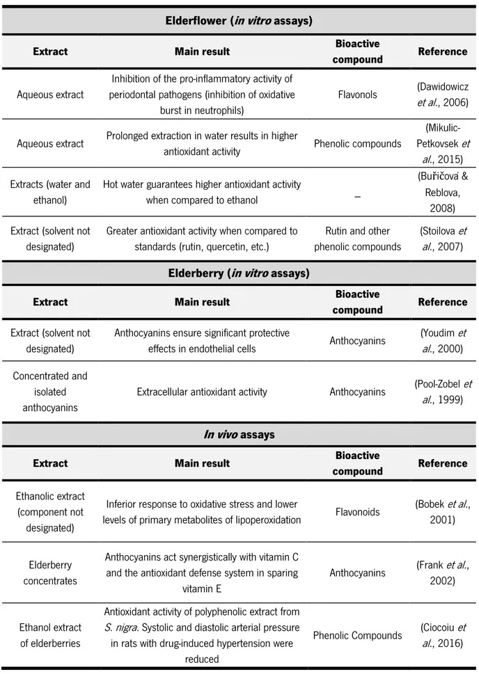
There is a high interest in plant-derived antioxidants to prevent the harmful effects of oxygen radicals and/or other reactive oxygen species (ROS) (Gülçin, 2012; Halliwell, 2013). Antioxidant activity is one of the most exploited features of S. nigra. These properties are often related to phenolic compounds, and especially anthocyanins and flavonols. However, it is relevant to point out possible interactions between extract components, which include non-phenolic compounds. The extraction conditions, and pre- and post-harvest conditions, might play a critical role on the result of the antioxidant capacity. Table 1 summarizes the main findings regarding the antioxidant activity of elderflowers and elderberries.
Dietary containing phenolic molecules may help to maintain the oxidative stress homeostasis of oxidative stress once consumed. However, during digestion (ingestion, absorption, assimilation) their stability and bioavailability might be affected (Zhou et al., 2016). It is described that there is a loss of elderberry bioactive composites occurs due to the digestion process (in vitro assay), that in the case of anthocyanins it can reach a 44% loss. Even so, the colon-digested aqueous extract decreases the excessive intracellular ROS production (22%) and oxidative damage (46%) in human colon cells (Olejnik et al., 2016).
Table 1. Relevant finding using in vitro and in vivo assays regarding to antioxidant activity using elderflower and elderberries extracts.
Elderflower (in vitro assays)
Extract Main result Bioactive
compound Reference
Aqueous extract
Inhibition of the pro-inflammatory activity of periodontal pathogens (inhibition of oxidative
burst in neutrophils)
Flavonols et al.(Dawidowicz , 2006)
Aqueous extract Prolonged extraction in water results in higher
antioxidant activity Phenolic compounds
(Mikulic-Petkovsek et
al., 2015) Extracts (water and
ethanol)
Hot water guarantees higher antioxidant activity
when compared to ethanol _
(Buřičová & Reblova,
2008) Extract (solvent not
designated)
Greater antioxidant activity when compared to standards (rutin, quercetin, etc.)
Rutin and other phenolic compounds
(Stoilova et al., 2007)
Elderberry (in vitro assays)
Extract Main result Bioactive
compound Reference
Extract (solvent not designated)
Anthocyanins ensure significant protective
effects in endothelial cells Anthocyanins
(Youdim et al., 2000) Concentrated and
isolated anthocyanins
Extracellular antioxidant activity Anthocyanins (Pool-Zobelal. et , 1999)
In vivo assays
Extract Main result Bioactive
compound Reference
Ethanolic extract (component not
designated)
Inferior response to oxidative stress and lower
levels of primary metabolites of lipoperoxidation Flavonoids
(Bobek et al., 2001)
Elderberry concentrates
Anthocyanins act synergistically with vitamin C and the antioxidant defense system in sparing
vitamin E
Anthocyanins (Frank et al., 2002)
Ethanol extract of elderberries
Antioxidant activity of polyphenolic extract from
S. nigra. Systolic and diastolic arterial pressure in rats with drug-induced hypertension were
reduced
Phenolic Compounds (Ciocoiual. et , 2016)
18
1.3.4.2. Anti-inflammatory Activity
Inflammation is associated to several human diseases, including, asthma, diabetes, allergy, multiple sclerosis, cardiovascular diseases, neurodegenerative disorders, and some types of cancer (Debnath et al., 2013; Mena et al., 2014). An important mechanism of organism self-protection involves inflammation, aiming to eliminate harmful stimuli such as damaged cells, irritants or pathogens (Ho et al., 2017). Usually, the inflammation process starts with the activation of monocytes and/or macrophages, which participate in the regulation of the inflammation by the release of several cytokines, such as tumor necrosis factor (TNF-α), interleukins (IL), and inflammatory mediators which include reactive oxygen species (ROS), nitric oxide (NO), and prostaglandin E2, which are produced by inducible nitric oxide synthase and cyclogenease-2 (COX2) (Ho et al., 2017).
Cytokines as TNF-α and IL(s) act as multipotential mediators on the cellular system, having an extensive diversity of biological activities. They can promote favorable or unfavorable effects on the host during immune response, depending on their local concentration, where the equilibrium between the inflammatory and anti-inflammatory cytokines will manage the outcome and the duration of the immune response (Barak et al., 2002).
It is described that elderberry aqueous extract downregulated the expression of mediators such as IL-1, IL-1β, IL-6 and TNF-α (Gorchakova et al., 2007; Olejnik et al., 2015). The elderflowers aqueous extract inhibits the macrophage production of pro-inflammatory cytokines and block the neutrophils activation. This may be due to the inhibition of activation of NF-κB and phosphatidylinositol-3-kinase (PI3K), an enzyme essential in the regulation of immunity and inflammation process. The bioactive composites responsible for the anti-inflammatory effects of elderflower aqueous extract are unidentified. However, the ability of the aqueous extract to inhibit PI3K seems to be mediated through quercetin (Yeşilada et al., 1997).
Modulation of NO production by macrophages and dendritic cells also have significative importance in inflammatory diseases. Ethanolic elderflowers extracts displayed an inhibitory activity on NO production in RAW cells and dendritic cells (Ho et al., 2017). In vivo experiments also validated the potential of elderflower inflammatory activity, when an 80% ethanol extract showed to have moderate anti-inflammatory activity in rats (Mascolo et al., 1987). Studies involving elderflower and elderberries extract and their role in the inflammation process are described in Table 2.
Table 2. Inflammation process involving the activation of monocytes and/or macrophages caused by pathogens
or harmful stimulus when exposed to elderflower or elderberries formulations. ↓ - downregulation; ↑ - activation/stimulation (Harokopakis et al., 2006; Ho et al., 2017; Olejnik et al., 2015).
Elderflower formulation Elderberries formulation
TNF-α ↓ ↓ IL-1α ↓ – IL-1β ↓ ↓ IL-6 – ↓ IL-10 – ↑ NO ↓ – H2O2 ↓ – O2⁻ ↓ – NF-κB ↓ –
The evidence of S. nigra berries and flowers influence on the anti-inflammatory and immunological pathways on humans is still scarce.
1.3.4.3. Anti-infective activity
Aqueous elderberry extracts (phenolic-type extracts) demonstrated antimicrobial activity, (fungi, bacteria) against human pathogenic microorganisms. The antimicrobial studies demonstrated that these extracts had antimicrobial activity against Staphylococcus aureus (resistant and methicillin-sensitive), Streptococcus mutans, Streptococcus pyogenes, Haemophilus influenza, Haemophilus parainfluenzae, Branhamella catarrhalis, and Helicobacter pylori in the conditions tested (Chatterjee et al., 2004; Izzo et al., 1995; Krawitz et al., 2011).
These experiments showed the ability of S. nigra to inhibit pathogens in vitro. However, it is essential to perform more studies, in order to understand which bacterial and fungal pathogens are vulnerable to S. nigra, and also their mechanisms of action.
One of the most interesting applications of S. nigra berries is associated with their ability to inhibit the influenza virus. Influenza virus A or B are responsible for an acute, febrile illness that happens in outbreaks varying severity every winter. Standardized elderberry extracts decreased hemagglutination and inhibited replication of numerous human and animal influenza viruses A and B in vitro (Zakay-Rones et al., 2004; Zakay-Rones et al., 1995).
20
Antiviral properties were also described against human immunodeficiency virus (1) on HIV-infected peripheral lymphocytes and herpes simplex virus (Vlachojannis et al., 2010). Flavonoids, including quercetin, cyanidin and petunidin, and proanthocyanins, present in european elderberry extract, bind to HIV-1 virions, blocking their mechanisms, preventing to host cells infection (Fink et al., 2009).
In vivo experiments performed on chimpanzees reinforced the in vitro conclusions regarding elderberry aqueous extract and its activity against influenza virus (Burge et al., 1999). The suggested action mechanism stimulates the immune system; inhibit hemagglutination of the influenza virus avoiding the adhesion of the virus to cell receptor; and present anti-inflammatory effect. Furthermore, the absence of side effects of this S. nigra formulations offers an alternative way for a safe treatment for influenza (Zakay-Rones et al., 2004).
1.3.4.4. Activity on colorectal cancer modulation
According to the literature, an aqueous elderberry polar extract can block the growth of a human colorectal adenocarcinoma cell line (HT29), presenting a IC50 of 130.3 µg of cyanidin-3-glucoside eq mL -1 (Jing et al., 2008). An aqueous acetone extracts, also with similar properties, was reported on Hepa
1c1c7 cells. This extract potentiates the induction of quinone reductase (QR) and inhibition COX2, which is indicative of anti-initiation and antipromotion properties, respectively (Thole et al., 2006).
The in vitro experiments reported the importance of including S. nigra preparations in diet to positively modulate colorectal cancer. It regulates inflammatory factors, anti-initiating and anti-promoting of tumoral factors, and oxidation processes. For this reason, tests to unveil the underlying anticancer mechanisms are needed (Thole et al., 2006).
1.3.4.5. Diabetes mellitus
Diabetes is mainly caused by a combination of insulin resistance and β-cell failure (pancreatic cells). It can be treated with insulin-sensitizing drugs that target the nuclear receptor peroxisome proliferator-activated receptor (PPARγ) (Christensen et al., 2010).
Several elderflower extracts (hexane, dichloromethane, methanol, ethyl acetate and water) demonstrated in vitro, an activation effect of PPAR (α, δ or γ), between 2.5 and 250 µg mL-1, without
stimulate adipocyte differentiation (Christensen et al., 2009; Christensen et al., 2010). The extracts had a positive effect on insulin-stimulated glucose uptake indicating that elderflowers have molecules with bioactivities comparable to those of partial PPARγ agonists (Christensen et al., 2009).
The exposure to elderflower lipophilic extract (dichloromethane extract 20 mg L-1) led to an increase
on glucose uptake by primary porcine myotubes and by mouse abdominal muscle in the absence of insulin. It was also observed a decrease on fat accumulation in Caenorhabditis elegans model (Bhattacharya et al., 2013), and a decrease on insulin secretion by clonal pancreatic β-cells (Gray et al., 2000).
Antidiabetic properties of elderberry extracts were also assessed in vivo. STZ-induced diabetic rats were supplemented with acidified 0.5% HCl-methanol polar extracts (phenolic-type extract), with doses ranging from 28 to 350 mg kg-1 body weight, and dichloromethane extracts (lipophilic type extracts) with
doses of 190 mg kg-1 body weight, for 4 to 16 weeks (Badescu et al., 2012; Badescu et al., 2015; Ciocoiu
et al., 2009; Ciocoiu et al., 2003; Groza et al., 2010; Groza et al., 2011; Salvador et al., 2016). The addition of these extracts to the rats dietary induced a decrease of glycemic serum levels and pro-inflammatory interleukins levels (namely, IL-6 and IL-1β) (Badescu et al., 2012; Ciocoiu et al., 2012; Ciocoiu et al., 2003; Groza et al., 2010; Salvador et al., 2016).
1.4. Solid-liquid extraction
Solid-liquid extraction is one of the oldest extraction techniques. Its principle results on the combination of a solid sample with a solvent in which the solute is soluble (Gertenbach, 2002). To perform a specific solid-liquid extraction several parameters must be taken into account, namely solvent nature, temperature, sample granulometry, partition coefficient and liquid-to-solid ratio (Gertenbach, 2002; Petronilho et al., 2014).
The use of solid-liquid extraction, also named maceration, involves the contact of the plant material (often powdered) with the solvent for a specific time that could range from few minutes to several days. This technique can be performed at room temperature or at higher temperatures to enhance extraction efficiency. This process is one of the most used on phenolic compounds extraction, including for the extraction of these compounds from S. nigra in which solvents like acidified methanol (with hydrochloric acid, acetic acid or formic acid) are used at room temperature (Lee & Finn, 2007; Veberic et al., 2007).
1.5. Chromatographic-based technologies
The wide spectrum of metabolites present on a given plant implies chemical characterization studies combining multiple chromatographic based platforms to increase metabolites coverage (Jorge et
22
al., 2016). Furthermore, the association between chromatographic devices and mass spectrometry (MS) brings together the high separation efficiency, selectivity and sensitivity (chromatography) with a high identification power of the spectrometric data (mass spectrometry) (Roessner-Tunali, 2007). Most representative techniques of this type include (ultra-)high pressure liquid chromatography-mass spectrometry ((U)HPLC-MS) (Grata et al., 2008; Toffali et al., 2011), gas chromatography-mass spectrometry (GC-MS) (Lisec et al., 2006) and comprehensive two-dimensional gas chromatography coupled with time-of-flight mass spectrometry (GC×MS-ToF) (Almstetter et al., 2012). Other techniques such as nuclear magnetic resonance, are also very powerful tools in metabolites structural elucidation (Kim et al., 2010).
1.5.1. One dimensional gas chromatography
GC-MS technology has long been used and improved for analysis of metabolites in plant species (Roessner-Tunali, 2007). The coupling of GC with electron impact ionization (EI) MS is perhaps the oldest hyphenated technique, being frequently denoted as the “gold standard”, as it is one of the most advanced, robust, and very sensitive technique for metabolite studies (Dettmer et al., 2007; Lisec et al., 2006).
The high reproducibility attained with GC-MS analysis is in part the result of the electron impact ionization (EI) method typically employed in GC-MS, where molecules interact with kinetically activated electrons with an acknowledged average standard energy of 70 eV (Jorge et al., 2016).
The operative principle of GC-MS system comprises the volatilization of the sample in a heated inlet port (injector), a chromatographic column responsible for the separation of the components in the sample, and detection of each constituent by the MS detector, being thus restricted to volatile and thermally stable composites (Kitson et al., 1996).
This method has already been used on the characterization of S. nigra plant, namely elderflowers (Kaack et al., 2006; Toulemonde & Richard, 1983) and elderberries (Kaack et al., 2005).
A schematic illustration of gas chromatograph coupled with a mass spectrometer utilized along this project is present in Figure 11.
Depending on the target compounds, derivatization reactions are occasionally necessary to convert analytes into volatile derivatives appropriate to be eluted from GC column without thermal decomposition (Orata, 2012). Derivatization procedure can as well improve detector response, peak resolution and peak symmetry (Orata, 2012).
Figure 11. One-dimensional gas chromatographic system.
Although one-dimensional chromatography (1D-GC) is extensively used in the qualitative and quantitative analysis of an extensive range of samples, providing high quality analytical data, occasionally the complexity of the samples surpasses the separation capacity of a single chromatographic column (Dettmer et al., 2013). When it occurs, peaks co-elution may happen, which complicate the identification and quantification of compounds. To overcome this problem, comprehensive two-dimensional gas chromatography (GC×GC) arises as a powerful solution, which guarantees an increase power resolution (Peter, 2016).
GC-based techniques also have some limitations. GC can only be utilized for low molecular weight (<1000 Da) molecules, which are either volatile at relative low temperatures, or that can be transformed into volatile derivatives (Roessner-Tunali, 2007).
1.5.2. High-performance liquid chromatography (HPLC)
High-performance liquid chromatography-mass spectrometry displays a major advantage over GC-MS techniques the potential to study thermolabile, polar metabolites (non-volatile), and high-molecular weight composites without previous derivatization (Jorge et al., 2016). The sample is initially dissolved in an appropriate solvent, and after being injected on HPLC system, the sample is then carried along a
24
chromatographic column by a liquid mobile phase. The choice of column, stationary phase and mobile phase are the key variables for the analytical results. Separation occurs as consequence of different interactions as liquid-solid adsorption, liquid-liquid partioning, ion exchange and size exclusion, and by solute/mobile-phase interactions (Harvey, 2000). In reverse phase chromatography, the more usually encountered system of HPLC, the stationary phase is non-polar and the mobile phase is polar. The separation of phenolic compounds by HPLC, a target chemical family in this project, is normally performed in an octadecyl bounded silica column (C18). The mobile phase employed for phenolic
compounds analysis, through reverse-phase HPLC, is normally composed by water and polar organic solvents (e.g. acetonitrile or methanol), and acetic, formic or phosphoric acids which are frequently added. Consequently, less polar composites develop a strong interaction with the stationary phase, and are more retained on the column when compared to the polar analytes (Jorge et al., 2016). A schematic illustration of a possible configuration of an HPLC instrument is shown in Figure 12.
One detector or more are located at the end of the column, being single-wavelength ultra-violet-visible spectrophotometers (UV-Vis) and multi-wavelength diode array sensors (DAD) the most common, as well as mass spectrometers Figure 12.
Electrospray ionization (ESI) is the most common ionization technique used on HPLC-MS systems, as it allows an effective transfer and ionization of analytes to the gas phase. Although, ESI is a soft-ionization method which, directly offers little structural data. This information can be attained by tandem mass spectrometry techniques (tandem-MSn) in which analytes ions are posteriorly fragmented.
The most common tandem-in-time instruments are ion-trap mass spectrometers (Jorge et al., 2016). HPLC systems have been widely applied on the analysis of phenolic compounds from different plant extracts (Dai & Mumper, 2010), including elderflowers (Mikulic-Petkovsek et al., 2015) and elderberries (Christensen et al., 2008).
Figure 12. High-performance liquid chromatography system.
1.5.3. Data processing and interpretation
Data processing and analysis is applied in order to capture relevant information required to formulate a scientific hypothesis. The chemical complexity of natural formulation, the absence of reference mass spectra for all the compounds and the intrinsic variability in each sample associated to the uniqueness of each organism, reinforces the data analysis importance. Data analysis comprises different strategies, which include: data pre-processing, pre-treatment, identification, quantification and processing to data post-processing, validation and interpretation of the data (Goodacre et al., 2007) (Table 3).
Data pre-processing and pre-treatment goal is to identify and delete extraneous variability features (human error, artifacts, instrument variation, etc.) from the inherent variations of the samples, displaying an essential role in data analysis (Goodacre et al., 2007). These include, data deconvolution, alignment, base-line correction, normalization, transformation and scaling (Goodacre et al., 2007).