ABSTRACT
Aim: To describe a challenging clinical case of a patient with a neurotrophic and exposure
corneal ulcer.
Case report: A 75-year-old male patient, with history of right eye (RE) limbic stem-cell
insuficiency due to complications of recurrent herpetic keratitis, underwent successful limbic stem-cell transplantation in 2008. In 2010, an uneventful penetrating keratoplasty was performed. After a cataract phacoemulsification surgery with intraocular lens implantation done in 2011, best corrected visual acuity was 20/20, and remained stable until 2015. In July 2015, the patient developed right facial nerve palsy and two months later, presented with an extensive central corneal ulcer, with a significant thinning of central stroma, without infection signs, but with an imminent risk of perforation. Treatment with topical ofloxacin and intensive ocular lubrification was started in association with permanent ocular oclusion. Due to lack of any clinical improvement, treatment with RGTA [Poli (carboximetilglucose) sulfate, dextrano T40] was started. After two weeks of treatment, a complete reepithelization and partial stromal filling was observed. Continued monitoring and treatment with artificial tears was maintained, with no recurrence observed.
Conclusion: There is an unmet need for a medical therapy that could help corneal neurotrophic
ulcers to heal. The presented clinical case shows that the approach of targeting extracellular matrix can be effective in the reepithelialization of neurotrophic and exposure corneal ulcer that do not respond to conventional treatments.
Key-words: corneal neurotrophic ulcer, corneal hypoesthesia, corneal exposure, RGTA.
RESUMO
Objetivo: Descrição de um caso clínico desafiante de doente com uma úlcera de córnea
neurotrófica e de exposição.
Relato de caso: Doente do sexo masculino, de 75 anos, com antecedentes de queratites herpéticas
de repetição no olho direito (OD), complicadas com o desenvolvimento de uma insuficiência límbica, foi submetido com sucesso a transplante de células límbicas em 2008. Em 2010 foi
submetido a queratoplastia penetrante e em 2011, após realização de cirurgia de catarata, apresentava uma melhor acuidade visual corrigida (MAVC) de 20/20. A MAVC manteve-se estável até Julho de 2015, altura em que desenvolveu paresia facial periférica à direita. Dois meses depois, o doente desenvolveu uma úlcera de córnea central extensa, com adelgaçamento significativo do estroma central, sem sinais de infeção, mas com risco iminente de perfuração. Foi iniciado tratamento tópico com ofloxacina, lubrificação intensiva e oclusão ocular contínua. Por ausência de melhoria clínica, foi iniciado tratamento tópico com um RGTA [Poli (carboximetilglucose) sulfato, dextrano T40]. Após duas semanas de tratamento, observou-se uma reepitelização completa e regeneração parcial do estroma. Foi mantida monitorização regular e tratamento com lágrimas artificiais, sem recidiva do quadro clínico.
Conclusão: Há uma grande necessidade de tratamentos médicos que possam ajudar na regeneração
de úlceras de córnea neurotróficas e de exposição. O caso clínico apresentado sugere que os fármacos que têm por alvo a matrix extracelular poderão ser eficazes na reepitelização de úlceras de córnea neurotróficas e de exposição que não respondem ao tratamento convencional.
Palavras-chave: úlcera de córnea neurotrófica, hipoestesia corneana, exposição da córnea, RGTA.
INTRODUCTION
Corneal lesions that progress towards stromal ulceration are an important clinical problem as they impose a considerable risk of corneal stromal melting and perforation.1
Many conditions can cause stromal ulceration, including neurotrophic keratitis. This last disease is associated to corneal hypoesthesia or total anesthesia and can be secondary to tumors that damage V nerve (neurinoma, meningioma), demyelinating disease (multiple sclerosis), cerebral infarct, aneurisms, herpes simplex or zoster. Other iatrogenic causes are the irradiation of the ocular surface, neurosurgery, corneal surgery or ciliary body ablation.1 These ulcers result from loss of the sensory innervation of the cornea, which leads to a decrease in the number of corneal stem cells, decreased metabolic and mitotic rates in the corneal epithelium, and reduced acetylcholine and choline acetyltransferase concentrations.2
Bell’s palsy (or idiopathic facial nerve palsy) increases the risk of corneal exposure. The visual problems associated with Bell´s palsy are a consequence of palpebral fissure widening, loss of the blink response and
orbicularis function and are exacerbated by gravitational effects.3
Neurotrophic and corneal exposure ulcers remain difficult to treat. The standard treatments consist of instillation of artificial tears, elimination of toxic agents, particularly preservatives, covering the eye with a patch or a soft contact lens, tarsorrhaphy, constructing a conjunctival flap, amniotic membranes and lamellar or penetrating keratoplasty.1 However, these procedures are often ineffective, and the outcome is usually a severe impairment of vision.
In the past few years, other approaches have been studied as the use of nerve growth factors, epidermal growth factor, human growth hormone, insulin-like growth factor-1 and ReGeneraTing Agents (RGTAs).
RGTAs constitute a new class of medicinal substance that enhances both speed and quality of tissue healing. RGTAs interact with and protect against proteolytic degradation of cellular signaling proteins as growth factors, cytokines, interleukins, colony stimulating factors, chemokines, neurotrophic factors etc., which are important for tissue healing.4 Some cases in literature have shown promising results in the treatment of corneal ulcers with these agents.
CLINICAL CASE
A 75-year-old male patient underwent limbic cell transplantation in the right eye (RE), in December 2008, due to herpetic corneal infection complications (Figure 1). Two years later, he was submitted to a penetrating keratoplasty and 8 months after, to cataract phacoemulsification surgery with intraocular lens implantation. After these procedures, the transplant remained functional, and in the July 2015, the best corrected visual acuity (BCVA) was 20/20.
Figure 1 - Limbic cell transplantation. Three months after the procedure, a stable corneal surface was maintained. However, the visual acuity was counting fingers, due to central corneal leucoma.
In November 2015, the patient had idiopathic right facial nerve palsy. In the initial phase, the patient presented an incomplete lid closure, and was treated with intensive lubrification and night occlusive therapy.
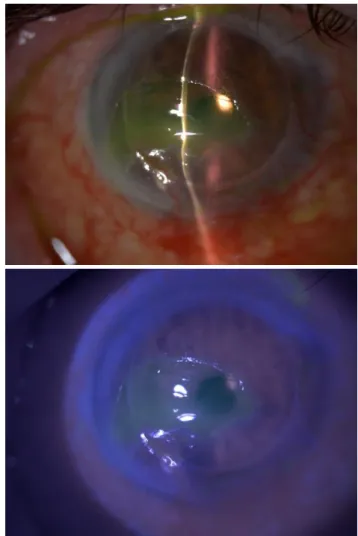
Two months later, in a medical appointment at the hospital, the patient mentioned a burning sensation and reddening in the RE, for 6 weeks. The BCVA was counting fingers. Slit lamp examination revealed an intense ciliary hyperemia with an extensive central corneal ulcer of 8 mm width by 4 mm height, without infection signs. A significant thinning of central stroma was seen, but Seidel test was negative (figure 2). Anterior chamber depth was preserved, without cells or flare. Treatment was immediately started with ofloxacin eye drops, ocular lubricants hourly and permanent ocular occlusion.
Figure 2 - a) Neurotrophic and exposure corneal ulcer. The large epithelial defect, with positive fluorescein staining (b), and thinning of central stroma are seen.
In subsequent visits the null effect of the treatment could be evidenced due to progression of the ulcer both in area and in depth, with an imminent risk of perforation. Considering the condition, it was decided to carry out treatment with RGTA [Poli (carboximetilglucose) sulfate, dextrano T40]. Before starting this treatment, a gently mechanical debridement of ulcer edges loose epithelium was performed with a cotton swab. RGTA eye drops were instilled in the morning, as the first eye drop, every 3 days, and the treatment was stopped after five applications.
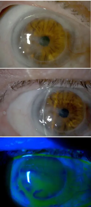
After the treatment, a complete reepithelization and partial stromal filling was observed. The risk of perforation was at this time minimal (figure 3). Seidel’s test remained negative.
In the following weeks, an intensive facial physiotherapeutic program was undertaken, and the palpebral function progressively recovered.
Figure 3 - a and b) Neurotrophic and exposure corneal ulcer after RGTA treatment. Complete epithelium healing, above a thin stromal layer. c) Although a pooling of fluorescein is seen, corneal staining is not observed.
At present, although the BCVA remains counting fingers, complete epithelization of the lesion with secondary corneal scarring and a total closure of eyelids can be seen. There are no signs of active inflammation.
In the control optical coherence tomography (OCT, figure 4), after 9 months, corneal epithelial layer is complete and a small stromal thickness increase is observed.
Figure 4 - OCT showing a complete epithelial layer over the stromal thinning area
Continued monitoring and treatment with artificial tears is maintained while waiting for a second penetrating keratoplasty, which may improve patient visual acuity.
DISCUSSION
The patient in this clinical case presents at least three causes that may have contributed to corneal ulceration: loss of corneal sensory innervations due to penetrating keratoplasty, previous corneal hypoesthesia due to herpetic corneal infection and exposure keratopathy caused by facial palsy.
These multiple etiology ulcers have devastating effects on the cornea, requiring a challenging approach. Moreover, the severity of stromal thinning in this patient, imposing an imminent risk of perforation, demanded an urgent “eye saving” measure.
At the moment, the efficacy of the medical treatments available for these situations are very limited. The treatment with intense lubrification and ocular occlusion was ineffective in this case. Autologous serum has shown to be effective in these cases.5 However, this treatment was not used in this case because it requires a weekly blood sampling and a specific preparation procedure, making it an expensive and laborious treatment.
The imminent risk of perforation, with the possibility of losing the eye, almost required an urgent "eye-saving" surgical procedure (tarsorrhaphy or amniotic membrane). In this case, tarsorrhaphy would have been a valid option as this surgery is suited to cases of combined V and VII cranial nerve paresis. Nevertheless, this procedure has been criticized for being cosmetically poor, often ineffective, and because of being associated to decreased peripheral vision.6 So, it was decided not perform it as an initial approach. Another valid option could have been amniotic membrane transplantation. This treatment has
been used efficiently for treating corneal ulcers and limbal stem cell defects. However, the heterologous origin of the amniotic membrane could be a significant problem, particularly in what concerns immune reactions and baseline morphology.7
A penetrating keratoplasty, even if associated with amniotic membrane transplantation or tarsorrhaphy, would be associated to a great risk of graft failure, due to the active inflammatory process. Thus, it was considered the possibility of using a safe and efficient drug-based treatment, to delay the need for this surgical intervention. With topical administration of RGTA, although the patient visual acuity couldn´t be restored, it was possible to resolve corneal ulcer and save the eye without any surgical intervention. The patient is now waiting for a penetrating keratoplasty, with a stable eye condition, having a much greater chance of a successful corneal transplant.
CONCLUSION
Neurotrophic and exposure corneal ulcers, particularly in patients with total corneal anesthesia, are among the most difficult ophthalmological conditions to treat, and may potentially result in blindness. In the absence of healing, they may progress towards corneal perforation or total de novo vascularization. Perforation is the final outcome of a process of cellular degradation resulting from chronic inflammation.8
There is an unmet need for a medical therapy that could help corneal neurotrophic ulcers to heal. The presented clinical case shows that the approach of targeting extracellular matrix can be effective in the reepithelialization of neurotrophic and corneal exposure ulcers that do not respond to usual treatments. As RGTAs contain no component of animal or biological origin, they are safe in terms of sterility and manufacturing origin. The efficiency and safety of this medical treatment make this approach especially appealing.
REFERENCES
1. Bonini S, Rama P, Olzi D, Lambiase A. Neurotrophic keratitis. Eye. 2003;17:989-95.
2. Sigelman S, Friedenwald JS. Mitotic and wound-healing activities of the corneal epithelium: effect of sensory denervation. Arch Ophthalmol 1954;52:46-57.
3. Rahman I., Sadiq S., Ophthalmic Management of Facial Nerve Palsy: A Review, Survey of ophthalmology, Volume 52, Number 2, 2007
4. Barritault D, Caruelle JP. Regenerating agents (RGTAs): a new therapeutic approach. Ann Pharm Fr. 2006 Mar;64(2):135-44.
5. Quinto GG, Campos M, Behrens A. Autologous serum for ocular surface diseases. Arq Bras Oftalmol. 2008;71:47–54.
6. Perusse P: Localized distichiasis after tarsorrhaphy. Am J Ophthalmol 114:104--5, 1992
7. Tsai RJ, Tseng SC. Human allograft limbal transplantation of corneal surface reconstruction. Cornea. 1994;13:389-400.
8. Mackie IA. Neuroparalytic keratitis. In: Fraunfelder FT, Roy FH, eds. Current ocular therapy 4. Philadelphia: W.B. Saunders, 1995:506
CONTACTO
Inês Carneiro Fernando Namora n°11 4470 289 Maia E-mail: inesmaria1215@hotmail.comThe author and co-authors do not have a financial interest/arrangement or affiliation with one or more organizations which could be perceived as a real or apparent conflict of interest in the context of the subject of this presentation.