UNIVERSIDADE DE LISBOA
FACULDADE DE CIÊNCIAS
DEPARTAMENTO DE BIOLOGIA VEGETAL
E
COLOGY OF
C
ONJUGATIVE
P
LASMIDS
Bernardino Machado Moreira
MESTRADO EM MICROBIOLOGIA APLICADA
2011
UNIVERSIDADE DE LISBOA
FACULDADE DE CIÊNCIAS
DEPARTAMENTO DE BIOLOGIA VEGETAL
E
COLOGY OF
C
ONJUGATIVE
P
LASMIDS
Dissertação para obtenção de grau de Mestre orientada por
Professor Doutor Francisco Dionísio, Centro de Biologia Ambiental
Professora Doutora Ana Maria Reis, Faculdade de Ciências da Universidade de
Lisboa
Bernardino Machado Moreira
MESTRADO EM MICROBIOLOGIA APLICADA
2011
E
COLOGY OF
C
ONJUGATIVE
P
LASMIDS
Bernardino Machado Moreira
2011
This thesis was fully performed at Center for Environmental Biology
(CBA - FCUL) under the direct supervision of Prof. Dr. Francisco
Dionísio and Prof. Dr. Ana Maria Reis in the scope of the Master in
Applied Microbiology of the Faculty of Sciences of the University of
1
ACKNOWLEDGEMENTS
I am very grateful to:
Francisco Dionísio, for giving me the opportunity to work on his group, as well as participating in highly interesting, motivating and challenging research projects. For his effort as my mentor, for guiding me all along this project, always facing me with new ideas and problematics, which allowed for this work to be fulfilled with success.
And to:
Ana Maria Reis, for her constant support and availability through the entire project, helping me fulfill all my objectives.
I am also grateful to:
Luís Carvalho and Silvia Mendonça, for providing me fundamental support, either in technical aspects or in data analysis, which proved to be of extreme importance through the entire project.
João Gama, Iolanda Domingues and Ana Marta de Matos, for being much more than just laboratory colleagues, being present for all the good and bad moments during this year.
I would also like to thank:
Inês Matias Reis, my girlfriend, for providing unconditional support ant patience, for being available to help in any way possible, and for brightening my days even when things were hard. I would like to express how important she was during all this, and how much I love her.
Her parents, that helped me in everything they could through the most difficult times.
My parents, that did everything they could and far beyond, so that I could finish this work without troubles.
To all my friends at Residência Universitária Ribeiro Santos, which provided me a funny and healthy environment to live in, and also supported me whenever I needed.
2
ABSTRACT
Bacterial conjugation is an important process which allows for the transference of plasmids between different bacteria. Plasmids may be useful for the bacteria in the way they carry genes that code for antibiotic resistance and other beneficial traits. Previous work has showed a great diversity of conjugation efficiency for several strains of Escherichia coli with the plasmid R1. We studied the conjugation efficiency of five plasmids (R16a, R702, RP4, R124 and R477-1) belonging to five different incompatibility groups. We observed a great diversity of conjugation efficiency, depending on the donor strain, on the recipient strain and on the plasmid to be transferred. Moreover, we observed that plasmids present in a donor strain influence the conjugation efficiency of a newly inserted plasmid.
Other studies showed that, although plasmids can be advantageous under certain conditions, they normally impose a cost to the host. That cost can be assumed as the plasmid’s virulence and it can evolve accordingly with models of evolution of virulence. It has been shown that the cost of harboring a plasmid can be minimized by means of co-evolution. Also, it can be influenced by epistatic interactions with the host’s genome or other plasmids. We observed that R16a didn’t impose fitness cost to any host, that R477-1 conferred and advantage to one donor strain and cost to other two strains. We could not pick a model of virulence evolution (trade-off hypothesis or within host competition hypothesis), to explain the different fitness values obtained. Epistatic interactions between plasmids and bacterial genome or other plasmids may also have influenced the fitness values we obtained. We also confirmed that through co-evolution, the cost for an E.coli strain to carry the plasmid R477-1 was compensated, and that this fitness change didn’t have a direct relationship with the conjugation efficiency for this plasmid-donor strain dyad.
3
RESUMO
A resistência a antibióticos constitui um grande desafio para a medicina moderna (Martinez et al, 2009). A aquisição de resistência a antibióticos pode resultar de mutações espontâneas em genes cromossomais, ou de eventos de transferência horizontal de genes que codifiquem para resistência a antibióticos.
A transferência horizontal de genes (THG) compreende processos que permitem a troca de genes entre dois organismos para além da herança de material genético da célula-mãe para a célula-filha. É um fenómeno de grande importância em termos da evolução de procariotas (Andam et al, 2010), que permite o surgimento de grande variabilidade e inovação metabólica ao nível dos organismos procariotas. A THG ganha assim importância em termos médicos, uma vez que é um grande veículo de disseminação de resistência a antibióticos.
Os principais processos de THG são a transformação, a transdução e a conjugação. A transformação consiste na absorção de ADN livre nas proximidades do microrganismo (Brigulla & Wackernagel, 2010). A transdução consiste no transporte de genes entre bactérias através de um bacteriófago (Skippington & Ragan, 2011). A conjugação consiste na transferência de plasmídeos entre bactérias em contacto estrito.
Os plasmídeos são elementos genéticos com replicação independente da do cromossoma da célula hospedeira. Estes não apresentam forma extracelular e, consequentemente, têm que ser transmitidos entre hospedeiros. Esta transferência pode dar-se aquando da divisão celular (transferência vertical) ou por um processo replicativo designado por conjugação (Willetts & Wilkins, 1984). O processo de conjugação envolve proteínas responsáveis pelo contacto célula a célula, e pela transferência do plasmídeo através das membranas celulares (Christie et al, 2005; Silverman, 1997). Uma bactéria pode albergar vários plasmídeos. Estes são classificados em grupos de incompatibilidade. Dois plasmídeos dizem-se incompatíveis sempre que, após a sua coexistência num mesmo hospedeiro, um deles não é mantido após a divisão celular, devido a interferência nos sistemas de controlo da replicação dos plasmídeos (Novick, 1987; Petersen, 2011). Sistemas de repressão da conjugação, tais como a exclusão de superfície (Frost et al, 1994) ou sistemas de restrição do hospedeiro (Jeltsch, 2003) podem funcionar como barreiras à conjugação.
A replicação e manutenção dos plasmídeos, bem como a expressão dos genes que estes possam conter, quando em situações em que a bactéria não beneficia dos produtos desses genes, implica uma diminuição do fitness da célula hospedeira, que se verifica na diminuição da taxa de crescimento (Dahlberg & Chao, 2003; Dionisio et al, 2005; Lenski & Bouma, 1987; Modi & Adams, 1991). Este custo pode ser reduzido por estratégias, tais como a co-evolução entre o plasmídeo e o hospedeiro (Dahlberg & Chao, 2003; Dionisio et al, 2005; Lenski & Bouma, 1987; Modi & Adams, 1991), a redução do número de cópias do plasmídeo por célula
4
(Dionisio et al, 2005), ou a redução das taxas de conjugação por sistemas que reprimam a síntese do aparato proteico associado à conjugação (Dionisio et al, 2002; Haft et al, 2009). O custo imposto por um plasmídeo pode ser interpretado como virulência para a célula hospedeira. Existem diversas hipóteses propostas para a evolução da virulência. Uma delas propõe que a virulência evolui tendo em conta o balanço final entre transmissão infecciosa e o dano provocado ao hospedeiro (Alizon et al, 2009; Anderson & May, 1982). Considerando os plasmídeos, o custo que este impõe ao hospedeiro evoluiria no sentido de se encontrar um ponto óptimo entre transmissão infecciosa (conjugação) e o custo causado ao hospedeiro. Outra hipótese propõe que o custo evolui maioritariamente devido a competição de diferentes genótipos patogénicos dentro do mesmo hospedeiro (co-infecção ou superinfecção). Assim, diferentes plasmídeos num mesmo hospedeiro competiriam entre si, sendo seleccionados os plasmídeos com maior capacidade da usar os recursos celulares, ainda que isso leve a uma diminuição global do fitness do hospedeiro. Mais ainda, foi demonstrado que os plasmídeos podem interagir com mutações em genes cromossomais, ou com outros plasmídeos, levando a alterações do fitness global do hospedeiro. Essa interacção é denominada epistasia (Silva et al, 2011).
Assim, tendo em conta a importância da THG, e particularmente da conjugação, como veículo de transmissão de genes de resistência a antibióticos, é de interesse estudar a dinâmica da transferência de plasmídeos entre microrganismos comensais, para melhor se entender como é que estes microrganismos contribuem para o espalhamento de plasmídeos. Este trabalho pretendeu portanto, estudar a eficácia de conjugação de cinco plasmídeos com genes de resistência a antibióticos (R16a, R124, R702, RP4 e R477-1), utilizando quatro estirpes de
Escherichia coli como estirpes dadoras do plasmídeo, e as mesmas estirpes recipientes do
plasmídeo usadas por Dionisio et al (2002).
Obteve-se uma grande variabilidade de eficácia de conjugação. Os valores obtidos abrangeram oito ordens de grandeza. Verificámos que a melhor estirpe dadora é a estirpe E.coli M3, a pior dadora é a estirpe E. coli Vdg435. A estirpe E.coli M1412 é a estirpe com menor capacidade para receber qualquer um dos plasmídeos utilizados. Esta variabilidade é influenciada pela estirpe dadora do plasmídeo, pela estirpe que o vai receber, pelo plasmídeo em si, e pelas interacções entre estes três factores.
Considerando também que Dionisio et al (2002) observaram que, após tratamento de todas as estirpes dadoras para remover todos os plasmídeos (curar as bactérias), as suas taxas de conjugação para o plasmídeo R1 eram homogéneas, independentemente da diferença observada antes da remoção dos plasmídeos, averiguámos o conteúdo em termos de plasmídeos das estirpes dadoras escolhidas para este trabalho. Verificámos que a estirpe
E.coli Vdg435, contém, para além dos plasmídeos que introduzimos, outros plasmídeos. As
5
introduzimos neste estudo. Inferimos assim que a presença de outros plasmídeos para além dos que introduzimos nas estirpes dadoras influencia a sua capacidade dadora.
Vimos também que a introdução dos cinco plasmídeos nas estirpes dadoras influencia o fitness destas: a introdução do plasmídeo R16a não altera o fitness das estirpes dadoras; a introdução dos plasmídeos R124, RP4 e R702 induz uma diminuição do fitness da estirpe E.coli Vdg435; o plasmídeo R477-1 impõe um custo de fitness nas estirpes E.coli M3 e M4, e confere uma vantagem à estirpe E.coli Vdg435. Estas alterações podem ser explicadas recorrendo a modelos que expliquem a evolução da virulência – competição dentro do hospedeiro, ou balanço entre virulência e transmissão infecciosa – ainda que nenhum dos dois modelos explique a totalidade dos resultados. Outra explicação passa pela ocorrência de interacções entre os plasmídeos inseridos nas estirpes dadoras e mutações nos cromossomas destas que causem resistência a antibióticos, ou com plasmídeos já existentes nas estirpes dadoras – epistasia. Este fenómeno pode explicar alguns dos valores obtidos (nomeadamente a vantagem conferida pelo plasmídeo R477-1 à estirpe dadora E.coli Vdg435).
Com o intuito de verificar se o custo provocado por um plasmídeo, bem como a sua evolução, podem influenciar a eficiência de conjugação desse plasmídeo, e tendo em conta que esse custo pode ser diminuído por co-evolução entre o plasmídeo e o hospedeiro, (Dahlberg & Chao, 2003; Dionisio et al, 2005; Lenski & Bouma, 1987; Modi & Adams, 1991) evoluímos a estirpe E.coli M3 com o plasmídeo R477-1 por cerca de 658 gerações. Verificámos que, de facto, ocorreu uma compensação do custo imposto pelo plasmídeo. Fizemos medições das taxas de conjugação do plasmídeo evoluído na estirpe dadora evoluída e verificámos que, para metade das estirpes dadoras, a taxa de conjugação aumentou significativamente. Para as restantes estirpes receptoras, não houve alteração significativa. Pudemos então concluir que não existe uma relação directa entre o custo que determinado plasmídeo impõe ao seu hospedeiro e a sua eficiência de conjugação.
6
INDEX
ACKNOWLEDGEMENTS ... 1 ABSTRACT ... 2 RESUMO ... 3 I. INTRODUCTION ... 71. Conjugative plasmids and their role in horizontal gene transfer ... 7
1.1 Horizontal gene transfer ... 7
1.2 Plasmids ... 7
1.3 Mechanism of conjugation ... 7
1.4 Plasmid incompatibility ... 8
1.5 Barriers to conjugation... 9
2. Plasmids as a cost to the host ... 9
2.1 Plasmids are costly ... 9
2.2 Cost reducing strategies ... 9
2.3 Cost as virulence ... 10
2.4 Epistasis involving plasmids ... 11
II. METHODS ... 13
1. Donor strain constructing ... 13
2. Conjugation rates ... 14
3. Strain evolution ... 15
4. Fitness assay ... 16
5. Gel electrophoresis... 17
6. Culture media and supplements ... 18
III. RESULTS ... 19 1. Conjugation rates ... 19 2. Fitness assay ... 24 3. Evolution assay ... 25 4. Gel electrophoresis... 26 IV. DISCUSSION ... 28 V. CONCLUSION ... 32 VI. REFERENCES ... 34
7
I.
INTRODUCTION
1. Conjugative plasmids and their role in horizontal gene transfer
1.1 Horizontal gene transfer
The breakout and dissemination of antibiotic resistance constitute a big challenge to modern medicine (Martinez et al, 2009). The genetic basis of antibiotic resistance involves chromosomal genes, as well as genes located in mobile genetic elements, such as plasmids (Martinez et al, 2009). The acquisition of antibiotic resistance results from two processes: i) spontaneous mutations on chromosomal genes or ii) horizontal gene transfer. Horizontal gene transfer (HGT) is the outcome of a series of processes that permit the transference of genes between two organisms in a way other than inheritance from the mother cell (vertical transmission). It is an evolutionary process of high importance in the evolution of prokaryotes (Andam et al, 2010). This process allows for the creation of biological novelty in a quick way, instead of possibly taking billions of years to occur (Jain et al, 2003). HGT is widespread among bacteria and leads to metabolic innovation (Skippington & Ragan, 2011). It is remarkably important since it allows the transfer of genes across species boundaries (Bansal et al, 2011). The genetic material transferred can range from small gene fragments (Chan et al, 2009; Gogarten et al, 2002; Yap et al, 1999) to operons (Olendzenski et al, 2000) or even superoperons that encode for full metabolic pathways (Igarashi et al, 2001) Normally, the transferred DNA consists of small fragments, often the length of one or several genes. HGT can take place by one of three major mechanisms: transformation, which consists in the active uptake of free DNA by the receiving cell (Brigulla & Wackernagel, 2010); transduction, that takes place when a bacteriophage works as a vector for the transportation of DNA between different bacteria (Skippington & Ragan, 2011); or conjugation, which consists in the transference of plasmids between cells in close contact.
1.2 Plasmids
Plasmids are genetic elements whose replication is independent from the bacterial chromosome replication, even though they use cellular machinery to replicate. They do not have an extracellular form, existing only as free DNA, in circular conformation (typically, but linear plasmids have also been documented) inside the host cell. Plasmids are considered different from the host’s chromosome in the way that they only code for non-essential functions to the host, even if these functions can actually be quite useful to them (Novick, 1987). In the current time, thousands of plasmids are known, and about 300 naturally occurring plasmids are associated with Escherichia coli strains alone.
8
Conjugation is the main process for HGT (Norman et al, 2009). It is a replicative process in which the recipient cell (the cell who is going to receive the plasmid) gets one or several copies of the plasmid present in the donor cell (the original host with the plasmid) (Willetts & Wilkins, 1984). For conjugation to be possible, an intimate contact between the donor and the recipient cells must exist (Grohmann et al, 2003). For conjugation to occur, the plasmid must have an origin of transfer, oriT, that is necessary to the beginning and to the conclusion of the conjugation process. The rest of conjugation apparatus is responsible for all the necessary steps, from DNA processing to its transfer between cells (Llosa & de la Cruz, 2005) .
On a general basis, the conjugation process involves a mating pair formation system for cell aggregation coupled to a DNA transfer apparatus. The majority of the proteins involved in conjugation are encoded in the plasmid, in the tra region. This region includes the mpf (mating pair formation) and the dtr (DNA processing and transport) genes (Christie et al, 2005; Silverman, 1997). The mpf encodes for proteins that are involved in the bacterial pilus formation and assembly of a type IV secretion system (T4SS, which is a multiprotein organelle necessary for horizontal transfer through the membranes of gram-negative bacteria).The conjugation process begins when the pilus (anchored in the donor cell surface) binds to the surface of the recipient cell. The pilus then retracts, making a stable cell-to-cell contact possible. Then, there is a signal which triggers the mobilization of DNA itself (Gomis-Ruth & Coll, 2006).The dtr gene encodes proteins involved in the process of preparing the DNA strand to be transferred: the formation of the relaxosome (complex formed by the oriT, the relaxase and one or more accessory nicking proteins) and its recruitment to the membrane transport pore (Llosa & de la Cruz, 2005). The DNA strand to be transferred is nicked in a unique site in the oriT region, which is typically less than 500 bp in size. This reaction is strand specific and is performed by a plasmid encoded relaxase in a reaction that covalently links the protein to the 5' terminus of the transfer intermediate (Luzhetskyy et al, 2006). The relaxosome is plasmid specific, because each relaxase is specific for a given oriT (Llosa & de la Cruz, 2005). The nucleoprotein complex is then transported to the recipient cell via a T4SS, with the DNA being actively pumped into the recipient cell by the type IV coupling protein (T4CP) (Christie, 2004; Llosa et al, 2002).
Some natural plasmids are only able to be transferred when in presence of another conjugative plasmid. They are referred as mobilizable plasmids and only contain an oriT, and carry genes for a relaxase, and one or more nicking-accessory proteins (Garcillan-Barcia et al, 2009).
1.4 Plasmid incompatibility
Two plasmids are said to be incompatible if upon coexistence inside the same host, at least one of them is not transferred (without the occurrence of external selection) during bacterial replication (Novick, 1987). Two plasmids from the same incompatibility group cannot be transferred from the same host, as they cannot coexist in a stable way within the same cell, and one of the replicons will disappear by chance, during cell division. The replication units of plasmids from the same incompatibility group will be similar, undistinguishable for the replication
9
and partition apparatus of the host cell. This functional equivalence will cause the replication units to interact with each other, blocking the replication of (at least) one of the plasmids (Petersen, 2011). Plasmids from different incompatibility groups can coexist in the same host (Phan & Wain, 2008). Considering the bacteria from the family Enterobacteriaceae, about 30 different incompatibility groups of plasmids are known (Zavil'gel'skii, 2000).
1.5 Barriers to conjugation
There are some barriers to conjugation. Surface exclusion is a mechanism that creates an efficient barrier to the entrance of plasmids whose transfer apparatus genes are similar to those present in plasmids aready within the host. Concerning the F plasmid, surface exclusion works in two levels: the protein TraT changes the bacterial cell surface, lowering the affinity with the F
pillus. On the other hand, the protein TraS fixes itself to the inner membrane and prevents
foreign DNA uptake by the cell (Frost et al, 1994). This system, although not universal, is fairly common in Gram-negative and Gram-positive bacteria. Another barrier to conjugation consists in restriction processes. If any given plasmid is susceptible to the host’s restriction systems (e.g. if it possesses the target sequences for the restriction system’s enzymes), it’s DNA will be broken into pieces and the frequency of transconjugants of that plasmid will be lowered (Jeltsch, 2003). However, the reduced size of (some) plasmids, alongside with the fact that DNA enters the cell in single-strand form, make it less probable that the restriction system acts on them.
2. Plasmids as a cost to the host
2.1 Plasmids are costly
Plasmids encode for several functions, such as antibiotic resistance, the ability to respond to pheromones, resistance to heavy metals (Ji & Silver, 1992), resistance to ultraviolet light (Tomich et al, 1979), bacteriocin production and resistance (Chak & James, 1984), among other traits. These functions can be highly advantageous for their host. They can provide key advantage for their host in adapting to new environments (e.g. addition of a new antibiotic), when competing against bacteria without those traits. However, this advantage will only be noticeable under conditions that favor bacteria with that function. When there is no selective pressure, and that function is not essential to cell survival, the plasmid in question will represent a cost to the host cell (De Gelder et al, 2007). Plasmids normally represent a cost to their host (Dahlberg & Chao, 2003; Dionisio et al, 2005; Lenski & Bouma, 1987; Modi & Adams, 1991). This cost can be seen as the reduction of the growth rate of the host. Besides additional traits coded by the plasmid, the cost can be imposed by the regulation and replication of the plasmid itself, which consumes time and cellular resources. The plasmid replication and the expression of its genes can interfere with the normal bacterial activity, and this cost is specific for each bacteria-plasmid association. This cost can vary from being undetectably small to as much as 40% (De Gelder et al, 2007).
10
Plasmids impose a fitness cost on their host. However, this cost can be minimized. The relationship between a plasmid and its host tends to evolve so that the cost of bearing the plasmid is minimized (Haft et al, 2009). There are several ways this can be achieved:
i) co-evolution between the plasmid and its host, leading to a reduction of the cost of bearing the plasmid, that is achieved by the occurrence of compensatory mutations, either in the host and in the plasmid (Dahlberg & Chao, 2003; Dionisio et al, 2005; Lenski & Bouma, 1987; Modi & Adams, 1991);
ii) reduction of the number of plasmids per cell that is, the plasmid copy number (Dionisio et al, 2005). A lower copy number would mean less metabolic cost due to plasmid replication, hence lowering the cost imposed by the plasmid on the host;
iii) reduction of the conjugation rates. In a population of plasmid-bearing cells, a plasmid with a lower rate of conjugation will be advantageous to the host, who will be able to replicate faster, hence having an advantage over the rest of the population. This reduction can be achieved by using a cis-encoded fertility inhibition system, fin, which inhibits the synthesis and assembly of the conjugation apparatus. This system has been described in plasmids from several incompatibility groups (F, I, X) and it can reduce conjugation of plasmid R1 1000 fold (Dionisio et al, 2002). Also for IncP plasmids, there are genes that repress the conjugation apparatus and, consequently, hinder their conjugation (Haft et al, 2009). It should be noted that under competition conditions, this evolution/minimization of the cost can be fastened (Dionisio et al, 2005).
2.3 Cost as virulence
Bacterial cells can harbor more than one plasmid, as long as they don’t belong to the same incompatibility group. Considering this, all the plasmids should interact amongst themselves, and the final outcome of this interaction will make the bacterial fitness increase or decrease. One can assume the cost imposed by a plasmid on the host cell to be the plasmid’s virulence (Smith, 2011). Several hypotheses have been proposed to explain evolution of virulence. The most prominent states that virulence will be determined mainly by the trade-off with infectious transmission (Alizon et al, 2009; Anderson & May, 1982). According to this hypothesis, the virulence caused by a pathogen would be an unavoidable side effect of the traits that increased its infectious transfer. An increase in virulence would limit the infection duration, either by causing the host to die or change its behavior, or by stimulating its immune system to clear the pathogen. Selection would favor pathogens which could better balance the damage caused to the host and its infectious transmission rates, in order to maximize the number of new infections caused by a single infected individual, on a totally susceptible population (Smith, 2011). Taking plasmids into account, one should expect that, accordingly with this hypothesis, plasmids with the best balance between virulence (as cost imposed to the host) and infectious transmission (conjugation efficiency), are selected in detriment of other
11
plasmids. In this way, the conjugation efficiency of a plasmid would evolve in order to find the better balance with the cost it imposed to the host, in order to maximize its transmission without impairing its host’s fitness too much. This hypothesis is also supported by the evidence that plasmids tend to reduce the cost they impose on their host by several generations of co-evolution (Dahlberg & Chao, 2003; Dionisio et al, 2005; Lenski & Bouma, 1987; Modi & Adams, 1991).
On the other hand, another approach to this problematic suggests virulence would be determined by competition among pathogens within the same host. This competition would take place whenever either a host is initially infected by multiple genotypes from the pathogen, or superinfection (secondary infection) takes place. Different genotypes (different pathogens) would then compete within the host, and the result of this competition would produce changes in the virulence of each pathogen. This competition could then lead to a higher cost, as competition for resources among the host would select for more virulent pathogens (Levin & Pimentel, 1981; Nowak & May, 1994). Considering plasmid virulence, the within host competition hypothesis would make sense in the cases which a bacterial cell hosts more than one plasmid. Bacteria could harbor more than one plasmid, as long as they are not part of the same incompatibility group. These plasmids could compete against each other, leading to an increase in terms of cost to the host, by (for instance) selecting plasmids with a quicker within-host replication. These plasmids would have an advantage, although proving more costly to their host. Also, as mentioned before, plasmids can prevent “superinfection” (the entry of another plasmid of the same incompatibility group), but this reduction isn’t absolute. Being so, superinfection with a different, more virulent form of a plasmid could take place, leading to within host competition, and, as said before, selection of the more virulent plasmid, although causing higher cost to the host.
2.4 Epistasis involving plasmids
Chromosomal mutations have been shown to impose a cost on the bacterial cell (Bjorkman et al, 1998). If that bacterium also hosts a plasmid, which imposes a cost to the host, one should expect that the final outcome in terms of fitness for that cell would be the sum of the costs of both the mutation and the plasmid. However, this final outcome may turn not to be just the sum of the plasmid and mutation influence. Silva et al (2011) has demonstrated that the interaction between antibiotic resistance mutations and conjugative plasmids (which also coded for antibiotic resistance) can result either in the reduction of the global fitness cost (that is, the global outcome is less costly than the sum of the two individual fitness costs) or also in the increase of global fitness cost. Remarkably, in some cases, the fitness reduction was so notorious that the strain carrying both resistance determinants was fitter than the strain carrying either the plasmid or the chromosomal mutation (Silva et al, 2011). This genetic interaction is called epistasis (Phillips, 2008). If we, for instance, consider antibiotic resistance determinants (deleterious if not in the presence of the respective antibiotic), epistasis can have two possible outcomes. A positive epistatic interaction would reduce the global fitness cost of harboring both
12
resistance determinants. In the case that this reduction is so pronounced that the actual fitness of the host is higher than the fitness of the host carrying the plasmid or the mutation, this interaction is named sign epistasis. On the other hand, if carrying both resistance determinants results in a bacterium less fit than the sum of the individual costs of each determinant, we are in presence of negative epistasis. Interestingly, this interaction was also shown to happen between two different plasmids inside the same bacterial cell. This interaction can prove to be either antagonistic or synergistic (Silva et al, 2011), and certainly plays an important role in the evolution and maintenance of plasmids among their hosts.
Having said that horizontal gene transfer, namely conjugation, is responsible for the spread of antibiotic resistance among bacterial populations, it is of medical interest to study this process in order to manage and (try to) avoid the spread of resistance, and, therefore, the loss of efficiency of the common antimicrobial treatments. Research has been done, bearing in mind pathogenic bacteria, plasmids and horizontal gene transfer between and within bacterial species. Considering that bacteria live in complex communities, composed of several different species of bacteria (e.g. a human being would have more than 400 bacterial species in the intestinal tract), the role of non-pathogenic (or commensal) bacteria as an antibiotic resistance gene (plasmid) reservoir or donor is of high importance, and still needs to be studied and clarified. So this work aims to study the rates of conjugation of five different plasmids among commensal bacteria (different strains of E.coli) in order to understand the role of commensal bacteria in the spread of antibiotic resistance.
13
II.
METHODS
1. Donor strain constructing
All the recipient strains and each of the donor strains (one per turn) (Table I) were grown overnight (37ºC, with agitation) in Luria Bertani (LB) media supplied with antibiotics in order to select both donor and recipient strains.
Table I: Escherichia coli strains used to build the new donor strains (conjugation rate assay). (Nal: nalidixic acid; Nitro: nitrofurantoin; Rif: rifampicin).
Strain: Role: Chromosome – encoded resistance
Escherichia coli VDG 380 Recipient Nal; Nitro
Escherichia coli VDG 411 Recipient Nal; Nitro
Escherichia coli VDG 435 Recipient Nal; Nitro
Escherichia coli M3 Recipient Nal; Nitro
Escherichia coli M4 Recipient Nal; Nitro
Escherichia coli M1412 Recipient Nal; Nitro
Escherichia coli C4705 Recipient Nal; Nitro
Escherichia coli C4720 Recipient Nal; Nitro
Escherichia coli C4734 Recipient Nal; Nitro
Escherichia coli K12 J35 Donor (RP4) Nal
Escherichia coli K12 J35-1 Donor (R702) Nal
Escherichia coli Donor (R16a) Rif
Escherichia coli WPS2 Donor (R124)
Escherichia coli WPS2 Donor (R477-1)
Then, cultures were transferred to fresh LB media, mixing 100 µL of each recipient strain with the donor strain, in a falcon tube. Negative controls were performed to ensure that there were no spontaneous mutant clones that could look like transconjugant bacteria. The mix was grown overnight at 37ºC (no agitation). Each mixture was then grown in LB media with agar (LA) supplied with antibiotics: in order to select transconjugant bacteria, a resistance marker from the recipient strain and one or more resistance markers from each plasmid were used. A clone from each transconjugant was picked and grown on LA supplied with the respective antibiotics, in order to purify and isolate each transconjugant (this step was performed two times). After purification, each transconjugant culture was stored at -20ºC (with 15% glycerol) in order to save stocks for further use.
This procedure was done using one donor strain carrying each plasmid and all recipient strains per round, in order to build new donor strains with each one of the five plasmids (Table II).
14
Table II: Plasmids introduced in the recipient strains to build the new donor strains to be used in further experiments (Amp: ampicillin, Tet: tetracycline; Kan: kanamycin; Sm: streptomycin; Su: sulphonamides).
Plasmid Incompatibility group Resistance
RP4 IncP1 Amp; Tet; Kan
R702 IncP Sm; Su; Tet; Kan
R16a IncA/C Amp; Su; Kan
R124 IncF IV Tet
R477-1 IncS Sm; Tet;
2. Conjugation rates
From each set of built donor strains (each plasmid [Table II]), only four strains were used (table III), according to the results obtained in previous work (Dionisio et al, 2002).
Table III: Escherichia coli strains used as plasmid donor and recipient in the conjugation rate assay (Nal: nalidixic acid; Nitro: nitrofurantoin; Rif: rifampicin; Fos: phosphomycin).
Strain: Role: Chromosome - encoded resistance
Escherichia coli VDG 435 Donor Nal; Nitro
Escherichia coli M3 Donor Nal; Nitro
Escherichia coli M4 Donor Nal; Nitro
Escherichia coli C4705 Donor Nal; Nitro
Escherichia coli VDG 380 Recipient Rif; Fos
Escherichia coli VDG 411 Recipient Rif; Fos
Escherichia coli VDG 435 Recipient Rif; Fos
Escherichia coli M3 Recipient Rif; Fos
Escherichia coli M4 Recipient Rif; Fos
Escherichia coli M1412 Recipient Rif; Fos
Escherichia coli C4705 Recipient Rif; Fos
Escherichia coli C4720 Recipient Rif; Fos
Escherichia coli C4734 Recipient Rif; Fos
Each donor strain and all recipient strains (Table III) were grown separately, overnight, with agitation, at 37ºC, in LB media supplied with the antibiotics necessary to select only for the donor or recipient strain, in each case. Then, each strain was grown in fresh LB media (previously heated to 37ºC) at 37ºC with agitation. Optical density for each culture was controlled (λ = 670 nm). Each culture was kept in ice once their optical density reached a value between 0.7 and 0.8. Then, 1 mL of the donor strain was mixed with 1 mL of each recipient
15
strain. Each mixture was filtered through a membrane with pores with diameter of 45 nm. Each membrane was then placed in LA media, and incubated for 100 min at 37ºC. Then, each membrane was transferred to a falcon tube with 5 mL of MgSO4 10
-2
M. Each tube with the membrane was vigorously agitated in order to release the cells from the membrane, and kept in ice so that conjugation would not go on. Serial dilutions were made from each tube with each membrane, and were plated in LA supplied with antibiotics (one or more resistance markers from the recipient strains and one or more markers from each plasmid to select for transconjugants, two resistance markers from the recipient strains to select for recipient bacteria and one or more resistance markers from each donor strain and one or more resistance markers from each plasmid to select for donor strains).
Conjugation rate (y) between each donor strain and recipient strains was then determined using the equation:
√
In this equation T is the number of transconjugant cfu, D is the number of donor cfu and R is the number of recipient cfu.
A variation of this assay was performed with the plasmid R477-1, in order to obtain higher transconjugant bacteria count. Everything was performed as described above, except for: the optical density at which each donor and recipient strain were used; the amount of donor and recipient bacteria mixed and filtered; and the incubation period of the membrane. All strains were grown to an optical density of 0,8 – 0.9 (λ = 670 nm). Then, 1.5 mL of each donor and recipient strains were mixed and filtered through a membrane with 45 nm diameter pores. Each membrane was incubated for 20 h in one LA plate at 37ºC. We made these changes in order to obtain measurable values, since the original protocol wouldn’t be sensitive enough to show lower conjugation rates. However, since the conjugation rate is calculated as a growth proportion between transconjugant and donor and recipient bacteria, the values obtained using this variation can be compared with the conjugation rates measured with the regular procedure.
3. Strain evolution
The E.coli strain M3 carrying the plasmid R477-1 was grown (37ºC, 170 rpm) in fresh LB media (10 mL in a falcon tube) with Streptomycin, Tetracycline and Nalidixic Acid. 5 µL of the culture were transferred to a new falcon tube with 10 mL of fresh LB media and the referred antibiotics. The bacteria were grown at 37ºC, with shaking (170 rpm). This step was repeated every 12 h,
16
for 30 days, corresponding to log2[10.000 µL/5 µL] * 30 days * 2 steps/day = 10.97*60 ≃ 658
generations. After each transfer, a portion of the grown culture was kept in order to save stocks of each evolution time.
4. Fitness assay
To estimate the fitness of each plasmid-bearing strain, we performed competition assays. All the donor strains (Table III), carrying each one of the five plasmids (Table II), the evolved strain, as well as the strains used as donor strains lacking the plasmids were competed against a reference strain, E.coli MG1655 Δara. Firstly, each of the strains to be tested, along with the reference strain, were grown in fresh LB media (37ºC, 170 rpm, overnight) with the necessary antibiotics to select for the desired strain (a chromosomal-encoded resistance and a plasmid-encoded resistance, except for the strains lacking plasmids, with which we only used a chromosomal-encoded resistance, and the reference strain, which was grown in the absence of antibiotics). 50 µL of each culture were then transferred to new fresh LB media, without antibiotics. Bacteria were then grown at 37ºC, overnight at 170 rpm. A mixture (1:1) of both the strain to be tested and the reference strain was then diluted in MgSO4 10
-2
M. 100 µL of the 10-4 dilution were then added to a falcon tube with 10 mL of fresh LB media, which was incubated for 24h at 37ºC with vigorous agitation (220 rpm) in order to prevent conjugation. From the original mixture, the appropriate dilutions were plated on TA agar supplemented with arabinose and Tetrazolium (TTZ) to determine the initial ratio of the strains. After the competition, the appropriate dilutions of the mixed culture were plated on TA agar supplemented with arabinose and TTZ, to obtain the final ratio for each competition.
The fitness of each strain was obtained as an indirect measure, considering the relative fitness of the strain to be tested and the reference strain. Let P, A and R be the strain with the plasmid, the strain without the plasmid and the reference strain, respectively. The cost that each plasmid confers to its host can be measured as a fitness ratio between strains P and A. The lower the fitness value, the higher is the cost of carrying the plasmid.
Considering this, the fitness of the strains P and A must be calculated. These values can be obtained as a measure of relative fitness (dimensionless quality that is calculated as the ratio of the growth rate of one strain relative to the cost of other strain, during a direct competition).
and
In which Pf, Af and Rf stand for the final frequencies of the strain with the plasmid, the strain
without the plasmid and the reference strain, respectively, and P0, A0, and R0 stand for the initial
frequencies of those strains. The cost caused to a strain by a plasmid can then be inferred as a simple ratio between WP and WA.
17
5. Gel electrophoresis
Strains (Table IV) were grown in fresh LB media (37ºC, 170 rpm, overnight) with the appropriate antibiotics to select for the respective plasmids, in order to achieve a high bacterial density. Table IV: Escherichia coli strains used in the electrophoresis performed to access the plasmid content of the strains used as donors, before and after inserting the plasmid used in the conjugation rate assay (Nal: nalidixic acid; Nitro: nitrofurantoin; Kan: kanamycin; Tet: tetracyclin).
Strain: Plasmid: Chromosome -
encoded resistance
Plasmid-encoded resistance
Escherichia coli VDG 435 None Nal; Nitro
Escherichia coli M3 None Nal; Nitro
Escherichia coli M4 None Nal; Nitro
Escherichia coli C4705 None Nal; Nitro
Escherichia coli VDG 435 R702 Nal; Nitro Kan; Tet
Escherichia coli M3 R702 Nal; Nitro Kan; Tet
Escherichia coli M4 R702 Nal; Nitro Kan; Tet
Escherichia coli C4705 R702 Nal; Nitro Kan; Tet
Escherichia coli K12 R702 Nal; Nitro Kan; Tet
Escherichia coli VDG 435 RP4 Nal; Nitro Kan; Tet
Escherichia coli M3 RP4 Nal; Nitro Kan; Tet
Escherichia coli M4 RP4 Nal; Nitro Kan; Tet
Escherichia coli C4705 RP4 Nal; Nitro Kan; Tet
Escherichia coli K12 RP4 Nal; Nitro Kan; Tet
1,5 mL of each culture was then centrifuged (8000 rpm, 2 min), resuspending the remaining pellet in 500µL of sucrose/tris/EDTA (STE) solution (the supernatant was rejected). The mixture was then centrifuged (8000 rpm, 2 min, rejecting the supernatant) and the pellet was resuspended with 100µL of glucose/tris/EDTA (GTE) solution supplemented with lysozyme (10 mg/mL) and RNAseA (5 mg/mL). 200µL of NaOH-SDS were added to each mixture, mixing by inversion. Each mixture was then placed in ice for 5 min. After that, we added 150µL of KAc to each mixture, mixing by inversion. The mixtures were then placed in ice for 5 min. Subsequently, we centrifuged the mixtures (17000 rpm, 10 min), keeping the supernatant and rejecting the pellet. We added 300µL of Isopropanol to each sample, mixing with the vortex. After 2 min, we centrifuged the samples (17000 rpm, 10 min), rejecting the supernatant. We then precipitated the DNA by adding 1 mL of ethanol (70% v/v) to each sample, and centrifuged the samples (8000 rpm, 2 min). We rejected the supernatant and left each sample at room temperature (each tube opened) in order to evaporate the remaining ethanol. We then
18
resuspended the pellet in 50µL of TE supplemented with RNAse (20 µg/mL) (Birnboim, 1983; Birnboim & Doly, 1979; Vinograd & Lebowitz, 1966).
The samples were placed at 37ºC for about 1 h, to homogenize each sample. We analyzed the samples by agarose (0,7% w/v) gel electrophoresis, running the samples at 100 V for 90 min. We visualized the gel by UV light after staining with GelRed and photographed it recurring to a transiluminator and a MicroDOC.
6. Culture media and supplements
All bacterial strains were grown in liquid Luria-Bertani (LB) medium at 37ºC with agitation. Solid media (LA) was obtained by the addition of agar (15 g/L). For growth and transconjugant selection, antibiotics were added as follows: 40 µg/mL of nalidixic acid, 100 µg/mL of rifampicin, 100 µg/mL of streptomycin, 20 µg/mL of tetracycline, 100 µg/mL of kanamycin, 100 µg/mL of ampicillin, 12 µg/mL of nitrofurantoin, 30 µg/mL of phosphomycin and 100 µg/mL of streptomycin. For the fitness assay, TA (tetrazolium arabinose) agar (15 g/L) were used. These plates were supplemented with arabinose (4 g/L) and tetrazolium.
19
III.
RESULTS
1. Conjugation rates
In this work, we aimed to measure conjugation rates of five different plasmids (R16a, R124, R702, RP4 and R477-1) using different E.coli strains either as donor or recipient strains. Dionisio et al (2002) have shown that E.coli Vdg435 is a very inefficient donor, M4 is a very good donor and M3 and C4705 were average donors of the plasmid R1. Therefore, we chose these four strains so that we could have a wide range of conjugation rates, in order to spot the influence of the donor strains more easily. The nine strains used as recipient strains (E.coli Vdg380, E.coli Vdg411, E.coli Vdg435, E.coli M3, E.coli M4, E.coli M1412, E.coli C4705, E.coli C4720 e E.coli C4734) were the same that were used by Dionisio et al (2002).
We have a wide range of conjugation rates (Tables V, VI, VII, VIII and IX). We noticed that one donor strain can be either good or bad donor, depending on the recipient strain. We also noticed that generally, the self-conjugation rates are not higher than the conjugation rates between different strains.
Table V: Log10 of the conjugation rates for the plasmid R16a (mean of three measurements). On
bold, values for self-transfer rate (conjugation between the same strain, as donor and recipient). Mean Donor Ability (MDA) and Mean Recipient Ability (MRA) were calculated using arithmetic average. SE stands for standard error.
Donor Recipient Vdg435 SE M3 SE M4 SE C4705 SE MRA Vdg380 -1,92 0,12 -1,32 0,22 -1,86 0,21 -1,46 0,11 -1,64 Vdg411 -2,54 0,35 -2,38 0,14 -2,72 0,16 -1,71 0,11 -2,34 Vdg435 -2,02 0,10 -2,20 0,09 -2,55 0,08 -4,02 0,55 -2,70 M3 -3,11 0,29 -2,02 0,15 -2,91 0,35 -1,51 0,22 -2,39 M4 -2,99 0,33 -2,30 0,23 -2,58 0,29 -1,55 0,11 -2,36 M1412 -6,67 0,20 -6,21 0,26 -7,26 0,16 -4,65 0,28 -6,20 C4705 -2,29 0,11 -1,47 0,18 -2,28 0,08 -1,23 0,13 -1,82 C4720 -2,84 0,10 -1,79 0,09 -2,57 0,03 -2,04 0,13 -2,31 C4734 -3,86 0,22 -2,32 0,11 -3,42 0,18 -3,15 0,09 -3,18 MDA -3,14 -2,45 -3,13 -2,37
20
Table VI: Log10 of the conjugation rates for the plasmid R124 (mean of three measurements).
On bold, values for self-transfer rate (conjugation between the same strain, as donor and recipient). Mean Donor Ability (MDA) and Mean Recipient Ability (MRA) were calculated using arithmetic average. SE stands for standard error.
Donor Recipient Vdg435 SE M3 SE M4 SE C4705 SE MRA Vdg380 -6,35 0,07 0,33 0,04 -0,36 0,16 -0,34 0,23 -1,68 Vdg411 -6,64 0,23 -0,43 0,02 -0,38 0,08 -0,38 0,18 -1,96 Vdg435 -5,53 0,11 -0,35 0,05 -0,44 0,10 -0,39 0,08 -1,68 M3 -7,63 0,09 -0,43 0,05 -2,63 0,08 -2,17 0,08 -3,22 M4 -6,40 0,14 -0,74 0,06 -0,18 0,05 -0,10 0,07 -1,86 M1412 -6,76 0,27 -6,73 0,11 -5,65 0,06 -7,37 0,09 -6,63 C4705 -5,40 0,03 -0,44 0,12 -0,48 0,03 -0,13 0,06 -1,61 C4720 -7,77 0,15 -0,77 0,12 -0,18 0,04 -0,45 0,04 -2,29 C4734 -6,63 0,13 -0,42 0,08 -0,21 0,13 -0,02 0,04 -1,82 MDA -6,57 -1,11 -1,17 -1,26
Table VII: Log10 of the conjugation rates for the plasmid R702 (mean of three measurements).
On bold, values for self-transfer rate (conjugation between the same strain, as donor and recipient). Mean Donor Ability (MDA) and Mean Recipient Ability (MRA) were calculated using arithmetic average. SE stands for standard error.
Donor Recipient Vdg435 SE M3 SE M4 SE C4705 SE MRA Vdg380 -1,95 0,23 0,07 0,02 -0,32 0,04 -0,33 0,05 -0,63 Vdg411 -2,15 0,04 -0,06 0,03 -0,40 0,13 -0,41 0,01 -0,75 Vdg435 -1,98 0,05 -0,59 0,06 -0,54 0,06 -0,86 0,28 -0,99 M3 -5,20 0,03 -0,36 0,15 -2,75 0,08 -2,51 0,03 -2,71 M4 -2,15 0,06 -0,45 0,19 -0,17 0,02 -0,38 0,05 -0,79 M1412 -7,46 0,11 -6,31 0,34 -4,00 0,30 -6,67 0,10 -6,11 C4705 -1,48 0,18 -0,06 0,06 -0,26 0,04 -0,47 0,06 -0,57 C4720 -3,82 0,13 -0,75 0,04 -0,48 0,21 -0,43 0,05 -1,37 C4734 -2,93 0,04 -0,57 0,18 -0,78 0,02 -0,76 0,07 -1,26 MDA -3,23 -1,01 -1,08 -1,42
Table VIII: Log10 of the conjugation rates for the plasmid RP4 (mean of three measurements).
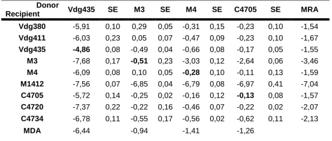
On bold, values for self-transfer rate (conjugation between the same strain, as donor and recipient). Mean Donor Ability (MDA) and Mean Recipient Ability (MRA) were calculated using arithmetic average. SE stands for standard error.
21 Donor Recipient Vdg435 SE M3 SE M4 SE C4705 SE MRA Vdg380 -5,91 0,10 0,29 0,05 -0,31 0,15 -0,23 0,10 -1,54 Vdg411 -6,03 0,23 0,05 0,07 -0,47 0,09 -0,23 0,10 -1,67 Vdg435 -4,86 0,08 -0,49 0,04 -0,66 0,08 -0,17 0,05 -1,55 M3 -7,68 0,17 -0,51 0,23 -3,03 0,12 -2,64 0,06 -3,46 M4 -6,09 0,08 0,10 0,05 -0,28 0,10 -0,11 0,13 -1,59 M1412 -7,56 0,07 -6,85 0,04 -6,79 0,08 -6,97 0,41 -7,04 C4705 -5,72 0,14 -0,25 0,02 -0,16 0,12 -0,13 0,08 -1,57 C4720 -7,37 0,22 -0,22 0,16 -0,46 0,07 -0,22 0,02 -2,07 C4734 -6,78 0,11 -0,55 0,17 -0,56 0,02 -0,62 0,11 -2,13 MDA -6,44 -0,94 -1,41 -1,26
Table IX: Log10 of the conjugation rates for the plasmid R477-1 (mean of three measurements).
On bold, values for self-transfer rate (conjugation between the same strain, as donor and recipient). Mean Donor Ability (MDA) and Mean Recipient Ability (MRA) were calculated using arithmetic average. SE stands for standard error.
Donor Recipient Vdg435 SE M3 SE M4 SE C4705 SE MRA Vdg380 -6,79 0,13 -6,11 0,36 -6,33 0,10 -7,64 0,46 -6,72 Vdg411 -7,75 0,06 -7,94 0,03 -7,36 0,04 -7,75 0,13 -7,70 Vdg435 -7,81 0,30 -6,86 0,13 -7,42 0,16 -6,51 0,07 -7,15 M3 -6,90 0,28 -8,18 0,08 -6,71 0,11 -6,14 0,02 -6,98 M4 -7,13 0,13 -7,94 0,18 -6,67 0,33 -6,91 0,16 -7,16 M1412 -8,31 0,09 -7,66 0,24 -7,38 0,19 -8,00 0,08 -7,84 C4705 -5,08 0,20 -4,30 0,27 -5,92 0,18 -7,71 0,22 -5,75 C4720 -6,12 0,06 -5,91 0,10 -5,86 0,52 -7,20 0,37 -6,27 C4734 -7,89 0,27 -6,17 0,21 -7,87 0,31 -6,34 0,14 -7,07 MDA -7,09 -6,79 -6,84 -7,13
The heterogeneity in conjugation rates depends on the donor strain, the recipient strain, and the plasmid, as well as the interactions between the three factors. (Table X).
The E.coli M3 strain is the one with higher donor ability, for all plasmids used (Figure 1). On the other hand, strain Vdg435 had the lowest donor ability amongst the four strains chosen. The
post hoc Tuckey test performed after the ANOVA test shows that strains M4 and C4705 have
no significant differences (p>0,05), which means that for a given plasmid and a given recipient strain, the conjugation rate achieved with both donor strains would be similar. Vdg435 and M3 strains were significantly different between themselves and from strains M4 and C4705, as well (p<0,001). These differences between the donor strains, even considering the fact that two strains are similar, contribute to the vast heterogeneity of conjugation rates obtained with this work.
22
Table X: three way ANOVA (donor strain, recipient strain and plasmid) performed with the values of the conjugation rates. D.f. stands for degrees of freedom, F accounts for the value of F test and p gives the level of significance.
Variation source D.f. F p
Donor strain 3 2857,45 0,00
Recipient strain 8 1417,22 0,00
Plasmid 4 5909,12 0,00
Donor strain*Recipient strain 24 40,41 0,00 Donor strain*Plasmid 12 580,74 0,00 Recipient strain*Plasmid 32 75,04 0,00 Donor strain*Recipient strain*Plasmid 96 23,98 0,00
Figure 1: Donor ability for the four E.coli strains used as donor strains, for the five plasmids. Each value was obtained as the average of the conjugation rates of each donor strain to the full set of recipient strains.
Bearing in mind the effect of the recipient strains, we could sort out differences that can help to explain the heterogeneity in the conjugation rates. The strain M1412 appears to have a poor recipient capability, for any of the given plasmids. The majority of the values obtained for this recipient strain were, in fact, given as the method’s detection limit, instead of being accurate conjugation rates (the transconjugant count was 0 colonies in those cases, so we considered
-8 -7 -6 -5 -4 -3 -2 -1 0 Vdg435 M3 M4 C4705 Log 10 Co n ju gation r ate
Donor E.coli strains
R16a R124 R702 RP4 R477-1
23
0,5 as the value to use). All the other strains had higher recipient capability. According the the
post hoc Tuckey test that we performed, almost all the strains had significantly different
recipient capabilities (p<0,05). However, the strain pairs E.coli Vdg411 and E.coli Vdg435, E.coli Vdg411 and E.coli C4720, E.coli Vdg411 and E.coli C4734, E.coli Vdg435 and E.coli M4, E.coli Vdg435 and E.coli C4720, E.coli M4 and E.coli C4720 had similar recipient capabilities for any of the given plasmids (p>0,05). In practice, this means that for each pair, any of the strains would receive a given plasmid from a given donor strain at a similar conjugation rate. Even considering these similarities, the recipient capabilities of the chosen strains are different enough to influence, in some way, the heterogeneity in the conjugation rates obtained (p<0.01, Table X).
We could also notice that the plasmid to be transferred can influence the conjugation rate itself (p<0.01, Table X). The plasmid R477-1 is conjugated at a very low rate (the lowest of the 5 plasmids, as seen on tables V to IX), as we could see by the low capability of all the donor strains to transfer the plasmid as well as the low capacity for all the recipient strains to receive this plasmid. The plasmid R16a also seemed to be transferred less easily than the other plasmids, excluding strain Vdg435 (Figure 1). In opposition, the plasmid R702 was the one to achieve the highest conjugation rates of the strains we tested. Plasmids RP4 and R124 showed similar conjugation patterns, either in donor or recipient capabilities (Figure 1 and 2).
Figure 2: Recipient ability for the nine E.coli strains used as recipient strains, for the five considered plasmids. Each value was obtained as the average of the conjugation rates of each recipient strain to the full set of donor strains.
A post hoc Tuckey test showed that, in fact, there is no significant difference between these two plasmids. This means that for any combination of donor and recipient strains, the conjugation
-9 -8 -7 -6 -5 -4 -3 -2 -1 0 Vdg380 Vdg411 Vdg435 M3 M4 M1412 C4705 C4720 C4734 Log 10 Co n ju gation r ate
Recipient E.coli strains
R16a R124 R702 RP4 R477-1
24
rate using one or the other plasmid would be similar. All the other three plasmids are significantly different between themselves and significantly different from RP4 and R124 as well, concerning the conjugation rates (Table XI). These results show that the plasmid, as well as the donor and recipient strains, influences the conjugation rates obtained in this work. Furthermore, and as it was said before, apart from the role that each one of the three factors considered plays in the final outcome of the conjugation rates, the interactions between them also matter (Table X). Since all the interactions (donor strain and recipient strain, donor strain and plasmid, recipient strain and plasmid, and all of the three together) have significant influence in the conjugation rates obtained, according to the ANOVA performed (p<0,001) we can tell that there is no main driving force, there is no main effect from one of the three factors considered in influencing the conjugation rates.
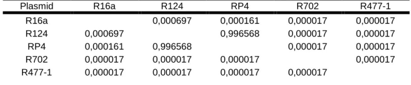
Table XI: Post Hoc Tuckey test performed after the three way ANOVA (donor strain, recipient strain, plasmid), considering the plasmids (D.f. = 360.00).
Plasmid R16a R124 RP4 R702 R477-1 R16a 0,000697 0,000161 0,000017 0,000017 R124 0,000697 0,996568 0,000017 0,000017 RP4 0,000161 0,996568 0,000017 0,000017 R702 0,000017 0,000017 0,000017 0,000017 R477-1 0,000017 0,000017 0,000017 0,000017 2. Fitness assay
In order to test for the existence of a relationship between the conjugation rate of each plasmid and the cost it imposes on its host (donor strains) we determined that fitness cost. To do that, we performed competitions of donor strains against a reference strain (E.coli MG1655 Δara). We could then calculate the indirect fitness of the plasmid donor strains, as a ratio between the fitness of the strains carrying the plasmid, and the fitness of the correspondent strain without the plasmid.
We observed that in over half the strains considered, the plasmid did not impose a fitness cost to its host. The plasmid R16a did not impose any statistically significant cost to any of the four hosts, in opposition to the plasmids RP4 and R477-1 that imposed a significant fitness cost in three and two of their hosts, respectively. Remarkably, the plasmid R477-1 conferred an advantage to the strain Vdg435. The strain’s fitness was higher carrying the plasmid than not carrying it. E.coli C4705 didn’t reveal any significant fitness alterations caused by any of the used plasmids (Figure 3).
25
Figure 3: Fitness values imposed by each plasmid in the E.coli strains used as donor strains in the conjugation assay. The error bars represent 2*standard error. Significant fitness values are marked with * at the top of the bars (Student T-test: p<0.05).
3. Evolution assay
In order to determine if the co-evolution of a bacterial strain and a given plasmid would or would not lower the cost of that strain to carry the plasmid, and to know if that cost alteration could affect the conjugation rates of that strain, we evolved a strain, E.coli M3, carrying the plasmid R477-1, for 30 days (approximately 658 generations), in the presence of selective pressure to ensure the maintenance of the plasmid (i.e. antibiotics for which the plasmid confers resistance). After the evolution assay, we measured the fitness of the new strain. We observed that the new evolved strain had an advantage over the original strain. The relative fitness of the evolved strain in relation to the original one was 1.07, significantly different from one (Student T-test: p=0.012). Having confirmed that the co-evolution had lowered the cost of carrying the plasmid to the donor strain, we proceeded to conjugate the new strain with the full set of 9 recipient strains used in the conjugation assay, in order to compare the results obtained, and access for differences caused by the changes of the plasmid cost to its host.
We obtained higher conjugation rates for the evolved strain for 5 out of the 9 recipient strains. (Table XII). None of the conjugation rates got lower with the co-evolution of the donor strain and the plasmid. For the remaining 4 recipient strains, the conjugation rates weren’t statistically different. These results then show that the fact that the cost of the donor strain to carry the plasmid has been lowered influenced its conjugation rates, at least for part of the lot of recipient strains used by us. We should also note that an increase in the fitness of the donor (i.e. reduction of the cost of carrying the plasmid) led to higher conjugation rates.
0,80 0,85 0,90 0,95 1,00 1,05 1,10 R16a R702 R124 RP4 R477-1 R e lativ e fi tn e ss Plasmid Vdg435 m3 m4 c4705 * * * * * * * *
26
Table XII: Log10 of the conjugation rates obtained using the evolved E.coli M3 strain with the
plasmid R477-1, and using its ancestral strain. A Student T-test was performed in order to access if the conjugation rates were statistically different between evolved and ancestral strains.
Recipient Donor Vdg380 Vdg411 Vdg435 M3 M4 M1412 C4705 C4720 C4734 Ancestral E.coli M3 (R477-1) -5,76 -7,90 -6,98 -8,27 -7,74 -7,93 -4,47 -6,03 -5,91 -5,74 -7,92 -7,01 -8,26 -7,79 -7,17 -4,66 -5,98 -6,02 -6,82 -8,00 -6,60 -8,02 -8,29 -7,88 -3,77 -5,71 -6,59 Evolved E.coli M3 (evolved R477-1) -3,46 -3,46 -5,49 -5,27 -6,40 -7,75 -4,63 -6,10 -6,64 -3,43 -3,42 -5,47 -4,89 -5,57 -7,74 -3,68 -5,85 -6,68 -3,85 -3,49 -5,80 -5,57 -5,30 -7,78 -3,67 -6,10 -6,58 P-value for Student T-test 0,01 0,00 0,00 0,00 0,01 0,73 0,51 0,45 0,16 4. Gel electrophoresis
In order to evaluate the plasmid content of the E.coli strains used as donors, we performed an electrophoresis to our strains after a treatment made to obtain plasmid DNA (alkaline lysis). In order to compare the content of each strain we used before and after the introduction of the plasmids to study (R702 and RP4), we treated both the donor strain with the plasmid and the same strain prior to the plasmid introduction. We also analysed E.coli K12 with plasmids R702 and RP4 as a reference for the plasmid position in the gel (this strain has no other plasmids than the ones we inserted). When analyzing strains Vdg435 (Figure 4A), we can infer that the strain itself has other plasmids in the cell, before the insertion of the plasmids we used. We also can see, when comparing the strains Vdg435 containing the plasmids R702 and RP4 with the strain with none of them, that the plasmid content of this strain was altered when we inserted each of the two plasmids. We can observe that the top band in the lane correspondent to E.coli Vdg435 (-) is missing in the lanes of the same strain with the plasmids R702 and RP4. This indicates that the insertion of R702 and RP4 in the considered strain forced some other plasmid out of the cell. On the other hand, the strains M3 (Figure 4A), M4 and C4705 (Figure 4B) had no apparent changes when both plasmids were inserted in the cells. When comparing them to the
E.coli K12 strains, we can see the upper and lower bands in both the reference strain and in the
tested strain. The M3, M4 and C4705 strains without the plasmid show no bands, suggesting that they have no other plasmids apart the ones we inserted.
27
Figure 4: Gel electrophoresis of the donor strains for the plasmids R702 and RP4. Figure 4A shows E.coli donor strains Vdg435 and M3, and E.coli K12 with both plasmids as a reference. M. corresponds to the DNA marker; K12 (R702) and K12 (RP4) correspond to E.coli K12 harboring plasmids R702 and RP4, respectively. Vdg435 (-), (R702) and (RP4) correspond to
E.coli Vdg435 Nal Nitro (Table I) and to E.coli Vdg435 harboring the plasmids R702 and RP4,
respectively. The same logic applies to M3 strains, and M4 and C4705 strains (Table I) shown in Figure 4B.
28
IV.
DISCUSSION
The results of this work indicate that there is a high variability in conjugation efficiency. For the plasmids and strains used, conjugation rates span eight orders of magnitude. This variability depends on the strain that works as the plasmid donor, on the plasmid itself, and on the strain that will receive the plasmid. The interaction amongst these three factors is also responsible for this variability.
From all the data collected, we can tell that E.coli Vdg435 is the worst donor strain of the four strains used, E.coli M1412 is the worst case in terms of recipient capability, and R477-1 is the plasmid with lower conjugation efficiency associated. Differently from what was observed by Dionisio et al (2002), the better donor strain was E.coli M3, instead of E.coli M4. These are the most important differences, in terms of conjugation efficiency, amongst our set of strains and plasmids (Tables V to IX). These differences can, in part, help to explain the wide range of conjugation rates we measured. The combination of a weak donor strain (a strain with lower plasmid donor efficiency) with a weak recipient strain (a strain with bad recipient efficiency) will lead to lower conjugation efficiency. On the other hand, if we choose particularly efficient strains (in terms of donor and recipient ability), we will get more efficient conjugation. The same goes for plasmids, with two plasmids (R477-1 and, to a lower extent, R16a) being associated with less efficient conjugation rates, when compared to the other plasmids we studied. We could also note that each one of the plasmids used has its own range of conjugation rates. These conjugation efficiency ranges are noticeably different amongst the plasmids we studied. Plasmids R124 and RP4 have similar intervals of conjugation efficiency, spanning almost six orders of magnitude. In opposition, plasmids R16a and R477-1 have quite narrow intervals of value of their conjugation efficiency.
It is important to understand the underlying causes for this diversity in conjugation efficiency. In order to try explaining this variability amongst conjugation rates, we performed essays to evaluate both the cellular content of the donor strains, and the costs that each plasmid imposes on the bacterial host.
Analyzing the conjugation efficiency of the strain E.coli Vdg435, we could notice that this was the weakest donor, for three of the five plasmids studied (Tables V to IX). One possible way to explain this particular difference lies on the cellular content, in terms of mobile genetic elements (in this case, plasmids) of the strains we used as donor strains. In a previous work (Dionisio et al, 2002), it has been shown that after curing bacteria (process by which the plasmids harbored by a bacteria are removed from it) and inserting a new plasmid into the different cured strains, the donor ability of each one would be similar, regardless of the differences prior to the curing process. Saying so, we can infer that the presence of other mobile genetic elements in the strains we used as donors may influence the donor efficiency of each one of them. After analyzing the plasmid content of our donor strains, by gel electrophoresis, we can tell that for all