FACULDADE DE FARMÁCIA
Development and characterization of novel
vehicles for topical drug delivery
Joana Marques Marto
Orientadora: Professora Doutora Helena Margarida Oliveira Marques Ribeiro
Co-orientadores: Professor Doutor António José Leitão das Neves Almeida
Dr.
Augusto Eduardo Vieira Borges OliveiraTese especialmente elaborada para a obtenção do grau de Doutor em Farmácia na
especialidade de Tecnologia Farmacêutica.
Development and characterization of novel vehicles for
topical drug delivery
Joana Marques Marto
Orientadora: Professora Doutora Helena Margarida Marques Ribeiro Co-orientadores: Professor Doutor António José Leitão das Neves Almeida
Dr. Augusto Eduardo Vieira Borges Oliveira
Tese especialmente elaborada para a obtenção do grau de Doutor em Farmácia na especialidade de Tecnologia Farmacêutica.
Júri
Presidente: Doutora Matilde da Luz dos Santos Duque da Fonseca e Castro, Professora Catedrática e Diretora da Faculdade de Farmácia da Universidade de Lisboa.
Vogais: Doutor João José Martins Simões de Sousa, Professor Associado com Agregação, Faculdade de Farmácia da Universidade de Coimbra;
Doutor Alberto António Caria Canelas Pais, Professor Associado com Agregação, Faculdade de Ciências e Tecnologia da Universidade de Coimbra;
Doutora Isabel Filipa Martins de Almeida, Professora Auxiliar, Faculdade de Farmácia do Porto;
Dr. Augusto Eduardo Vieira Borges Oliveira, Director de Assuntos Regulamentares, Laboratórios Atral. S.A., na qualidade de especialista de reconhecido mérito e competência, Coorientador;
Doutora Helena Margarida Oliveira Marques Ribeiro, Professora Associada, Faculdade de Farmácia, Universidade de Lisboa; Orientadora;
Doutor Luis Filipe Baptista Pleno Gouveia, Professor Auxiliar, Faculdade de Farmácia da Universidade de Lisboa.
“Se não sais de ti,
não chegas a saber quem és.”
vii
Table of Contents
Agradecimentos……… ... xv Abstract……… ... xix Resumo……… ... xxi List of Figures……… .... xxvList of Tables……… .... xxxi
Abbreviations & Symbols……… ... xxxv
Aims and Organisation of the Thesis………. xxxvii
Chapter 1 - General Introduction……….. .. ….1
Graphical abstract……… .. 5
Highlights………... 5
1 Introduction ... 7
1.1 Starch: functional characteristics and relevance ... 7
1.1.1 Modified starch: a strategy to prepare high performance starch ... 10
1.1.2 Starches: from granules to novel applications ... 10
1.2 Skin: the epidermal barrier ... 11
1.3 Topical delivery systems ... 15
1.3.1 Conventional topical delivery systems ... 16
1.3.1.1 Emulsions ... 16
1.3.1.2 Gels ... 20
1.3.1.3 Starch in personal care: a multifunctional ingredient ... 24
1.3.2 Non-conventional topical delivery systems ... 28
1.3.2.1 Polymeric nanopartciles ... 28
2 Conclusions ... 35
viii
Chapter 2 - Preformulation studies on native and modified starches ……….. . 43
Graphical abstract……….… 45
Highlights………... . 45
1 Introduction ... 47
2 Material and methods ... 49
2.1 Materials ... 49
2.2 Methods ... 49
2.2.1 Starch granules characterization ... 49
2.2.2 Sample preparation ... 50
2.2.3 Structure analysis of starch aqueous solutions ... 50
2.2.3 Biocompatibility assay ... 51
3 Results and discussion ... 52
3.1 Starch granules ... 53
3.1.1 Particle size measurements and morphology of starch granules ... 53
3.1.2 Wettability measurements ... 56
3.2 Structure analysis of starch aqueous solutions ... 57
3.2.1 Dynamic and oscillation measurements ... 57
3.2.2 Thermoanalytical measurements and hot stage microscopy ... 58
3.2.3 Steady state fluorescence measurements ... 61
3.3 Biocompatibility assay ... 63
4 Conclusions ... 64
5 References ... 65
Chapter 3 - Starch-based Pickering emulsion……… 67
Section 1 - Starch-based Pickering emulsions for topical drug delivery: a QbD approach 69 Graphical abstract……….… 71
Highlights………... . 71
1 Introduction ... 73
2 Material and methods ... 74
2.1 Materials ... 74
2.2 Methods ... 74
2.2.1 Characterization of starch granules ... 74
2.2.2 Preparation of the starch-stabilized emulsions (ASt-emulsion) ... 75
2.2.3 Identification of Quality Target Product Profile (QTPP) and Critical Quality Attributes (CQAs) ... 75
ix
2.2.4 Risk analysis of CQAs ... 76
2.2.5 Design space assessment ... 76
2.2.6 Stability of w/o ASt-emulsions... 77
2.2.7 ASt-emulsions’ mechanical and structure properties ... 78
2.2.8 In vitro cytotoxicity studies ... 79
2.2.9 Antimicrobial activity ... 79
3 Results and discussion ... 80
3.1 Characterization of starch granules ... 80
3.1.1 Particle size measurements of ASt granules ... 80
3.1.2 ASt wettability ... 80
3.1.3 Type of emulsion ... 81
3.1.4 Risk analysis of CQAs ... 82
3.1.5 Response surface analysis ... 84
3.1.6 Design space ... 87
3.2 Characterization of ASt-emulsions ... 88
3.2.1 Stability of ASt-emulsions ... 89
3.2.2 ASt-emulsions’ mechanical and structure properties ... 90
3.2.3 In vitro cytotoxicity studies ... 95
3.2.4 Antimicrobial activity ... 96
3.2.5 Microstructure of ASt-emulsions ... 97
4 Conclusions ... 98
5 References ... 99
Section 2 - Minocycline-loaded starch-based Pickering emulsions: in vitro and in vivo studies……….…10 3 Graphical abstract………. . 105
Highlights……… . ….105
1 Introduction ... 107
2 Material and methods ... 108
2.1 Materials ... 108
2.2 Methods ... 108
2.2.1 Preparation of MH-loaded starch-stabilized emulsions... 108
2.2.2 Microscopic aspect of MHASt-emulsions ... 109
2.2.3 Stability of MHASt-emulsions ... 109
x
2.2.5 Topical delivery studies of MH ... 111
2.2.6 In vitro cytotoxicity studies ... 112
2.2.7 In vitro determination of the antibacterial activity of MHASt-emulsions ………..112
2.2.8 In vivo studies ... 114
2.2.9 Statistical analysis ... 115
3 Results and discussion ... 116
3.1 Formulation development ... 116
3.1.1 Drug substance: MH as a model drug ... 116
3.1.2 Optimization of MHASt-emulsions ... 117
3.1.3 Stability of MHASt-emulsions ... 117
3.2 In vitro release studies ... 118
3.3 Topical delivery studies of MH ... 122
3.4 In vitro studies ... 124
3.4.1 In vitro cytotoxicity studies ... 124
3.4.2 Scratch wound healing migration assay ... 125
3.4.3 In vitro determination of the antibacterial activity of MH in ASt-emulsions ……… . …127
3.4.4 Skin adapted agar diffusion test ... 127
3.5 In vivo studies ... 129
3.5.1 In vivo antibacterial activity studies: tape-stripping infection model ... 129
3.5.2 Skin histology ... 131
4 Conclusions ... 133
5 References ... 134
Chapter 4 - Starch-based nanocapsules………139
Section 1 - A Quality by Design (QbD) approach on starch-based nanocapsules: a promising platform for topical drug delivery……….141
Graphical abstract………. . 143
Highlights……… . ….143
1 Introduction ... 145
2 Materials and methods ... 146
2.1 Materials ... 146
2.2 Methods ... 146
xi 2.2.2 Identification of quality target product profile (QTPP) and critical quality
attributes (CQAs) ... 148
2.2.3 Risk Analysis of CQAs ... 148
2.2.4 Response surface analysis ... 148
2.2.5 Physical stability of StNC ... 149
2.2.6 In vitro cytotoxicity studies ... 150
3 Results and discussion ... 151
3.1 Quality by design approach ... 151
3.1.1 Risk analysis of CQAs ... 151
3.1.2 Response surface analysis ... 152
3.1.3 Stability of the StNC ... 157
3.1.4 Physicochemical characterization... 158
3.1.5 Nanostructure of the StNC ... 160
3.1.6 In vitro studies of interactions between StNC and cells ... 163
4 Conclusion ... 164
5 References ... 165
Section 2 - Starch nanocapsules containing a novel neutrophil elastase inhibitor with improved pharmaceutical performance………. .167
Graphical abstract………. . 169
Highlights……… . ….169
1 Introduction ... 171
2 Materials and methods ... 172
2.1 Materials ... 172
2.2 Methods ... 172
2.2.1 HNE inhibitor synthesis ... 172
2.2.2 Biological assays ... 173
2.2.3 Preparation of neutrophil elastase inhibitor-loaded starch-based nanocapsules (StNC ER143) ... 174
2.2.4 Particle size analysis and zeta potential measurements ... 174
2.2.5 Encapsulation efficiency and drug loading ... 174
2.2.6 Fourier transform infrared spectroscopy (FTIR) ... 175
2.2.7 Differential scanning calorimetry (DSC) ... 175
2.2.8 Quality by design (QbD) approach ... 175
2.2.9 In vitro ER143 release studies from StNC ... 177
xii
2.2.11 In vivo anti-inflammatory activity studies ... 178
2.2.12 Mouse ear histology ... 179
2.2.13 Statistical analysis ... 180
3 Results and discussion ... 180
3.1 Synthesis of HNE inhibitor ER143 ... 180
3.2 Biological assays ... 181
3.2.1 Enzymatic inhibition assay ... 181
3.2.2 Fluorescence microscopy with human neutrophils ... 181
3.3 Quality by design approach ... 182
3.3.1 Risk analysis of CQAs ... 182
3.3.2 Response surface analysis ... 182
3.4 ER143-StNC interaction by DSC and FTIR... 186
3.5 In vitro ER143 release studies from StNC ... 188
3.6 Topical delivery studies of StNC ER143 ... 190
3.6.1 In vitro skin permeation and retention ... 190
3.7 In vivo studies ... 194
4 Conclusions ... 197
5 References ... 198
Chapter 5 - Starch-based vehicles: a safety and effective platform for dermatological purpose………. ……….201
Graphical abstract………. . 203
Highlights……… . .203
1 Introduction ... 205
2 Materials and methods ... 206
2.1 Materials ... 206
2.2 Methods ... 206
2.2.1 Preparation of starch-based vehicles (St-BV) ... 206
2.2.2 Safety assessment of St-BV ... 207
2.2.3 EpiSkin™ assay ... 208
2.2.4 In vivo studies ... 209
2.2.5 Statistical analysis ... 210
3 Results and discussion ... 210
3.1 Safety assessment of St-BV ... 210
xiii 3.1.2 Exposure assessment ... 215 3.1.3 Dose-response assessment ... 216 3.1.4 Risk characterization ... 217 3.2 EpiSkin™ assay ... 217 3.2 In vivo studies ... 217 4 Conclusion ... 220 5 References ... 221
Chapter 6 - Pickering emulsion sunscreen………225
Graphical abstract………. . 229
Highlights……… . ….229
1 Introduction ... 231
2 Materials and methods ... 234
2.1 Materials ... 234
2.2 Methods ... 235
2.2.1 Solid particles (SP) – mTiO2, ZnO and ASt ... 235
2.2.2 Natural oils ... 235
2.2.3 Sunscreen formulations – PhotoMel 1 (PM1) and PhotoMel 2(PM2) ... 235
2.2.4 Efficacy of PM1 and PM2 ... 236
2.2.5 Physicochemical and microbiological stability ... 237
2.2.6 UV degradation studies ... 238
2.2.7 Characterization studies ... 238
2.2.8 Topical delivery studies ... 239
2.2.9 In vitro cytotoxicity studies ... 239
2.2.10 In vitro EpiSkin™ ... 240
2.2.11 Safety assessment ... 240
2.2.12 In vivo studies ... 241
2.2.13 Statistical analysis ... 241
3 Results and discussion ... 242
3.1 Solid particles (SP) ... 242
3.1.1 Wettability measurements ... 242
3.1.2 Particle size distribution ... 243
3.2 Natural oils ... 243
3.2.1 SPF measurement ... 243
xiv
3.3.1 SPF measurement ... 245
3.3.2 In vitro sun product water resistance ... 245
3.3.3 Physical and chemical stability ... 246
3.4 UV degradation studies ... 247
3.5 Characterization studies ... 248
3.5.1 Microscopy analysis ... 248
3.5.2 Structural analysis ... 249
3.6 Topical delivery studies ... 253
3.6.1 Skin permeation and retention... 253
3.7 In vitro ROS assay ... 255
3.8 In vitro EpiSkin™... 257 3.9 Safety assessment ... 257 3.9.1 Hazard identification ... 257 3.9.2 Exposure assessment ... 260 3.9.3 Dose-response assessment ... 260 3.9.4 Risk characterization ... 261 3.10 In vivo studies ... 261 3.10.1 HRIPT ... 261
3.10.2 In vivo sun product water resistance ... 261
4 Conclusions ... 263
5 References ... 264
Chapter 7 - Concluding remarks and future work……… . …..271
1 Concluding remarks and future work ... 273
1.1 Concluding remarks ... 273
xv
Agradecimentos
A finalização dos estudos conducentes ao grau de doutor compreenderam desafios intelectuais e pessoais que só foram possíveis de ultrapassar com a entreajuda de todos aqueles que contribuíram para este árduo e longo caminho. É, assim, chegada a hora de expressar os meus mais sinceros agradecimentos a todos os que me orientaram, acolheram e ajudaram na concretização desta aspiração pessoal e que tornaram possível a realização desta dissertação.
À Professora Doutora Helena Margarida Ribeiro, minha orientadora principal, agradeço especialmente pela exemplar orientação que me permitiu crescer cientificamente e, não menos importante, crescer pessoalmente. Agradeço a disponibilidade com que me recebeu e me integrou no seu grupo de trabalho desde o primeiro momento. Ao longo destes anos, agradeço a recetividade constante e a sábia capacidade pedagógica e científica que me permitiram superar dúvidas e reformular decisões. Não posso por isso deixar de destacar a visão crítica e sempre positiva que de uma forma muito clara, objetiva e motivadora conduziram ao aperfeiçoamento do presente trabalho. A sua simpatia, boa disposição, plena dedicação e interesse, permitiram o melhor ambiente que alguém pode desejar enquanto desenvolve um trabalho desta complexidade. Saliento ainda o privilégio que sinto pela amizade que construímos e que se projetará muito para além deste tempo.
Ao Professor Doutor António José Leitão das Neves Almeida, co-orientador desta tese, quero expressar o meu sincero agradecimento pela excelência científica e rigor de desempenho. Agradeço a motivação, a dedicação e a disponibilidade constantes. Agradeço os ensinamentos, as sugestões pertinentes, a visão crítica, clara, oportuna e sempre construtiva no decurso deste trabalho. Saliento a sua conduta humana, apoio e amizade sempre demonstradas em momentos decisivos que resultaram sempre num forte estímulo para prosseguir o caminho que culmina nesta tese.
xvi
Ao Dr. Eduardo Oliveira, meu co-orientador científico, quero manifestar o meu agradecimento, realçando a excelência do seu desempenho, enquanto orientador industrial desta tese. Agradeço o apoio e as circunstâncias que me proporcionaram realizar este trabalho, bem como as discussões científicas que me ajudaram a orientar esta tese para necessidades reais da indústria farmacêutica Portuguesa.
Ao Professor Doutor Luis Filipe Gouveia, porque me quis honrar com o seu apoio, pelos conselhos e diálogos científicos, pelas adequadas sugestões formuladas, quero agradecer a sua visão critica e construtiva em momentos decisivos que me ajudaram a atingir os objetivos propostos. O meu reconhecimento para toda a vida.
À Doutora Lídia Maria Diogo Gonçalves, agradeço o rigor pelas boas práticas, chancela do seu trabalho de excelência, e a inteira disponibilidade sempre demonstrada. As suas palavras de encorajamento e de grande amizade revelaram-se determinantes nesta etapa que agora termina.
À Professora Doutora Aida Duarte, expresso os meus agradecimentos pela disponibilidade e partilha de conhecimentos. As discussões científicas críticas contribuíram inquestionavelmente para o aperfeiçoamento da minha forma de trabalhar e pensar. Com um sentido maternal, preocupado e acima de tudo dinâmico, inteligente, objectivo e empreendedor, é inigualável o modo como sempre se e nos destacou.
À Dra. Ana Salgado reconheço o papel determinante em todo o caminho percorrido, a nível profissional e pessoal. Um muito obrigado pela amizade e camaradagem.
À Doutora Sandra Simões e Doutor Pedro Pinto agradeço a colaboração pronta e rigorosa que me dispensaram na execução de alguns trabalhos laboratoriais, bem como o apoio e amizade demostrados.
Ao espírito de grupo, amizade e sentido de entreajuda de todos os colegas da Tecnologia Farmacêutica, e de um modo especial, e sem ordem em particular, à Sara Raposo, Rui Lopes, Diana Gaspar, Maria Paisana, Joana Pinto, Inês Ferreira, Paulo Roque Lino, Ana Matos, Filipa Silva, Joana Bicho, Gonçalo Oliveira, Giuliana Mancini, Andreia Ascenso, Diana Rafael, Ana Cadete, Lara Figueiredo, Ana Varela, Joana Silva, Filipa Guilherme, Ana Neves, Carla Vitorino, Alice Gelpi, Cecilia Sangali, Liliana Aranha, Diogo Baltazar,
xvii Barbara Gregori, Margherita Alegro, João Quintas, André Sá Couto, Nélio Drummond e Ana Costa pelo contínuo apoio que me ajudaram a ultrapassar as fases mais difíceis.
À ADEIM, em especial a Dra. Alexandra Silva e Paula Machado por todo o apoio, espírito de grupo e amizade com que sempre me brindaram e enriqueceram.
À Engenheira Carla Eleuterio, D. Fernanda Carvalho, D. Henriqueta Pinto e D. Fernanda Oliveira, agradeço o bom acolhimento e disponibilidade continuada, que concederam às minhas múltiplas solicitações.
À Doutora Susana Dias Lucas e ao Professor Doutor Rui Moreira um agradecimento pela possibilidade de colaboração com o MedChem Group do iMed.ULisboa, e em especial ao Eduardo Ruivo pelo desenvolvimento da molécula utilizada no Capitulo 4, Secção 2 e no esclarecimento de questões relacionada com a mesma. Agradeço ainda todo o apoio, partilha, paciência, sorrisos e alegrias com que diariamente me enriqueceram este percurso e com que me ajudaram a superar as etapas mais difíceis.
Ao Professor Doutor Alberto Pais Canelas do Departamento de Química da Universidade de Coimbra, agradeço a disponibilidade e o interesse manifestado por este trabalho. Ao Professor Doutor Filipe Antunes, Professor Doutor João Pina e Doutor Sérgio Silva agradeço todos os ensinamentos, esclarecimentos, sugestões e diálogos científicos que em muito contribuíram para a qualidade desta tese. À Doutora Sandra Nunes e Dra. Tânia Firmino não posso deixar de agradecer a contagiante boa disposição que proporcionaram a vivência de momentos de descontração únicos.
À Professora Doutora Vera Isaac do Laboratório de Cosmetologia da Faculdade de Ciências Farmacêuticas da UNESP, agradeço a simpatia e amizade com que me recebeu em Araraquara e o interesse demonstrado na colaboração do meu trabalho. Também à Doutora Bruna Chiari-Andreo apresento o meu muito obrigada por toda a disponibilidade no laboratório e no esclarecimento de questões relacionadas com as mesmas, por todas as considerações científicas e sugestões que culminaram no trabalho publicado e que será apresentado nesta tese.
A Doutora Tania Carvalho, Ana Margarida Santos, Andreia Pinto e Bruna Almeida do Laboratório de Histologia e Patologia Comparada no Instituto de Medicina Molecular pelo trabalho de histologia. Agradeço-lhes as valiosas observações e sugestões para a melhor
xviii
análise das amostras e os esclarecimentos prestados sobre as mesmas sempre que uma dúvida surgia.
O meu agradecimento também aos estudantes Inês Jorge, Patrícia Manteigas e Joice Lana pela sua contribuição na realização do intenso trabalho de laboratório.
À Professora Doutora Ana Francisca Bettencourt e à Professora Maria João Silva por todo o carinho, bem como apoio e amizade demostrados.
À Dra. Inês Casais pelo design gráfico da presente dissertação.
À empresa DS Produtos Químicos Lda., em especial ao Sr. Óscar Brás, pela disponibilização de várias matérias-primas e ajuda em algumas questões científicas. À direcção e restantes membros do Departamento de Farmácia Galénica e Tecnologia Farmacêutica e ao Departamento de Microbiologia bem como do grupo Nano2B, antigo NanoDDS e da FFULisboa por sempre ter encontrado as melhores condições para um correcto desenrolar de trabalhos bem como pelo ambiente cientificamente rico e potenciador de conhecimento.
Aos meus amigos e em especial ao André, pela sua inestimável paciência, tempo interminável... e contenção das minhas angústias finais.
À minha Mãe, Pai e família. Ao apoio incondicional que me deram ao encarar este projecto e com o qual me continuam ajudar a avançar sempre em frente, fomentando a conclusão dos objectivos a que me proponho, sempre rumo a um crescimento saudável e feliz. São as pessoas que me enchem o peito a razão do meu viver e estou-lhes e estarei sempre eternamente agradecida!
Aos Laboratórios Atral S.A., agradeço as condições económicas que me proporcionaram realizar esta tese bem como todo o apoio dispensado.
À Fundação para a Ciência e Tecnologia agradeço a bolsa de doutoramento SFRH/BDE/51599/2011 que nos permitiu efectuar o presente conjunto de trabalhos.
xix
Abstract
Topical drug delivery is challenging since the skin acts as a natural and protective barrier. The skin has been an important route for drug delivery when topical, local or systemic effects are desired. The outcome of topical dermatological drug treatment is significantly influenced by the choice of vehicle. In recent years there has been an increased interest in developing improved delivery systems and, exploring new ways of using approved excipients, such as, starch. Due to its unique properties, starch has been extensively used in various topical pharmaceutical application, i.e. as a sensorial enhancer, a stabilizer and drug delivery polymer, providing protection and control release of the drug molecule.
The base-concept of this study was to develop and characterize novel starch-based vehicles for dermatological application, easily scaled-up to industry and produced by methods that can allow the decrease of production costs, and further investigate the resulting systems behavior in in vitro and in vivo conditions.
Starch-based vehicles were prepared successfully using QbD approach with the understanding of the high risk process and formulation parameters involved and optimized design space with a multifactorial combination of critical parameters to obtain predetermined specifications. Three different model drugs were incorporated into the optimized starch-based vehicles. Minocycline hydrochloride (MH) was incorporated in Pickering emulsions, a human neutrophil elastase inhibitor (ER143), a new molecule developed by the MedChem Group at iMed.ULisboa, was encapsulated into starch nanocapsules and melatonin was added on Pickering emulsions sunscreen, in order to fully characterize these new formulations, and further study its topical delivery and in vitro and in vivo efficacy.
The in vitro antibacterial activity studies for Pickering emulsions containing MH revealed that the released drug exceeded the minimum inhibitory concentration of MH against S.aureus. In
xx
vitro release studies showed a prolonged release of the MH, with an initial burst effect. Regarding in vitro permeation studies, MH does not pass through the entire skin layer, suggesting a minimal potential for the systemic absorption of the MH upon topical administration. In vivo results showed that topical administration of MH was effective in S. aureus superficial infections treatment.
Starch nanocapsules presented a mean particle size ranging from 200 to 250 nm and a positive zeta potential. In vitro permeation studies showed that the starch nanocapsules were suitable for the delivery of ER143, allowing a high control of the drug release, contributing to a high skin retention and/or permeation profiles of ER143. In vivo results showed that erythema and edema were attenuated in 98%, following the local application of ER143-loaded starch nanocapsules.
Regarding Pickering emulsions sunscreen, formulation studies demonstrated that starch particles presented no intrinsic photoprotection properties, they proved to be a sun protection factor promoter by a synergistic effect. Besides the excellent sunscreen activity confirmed by in vitro and in vivo results, the final formulations proved to be also suitable for topical use according to the rheological assessment and stability throughout the study period (3 months). Additionally, the safety and biological effects of the placebos (vehicles without drug) was assessed by using both in vitro and in vivo studies, as an adequate equilibrium between the safety and efficacy effects.
Overall, these findings highlight the starch-based vehicles as promising for the development of topical delivery systems, covering innovative therapeutic approaches.
Keywords: New delivery systems; Pickering emulsions; quality by design; starch; polymeric nanocapsules; topical delivery.
xxi
Resumo
A pele revela-se particularmente adequada para a aplicação tópica de fármacos ao favorecer o estabelecimento de um contacto íntimo do fármaco com o tecido-alvo a tratar e por dessa aplicação decorrer habitualmente um mínimo de efeitos sistémicos. No entanto, a administração tópica de fármacos continua a ser um desafio, uma vez que a terapêutica das doenças da pele está condicionada por duas características fundamentais deste órgão: a sua função de barreira, determinada pela estrutura da camada córnea que, por si, constitui o factor limitante da absorção percutânea e a sua sensibilidade ao contacto repetido com substâncias químicas. Normalmente as reacções adversas resultantes da aplicação tópica de fármacos não são imputáveis exclusivamente às substâncias activas utilizadas, sendo-o também aos excipientes, agentes conservantes ou substâncias aromáticas incluídos na preparação. Por esta razão impõe-se a investigação de novos excipientes ou o melhoramento dos existentes através da aplicação de novas abordagens.
São actualmente bem conhecidos os factores que condicionam a absorção e a difusão dos fármacos aplicados topicamente. Em primeiro lugar, a estrutura e características físico-químicas da substância activa e, seguidamente o tipo de excipiente que vai influenciar o grau de penetração do fármaco ativo, podendo ainda melhorar o grau de hidratação da pele e ter efeito emoliente e protector. Contudo, para terem actividade terapêutica, os fármacos têm que ultrapassar a camada córnea, o que consiste no transporte de uma substância para uma determinada camada da pele. A maior parte dos fármacos não consegue permear a pele, pelo que é necessário um veículo para os transportar ou para aumentar a libertação no local de ação.
Assim, o desenvolvimento e melhoramento de novas formulações para aplicação cutânea é uma estratégia interessante para a administração de fármacos cuja acção é local ou
xxii
transdérmica, representando uma alternativa que permite superar aspectos indesejados relacionados com as características farmacocinéticas e farmacodinâmicas dos fármacos. O desenvolvimento de novas formas farmacêuticas implica um esforço considerável que é muitas vezes consumido na elaboração de especificações rigorosas de forma a garantir a sua estabilidade física, química e microbiológica. Estes novos produtos, concebidos para aplicação tópica de acordo com critérios específicos têm, até ao presente, sido produzidos em escala laboratorial ou em equipamentos de escala piloto. Para além dos requisitos de eficácia e segurança do medicamento, a facilidade de produção, de uma forma reprodutível, em equipamento industrial de produção rápida e económica é, frequentemente um factor que diferencia um produto com sucesso de outro que é encarado como uma curiosidade científica. Para ter sucesso o produto tem que ser capaz de ser processado e embalado em grande escala. Na instalação piloto uma fórmula é traduzida num produto viável e robusto pelo desenvolvimento de um método de produção fiável e prático que permita uma transição fácil entre o laboratório e uma instalação industrial.
Assim, a optimização de formulações e a utilização de sistemas transportadores constitui uma estratégia válida, nomeadamente com recurso a novos veículos, produzidos com excipientes devidamente regulamentados como os lípidos sólidos, o alginato e o amido.
Devido às suas propriedades únicas, o amido tem sido amplamente utilizado em diversas aplicações farmacêuticas. No entanto, uma potencial aplicação tem sido pouco explorada: a sua utilização em formas farmacêuticas para aplicação tópica. Devido à multifuncionalidade do amido, muitos desenvolvimentos são esperados nesta área, o que pode representar uma visão renovada para a indústria farmacêutica. Assim, um desenvolvimento farmacêutico racional, que integre formulações simples, de baixo custo e facilmente traspostas para a escala industrial, irá ajudar no desenvolvimento de veículos adequados.
Este projeto teve como principais pressupostos o desenvolvimento e caracterização de formulações com veículos inovadores, seguros, de menor custo e com vantagens terapêuticas para uso dermatológico e, de valor acrescentado para a empresa financiadora.
Foram desenvolvidos e optimizados vários veículos à base de amido, utilizando abordagem quality by design (QbD) e de acordo com a ICH Q8(R2). Após desenvolvidos e detalhadamente caracterizados, recorrendo a técnicas de DSC, FTIR, microscopia, fluorescência, ângulos de contacto, reologia, biocompatibilidade, o objetivo foi estudar os
xxiii perfis de libertação e permeação através de membranas sintéticas e pele de leitão, e realizar estudos in vivo de eficácia, comparando os resultados obtidos com a formulação de referência no mercado.
Finalmente, os efeitos biológicos e a avaliação de segurança foram realizados para os veículos de acordo com a legislação europeia de cosméticos em vigor.
Foram seleccionados 3 fármacos modelo para os diferentes veículos desenvolvidos e optimizados. Um antibiótico, cloridrato de minociclina (MH) foi incorporado em emulsões de Pickering, um inibidor da elastase de neutrófilos humanos (ER143), uma nova molécula desenvolvida pelo grupo de investigação MedChem do iMed.ULisboa, foi encapsulado em nanocápsulas de amido e por fim, a melatonina, um potente antioxidante, foi incorporada num protetor solar, utilizando como forma farmacêutica as emulsões de Pickering. Os fármacos modelo incorporados nos diferentes veículos foram utilizados com o intuito de caracterizar intensivamente os novos medicamentos/produtos de saúde desenvolvidos, e compreender os mecanismos de libertação e permeação, assim como estudar a sua eficácia terapêutica in vitro e in vivo.
Relativamente as emulsões de Pickering contendo MH, os estudos in vitro para determinar a actividade antibacteriana revelaram que o fármaco libertado das formulações é superior a concentração mínima inibitória contra Staphylococcus aureus. Estudos de libertação in vitro revelaram que o perfil de libertação do fármaco depende da concentração do agente estabilizador, grânulos de amido e da natureza da fase externa da emulsão. Todas as formulações estudadas exibem uma libertação controlada do fármaco. Estudos de permeação in vitro demostraram que as formulações em estudo tem pouca influencia na permeação do MH, dado que o MH não permeou a pele, revelando possuir um perfil de segurança adequado para os antibióticos de aplicação tópica. Resultados in vivo com murganhos demostraram que a administração tópica de MH foi eficaz no tratamento de infecções superficiais por S. aureus. As nanocápsulas de amido desenvolvidas e optimizadas apresentam um tamanho médio de partícula entre 200 a 250 nm e um potencial zeta positivo (> 25 mV). A abordagem QbD permitiu compreender a formação deste veículo e concluir que a concentração de tensioactivo (não iónico) e a quantidade de lípido (núcleo oleoso) influenciam o tamanho da nanocápsula. Estudos de permeação in vitro demonstraram que o veículo desenvolvido era adequado para a administração tópica de ER143, permitindo um elevado controlo da libertação da molécula, o
xxiv
que contribui para uma elevada retenção da pele. Os resultados obtidos no ensaio de tape stripping em pele de leitão, confirmaram os resultados, demostrando que a quantidade de ER143 que atingiu as camadas viáveis da pele é baixa (10.64 %), ficando parte do fármaco retido na camada córnea (22.70 %). No entanto, os estudos in vivo demostraram que as formulações desenvolvidas diminuíram o edema e o eritema na orelha do murganho em mais de 92 %. Adicionalmente, foi demostrado que a eficácia das formulações é semelhante à da formulação comercial relativamente aos estudos da actividade anti-inflamatória.
Relativamente ao protector solar desenvolvido e, utilizando o conceito de emulsões de Pickering, foi possível obter uma emulsão estabilizada por filtros solares físicos (dióxido de titânio e oxido de zinco) e grânulos de amido. A melatonina, um poderoso antioxidante que retarda o envelhecimento cutâneo, é uma molécula extremamente instável quando exposta à radiação solar, sendo um desafio a sua estabilização química num protector solar. No entanto, estudos in vitro de fotoestabilidade provaram que a tecnologia de emulsões de Pickering permitiu estabilizar quimicamente a melatonina, mantendo-a com as suas características durante a exposição solar. Estudos in vitro de determinação do fator de protecção solar (FPS) demonstraram que o amido não possui propriedades intrínsecas de fotoproteção, no entanto, provou ser um promotor do FPS, por efeito sinérgico. Além da excelente protecção solar (FPS 50+) confirmada pela determinação in vitro, os resultados in vivo, evidenciaram que as formulações finais foram adequadas para aplicação tópica. Todos os resultados revelaram um excelente compromisso entre a estabilidade, proteção contra radiação ultravioleta, eficácia, segurança e cosmeticidade.
Os estudos dos efeitos biológicos (perda de água trans epidérmica, corneometria, sebometria e microcirculação) dos veículos estudados permitiram concluir que estes contribuíram para o aumento da hidratação e da microcirculação. Adicionalmente a avaliação de segurança concluiu que estes veículos são seguros para aplicação tópica nas condições previstas.
No geral, estes resultados evidenciam os veículos à base de amido como promissores para o desenvolvimento de sistemas de libertação tópica, podendo ser utilizados em abordagens terapêuticas inovadoras.
Palavras-chave: Administração tópica; amido; emulsões de Pickering; nanocapsulas de amido; novos veículos; quality by design.
xxv
List of Figures
Figure Page
Chapter 1
1.1 Structural formula of starch………. 7 1.2 Starch granules composition [5, 9]…..……… 8 1.3 Skin structure (adapted from [22]…..……….... 11 1.4 Possible transport pathways through the stratum corneum (adapted
from [33])………. 12
1.5 Some methods for optimizing transdermal drug delivery (adapted from
[32, 35-37])….……….. 13
1.6 Surfactant-based emulsion (left) and a Pickering emulsion (right)…….. 16 1.7 Mechanism of starch-based gel formation………... 21
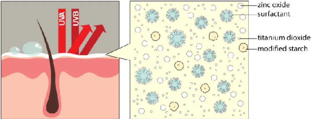
1.8 Mechanism of action of sunscreens/solid particles with UV
radiation……… 27
1.9 Schematic representation of nanospheres and nanocapsules (adapted
from [88])………..………... 28
1.10 Different mechanisms of drug incorporation into polymeric
nanoparticles [84, 88]………... 29
1.11 Film formation associated with the occlusion effect of nanoparticles
(adapted from [85])……….……. 30
Chapter 2
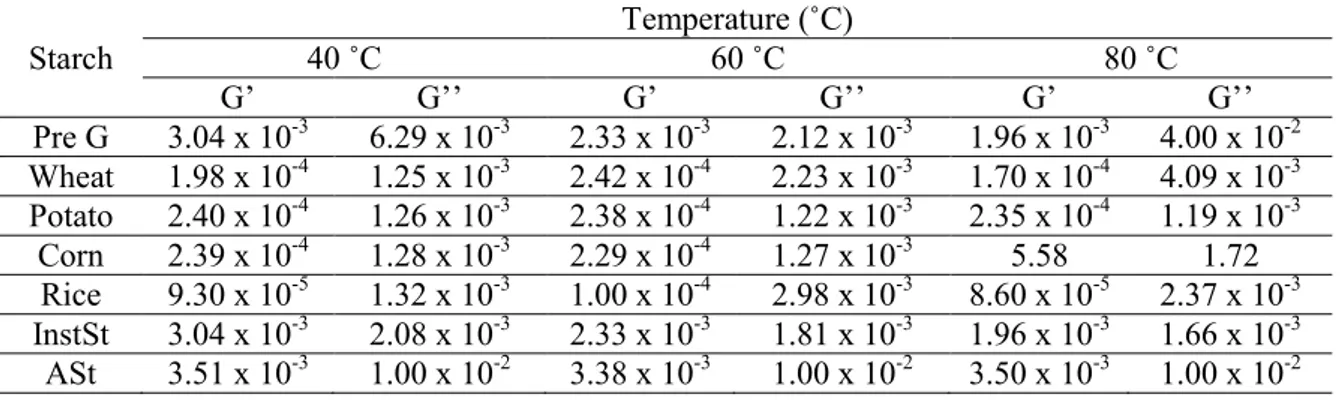
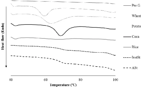
2.1 (a) Viscosity of different starches at different temperatures. (b) Dependence of storage (G’) and (c) loss (G’’) moduli for different starches at different temperatures……… 58 2.2 DSC scanning thermograms of different starches……… 59 2.3 Steady-state fluorescence emission anisotropy values for
1,6-diphenyl-1,3,5-hexatriene (DPH) in the starch aqueous solutions collected as a
function of temperature……… 62
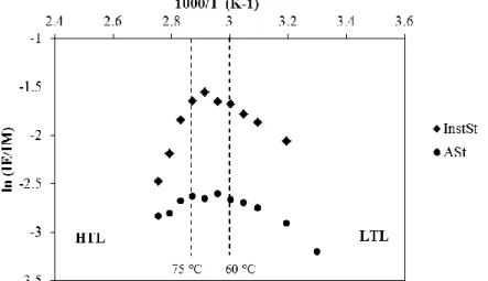
2.4 Arrenhius plots for the excimer-to-monomer intensity ratio (IE/IM) of
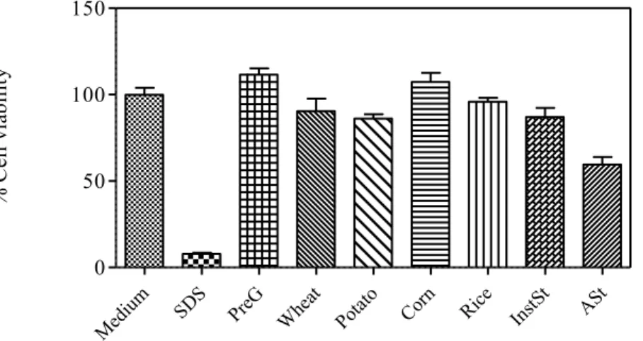
pyrene in aqueous solutions of the starch derivatives InstSt and ASt…. 62 2.5 Viability of HaCaT cells after 48 h of incubation with different
starches at concentration of 2 mg/ml (mean ± SD, n=5). SDS - sodium
dodecyl sulfate………. 63
2.6 Membrane integrity of HaCaT cells after 48 h of incubation with different starches at concentration of 2 mg/ml (mean ± SD, n=5). PI - propidium iodide; SDS - sodium dodecyl sulfate……….…….. 64
xxvi
Chapter 3 Section 1
3.1.1 Morphology of ASt under (a) bright-field. (b) sobel filter and (c) polarized light. Scale Bar = 50µm………... 80 3.1.2 Contact angles of aluminium starch octenylsuccinate in (a) water, (b)
LP and (c) CT……….……..… 81
3.1.3 Emulsion stabilised by starch granules dyed with methylene blue
(Scale bar = 100 µm)………... 82
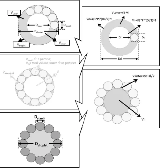
3.1.4 Ishikawa diagram illustrating factors that may have impact on the droplet size of an ASt-emulsion………... 82 3.1.5 Schematic representation of the rationale to calculate the total amount
of ASt needed to fully cover the aqueous droplets……….…. 84 3.1.6 Isoresponse curves (graph floor) and response surface plots of relative
size distribution (µm), respectively: (a) d(50) (b) d(90) and (c) Span, for process optimization using LP as the oil phase, (d) d(50), (e) d(90) and (f) Span, for formula optimization using LP as the oil phase and (g) d(50), (h) d(90) and (i) Span, for formula optimization using CT as the
oil phase………..……… 86
3.1.7 Plots evidence the DS for (a) the process and (b) the formula with (b.1) LP and (b.2) CT as the oil phase, respectively………. 88 3.1.8 Dependence of storage (G’) and loss (G’’) moduli with shear stress for
ASt-emulsion a) LP and b) CT. All the experiments were carried out at
25ºC………..…… 91
3.1.9 DSC thermograms of ASt-emulsions: (a) LP and (b) CT with photomicrographs of α-ASt-emulsions during a heating program with 10 °C/min between 30 °C and 120 °C. At 60 ºC (1), at 70 ºC (2) and at 80 ºC (3) (Scale bar = 200 µm)……… 94 3.1.10 Micrographs of (above) LP and (below) CT emulsions after 1 week of
preparation (Scale bar = 200 µm)……… 95 3.1.11 Cell viability of HaCaT cells after 24 h of incubation with
ASt-emulsions, emulsions stabilized by surfactants (CE) and positive control (SDS - sodium dodecyl sulfate) (mean ± SD, n=10)………….. 96 3.1.12 Structure of the proposed model for w/o emulsion stabilized by ASt
granules. a) water-soluble dye methylene blue, which only stained the disperse droplets, demonstrating that a w/o emulsion has been formed; b) water droplet; c) molecules involved in the interfacial
phenomenon………. 98
Section 2
3.2.1 Chemical structure of MH [22].………...…… 116 3.2.2 Micrographs of (above) LP and (below) CT MHASt-emulsions after 1
week of preparation (Scale bar = 200 µm). ASt - Aluminum starch octenylsuccinate………..………..… 118 3.2.3 Release profile and fitting curve of Korsmeyer-Peppas model for MH
from MHASt-emulsions through Tuffryn® membrane in water at 37 ºC (mean SD, n = 6). FC – Fitting curve; LP – MHASt-emulsions with LP; CT - MHASt-emulsions with CT; - α – 2.5 % ASt; 0 – 5 % ASt; α
xxvii 3.2.4 Viability of HaCaT cells after 24 h of incubation with MH at the
concentration of 525 μg/ml, either in the free form or incorporated into the emulsions (a) LP (b) CT; - α – 2.5 % ASt; 0 – 5 % ASt; α – 7.5% ASt; SDS - sodium dodecyl sulfate (mean ± SD, n=10) (* p < 0.05)… 125 3.2.5 Representative time-lapse images of HaCaT keratinocyte scratch
assays immediately after the scratches had been made and then after 48 h in the presence of 0-ASt-emulsions with LP (A, G), emulsions with LP (B, H), 0-ASt-emulsions with CT (C, I), 0-MHASt-emulsions with CT (D, J), MH solution (E, K) or control medium (F, L). The cells were allowed to migrate for 48 h, fixed and photographed. Outlines of the original wounds are marked with dashed lines. Original magnification 40×……….... 126 3.2.6 Skin adapted agar diffusion test cross sections. A - Drug skin
permeation (black arrows) inhibits S. aureus growth and an inhibition zone is observed around the skin disk. B - Drug skin retention (black arrows) does not inhibit S. aureus growth. No inhibition zone is observed under the skin neither around the skin disk………..………… 127 3.2.7 Skin adapted agar diffusion test. MHS – Solution of MH ranging from
17 to 545 µg/ml. MHLP – MHASt-emulsion with liquid paraffin; MHCT – MHASt-emulsions with caprylic/capric acid triglyceride…… 128 3.2.8 Effect of different formulations on antibacterial activity (a)
MHASt-emulsion with liquid paraffin; (b) MHASt-MHASt-emulsion with caprylic/capric acid triglyceride; - α – 2.5 % ASt; 0 – 5 % ASt; α – 7.5% ASt. Skin discs were infected with S. aureus ATCC 6538. The number of bacteria (cfu/ml) from each skin disc is represented by the symbol for the corresponding experimental group. The median value of the data for each group is shown as a horizontal bar (mean ± SD, n=4).. 129 3.2.9 Effect of different formulations on anti-bacterial activity. Tape-stripped
mice were infected with S. aureus ATCC 6538. The number of bacteria (cfu/ml) extracted from each mouse is represented by the symbol for the corresponding experimental group. The median value of the data for each group is shown as a horizontal bar. CS: Commercial solution; LP 0: 0-ASt-emulsions with LP; MHLP 0: 0-MHASt-emulsions with LP; CT 0: 0-ASt-emulsions with CT; MHCT 0: 0-MHASt-emulsions with CT………...……….. 130 3.2.10 Hematoxylin and eosin-stained sections of tape-stripping lesions in
BALB/c mice, followed by epicutaneous bacterial infection; experimental groups included: untreated; CT0: 0-ASt-emulsions with CT; MHCT0: 0-MHASt-emulsions with CT; LP0: 0-ASt-emulsions with LP; MHLP0: 0-MHASt-emulsions with LP; CS: Commercial
solution……….……… 132
Chapter 4 Section 1
4.1.1 Ishikawa diagram illustrating factors that may have impact on the physicochemical characterization of StNC……….. 152 4.1.2 Isoresponse curves (graph floor) and response surface plots on relative
particle size distribution and zeta potential………. 155 4.1.3 Overlay plot evidence the DS for the optimization study……..……… 156
xxviii
4.1.4 FTIR spectra of (a) pregelatinized modified starch, (b) cetrimide and
(c) StNC………..…… 159
4.1.5 DSC thermograms of capric/caprylic triglycerides, cetrimide, Tween®
80, pregelatinized modified starch and StNC……….…. 160 4.1.6 (a) Possible schematic representation for the structure of the StNC. 1)
General representation of a suspension of StNC containing nanocapsules in the water phase; 2) schematic representation of a StNC; 3) schematic representation of the molecules involved in the interfacial phenomenon. (b1 and 2) TEM micrographs of optimized StNC in two different magnifications. (c) AFM images for the optimized StNC: (1) height image and respectively error image (2), (3) 3D representation of a height image and (4 and 5) examples of cross-section height profiles performed to quantitatively measured the width of the particles. (6) The right-down panel shows the histogram of the width of the nanoparticles after performing about 180 cross-section profiles of different nanoparticles ……….…... 162 4.1.7 Confocal imaging of HaCat cells incubated with Cou6-labelled StNC.
(a) nuclei were stained with DAPI (blue); (b) Cou6-labelled StNC (green); (c) actin were stained with rhodamine phalloidin; (d) overlapping of the three channels (Scale bar: 25µm)………...………… 164
Section 2
4.2.1 Chemical structure of ER143……….…. 173 4.2.2 Synthetic approach to ER143 and mechanism of action against HNE... 180 4.2.3 (a) The IC50 curve for ER143 (IC50= 0.67 ± 0.19 nM); (b) Plots of
progress curves for HNE inhibition by 0.15 to 1.22 nM of compound ER143. No time dependent inhibition was observed and lines indicate
linear best fits………... 181
4.2.4 Fluorescence microscopy micrograph of human neutrophils incubated with (a) PBS and (b) ER143 (Scale bar: 10 µm)………...……..…. 181 4.2.5 Ishikawa diagram illustrating factors that may have impact on the
physicochemical characterization and in vivo efficacy of StNC
ER143……….. 182
4.2.6 Isoresponse curves (graph floor) and response surface plots on relative particle size distribution (µm), respectively, d(10), d(50), d(90) and
Span………. 185
4.2.7 Isoresponse curves (graph floor) and response surface plots on relative zeta potential, encapsulation efficiency and drug loading………….…. 185 4.2.8 Overlay plot evidence the DS for the optimization study…………..…. 186 4.2.9 DSC thermograms of (a) caprylic/capric triglycerides, (b) cetrimide, (c)
Tween® 80, (d) pregelatinized modified starch, (e) StNC, (f) ER143
and (g) StNC ER143……….. 187
4.2.10 FTIR spectra of (a) cetrimide, (b) pregelatinized modified starch, (c) StNC, (d) ER143 and (e) StNC ER143……….. 188 4.2.11 Release profile and fitting curve of Weibull model for ER143 from
StNC in water:ethanol (7:3) at 37 ºC (mean SD, n = 6)…………... 189 4.2.12 Permeation profile of ER 143 from StNC ER143 and a solution of
ER143 in water:ethanol (7:3) through newborn pig skin at 37 °C (mean
xxix 4.2.13 Penetration of StNC ER143 and ER143 solution (ER143 Sol) in the SC
(from tape stripping, TS) and viable skin layers (epidermis and dermis - ED) after 24h. Inner graphic: Penetration of StNC ER143 and ER143 in the different tape strip layers (TS) and in the viable skin layers (ED). Statistical analysis was performed using one-way ANOVA (p ˂ 0.05)
(mean SD, n=6)………. 193
4.2.14 Effect of treatment with StNC, StNC ER143, ER143 solution and commercial lotion (CL) on the percentage of inhibition of the edema on a mouse ear, challenged with croton oil (mean ± SD, n=6)……….... 195 4.2.15 Representative hematoxylin and eosin-stained sections of ear pinna of
mice challenged with different samples and their solvents: (Negative control) unchallenged ear; (Positive control) ear from mouse challenged with croton-oil in the absence of any treatment; (StNC) ear from mouse challenged with croton-oil post-treated with StNC; (StNC ER143) ear from mouse challenged with croton-oil post-treated with StNC ER143; (ER143 Sol) ear from mouse challenged with croton-oil post-treated with ER143 solution; (CL) ear from mouse challenged with croton-oil post-treated with commercial lotion (CL)……….. 196
Chapter5
5.1 Comparison of (a) TEWL and (b) skin hydration values in terms of capacitance during 28 days between St-BV and control; c) skin's microcirculation after application of methyl nicotinate measured using a two-probe laser Doppler perfusion monitor (mean ± SD, n=20); d)
sensory profile of St-BV………. 219
Chapter 6
6.1 UV degradation studies of melatonin solution (Mel Sol) and melatonin
formulations (PM1 and PM2)……… 248
6.2 Micrographs of (a) PM1 under bright-field, (b) PM1 under polarized light, (c) PM2 under bright-field and (d) PM2 under polarized light
(Scale bar = 50µm)……….. 249
6.3 (a) Flow curves, (b) frequency sweep plot and (c) creep and recovery plot of PM1 and PM2 emulsions……….. 250 6.4 Schematic representation of a w/o Pickering emulsion (PM2) proposed
by this research work………... 252
6.5 Permeation profile of melatonin from PM1, PM2 and Mel Solution (Sol) through newborn pig skin (mean ± SD, n=6)………. 253 6.6 Penetration of PM1, PM2 formulations and melatonin solution (Mel
Sol) in the SC (from tape stripping, TS) and viable skin layers (epidermis and dermis) after 24h. Inner graphic: Penetration of PM1, PM2 and Mel Sol in the different tape strip layers (TS) and in the viable skin layers (ED). Statistical analysis was performed using one-way ANOVA with Tukey’s post hoc test (p ˂ 0.05) (mean SD, n = 6)……….. 254 6.7 Relative ROS determination of HaCat cell line measured by the
H2-DCFDA assay. Melatonin concentration is 1% (w/w) in all cases. Statistical analysis was performed using one-way ANOVA with Tukey’s post hoc test (p ˂ 0.05) (mean SD, n = 6)…………..……. 256
xxx
6.8 Histograms of skin whiteness (%) resulting from sunscreens applied on dry skin and cross-polarized images of two sunscreens applied to the volar forearm of a subject: Dark grey - bare skin; light grey - fresh sunscreen applications with 30 min air drying; and grey - sunscreen after 40 min water immersion………. 262
Chapter 7
7.1 Hypothetical microstructure of the Pickering emulsion stabilized by
xxxi
List of Tables
Table Page
Chapter 1
1.1 Characteristics of starch granules from different botanical sources [3-6,
8-12])……….………... 9
1.2 Pickering emulsions for topical drug delivery……….….……. 19 1.3 Gelatinization properties of native starches (adapted from
[60])……… 21
1.4 Pharmaceutical starch gels for topical delivery………..……... 25 1.5 Nanoparticles for topical drug delivery………..…... 34
Chapter 2
2.1 Optical micrographs of native and modified starch granules and characteristics of starch granules from different botanical sources [7, 9,
18, 21-26]…. ………. 55
2.2 Contact angle of water, liquid paraffin and caprylic/capric acid triglycerides with starches (mean ± SD, n=6)………..….. 56 2.3 Storage modulus (G’) and loss modulus (G’’) as a function of the
temperature, for different starches………. 58 2.4 Micrographs of different starches at different temperatures (25 and 70
°C) during the heating process………...… 60 Chapter 3
Section 1
3.1.1 QTPP of ASt-emulsions……….…….... 76 3.1.2 Formula and process CCD matrix and experimental matrix………... 77 3.1.3 Summary of regression analysis results for measured responses, for
process and formula optimization………..… 85 3.1.4 Apparent viscosity values were obtained at a shear rate of 1s-1, and
storage modulus (G’) and loss modulus (G’’) were obtained at a shear stress of 10 Pa (mean ± SD, n=2). ………..…….…. 90 3.1.5 Mechanical properties of the Pickemulsions extracted from the TPA
mode (mean ± SD, n=3)………..…... 93
xxxii
Section 2
3.2.1 Qualitative and quantitative composition of the optimized
MHASt-emulsions………... 108
3.2.2 Histopathological criteria used for wound healing staging and for
semi-quantitative analysis of inflammation [21]……… 115 3.2.3 Physicochemical properties of MH according to [22] and [23]…….…... 117 3.2.4 Kinetic parameters obtained after fitting the release data from the
MHASt-emulsions with LP to different release models (mean ± SD, n=6)……… 121 3.2.5 Kinetic parameters obtained after fitting the release data from the
MHASt-emulsions with CT to different release models (mean ± SD, n=6)……….... 122 3.2.6 Comparison of zone of inhibition produced by MHASt-emulsions after
24 h of incubation (mean ± SD, n=3)……… 127
3.2.7 Bacterial counts obtained in the various treatment groups……… 130 3.2.8 Histopathological analysis: semi-quantification of epidermal e dermal
healing stage, and severity of inflammatory cell infiltration [21]………. 131 Chapter 4
Section 1
4.1.1 QTPP of StNC………...… 148
4.1.2 CCD matrix and experimental matrix………... 149 4.1.3 Summary of ANOVA and lack of fit for testing models………... 153 4.1.4 Summary of regression analysis results for measured responses, for
formula optimization………. 153
Section 2
4.2.1 QTPP of StNC ER143……….. 176
4.2.2 Experimental design conditions, design and experimental matrixes….… 177 4.2.3 Summary of ANOVA and lack of fit for testing models……….. 183 4.2.4 Summary of regression analysis results for measured responses, for
formula optimization………. 183
4.2.5 Kinetic parameters obtained after fitting the release data from the StNC ER143 to different release models (mean ± SD, n=6)………... 190 4.2.6 Permeation flux, Kp and lag time of ER143 through newborn pig skin
membrane for StNC ER143 and ER143 solution (mean ± SD, n= 6)…. 191 Chapter 5
5.1 Qualitative and quantitative composition of the optimized St-BV…...… 207 5.2 Chemical properties of the ingredients presented in the St-BV……….... 211 5.3 Summary of the biological safety of the ingredients………. 213 5.4 Exposure data of ASt-emulsions ingredients………... 216 5.5 Exposure data of StNC formulation ingredients……….….. 216
xxxiii Chapter 6
6.1 Qualitative and quantitative composition of the final formulations…….. 236 6.2 Contact angle of water, liquid paraffin and green coffee oil with mTiO2,
ZnO and ASt (mean ± SD, n=3)……….. 242 6.3 Particle size distribution of the different SP proposed (mean ± SD,
n=6)………... 243
6.4 SPF found for the natural oils (mean ± SD, n=3)………. 244
6.5 In vitro and in vivo efficacy tests of the PM1 and PM2………... 245
6.6 Droplet size distribution of the PM1 and PM2 emulsions (mean ± SD; n= 625) and percentage of melatonin recovered in batches 1 and 2 (mean ± SD; n=3) stored at 25 ± 2 °C and 40 ± 2 °C during 90 days,
respectively……… 247
6.7 Mechanical properties of the emulsions extracted from the TPA mode in batches 1 and 2 (mean ± SD, n=3)………. 252 6.8 Chemical properties of the ingredients presented in the PM1 and PM2... 257 6.9 Summary of the biological safety of the ingredients………. 259 6.10 Exposure data of formulation ingredients………. 260
xxxv
Abbreviations & Symbols
AFM Atomic force microscopy AIC Akaike Information Criterion
AM Amylose
ANOVA Analysis of variance AP Amylopectin
ASt Aluminum starch octenylsuccinate ATCC American type culture collection ca. Approximately
Bw Body weight
CCD Central Composite Design
CLSM Confocal laser scanning microscopy CT Caprylic/capric triglyceride
CQAs Critical quality attributes DDM Disc diffusion method DE Dissolution efficiency DL Drug loading
DMSO Dimethyl sulfoxide
DoE Design of Experiments DPH 1,6-diphenyl-1,3,5-hexatriene DSC Differential scanning calorimetry e.g. For example
ER143 Novel synthetic HNE inhibitor EE Encapsulation efficiency EMA European Medicines Agency EP European Pharmacopoeia FDA Food and Drug Administration
FTIR Fourier transform infrared spectroscopy GRAS Generally regarded as safe
G’ Storage modulus G´´ Loss modulus
H&E Hematoxylin and eosin HLB Hydrophilic lipophilic balance
HNE Human neutrophil elastase
HPLC High-performance liquid chromatography HRIPT Human repeated insult patch test
ICH International Conference on Harmonization IC50 Half maximal inhibitory concentration
InstSt Pregelatinized modified starch ISO International Standard Organisation LD50 Median lethal dose
LP Liquid paraffin J Permeation flux
xxxvi
JSS Steady-state flux
K Partition coefficient Kp Permeability coefficient
MH Minocycline hydrochloride
MHASt Pickering emulsions containing minocycline hydrochloride MIC Minimum inhibitory concentration
MoS Margin of safety
MPF Monochromatic protection factor MSC Model selection criterion
MTT 3-[4,5-dimethylthiazol-2-yl]-2, 5-diphenyltetrazolium bromide NOAEL No observed (adverse) effect level
PAR Proven Acceptable Range PBS Phosphate buffer saline PI Propidium Iodide PreG Pregelatinized starch QbD Quality by design
QTPP Quality target product profile RH Relative humidity
ROS Reactive oxygen species SC Stratum corneum
SCCS Scientific Committee on Consumer Safety SD Standard deviation
SDS Sodium dodecyl sulfate SED Systemic exposure dose SEM Scanning electron microscope SLS Sodium lauryl sulfate
SPF Sun protection factor StNC Starch-based nanocapsules St-BV Starch-based vehicles
TEM Transmission electron microscopy TEWL Trans-epidermal water loss
TPA Texture profile analysis UV Ultraviolet
VE Viable epidermis
WRR Water resistance retention ZP Zeta potential
xxxvii
Aims and Organization of the Thesis
The research project leading to this thesis, which began 1st February 2012, integrated the development of innovative starch-based topical systems for the delivery of model drugs and was especially oriented to meet the industrial needs of a pharmaceutical Portuguese company. All the research and scientific work, results from a joint partnership between a Portuguese pharmaceutical company – Laboratórios Atral S.A and the Faculty of Pharmacy of the University of Lisbon, Portugal. The financial support of the entire research project, including the PhD grant, was equally shared by Laboratorios Atral S.A and by Portuguese Foundation for Science and Technology (SFRH/BDE/51599/2011), between February 2012 and January 2016.
The experimental work that supports this thesis was performed at the Departamento de Farmácia Galénica e Tecnologia Farmacêutica of the Faculty of Pharmacy of the University of Lisbon, with the exception of the sun protection factor determination of the Pickering emulsion sunscreen that was performed at Laboratório de Cosmetologia of the Faculty of Pharmacy of the UNESP (São Paulo, Brazil) and, rheological and fluorescence studies that was performed at Faculdade de Ciências e Tecnologia da Universidade de Coimbra, Departamento de Química.
Considering the increase of the complexity and competitiveness of the pharmaceutical market, it is of high importance for all pharmaceutical companies to pursuit the development of innovative pharmaceutical forms and products, in order to guarantee the quality of the products and, consequently, to strengthen the position of the companies in the market. In this sense, Laboratorios Atral S.A needs to be constantly alert to the feedback from consumers, as well as to the market developments in order to detect gaps, new opportunities and/or possible ways of improving their products. Thus, it is very important for Laboratorios Atral S.A to improve their products, as well as to develop new products.
Topical drug delivery is a challenging area, presenting many known advantages, such as avoiding the first passage effect and also a local therapeutic effect. Despite all these
xxxviii
advantages, topical drug delivery remains limited to a narrow range of drugs, since skin act as a barrier in the delivery of many molecules at a therapeutic level. To overcome this obstacle, the most favored strategy is to select suitable vehicles for dermatologic therapy, such as, emulsions, gels and, more recently, nanoparticulate systems. In recent years there has been an increased interest in developing improved delivery systems and, exploring new ways of using approved excipients, such as, starch. Exploring innovative applications for approved excipients with a history of safe use in medicine is a smart strategy to obtain improved medicinal products. Due to its unique properties, starch has been extensively used in various topical pharmaceutical applications.
The current and emerging approaches of optimizing the topical delivery of dermatological agents include the use of chemical enhancers, liposomes, nanoparticulate carriers, iontophoresis, ultrasound, among others. These delivery approaches are a significant improvement over conventional systems (creams, lotions, ointments and pastes) and have the potential to enhance efficacy and tolerability, improve patient compliance, and also fulfill other unmet needs of the topical dermatological market.
Thus, the emphasis of this project relayed on the development and characterization of starch-based vehicles for dermatological application. During the formulation development, the aim was also to develop vehicles that are physically stable, including the minimum number of excipients, and requiring as little energy as possible during their preparation, i.e. by using a cold process.
Additionally, the safety and biological effects of the placebos (product without drug) was assessed by using both in vitro and in vivo studies, as an adequate equilibrium between the safety and efficacy effects.
The thesis is organized as a collection of chapters, each one in the format of a research article that has been published or submitted for publication. The first chapter is introductory to the work, comprising a book chapter that contains the state-of-the-art. The following chapters have the structure of research articles.
Therefore, this thesis is organized as follows:
Chapter 1 consists on a literature review about the main functions of the skin, a detailed description about the physiology and anatomy of the main barrier for the percutaneous absorption – the stratum corneum (SC), as well as the main diffusion routes through the skin. This chapter provides a brief overview of starch historical, structural and chemical background, and its used in the pharmaceutical and cosmetic field. A summary of
starch-xxxix based topical vehicles for dermal delivery of model drugs is also given, as well as the role and their effect on the medicine and cosmetic product performance.
Chapter 2 describes the effect of the temperature on the physicochemical properties of five different native types of starch (rice, wheat, potato, corn and pregelatinized starch) and two different modified types of starch. Thus, seven starches were studied by means of differential scanning calorimetry (DSC), hot-stage microscopy and rheology. Starches were also evaluated concerning biocompatibility under in vitro conditions. Micropolarity and aggregation of the starch chains were monitored by fluorescence spectroscopic technique.
Chapter 3 describes the pharmaceutical development of Pickering emulsions stabilized by starch granules according to the guideline ICH Q8 (R2). It is divided in two sections:
In Section 1 a QbD approach was applied to the development of Pickering emulsions stabilized by starch granules, using a cold emulsification process. Stability studies, mechanical and rheological evaluation, in vitro cytotoxicity tests and microbiological studies are presented.
Section 2 describes the loading of minocycline hydrochloride into the previously optimized emulsions. Studies include preparation, in vitro antibacterial studies, in vitro cytotoxicity tests and in vitro release and permeation studies, as well as, in vivo antibacterial studies.
Chapter 4 describes the pharmaceutical development of starch nanocapsules according to the guideline ICH Q8 (R2). It is divided in two sections:
In Section 1 the role of the different factors that affect starch nanocapsule size distribution and zeta potential prepared by the emulsification–solvent evaporation method was assessed using a QbD approach. An optimal formulation was selected and fully characterized in terms of molecular interactions (DSC and FTIR), morphology (TEM and AFM), as well as in vitro cell uptake studies.
xl
Section 2 describes the formula optimization of ER143-loaded starch nanocapsules. This section also presents the releasing profile and skin permeation and penetration of ER143–loaded starch nanocapsules, as well as the in vivo anti-inflammatory studies. The formulation optimized was selected to the complete physical characterization. Chapter 5 describes the safety assessment of starch-based vehicles (Pickering emulsions and starch nanocapsules) as well as its biological effects on human volunteers, as starch-based vehicles have also potential for being marketed as a cosmetic product.
Chapter 6 describes the development of an innovative sunscreen formulation based on Pickering emulsions concept, stabilized by physical UV filters and starch associated to melatonin as a key strategy for prevention against UV-induced skin damage. The formulations were characterized in terms of mechanical, physical and chemical stability by a thorough pharmaceutical control. In addition, the sun protection factor (SPF) and topical delivery were also evaluated, as well as the in vitro and in vivo biological properties of the final formulations.
Chapter 7 summarizes the highlights of the thesis regarding the experimental results, the impact of the work in the industrial field and near-future perspectives.
General Introduction
2
3
Novel starch-derived
topical delivery systems
This chapter was adapted from the in press chapter book in:
J Marto, I Jorge, AJ Almeida, HM Ribeiro. Novel starch-based topical delivery systems, in Carrier-Mediated Dermal Delivery: Applications. in: the Prevention and Treatment of Skin Disorders. Ascenso A, et al., Editors. 2016. Pan Stanford Publishing. II.1.
4
5
Graphical Abstract
Highlights:
Starch is a GRAS excipient with endless advantages, that could potentially grant a renewed vision for the pharmaceutical and cosmetic industry.
Starch has been extensively used in various topical pharmaceutical applications as a sensorial enhancer, stabilizer and drug delivery polymer.
The improvement of the native starch originates modified starches with better physicochemical properties, allowing its use as a more efficient pharmaceutical and cosmetic excipient.
Starch-based vehicles as a smart strategy to obtain improved medicinal products.
Starch-based gel Starch-based cosmetics
Starch-based nanocapsules Starch-based Pickering
6
![Fig. 1.4 - Possible transport pathways through the stratum corneum (adapted from [33])](https://thumb-eu.123doks.com/thumbv2/123dok_br/19186370.947750/52.892.262.612.607.901/fig-possible-transport-pathways-stratum-corneum-adapted.webp)
![Fig. 1.5 - Some methods for optimizing transdermal drug delivery (adapted from [32, 35- 35-37])](https://thumb-eu.123doks.com/thumbv2/123dok_br/19186370.947750/53.892.163.750.426.724/fig-some-methods-optimizing-transdermal-drug-delivery-adapted.webp)
![Table 1.3 - Gelatinization properties of native starches (adapted from [60]).](https://thumb-eu.123doks.com/thumbv2/123dok_br/19186370.947750/61.892.132.815.556.1013/table-gelatinization-properties-native-starches-adapted.webp)
![Fig. 1.11 - Film formation associated with the occlusion effect of nanoparticles (adapted from [85])](https://thumb-eu.123doks.com/thumbv2/123dok_br/19186370.947750/70.892.166.708.820.1068/fig-film-formation-associated-occlusion-effect-nanoparticles-adapted.webp)