Article
J. Braz. Chem. Soc., Vol. 22, No. 6, 1111-1117, 2011. Printed in Brazil - ©2011 Sociedade Brasileira de Química
0103 - 5053 $6.00+0.00
A
*e-mail: [email protected]
Chemical Constituents Isolated from the Bark of
Guatteria blepharophylla
(Annonaceae) and their Antiproliferative and Antimicrobial Activities
Emmanoel V. Costa,a,b Francisco de Assis Marques,a Maria Lúcia B. Pinheiro,c Raquel M. Braga,d Camila Delarmelina,e Marta Cristina T. Duarte,e Ana Lúcia T. G. Ruiz,f
João Ernesto de Carvalhof and Beatriz H. L. N. S. Maia*,a
aDepartamento de Química, Universidade Federal do Paraná, CP 19081, 81531-990 Curitiba-PR, Brazil
bDepartamento de Química, Universidade Federal de Sergipe, 49100-000 São Cristovão-SE, Brazil
cDepartamento de Química, Universidade Federal do Amazonas, 69077-000 Manaus-AM, Brazil
dInstituto de Química, Universidade Estadual de Campinas, CP 6154, 13083-970 Campinas-SP, Brazil
eDivisão de Microbiologia and fDivisão de Farmacologia e Toxicologia, CPQBA, Universidade
Estadual de Campinas, CP 6171, 13083-970 Campinas-SP, Brazil
O estudo itoquímico das cascas de Guatteria blepharophylla (Mart.) Mart. forneceu 12 compostos, sendo dois sesquiterpenos, óxido de carioileno (1) e espatulenol (3), uma xantona, liquexantona (2), uma mistura de esteróides, β-sitosterol (4) e estigmasterol (5), e sete alcalóides isoquinolínicos, O-metilmoschatolina (6), lysicamina (7), nornuciferina (8), liriodenina (9), isocoreximina (10), subsessilina (11) e isomoschatolina (12). Suas estruturas foram determinadas através de métodos espectroscópicos. Os compostos 1-6, 11 e 12 são reportados pela primeira vez nesta espécie. Os dados de RMN 13C (ressonância magnética nuclear) para os compostos 11 e 12
são descritos pela primeira vez na literatura. As atividades antiproliferativa em linhagens de células tumorais humanas e antimicrobiana foram investigadas para os compostos majoritários. O composto
9 mostrou signiicativa atividade contra linhagens de células de mama (MCF-7, Michigan Cancer Foundation-7), superior ao controle positivo doxorrubicina. O composto 12 apresentou atividade antifúngica similar ao controle positivo nistatina contra Candida albicans.
Phytochemical study of the bark of Guatteria blepharophylla (Mart.) Mart. afforded twelve compounds, namely two sesquiterpenes, caryophyllene oxide (1) and spathulenol (3), one xanthone, lichexanthone (2), a mixture of steroids, β-sitosterol (4), and stigmasterol (5), and seven isoquinoline alkaloids, O-methylmoschatoline (6), lysicamine (7), nornuciferine (8), liriodenine (9), isocoreximine (10), subsessiline (11), and isomoschatoline (12). Their structures were established on the basis of spectroscopic methods. Compounds 1-6, 11 and 12 were reported for the irst time in this species. The 13C NMR (nuclear magnetic resonance) data for the compounds 11 and 12 are
described for the irst time in the literature. The antiproliferative activity against human tumour cell lines and antimicrobial activities were investigated for the major compounds. Compound 9
showed signiicant activity against cell lines of breast (MCF-7, Michigan Cancer Foundation-7), superior to the positive control doxorubicin. Compound 12 presented antifungal activity similar to the positive control nystatin against Candida albicans.
Keywords: Guatteria blepharophylla, antifungal and antiproliferative activities, alkaloids, terpenes, xanthone
Introduction
The genus Guatteria Ruiz & Pav. contains close to 290 species and is the largest genus within the Annonaceae
family.1 Species of Guatteria are frequent constituents of
Neotropical (lowland) forests, and it is widely distributed throughout Mesoamerica, the Caribbean and tropical South America.1
Chemical Constituents Isolated from the Bark of Guatteria blepharophylla (Annonaceae) J. Braz. Chem. Soc.
1112
and pharmacological investigations on some species of this family, including Guatteria, have indicated the presence of important bioactive compounds, exhibiting many pharmacological activities, such as, cytotoxicity against human tumor cell lines,3-5 antimicrobial,5,6-9 and antiparasitic
properties, particularly against Leishmania sp.,3,7,10-12
Plasmodium falciparum 5,12,13 and Trypanosoma sp.3,12,14
Despite the importance of annonaceous members in folk medicine, the number of species that have been chemically investigated is still very small. One of them is Guatteria blepharophylla Mart in Mart, a small tropical tree that occurs in the Amazonian region (Brazil, Peru, Guyana, Ecuador and Venezuela).1 In Brazil this species
is common in Amazonas and Pará states, where it is known as “envireira”.15 Previous phytochemical studies
on this species described the isolation and identiication of essential oils and isoquinoline alkaloids.8,15,16
In continuation of our research on bioactive compounds from Amazonian annonaceous plants, we report herein the phytochemical study of the bark of G. blepharophylla and the evaluation of antiproliferative and antimicrobial properties of the main isolated compounds.
Experimental
General experimental procedures
Melting points were determined on a Quimis Q-340S23 micromelting point apparatus. UV-Vis spectra were obtained in CH3OHon a Hewlett-Packard HP 8452A diode array
spectrophotometer. IR spectra were acquired on a BIORAD FTS-3500 GX spectrophotometer. Mass spectra were recorded on a Varian Saturn 2000 spectrometer operating at 70 eV. NMR data, 1D and 2D, were recorded at 293 K in CDCl3 or CDCl3 + CD3OD or CD3OD on a Bruker Avance
DRX 400 and Brucker ARX 200 spectrometers. The spectrometers were equipped with a 5 mm multinuclear direct detection probe with z-gradient. One-bond and long-range 1H-13C correlation (HSQC, (heteronuclear single
quantum correlation) and HMBC (heteronuclear multiple bond coherence), respectively) experiments were optimized for an average coupling constants 1J
(C,H) and LRJ
(C,H) of 140
and 8 Hz, respectively. All 1H and 13C NMR chemical shifts
(d) are given in ppm related to the TMS (tetramethylsilane) signal at 0.00 ppm as internal reference and the coupling constants (J) in Hz. Silica gel 60 (70-230 mesh) was used for column chromatography, while silica gel 60 F254 were used
for analytical (0.25 mm), and preparative (1.00 mm) TLC. Compounds were visualized by exposure under UV254/366
light, spraying p-anisaldeyde reagent followed by heating on a hot plate, or spraying with Dragendorff’s reagent.
Plant material
The bark ofG. blepharophylla was collected in January 2005 on the campus of the Universidade Federal do Amazonas (UFAM), Manaus-AM, Brazil, and identiied by the taxonomist Prof. Dr. A. C. Webber from UFAM. A voucher specimen (number 7340) has been deposited at the Herbarium of the UFAM. After identification, G. blepharophylla bark was dried at room temperature and inely powdered.
Extraction and isolation of the chemical constituents
The dried powdered bark (1500 g) of G. blepharophylla was successively extracted with n-hexane followed by MeOH to yield n-hexane (28.18 g) and MeOH (212.50 g) extracts.
The n-hexane extract (3.50 g) was subjected to silica gel column chromatography eluted with the gradient systems: petroleum ether-CH2Cl2 from 100:0 to 10:90 (v/v)
followed by CH2Cl2-EtOAc from 100:0 to 10:90 (v/v), and
EtOAc-MeOH from 100:0 to 80:20 (v/v), affording 295 fractions (each 15 mL). The eluted fractions were evaluated and pooled according to TLC analysis, to afford 24 groups of fractions. Fraction 4 (340.7 mg) was submitted to further silica gel column chromatography eluted with the gradient systems: petroleum ether-CH2Cl2 from 100:0 to 10:90 (v/v),
and CH2Cl2-EtOAc from 100:0 to 10:90 (v/v), yielding 40
subfractions that were pooled in nine subfractions according to TLC analysis. Subfraction 4.4 (154.7 mg) was puriied by preparative TLC eluted with petroleum ether-EtOAc (90:10, v/v, two times) to give 1 (56.7 mg) and 2 (20.0 mg). Fraction 5 (187.2 mg) was also submitted to another silica gel column chromatography using the same methodology described for fraction 4 affording 40 new subsfractions that were pooled in nine groups of subfractions, according to TLC analysis. Subfractions 5.3 (14.0 mg) and 5.4 (55.7 mg) were puriied by preparative TLC eluted with petroleum ether-EtOAc (90:10, v/v, two times) to give 2
(3.0 mg), and 3 (16.8 mg), respectively. Fraction 6 were also submitted to silica gel column chromatography using the same methodology above yielding 56 subfractions that were pooled in ten groups of subfractions, according to TLC analysis. Subfraction 6.6 was puriied by preparative TLC eluted with petroleum ether-EtOAc (80:20, v/v, two times) to yield a mixture of 4 and 5 (36.1 mg).
TLC investigations indicated a high concentration of alkaloids in the MeOH extract. Therefore, an aliquot of the MeOH extract (210.0 g) was initially subjected to an acid-base extraction10 to give the CH
2Cl2 alkaloid fraction
Costa et al. 1113 Vol. 22, No. 6, 2011
alkaloid fraction (1.0 g) was subjected to 10% NaHCO3
treated silica gel column chromatography10 eluted with
the gradient systems: petroleum ether-CH2Cl2 from 100:0
to 10:90 (v/v) followed by CH2Cl2-EtOAc from 100:0 to
10:90 (v/v), and EtOAc-MeOH from 100:0 to 50:50 (v/v), yielding 97 fractions (each 25 mL). The eluted fractions were evaluated and pooled according to TLC analysis, to afford 13 groups of fractions. Fraction 4 (112.8 mg) was puriied by preparative TLC eluted with CH2Cl2-MeOH
(95:05, v/v, two times) to give 6 (13.0 mg), 7 (7.0 mg), and 8 (8.0 mg). Fraction 5 (87.0 mg) was also puriied by preparative TLC eluted with CH2Cl2-MeOH (95:05, v/v,
two times) to yield 7 (12.6 mg), 8 (2.8 mg) and 9 (2.0 mg). Fraction 6 (282.3 mg) was also submitted to another silica gel column chromatography using the same methodology above affording 27 new subsfractions that were pooled in six groups of subfractions, according to TLC analysis. Subfraction 6.3 (101.5 mg) was puriied by preparative TLC eluted with CH2Cl2-MeOH (90:10, v/v, two times) to give
10 (9.0 mg). Fraction 8 was puriied by preparative TLC eluted with CH2Cl2-MeOH (95:05, v/v, three times) to give
11 (12.7 mg). Fraction 10 was also puriied by preparative TLC eluted with CH2Cl2-MeOH (90:10, v/v, three times)
to give 12 (31.2 mg).
Subsessiline (11)
Orange needles (CHCl3:MeOH 2:1); UV λmax/nm
(MeOH) (log ε) 204 (3.89), 238 (3.41), 278 (3.54), 472 (2.95); IR νmax/cm-1 (KBr) 3431, 3132, 3097, 2955, 2934,
2855, 1646, 1602, 1582, 1543, 1492, 1466, 1394, 1380, 1326, 1258, 1206, 1087, 1055, 1015, 994, 872; 1H and 13C NMR data: Table 2.
Isomoschatoline (12)
Blue needles (CHCl3:MeOH 2:1); UV λmax/nm (MeOH)
(log ε) 204 (4.23), 238 (3.84), 282 (4.01), 304 (3.93), 468 (3.15), 595 (3.09); IR νmax/cm-1 (KBr) 3325, 2938, 2843,
1660, 1614, 1596, 1578, 1541, 1494, 1410, 1367, 1337, 1300, 1266, 1202, 1042, 996, 801, 691; 1H and 13C NMR
data: Table 2.
In vitro anticancer activity assay
Human tumour cell lines UACC-62 (melanoma), MCF-7 (breast), NCI-H460 (lung, non-small cells), OVCAR-03 (ovarian), PC-3 (prostate), HT-29 (colon), 786-0 (renal), K562 (leukemia) and NCI-ADR/RES (ovarian expressing phenotype multiple drugs resistance) were kindly provided by National Cancer Institute (NCI). Stock cultures were grown in medium containing 5 mL RPMI 1640 (GIBCO® BRL) supplemented with 5% fetal bovine serum.
Penicillin:streptomycin (1000 μg mL-1:1000 UI mL-1,
1 mL L-1) was added to experimental cultures. Cells in
96 well plates (100 μL cells well-1) were exposed to sample concentrations in DMSO/RPMI (0.25, 2.5, 25 and 250 μg mL-1) (DMSO, dimethyl sulfoxide) at 37 °C,
5% of CO2 in air for 48 h. Final DMSO concentration did
not affect cell viability. Afterwards, cells were ixed with 50% trichloroacetic acid and cell proliferation determined by spectrophotometric quantiication (540 nm) of cellular protein content using sulforhodamine B assay. Using the concentration-response curve for each cell line, TGI (concentration that produces total growth inhibition or cytostatic effect)17 was determined through non-linear
regression analysis (Table 1) using software ORIGIN 7.5 (OriginLab Corporation).
In vitro antimicrobial activity
The growth inhibitory activity of the crude extracts, fractions and isolated compounds was tested against 11 microorganisms (Bacillus subtilis ATCC 5061, Candida albicans ATCC 10231, Enterococcus faecium CCT 5079, Enterococcus hirae ATCC 10541, Escherichia coli ATCC 11775, Micrococcus luteus ATCC 4698, Pseudomonas aeruginosa ATCC 13388, Rhodococcus equi ATCC 6939, Salmonella choleraesuis ATCC 10708, Staphylococcus aureus ATCC 6538 and Staphylococcus epidermidis ATCC 12228).
The bacteria strains were cultured overnight at 36 oC
in Nutrient Agar (Merck), while C. albicans was cultured in Saboraud Dextrose Agar. Inoculum for the assays was prepared by diluting a scraped cell mass in 0.85% NaCl solution, adjusted to McFarland scale 0.5 and conirmed by spectrophotometer reading at 580 nm. Cell suspensions were inally diluted to 104 CFU mL-1 for using in the activity
assays. MIC (minimal inhibitory concentration) tests were carried out according to Eloff,18 using Müller-Hinton
broth on a tissue-culture test plate (96 wells). The stock solution crude extracts, fractions and isolated compounds were diluted and transferred into the irst well, and serial dilutions were made so that concentrations in the range of 1.0-0.015 mg mL-1 were obtained. Chloramphenicol
and nystatin (Merck) were used as the reference antibiotic control in the range of 0.25-0.002 mg mL-1. The inoculum
was added to all wells, and the plates were incubated at 36 oC for 48 h. Each concentration was screened in triplicate.
Chemical Constituents Isolated from the Bark of Guatteria blepharophylla (Annonaceae) J. Braz. Chem. Soc.
1114
Results and Discussion
Once n-hexane and MeOH crude extracts were found to have antiproliferative activity (Table 1), these extracts were subjected to successive chromatographic fractionations as described in the Experimental section leading to the isolation of the chemical constituents 1-12 (Figure 1).
Compounds 1-10 were identiied by comparison of their spectroscopic data with those reported in the literature as caryophyllene oxide19 (1), lichexanthone20 (2), spathulenol19
(3), a mixture of β-sitosterol21 (4), and stigmasterol21
(5), O-methylmoschatoline22,23 (6), lysicamine22,23 (7),
nornuciferine22 (8), liriodenine6,23 (9), and isocoreximine15
(10). Compounds 7-10 were recently found on the stem of this species,15 while compounds 1-6, 11 and 12 were
reported for the irst time in this specie. Compound 2 is related for the irst time in the genus Guatteria, and it is the second report in the Annonaceae, previously isolated from the roots of Rollinia leptopetala.24
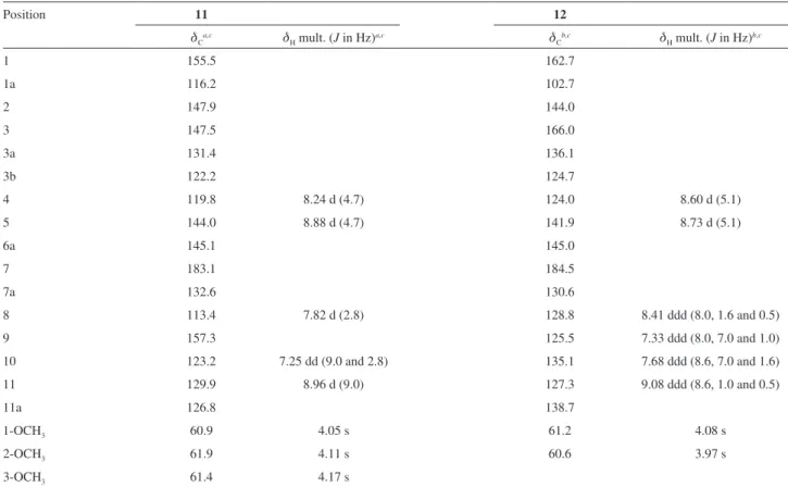
Compound 11 was obtained as an orange amorphous powder which was positive to the Dragendorff’s test. Its UV-vis spectrum showed absorption peaks at 204, 238,
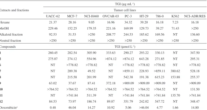
Table 1. Antiproliferative activity of extracts, fractions and major compounds against cancer cell lines
Extracts and fractions
TGI (µg mL-1)
Tumor cell lines
UACC-62 MCF-7 NCI-H460 OVCAR-03 PC-3 HT-29 786-0 K562 NCI-ADR/RES
Hexane 21.37 28.16 9.05 16.96 34.32 39.20 16.18 7.23 16.18
MeOH 229.46 152.25 179.35 221.16 169.99 129.73 39.27 71.43 >250
Alkaloid fraction 92.53 51.53 >250 208.77 244.53 185.62 169.56 NT 136.60
Neutral fraction >250 >250 >250 >250 >250 >250 >250 >250 >250
Compounds TGI (µmol L-1)
1 260.45 282.54 305.90 333.63 290.27 293.22 330.13 NT 347.50
2 275.07 274.12 554.96 >874.12 >874.12 643.28 271.85 NT 295.31
6 NT >778.82 >778.82 NT >778.82 >778.82 >778.82 NT >778.82
7 NT 289.38 49.52 NT >859.11 228.93 >859.11 380.62 528.18
8 NT 215.58 201.99 NT 542.38 191.38 615.23 153.88 255.37
9 63.02 37.67 87.41 372.18 >909.09 >909.09 >909.09 NT >909.09
10 >764.52 >764.52 >764.52 >764.52 >764.52 >764.52 >764.52 NT 131.50
11 NT >741.84 511.39 NT >741.84 >741.84 >741.84 135.70 >741.84
12 84.53 73.97 186.74 89.87 351.79 242.02 547.72 NT 348.47
Doxorubicina 0.48 46.04 14.27 10.92 5.06 >46.04 4.77 1.66 14.80
a Positive control; NT (Not tested); UACC-62 (melanoma), MCF-7 (breast), NCI-H460 (lung, non-small cells), OVCAR-03 (ovarian), PC-3 (prostate), HT-29
(colon), 786-0 (renal), K562 (leukemia), and NCI-ADR/RES (ovarian expressing phenotype multiple drugs resistance); TGI (Total Growth Inhibition).
Costa et al. 1115 Vol. 22, No. 6, 2011
278 and 472 nm, which are typical of molecules with an oxoaporphine skeleton. The IR spectrum showed absorption bands at 3431 and 1646 cm-1, characteristic of phenolic
hydroxyl and carbonyl groups. The 1H NMR spectrum
indicated a tetrasubstituted-oxoaporphine alkaloid. Its
1H NMR spectrum revealed the presence of a pair of
doublets at d 8.88 and d 8.24 (H-5 and H-4, J 4.7 Hz, respectively), which was consistent with the presence of a pyridine system. Additionally, it was found to have a spin system consisting of three hydrogens with the signals at
d 8.96 (1H, d, J 9.0 Hz), d 7.82 (1H, d, J 2.8 Hz), and d 7.25 (1H, dd, J 9.0 and 2.8 Hz), which were attributed to H-11, H-8, and H-10, respectively, and indicated substitution in the D ring. The signals at d 4.05, d 4.11 and d 4.17 (each 3H, s) were assigned to three methoxyl groups located in the A ring, according to long-range 1H-13C correlation in
the HMBC NMR experiment (Table 2). The presence of a hydroxyl group in the molecule located in the D ring at C-9 was established on the basis of the long-range 1H-13C
correlation of the hydrogen H-11 at d 8.96 with the carbon C-9 at d 157.3, which showed no correlation with the methoxyl groups.
The 13C NMR experiment along with HSQC and HMBC
experiments allowed to verify the presence of 19 carbons,
including a carbonyl group at d 183.1, 15 aromatic carbons between d 157.3-113.4, and three methoxyl groups at
d 61.9, d 61.4, and d 60.9, which was in agreement with structure 11 (Table 2). The hydrogen H-8 at d 7.82 showed strong long-range 1H-13C correlation with the carbon C-7
at d 183.1, which conirmed the oxoaporphine skeleton. According with 1H and 13C NMR 1D/2D this compound was
identiied as the oxoaporphine alkaloid known subsessiline. This compound has been found only in two species of Annonaceae Guatteria ouregou25 and G. subsessilis,26 but
the physical data are incomplete. The 13C NMR data are
reported for the irst time. The complete physical data for this compound are described in this work.
Compound 12 was obtained as a blue amorphous powder. Its UV-Vis spectrum showed absorption peaks at 204, 238, 282, 304, 468 and 595 nm, which are typical of molecules with an oxoaporphine skeleton. The IR spectrum showed absorption bands at 3325 and 1660 cm-1,
characteristic of phenolic hydroxyl and carbonyl groups. The 1H and 13C NMR spectra of 12 were very similar to
those of 11, except for the absence of a methoxyl signal, which was replaced by a hydroxyl group, and absence of substitution in the D ring, in structure 12 according to the IR spectrum and NMR data (Table 2). The absence of
Table 2. NMR spectroscopy data (400 MHz) for compounds 11 and 12
Position 11 12
dCa,c dH mult. (J in Hz)a,c dCb,c dH mult. (J in Hz)b,c
1 155.5 162.7
1a 116.2 102.7
2 147.9 144.0
3 147.5 166.0
3a 131.4 136.1
3b 122.2 124.7
4 119.8 8.24 d (4.7) 124.0 8.60 d (5.1)
5 144.0 8.88 d (4.7) 141.9 8.73 d (5.1)
6a 145.1 145.0
7 183.1 184.5
7a 132.6 130.6
8 113.4 7.82 d (2.8) 128.8 8.41 ddd (8.0, 1.6 and 0.5)
9 157.3 125.5 7.33 ddd (8.0, 7.0 and 1.0)
10 123.2 7.25 dd (9.0 and 2.8) 135.1 7.68 ddd (8.6, 7.0 and 1.6)
11 129.9 8.96 d (9.0) 127.3 9.08 ddd (8.6, 1.0 and 0.5)
11a 126.8 138.7
1-OCH3 60.9 4.05 s 61.2 4.08 s
2-OCH3 61.9 4.11 s 60.6 3.97 s
3-OCH3 61.4 4.17 s
The experiments were obtained at 293 K and TMS as internal reference (0.00 ppm) in a CDCl
3 + drops of CD3OD or b CD3OD. c Long-range 1H-13C HMBC
Chemical Constituents Isolated from the Bark of Guatteria blepharophylla (Annonaceae) J. Braz. Chem. Soc.
1116
substitution in the D ring was conirmed by the presence of four adjacent aromatic hydrogens at d 9.08 (1H, ddd, J 8.6, 1.0 and 0.5 Hz), d 8.41 (1H, ddd, J 8.0, 1.6 and 0.5 Hz),
d 7.68 (1H, ddd, J 8.6, 7.0 and 1.6 Hz), and d 7.33 (1H, ddd, J 8.0, 7,0 and 1.0 Hz) which were attributed to H-11, H-8, H-10, and H-9, respectively (Table 2). The position of the hydroxyl group at C-3 was established on the basis of the long-range 1H-13C correlation of the hydrogen H-4
at d 8.60 with the carbon C-3 at d 166.0, which showed no correlation with the methoxyl groups. According with
1H and 13C NMR 1D/2D, this compound was identiied
as the oxoaporphine alkaloid known isomoschatoline. As well as observed for 11, the physical data published for this compound are incomplete and presented in this work. The
13C NMR data is reported for the irst time. This compound
has been found only in three species of Annonaceae Uvaria mocoli,27 Guatteria melosma and Cleistopholis patens.28
The isolated compounds 1-2 and 6-12 were evaluated for in vitro antiproliferative activity against nine human tumour cell lines (Table 1), while compounds 6, 7, 10 and 12 were also tested for antimicrobial activity. Compound 9 showed the higher antiproliferative activity for breast (MCF-7) with a TGI value of 37.67 µmol L-1, more active than
positive control doxorubicin (TGI value of 46.04 µmol L-1).
Compound 7 presented signiicant antiproliferative activity for lung, non-small cells (NCI-H460) with a TGI value of 49.52 µmol L-1 (doxorubicin, TGI value of 14.27 µmol L-1),
while compound 12 showed signiicant activity for breast (MCF-7) with TGI value of 73.97 µmol L-1. Compound
10 showed selective activity for ovarian expressing phenotype for multiple drug resistance (NCI-ADR/RES) with a TGI value of 131.50 µmol L-1, but was less
active than doxorubicin (TGI value of 14.80 µmol L-1
(Table 1). Compounds 9 and 12 also showed signiicant antiproliferative activity against different tumor cell lines with TGI values below to 100 µmol L-1 (Table 1).
It is important to notice that compounds 6, 7, 9, 11
and 12 shared the same basic skeleton with different substitution patterns. This way, our results suggested that a methoxylated substitute at R3 reduces antiproliferative
activity (compound 11) or even results in an inactive compounds as 6, but a hydroxyl group at R3 favored the
activity (compound 12). The best results were obtained for methylenedioxy group (compound 9) or methoxy groups (compounds 7 and 12) at R1 and R2.
No signiicant antibacterial activity was observed for the tested compounds. The only signiicant antimicrobial result was observed for compound 12 that showed antifungal activity against C. albicans (MIC value of 50.81 µmol L-1)
similar to the positive control nystatin (MIC value of 54.00 µmol L-1).
Conclusion
T h e c h e m i c a l i nve s t i ga t i o n o f t h e b a r k o f G. blepharophylla has resulted in the isolation of several compounds common in the taxon Guatteria, such as, 1,
3, 6, 7 and 9 that could be considered chemotaxonomic markers of this genus. Compounds 1-6, 11 and 12 are being reported for the irst time in this species, and are important for the chemotaxonomy of Annonaceae family. The signiicant in vitro antiproliferative results obtained against several human tumour cell lines and antimicrobial activities demonstrated by the major compounds indicate that this species is a natural source of biologically active compounds. Compound 12 showed signiicant antifungal and antiproliferative activities against C. albicans and human tumor cell lines of breast (MCF-7). Therefore, studies involving mechanisms of action are necessary to fully understand its biological signiicance.
Supplementary Information
Supplementary data including physical data for compounds 1-10 and 1H, 13C NMR, HSQC and HMBC
for compounds 11 and 12 are available free of charge at http://jbcs.sbq.org.br as a PDF ile.
Acknowledgements
The authors are grateful to Prof. Dr. A. C. Webber of the Universidade Federal do Amazonas (UFAM) for the botanical identiication, as well as, to CAPES, CNPq and Fundação Araucária for inancial support.
References
1. Erkens, R. H. J.; Maas, P. J. M.; Rodriguésia 2008, 59, 401. 2. Corrêa, M. P.; Dicionário das Plantas Úteis do Brasil e das
Exóticas Cultivadas, IBDF: Rio de Janeiro, Brasil, 1984. 3. Silva, D. B.; Tulli, E. C. O.; Militão, G. C. G.; Costa-Lotufo,
L. V.; Pessoa, C.; Moraes, M. O.; Albuquerque, S.; Siqueira, J. M.; Phytomedicine 2009, 16, 1059.
4. Hsieh, T.-J.; Chang, F.-R.; Chia, Y.-C.; Chen, C.-Y.; Chiu, H.-F.; Wu, Y.-C.; J. Nat. Prod. 2001, 64, 616.
5. Muhammad, I.; Dunbar, D. C.; Takamatsu, S.; Walker, L. A.; Clark, A. M.; J. Nat. Prod. 2001, 64, 559.
6. Costa, E. V.; Marques, F. A.; Pinheiro, M. L. B.; Vaz, N. P.; Duarte, M. C. T.; Delarmelina, C.; Braga, R. M.; Maia, B. H. L. N. S.; J. Nat. Prod. 2009a, 72, 1516.
Costa et al. 1117 Vol. 22, No. 6, 2011
8. Costa, E. V.; Teixeira, S. D.; Marques, F. A.; Duarte, M. C. T.; Delarmelina, C.; Pinheiro, M. L. B.; Trigo, J. R.; Maia, B. H. L. N. S.; Phytochemistry 2008, 69, 1895.
9. Costa, E. V.; Pinheiro, M. L. B.; Barison, A.; Campos, F. R.; Salvador, M. J.; Maia, B. H. L. N. S.; Cabral, E. C.; Eberlin, M. N.; J. Nat. Prod. 2010, 73, 1180.
10. Costa, E. V.; Pinheiro, M. L. B.; Xavier, C. M.; Silva, J. R. A.; Amaral, A. C. F.; Souza, A. D. L.; Barison, A.; Campos, F. R.; Ferreira, A. G.; Machado, G. M. C.; Leon, L. L. P. J.; J. Nat. Prod. 2006, 69, 292.
11. Montenegro, H.; Gutiérrez, M.; Romero, L. I.; Ortega-Barría, E.; Capson, T. L.; Rios, L.C.; Planta Med. 2003, 69, 677. 12. Mahiou, V.; Roblot, F.; Fournet, A.; Hocquemiller, R.;
Phytochemistry 2000, 54, 709.
13. Boyom, F. F.; Ngouana, V.; Zollo, P. H. A.; Menut, C.; Bessiere, J. M.; Gut, J.; Rosenthal, P. J.; Phytochemistry 2003, 64, 1269. 14. Waechter, A.-I.; Cavé, A.; Hocquemiller; R.; Bories, C.; Muñoz,
V.; Fournet, A.; Phytother. Res. 1999, 13, 175.
15. Costa, E. V.; Pinheiro, M. L. B.; Marques, F. A.; Braga, R. M.; Maia, B. H. L. N. S.; Biochem. Syst. Ecol. 2009c, 37, 43. 16. Maia, J. G. S.; Andrade, E. H. A.; Carreira, L. M. M.; Oliveira,
J.; Araújo, J. S.; Flavour Frag. J. 2005, 20, 478. 17. Shoemaker, R. H.; Nat. Rev. Cancer 2006, 6, 813. 18. Ellof, J. N.; Planta Med. 1998, 64, 711.
19. Ragasa, C. Y.; Ganzon, J.; Hoileña, J.; Tamboong, B.; Rideout, J. A.; Chem. Pharm. Bull. 2003, 51, 1208.
20. Micheletti, A. C.; Beatriz, A.; Lima, D. P.; Honda, N. K.; Pessoa, C. O.; Moraes, M. O.; Lotufo, L. V.; Magalhães, H. I. F.; Carvalho, N. C. P.; Quim. Nova 2009, 32, 12.
21. Facundo, V. A.; Polli, A. R.; Rodrigues, R. V.; Militão, J. S. L. T.; Stabelli, R. G.; Cardoso, C. T.; Acta Amaz. 2008, 38, 733. 22. Guinaudeau, H.; Leboeuf, M.; Cavé, A.; J. Nat. Prod. 1983, 46,
761.
23. Harrigan, G. G.; Gunatilaka, A. A. L.; Kingston, D. G. I.; Chan, G. W.; Johnson, R. K.; J. Nat. Prod. 1994, 57, 68.
24. Arriaga, A. M. C.; Feitosa, E. M. A.; Lemos, T. L. G.; Santiago, G. M. P.; Lima, J. Q.; De Oliveira, M. C. F.; Vasconcelos, J. N. E.; Rodrigues, F. E. A.; Gomes, T. B. M.; Braz-Filho, R.; Nat. Prod. Commun. 2008, 3, 1687.
25. Cortes, D.; Hocquemiller, R.; Leboeuf, M.; Cavé, A.; Morreti, C.; J. Nat. Prod. 1986, 49, 878.
26. Hasegawa, M.; Sojo, M.; Lira, A.; Marques, C.; Acta Cient. Venez. 1972, 23, 165.
27. Fleischer, T. C.; Waigh, R. D.; Waterman, P. G.; Phytochemistry
1998, 47, 1387.
28. Atti, S. A.-E.; Ammar, H. A.; Phoebe Jr., C.H.; Schiff Jr., P. L.; Slatkin, D. J.; J. Nat. Prod. 1982, 45, 476.
Submitted: July 14, 2010 Published online: February 17, 2011
Supplementary Information
S
I
J. Braz. Chem. Soc., Vol. 22, No. 6, S1-S15, 2011.Printed in Brazil - ©2011 Sociedade Brasileira de Química 0103 - 5053 $6.00+0.00
*e-mail: [email protected]
Chemical Constituents Isolated from the Bark of
Guatteria blepharophylla
(Annonaceae) and their Antiproliferative and Antimicrobial Activities
Emmanoel V. Costa,a,b Francisco de Assis Marques,a Maria Lúcia B. Pinheiro,c
Raquel M. Braga,d Camila Delarmelina,e Marta Cristina T. Duarte,e Ana Lúcia T. G. Ruiz,f
João Ernesto de Carvalhof and Beatriz H. L. N. S. Maia*,a
aDepartamento de Química, Universidade Federal do Paraná, CP 19081, 81531-990 Curitiba-PR, Brazil
bDepartamento de Química, Universidade Federal de Sergipe, 49100-000 São Cristovão-SE, Brazil
cDepartamento de Química, Universidade Federal do Amazonas, 69077-000 Manaus-AM, Brazil
dInstituto de Química, Universidade Estadual de Campinas, CP 6154, 13083-970 Campinas-SP, Brazil
eDivisão de Microbiologia and fDivisão de Farmacologia e Toxicologia, CPQBA, Universidade
Estadual de Campinas, CP 6171, 13083-970 Campinas-SP, Brazil
Table S1. Chemical constituents isolated from the bark of Guatteria blepharophylla and the respective morphology and data spectra numbering (Figure S_)
Caryophyllene oxide (1): Colorless oil. EI-MS m/z 220 [M]+. 1H NMR (S1). 13C NMR (S2).
Lichexanthone (2): Light yellow needles (CHCl3). Mp 189-190 oC. 1H NMR (S3). 13C NMR (S4).
Spathulenol (3): Colorless oil. EI-MS m/z 220 [M]+. 1H NMR (S5). 13C NMR (S6).
Mixture ofβ-sitosterol (4) and stigmasterol (5): White needles (Hexane:CH2Cl2 2:1). 1H NMR (S7). 13C NMR (S8).
O-methylmoschatoline (6): Orange needles (CHCl3); mp 182-183 oC. 1H NMR (S9). 13C NMR (S10).
Lysicamine (7): Yellow needles (CHCl3); mp 186-187 oC. 1H NMR (S11). 13C NMR (S12).
Nornuciferine (8): Brown amorphous solid. 1H NMR (S13). 13C NMR (S14).
Liriodenine (9): Yellow needles (CHCl3:MeOH 2:1); mp 279-280 oC. 1H NMR (S15). HSQC (S16). HMBC (S17).
Isocoreximine (10): Light yellowish prisms (CHCl3:MeOH 2:1); mp 241-242 oC. 1H NMR (S18). 13C NMR (S19).
Subsessiline (11): Orange needles (CHCl3:MeOH 2:1). 1H NMR (S20). 13C NMR (S21). HSQC (S22). HMBC (S23).
Chemical Constituents Isolated from the Bark of Guatteria blepharophylla (Annonaceae) J. Braz. Chem. Soc.
S2
Figure S1. 1H NMR spectrum of compound 1 in CDCl
3 at 400 MHz.
Figure S2. 13C{1H} NMR spectrum of compound 1 in CDCl
Costa et al. S3 Vol. 22, No. 6, 2011
Figure S3. 1H NMR spectrum of compound 2 in CDCl
3 at 400 MHz.
Figure S4. 13C{1H} NMR spectrum of compound 2 in CDCl
Chemical Constituents Isolated from the Bark of Guatteria blepharophylla (Annonaceae) J. Braz. Chem. Soc.
S4
Figure S5. 1H NMR spectrum of compound 3 in CDCl
3 at 400 MHz.
Figure S6. 13C{1H} NMR spectrum of compound 3 in CDCl
Costa et al. S5 Vol. 22, No. 6, 2011
Figure S7. 1H NMR spectrum of the mixture of compounds 4 and 5 in CDCl
3 at 200 MHz.
Figure S8. 13C{1H} NMR spectrum of the mixture of compounds 4 and 5 in CDCl
Chemical Constituents Isolated from the Bark of Guatteria blepharophylla (Annonaceae) J. Braz. Chem. Soc.
S6
Figure S9. 1H NMR spectrum of compound 6 in CDCl
3 at 400 MHz.
Figure S10. 13C{1H} NMR spectrum of compound 6 in CDCl
Costa et al. S7 Vol. 22, No. 6, 2011
Figure S11. 1H NMR spectrum of compound 7 in CDCl
3 at 400 MHz.
Figure S12. 13C{1H} NMR spectrum of compound 7 in CDCl
Chemical Constituents Isolated from the Bark of Guatteria blepharophylla (Annonaceae) J. Braz. Chem. Soc.
S8
Figure S13. 1H NMR spectrum of compound 8 in CDCl
3 at 400 MHz.
Figure S14. 13C{1H} NMR spectrum of compound 8 in CDCl
Costa et al. S9 Vol. 22, No. 6, 2011
Figure S15. 1H NMR spectrum of compound 9 in CDCl
3 at 400 MHz.
Figure S16. 1H-13C one-bond correlation map from HSQC NMR experiment of compound 9 in CDCl
Chemical Constituents Isolated from the Bark of Guatteria blepharophylla (Annonaceae) J. Braz. Chem. Soc.
S10
Figure S17. 1H-13C long-range correlation map from HMBC NMR experiment of compound 9 in CDCl
3 at 400 and 100 MHz.
Figure S18. 1H NMR spectrum of compound 10 in CDCl
Costa et al. S11 Vol. 22, No. 6, 2011
Figure S19. 13C{1H} NMR spectrum of compound 10 in CDCl
3 + drops of CD3OD at 100 MHz.
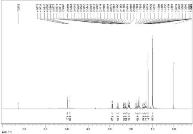
Figure S20. 1H NMR spectrum of compound 11 in CDCl
Chemical Constituents Isolated from the Bark of Guatteria blepharophylla (Annonaceae) J. Braz. Chem. Soc.
S12
Figure S21. 13C{1H} NMR spectrum of compound 11 in CDCl
3 + drops of CD3OD at 100 MHz.
Figure S22. 1H-13C one-bond correlation map from HSQC NMR experiment of compound 11 in CDCl
Costa et al. S13 Vol. 22, No. 6, 2011
Figure S23. 1H-13C long-range correlation map from HMBC NMR experiment of compound 11 in CDCl
3 + drops of CD3OD at 400 and 100 MHz.
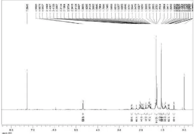
Figure S24. 1H NMR spectrum of compound 12 in CD
Chemical Constituents Isolated from the Bark of Guatteria blepharophylla (Annonaceae) J. Braz. Chem. Soc.
S14
Figure S25. 13C{1H} NMR spectrum of compound 12 in CD
3OD at 100 MHz.
Figure S26. 1H-13C one-bond correlation map from HSQC NMR experiment of compound 12 in CD
Costa et al. S15 Vol. 22, No. 6, 2011
Figure S27.1H-13C long-range correlation map from HMBC NMR experiment of compound 12 in CD