Hypoxia Pretreatment of Bone Marrow
Mesenchymal Stem Cells Facilitates
Angiogenesis by Improving the Function of
Endothelial Cells in Diabetic Rats with Lower
Ischemia
Jiejie Liu1☯, Haojie Hao1☯, Lei Xia3☯, Dongdong Ti1, Hong Huang1, Liang Dong1, Chuan Tong1, Qian Hou1, Yali Zhao2, Huiling Liu1, Xiaobing Fu1*, Weidong Han1*
1Institute of Basic Medicine Science, College of Life Science, Chinese PLA General Hospital, Beijing, China,2Central laboratory, Hainan branch of Chinese PLA General Hospital, Sanya, China,3Department of Medical Administration, Chinese PLA General Hospital, Beijing, China
☯These authors contributed equally to this work.
*[email protected](WH); ([email protected](XF)
Abstract
Endothelial dysfunction induced by unordered metabolism results in vascular reconstruc-tion challenges in diabetic lower limb ischemia (DLLI). Mesenchymal stem cells (MSCs) are multipotent secretory cells that are suitable for clinical DLLI treatment, but their use has been hampered by poor survival after injection. Hypoxia can significantly enhance the ca-pacity of MSCs to secrete angiogenic factors. We investigated transient hypoxia pretreat-ment of MSCs to facilitate revascularization in DLLI. Rat bone marrow MSCs (BM-MSCs) were cultured at different oxygen concentrations for varying time periods. The results indi-cated that transient pretreatment (5% O2, 48 h) not only increased the expression of
VEGF-1α, ANG, HIF-1αand MMP-9 in BM-MSCs as assessed by real-time RT-PCR, but also in-creased the expression of Bcl-2 as determined by western blotting. The transplantation of pretreated BM-MSCs into rats with DLLI demonstrated accelerated vascular reconstruction when assayed by angiography and immunohistochemistry. CM-Dil-labeled tracer experi-ments indicated that the survival of BM-MSCs was significantly improved, with approximate-ly 5% of the injected cells remaining alive at 14 days. The expression levels of VEGF-1α, MMP-9 and VEGF-R were significantly increased, and the expression of pAKT was up-reg-ulated in ischemic muscle. Double immunofluorescence studies confirmed that the pre-treated BM-MSCs promoted the proliferation and inhibited the apoptosis of endothelial cells. In vitro, pretreated BM-MSCs increased the migratory and tube forming capacity of endothelial cells (ECs). Hypoxia pretreatment of BM-MSCs significantly improved angio-genesis in response to tissue ischemia by ameliorating endothelial cell dysfunction and is a promising therapeutic treatment for DLLI.
OPEN ACCESS
Citation:Liu J, Hao H, Xia L, Ti D, Huang H, Dong L, et al. (2015) Hypoxia Pretreatment of Bone Marrow Mesenchymal Stem Cells Facilitates Angiogenesis by Improving the Function of Endothelial Cells in Diabetic Rats with Lower Ischemia. PLoS ONE 10(5): e0126715. doi:10.1371/journal.pone.0126715
Academic Editor:Giovanni Camussi, University of Torino, ITALY
Received:September 19, 2014
Accepted:April 7, 2015
Published:May 21, 2015
Copyright:© 2015 Liu et al. This is an open access article distributed under the terms of theCreative Commons Attribution License, which permits unrestricted use, distribution, and reproduction in any medium, provided the original author and source are credited.
Data Availability Statement:All relevant data are within the paper and its Supporting Information files.
Introduction
Lower limb ischemia, a common complication in Type 2 diabetes (T2D), afflicts approximately 15% of the diabetic patient population worldwide [1,2]. The dysregulation of vascular remod-eling and vascular growth is induced by hyperglycemia and results in damage to endothelial cells (ECs) [3,4]. The dysfunction of ECs generates decreased responsiveness to ischemic/hyp-oxic stimuli, impaired or abnormal neovascularization, and a lack of endothelial regeneration and is associated with peripheral microvascular complications in diabetes [5]. Thus, a key strat-egy for restoring blood flow is the repair of dysfunctional ECs.
Previous reports have demonstrated the positive effects of ECs in vascular network recon-struction in terms of angiogenesis for lower limb ischemic lesions [6]. The complicated patho-physiological changes that occur as a result of diabetes could exacerbate the functional damage of ECs, contributing to impaired re-endothelialization and vascular recovery. Some strategies attempt to recover the mobilization, adhesion, tube formation and proliferation of ECs by modifying relevant molecular pathways, including the eNOS, CXCR-4 and hemeoxygenase-1 (HO-1) pathways [7–9]. Other proangiogenesis factors such as VEGF, HGF, and proangiogen-esis protein MMP-9 repair endothelial dysfunction by improving the ability of ECs to migrate and proliferate [10–12]. Enhanced angiogenesis after VEGF treatment was accompanied by the increased expression of the adhesion protein N-cadherin, which mediates endothelial-pericytic interactions, in ischemic brain microvessels [13]. However, continuous hyperglycemia causes defective VEGF signaling, including the inactivation of the VEGF receptor FLK-1 [14], which affects endothelial growth and migration as well as monocytes and ECs recruitment and re-leased from bone marrow in DLLI. Due to defective VEGF signaling that results in the low bio-availability of VEGF, the effect of providing VEGF to promote angiogenesis should
further explored.
MSCs are able to function as paracrine or autocrine signals that induce the release of proan-giogenic cytokines, resulting in the stimulation of resident ECs to migrate, differentiate, and proliferatein situ, and are thought to influence neovascularization.In vitro, MSCs secrete
VEGF and other angiogenic factors that can promote endothelial cell migration, proliferation and tube formation [15].In vivo, the mechanisms underlying MSCs improvement of cardiac
functions may involve neovascularization that induced by the parasecretion of growth factors in myocardial infarcted rats with occlusion of the left anterior descending artery [16]. Further-more, to increase the therapeutic potential of MSCs, alternative methods have been investigat-ed to enhance their ability to secrete proangiogenic factors. For instance, the transplantation of VEGF-1αgene-transfected MSCs improved the treatment of myocardial perfusion and the res-toration of heart function in ischemic heart models of inbred rats [17,18]. To avoid the risk of genetic modification, overexpressed the angiogenic factors on MSCs to promote angiogenesis after DLLI treatment.
The O2concentration of MSCs in the bone marrow is 2%-7% under physiologic conditions
[19,20].In vitro, MSCs are cultured at an ambient O2concentration (21% O2), which is
unsuit-able for secretion activity. Once the cells that expandedin vitrowere transplanted and localized
to the ischemic tissue, the MSCs encountered severely hypoxic conditions, ranging from 0.4% to 2.3% O2, which often resulted in apoptosis [21]. Previous studies have demonstrated that
hypoxia-induced apoptosis can be circumvented by preconditioning cells in less severe hypoxic conditions for a period of time before exposing them to severe ischemia at the site of injury in other cell types. MSCs exposed to a hypoxic cultural environment at 0.5% O2after 24 h and 48
h were in a state of prosurvival and adaptation [22]. Research exploring the mechanism shows that MSCs cultured under hypoxic conditions and transplanted to a subcutaneous polyvinyl al-cohol sponge model exhibited increased paracrine production of VEGF and ANG [23]. The Competing Interests:All authors have declared that
reports suggested that, in addition to maintaining their viability when cultured in physiologic niche conditions, MSCs also increased their secretion after an initial lag phase [24,25]. There-fore, an exploration of the effects of an appropriate physiological environment on the ability of MSCs to secrete proangiogenic factors and maintain quiescence when transplanted into ische-mic tissue is worthwhile.
In our study, we identified that a suitable physiologic niche treatment of 5% O2for 48 h for
BM-MSCs significantly increased the secretion of proangiogenesis factors and enhanced angio-genesis in ischemic tissue by ameliorating endothelial dysfunction for DLLI treatment.
Materials and Methods
Isolation and culture of BM-MSCs
Sprague–Dawley (SD) rats were purchased from the Chinese PLA General Hospital and were housed under specific pathogen-free conditions in the animal center of Chinese PLA General Hospital. All animal experiments were carried out in accordance with the National Institutes of Health Guide for Care and Use of Laboratory Animals and were approved by the Animal Care and Use Committee at the Chinese PLA General Hospital. MSCs were generated from the bone marrow of adult male SD rats, aged 4–6 weeks and weighing 120–150 g, according to a previously published protocol [26]. Briefly, the bone marrow aspirate was obtained by flushing the rat femurs and tibias with fetal bovine serum (FBS)-free low glucose (LG)-DMEM medium (Invitrogen, Carlsbad, USA). Then, the aspirate samples were centrifuged and re-suspended in LG-DMEM medium with 10% FBS and incubated at 37°C in a humidified chamber with 5% CO2. The culture media was completely replaced every 3 days, and the non-adherent cells were
discarded. The MSCs were recognized by their ability to proliferate in culture with an attached well-spread morphology. Once the cells were more than 80% confluent, the adherent cells were detached and re-plated at a 1:3 dilution in culture flasks.
Cell apoptosis by flow cytometry
For hypoxia treatment, the cells were subcultured at a 1:4 dilution and cultured for 3 days until confluent. Fresh complete medium was added prior hypoxia treatment, which was performed in a well characterized, finely controlled pro-C-O—chamber (Thermo) for 24 h. The oxygen concentration in the chamber was maintained at 2%, 5% or 7% with a residual gas mixture composed of 5% CO2and balanced nitrogen. After hypoxia pre-conditioning, the MSCs were
collected, washed with PBS, resuspended in PBS at 1×10⁶/mL, and stained with Annexin V and propidium iodide solution (PI, BD Biosciences Pharmingen, San Diego, USA) for 15 min in the dark. Apoptotic cells were then analyzed by flow cytometry (BD Biosciences Pharmingen, San Diego, USA).
Real-time RT-PCR
Rat diabetic and ischemic hind limb model
To induce moderate diabetes, SD rats (8-weeks old, n = 25) were injected intraperitoneally with 65 mg/kg of streptozotocin (STZ, Sigma, St. Louis, MO, USA) in 0.9% sterile saline daily for 3 days. Blood glucose levels were monitored at 0, 3, 5, and 7 days after the initial injection. Rats with glucose levels less than 250 mg/dL after 3 days of STZ treatment were excluded from further studies. The diabetic rats then underwent unilateral hind limb ischemia 7 days after the initial STZ injection. Briefly, after intraperitoneal injection of 3% sodium pentobarbital anes-thesia, a longitudinal incision was made along the left medial thigh to allow the isolation, liga-tion, and excision of the femoral artery from its origin directly above the inguinal ligament to its bifurcation at the origin of the popliteal and saphenous arteries.
Local transplantation of BM-MSCs
BM-MSCs for transplantation were labeled with CM-Dil (Invitrogen, Carlsbad, USA) accord-ing to the manufacturer's instructions. The ischemic, diabetic animals were immediately ran-domly assigned into 3 groups and received intramuscular injections (100μl in saline) of either
4×10⁶of hypoxia-treated, Dil-labeled BM-MSCs; normoxic, Dil-labeled BM-MSCs; or a blank sample as a negative control. The injection points were evenly distributed among six injections in the ischemic muscle along the femoral artery. The skin incision was closed with 2–0 inter-rupted silk sutures (Ethicon), and all animals were closely monitored during the
postoperative period.
Angiography
The SD rats were anesthetized with sodium pentobarbital and fixed to the sample table. The ab-dominal cavity was opened, and a catheter was inserted downward from the abab-dominal aorta for the injection of the contrast medium. The animals were transferred onto an OEC9800 (GE, USA), and the X-ray was adjusted to the target area, followed by the injection of contrast agent through the indwelling catheter and instantaneous imaging.
Laser Doppler perfusion imaging
Blood flow in DLLI was mapped with a high resolution pericam PSI Laser Doppler perfusion imager (Perimed Inc., NorthRoyalton). The parameter settings during measurement were as following: scanning area, 50 mm×50 mm; high resolution; distance between the scanner head and wound, 15 cm; temperature. The measurement of the image and perfusion value was car-ried out with LDISOFT software.
Table 1. List of primers used for real-time RT-PCR.
Gene name Forward(5’- 3’) Reverse(5’- 3’)
PDGF GTCCAGGTGAGGTTAGAGG CACGGAGGAGAACAAAGAC
HIF-1α-F AAGTCTAGGGATGCAGCAC CAAGATCACCAGCATCTAG
VEGF-1a CAGCTATTGCCGTCCAATTGA CCAGGGCTTCATCATTGCA
Mmp-9 TGACATCTATGCAATGGGCTTAGTAT CTTGGTGGATTVVGCCAAT
HGF GAGGAGAAACGCAAACAG ACGACCAGGAACAATGAC
β-actin GAG ACC TTC AAC ACC CCA GCC AATGTCACGCACGATTTCCC
Western blot analysis
Muscle tissues and co-cultured cells were lysed in protein lysis buffer (Sigma, St. Louis, MO, USA). The protein lysates were loaded onto 8%-15% sodium dodecyl sulfate polyacrylamide gels, and then, the separated proteins were transferred to polyvinylidene difluoride (PVDF) membranes, blocked, and incubated overnight at 4°C with primary antibodies. The membranes were washed and then incubated with a secondary antibody for 1 hour at room temperature using an orbital shaker. After washing, the bands were detected using an enhanced chemilumi-nescence reagent (Santa Cruz Biotechnology, Santa Cruz, USA).
Histological analysis and immunohistochemistry
The rats were sacrificed by the intraperitoneal administration of an overdose of pentobarbital at the indicated time points, and the ischemic thigh muscles were carefully excised and post-fixed in 4% paraformaldehyde or paraffin. For morphometric analysis, the muscle fiber num-ber and size were examined in sections stained with hematoxylin-eosin, with the averaging of the counts from 10 separate fields in 5 different areas of each specimen. For histological analy-sis of vascularization and cell surface markers, 5-mm paraffin sections were incubated for 16 h at 4°C in a 1:250 dilution of an vWF antibody (Abcam., Cambridge, USA) with an anti-body diluent (Invitrogen, Carlsbad, USA) and then for 60 min at RT with a 1:300 dilution of a biotinylated goat anti-rabbit IgG antibody (Invitrogen, Carlsbad, USA) with PBS, followed by development with DAB (Sigma-Aldrich, St. Louis, USA). Five fields from each tissue section were randomly selected, and the number of microvessels was counted. To analyze the mecha-nism through which the injected BM-MSCs promoted angiogenesis, serial frozen section were incubated overnight at 4°C with rabbit antibodies against either vWF (1:150 dilution in anti-body diluent) or Ki67 (1:100 in antianti-body diluent; BD Biosciences Pharmingen, San Diego, USA); then, the sections were incubated with FITC-conjugated goat anti-rabbit IgG (1:200 di-lution in PBS-NGS; Invitrogen, Carlsbad, USA) or FITC-conjugated goat anti-mouse IgG at 37°C for 1 h.
TUNEL staining
Apoptotic cells were identified by terminal dUTP nick-end labeling (TUNEL) staining accord-ing to the manufacturer's protocol usaccord-ing a commercially available kit (Molecular Probes, USA). The sections were counterstained with Hoechst 33342 (Sigma-Aldrich, St. Louis, USA) to visu-alize all nuclei and were viewed under a fluorescence microscope (Olympus).
Construction of siRNA-expressing vectors
VEGF-1α-specific target sequences were chosen according to online siRNA tools from Invitro-gen using the VEGF-1αreference sequence. The target sequences for VEGF-1αare as follows: sense1 5'-CACCGGGATGACCTTGTAGTGAAGGCGAACCTTCACTACAAGGTCATCC C-3'; sense2 5'- CACCGCTGTGCCTTTCACAGTTTCTCGAAAGAAACTGTGAAAGGCA CAGC-3';、antisense 5'-CACCGATACAGTAGTAGTAGGACCTGCGAACAGGTCCTAC
TACTACTGTA-3'. The siRNAs were chemically synthesized, and a lentiviral vector was con-structed as described by the Invitrogen, Carlsbad, USA lentiviral vector protocol. The correct insertion of the specific siRNA was confirmed by sequencing (Invitrogen, Carlsbad, USA).
Cell migration and tube formation assay
Type Culture Collection (CCTCC) in Wuhan. The cells were cultured in RPMI 1640 medium complemented with 10% heat-inactivated fetal bovine serum, penicillin (100 IU/ml), strepto-mycin (100 mg/ml) and 2 mM L-glutamine in a humidified CO2incubator with 5% CO2at
37°C. HUVECs were grown in a 24-well dish, and wounds were inflicted by dragging a sterile pipette tip across the monolayer, creating a 350 mm cell-free path. The medium was then re-placed with conditioned medium (CM) from BM-MSCs cultured in LG-DMEM under nor-moxia for 48 h (N-CM) or hypoxia for 48 h (H-CM) or from siVEGF-1αtransfected BM-MSCs cultured under hypoxia (siVEGF-H-CM). After 12 h under normoxic conditions, the wound area/original wound area ratio was calculated using GraphPad Prism 5 Software. The vascular tube formation assay was performed by plating HUVECs in one well of a 96-well plate precoated with 40 mL of Matrigel (BD Biosciences Pharmingen, San Diego, USA); adding N-CM, H-CM, or siVEGF-H-CM; and then incubating the plates for 16 h at 37°C under nor-moxic conditions. Then, tube formation was examined under an inverted phase-contrast mi-croscope, and the vessel length was analyzed.
Statistical analysis
Statistics were performed using GraphPad Prism 5 Software. The data, collected from at least three independent experiments, are expressed as the means with standard errors. Statistical comparisons were made using one-way analysis of variance, and the results were considered to be significant statistically whenP<0.05.
Results
The characterization of rat BM-MSCs cultured under hypoxic conditions
To characterize rat BM-MSCs cultured under hypoxic conditions, BM-MSCs were isolated and cultured with different oxygen concentrations for different times. The results demonstrated that the expression of VEGF-1a, HIF-1a, HGF, bFGF, MMP-9, and PDGF was enhanced in
BM-MSCs cultured under hypoxia (2%, 5%, or 7%), as determined by real time RT-PCR. How-ever, we found that the expression of those genes was transient, with gene expression most markedly increased in BM-MSCs cultured in 5% O2for 48 h. In particular, the expression of
VEGF-1a and HIF was highly upregulated, compared to the other genes (Fig 1A). Western blot-ting showed that the expression of VEGF-1a was consistent with the gene expression results (Fig 1B). For this reason and because VEGF-1a and HIF play important roles in promoting angio-genesis, the BM-MSC pretreatment condition of 5% O2for 48 h was chosen for this study. We
further observed the transient effect of pretreatment with 5% O2for 48 h on BM-MSC apoptosis
by flow cytometry. The results indicated that the percentage of positive (Annexin V+, PI+) cells after hypoxia pretreatment was similar to those cultured under normoxic conditions (Fig 1D). Moreover, hypoxic pretreatment had no effect on the cell phenotype or adipogenic and osteo-genic differentiation abilities. We further examined the phenotypic changes of BM-MSCs under 5% oxygen pretreatment and found that the phenotype of BM-MSCs showed no obvious change after hypoxic preconditioning (S1 Fig). Western blotting indicated that pretreated BM-MSCs ex-hibited enhanced expression of the anti-apoptotic protein Bcl-2 (1:500, Cell Signaling), while the expression of apoptotic protein caspase-3 (1:500, Cell Signaling) was not altered (Fig 1C).
Hypoxia pretreatment of BM-MSCs improves the function of HUVECs in
vitro
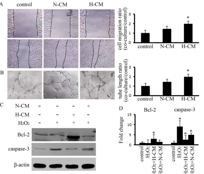
Furthermore, we observed the effects of hypoxia pretreatment of BM-MSCs on ECsin vitro.
concentrated 10 times and were used to evaluate the paracrine effect of BM-MSCs on HUVECs as well as the function of CMin vivo(S2 Fig). The results indicated that non-concentrated CM
from normoxic BM-MSCs (N-CM) increased the capacity of migration and tube formation on HUVECs as compared to the control group, and this capacity was significantly increased after treatment with CM from hypoxia-pretreated BM-MSCs (H-CM) (Fig2Aand2B). Increasing the anti-apoptotic function of ECs is important for angiogenesis. Therefore, we investigated the effects of CM on HUVECs apoptosis. The results indicated that H-CM downregulated the expression of caspase-3, which is induced at 4 h by H2O2, and upregulated the expression of
Bcl-2 (Fig2Cand2D). Therefore, H-CM improved the function of HUVECsin vitro.
Hypoxia-pretreated BM-MSCs activate VEGF/AKT signaling in HUVECs
The expressions of VEGF/AKT-related proteins in endothelial cells were affected by hypoxia pretreatment of BM-MSCs, as assessed by western blot. The results indicated that the
Fig 1. The characterization of rat BM-MSCs cultured under hypoxic conditions.(A) The expression of VEGF-1α, HIF-1α, ANG-1, bFGF, and MMP-9 were significantly increased in BM-MSCs receiving hypoxia pretreatment for 24 h compared to normoxic controls. At 48 h or 72 h, hypoxia pretreatment at 5% O2continuously increased the expression of these genes. (B) The expression of the VEGF-1a protein was significantly increased in BM-MSCs after hypoxia pretreatment. (C) The expression of the anti-apoptosis protein Bcl-2 was enhanced by western blot.*indicatesP<0.05 versus the normoxia group, and (D)
the apoptosis of BM-MSCs was analyzed by flow cytometry for cells cultured with 5% O2. At 48 h, no changes were observedin vitro,.
expression of pAKT and VEGF-R in ECs was upregulated with H-CM. However, when the ex-pression of VEGF-1αwas silenced in BM-MSCs by lentivirus-mediated siRNA (Fig 3A), the upregulated expression of VEGFR and pAKT was suppressed in HUVECs treated with H-siVEGF-CM. (Fig 3B). Furthermore, the capacity for tube formation by HUVECs was inhibited by H-siVEGF-CM treatment (Fig 3C). The expression of VEGF/AKT-related protein in ECs was activated by BM-MSCs, and the capacity for angiogenesis was improved.
Hypoxia pretreatment of BM-MSCs promotes angiogenesis in DLLI rats
The DLLI model, established by STZ-induced diabetic rats with ligation of the femoral artery, was confirmed by angiography (Fig 4A). Hypoxia-pretreated BM-MSCs were injected into is-chemic muscle tissue along the artery with multiple local injection points, and normoxic BM-MSCs and saline were used in the control groups. Seven days after cell transplantation,
Fig 2. Hypoxia pretreatment of BM-MSCs enhanced the capacity for angiogenesis and reduced apoptosis in HUVECs.(A) Hypoxia pretreatment of BM-MSCs enhanced the capacity for migration by HUVECs after 12 h, as compared to the control group; the efficiency of cell migration was detected.* indicatesP<0.05 versus control group. (B) Hypoxia pretreatment of BM-MSCs increased the ability for tubular formation by HUVECs after 24 h, as compared to the control group; the average length of the tubular formations was calculated.*indicatesP<0.05 versus control group. (C,D) H2O2-treated HUVECs
exhibited high expression of caspase-3. The CM from hypoxia-pretreated BM-MSCs depressed the expression of caspase-3 and enhanced the expression of Bcl-2.*indicatesP<0.05 versus control group.
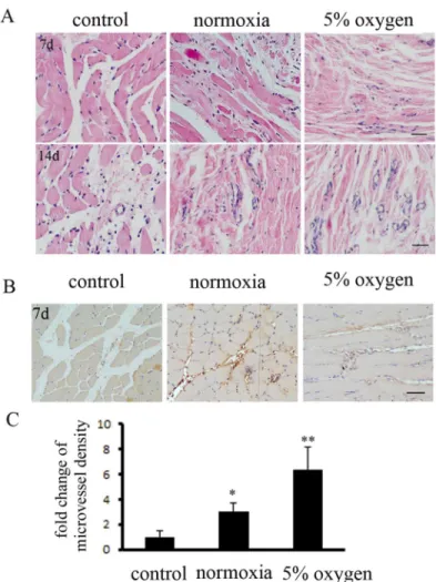
angiography indicated that neovascularization was found at ischemic tissue in the hypoxia pre-treatment group, and that the mean length of the capillary was greater than that of the control groups. At day 14, a renascent vascular network had been formed at the femoral artery break away site in the hypoxia-pretreated group (Fig4B–4D). Laser Doppler perfusion imaging was performed 14 days postoperatively to detect the blood perfusion of the ischemia of lower ex-tremities. Compared with the hypoxia group, perfusion was decreased in the normoxia and control groups (Fig4Eand4F). In the process of cell therapy, body weight, blood glucose, and glycosylated hemoglobin (HbA1c) levels were measured, and the results showed insignificant differences in local transplantation in the BM-MSCs and control groups (Table 2).
The ischemic muscle tissues were collected for paraffin sections. H&E staining indicated that hypoxia pretreatment of BM-MSCs significantly repaired muscle fibers in ischemic tissue at 7 d and 14 d, compared to the other groups (Fig 5A). Next, immunohistochemical staining demonstrated that the mature microvessel density, counted by immunostaining of endothelial cells marked with anti- CD31 antibodyies, was markedly increased in the hypoxia pretreatment BM-MSCs group at 7 d (Fig5Band5C). Hypoxia pretreatment of BM-MSCs increased the re-pair of ischemic tissues in DLLI.
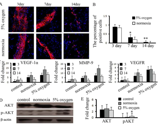
Hypoxia pretreatment enhances the survival of BM-MSCs and promotes
the expression of angiogenic factors in ischemic tissue
We confirmed that hypoxia pretreatment of BM-MSCs activated the expression of anti-apo-ptotic proteinsin vitro. Based on this result, we analyzed the survival of BM-MSCsin vivo. The
cell tracer results indicated that there were a similar number of cells in the three groups at 3 d.
Fig 3. Hypoxia pretreatment of BM-MSCs modulates VEGF/AKT signaling in HUVECs.(A) The expression of VEGF-1αwas suppressed with lentiviral-mediated siRNA-VEGF-1α(siVEGF). (B) CM generated from hypoxia pretreated BM-MSCs treated with siVEGF downregulated the expression of pAKT and VEGFR in HUVECs, as determined by western blot. (C) The capacity of HUVECs to form tubes was inhibited by CM from VEGF-1α-silenced BM-MSCs.
Fig 4. Hypoxia pretreatment of BM-MSCs promoted angiogenesis in ischemic muscle, as evaluated by angiography.(A) We presented the block diagram of study for BM-MSCs transplanted to treat DLLI. (B) The model of DLLI was developed and confirmed by angiography. (C) Hypoxia pretreatment of BM-MSCs locally transplanted to ischemic muscle markedly enhanced the microvascular density, compared to the control group. (D) The average length of the capillaries was detected.n= 4.*indicatesP<0.05 versus the 7 d
Thereafter, the survival of the hypoxia-pretreated BM-MSCs was higher than that of the nor-moxia group. Until day 14, approximately 5% of the hypoxia-pretreated BM-MSCs remained, whereas no surviving cells were observed in the normoxia group (Fig6Aand6B). We further detected the survival of BM-MSCs in non-ischemic muscle, the results shows that BM-MSCs reduced survival ability compared with the ischemia group (S3 Fig). We also detected the
Table 2. The biochemical index of the rats.
Bodyweight(g) bloodglucose(mmol/L) HbA1c(%)
group 3day 7day 14day 3day 7day 14day 3day 7day 14day
control 251±3 228±2 180±5 28±2 30±3 29±3 11.8±0.3 12.1±0.2 12.2±0.3 normoxia 251±5 225±4 182±3 27±2 27±3 30±1 12.1±0.3 11.9±0.2 11.9±0.4 hypoxia 251±4 232±4 175±7 30±5 29±4 29±1 12.0±0.4 12.0±0.2 12.0±0.1
doi:10.1371/journal.pone.0126715.t002
Fig 5. Hypoxia pretreatment of BM-MSCs promoted angiogenesis in DLLI muscle, as assessed by immunohistochemical staining.(A) Histological analysis of muscle tissues was performed by immunostaining. The specimens were stained with H&E (hematoxylin-eosin). Hypoxia pretreatment of BM-MSCs accelerated the repair of muscle fibers at day 7. At day 14, the hypoxia pretreatment BM-MSCs group exhibited a de novo rebound in the microvasculature. (B) Immunohistochemical staining with a specific antibody recognizing the endothelial marker CD31 revealed the presence of more mature vessels (muscle), predominantly in the hypoxia pretreatment BM-MSCs group rather than in the control group, and (C) the number of vessels was measured.*,**indicateP<0.05, 0.01 versus control group, respectively.
expression of antigenic factors in the ischemic tissues. The results indicated that the expression of angiogenic factors was significantly increased in the hypoxia-pretreated BM-MSC group at days 3, 7, and 14 (Fig 6C). However, while the survival of BM-MSCs was enhanced by hypoxia pretreatment in vivo, we examined the expression of downstream target proteins mediated by proangiogenesis factors and found that the expression of pAKT (1:400, Cell Signaling) was upregulated and that the expression of AKT (1:600, Cell Signaling) exhibited no changes at day 7 (Fig6Dand6E). Hypoxia pretreatment of BM-MSCs promoted the survival ability of the in-jected cells, increased the expression of angiogenic factors in ischemic tissue and activated the expression of angiogenesis-related signals.
Hypoxia pretreatment of BM-MSCs improves endothelial function in
ischemic tissues
We further analyzed the function of endothelial cells with BM-MSCs in ischemic tissue by the hypoxia pretreatment of BM-MSCs. Greater numbers of ECs triple-stained for proliferation and apoptosis were found in the ischemic tissues of DLLI rats administered normoxia-or hyp-oxia-pretreated BM-MSCs, compared with the saline group (Fig7Aand7B). The results indi-cated that the number of positive cells triple-stained with vWF/Ki67/Hoechst 33342 were significantly greater in the hypoxia-pretreated BM-MSC group than in the normoxia and saline groups. In contrast, the number of vWF/TUNEL/Hoechst 33342 triple-stained positive cells decreased in the hypoxia pretreatment group compared with the normoxia and saline groups (Fig7Cand7D). The expression of apoptotic and anti-apoptotic proteins was also detected in the ischemic tissue of DLLI rats by western blotting. The results suggested that the expression of Caspase-3 was downregulated in the hypoxia-pretreated BM-MSC group, while the expres-sion of Bcl-2 was upregulated (Fig7Eand7F). The results demonstrated that hypoxic pretreat-ment of BM-MSCs promoted ECs proliferation and decreased apoptosis of ECs.
Discussion
In this study, we confirmed that BM-MSCs cultured with 5% O2for 48 h exhibited significantly
improved capacity for the secretion of angiogenic factors. Local transplantation of hypoxia-pretreated BM-MSCs significantly increased the survival of the BM-MSCs in ischemic muscle, markedly increased angiogenesis and improved the proliferation of endothelial cells in ische-mic muscle (Fig 8)In vitro, we also confirmed that the hypoxia pretreatment of BM-MSCs
in-creased angiogenesis by ECs.
Currently, endothelial dysfunction is a systemic pathological condition that can be broadly defined as an imbalance between vasodilating and vasoconstricting substances manufactured by the endothelium or inclusive functions of the endothelium [27]. Diabetic mellitus generates the disordered metabolism of proteins and fats due to inappropriate production of insulin, a blood glucose regulator, or resistance to insulin that results in high blood glucose levels, or hy-perglycemia [28]. Over time, elevated glucose levels in the bloodstream can lead to the im-paired function of the vessel endothelial cells [29]. Nevertheless, owing to endothelial
dysfunction, thede novoendothelial cells cannot expand, leading to difficulties in
neovasculari-zation or angiogenesis that are also interchangeably associated with vasculogenesis, which pri-marily refers to the developmental formation of vascular structures. A previous study
Mesenchymal stem cells have been isolated from bone marrow, adipose tissue, and other adult tissues [32]. However, the number of MSCs contained in tissues is low, and the cells must be expandedin vitrofor transplantation. In this process, BM-MSCs usually encounter two
dis-tinct environmental conditions. One is thein vitroculture environment (from isolation to
transplantation) and the other is the physiological conditionsin vivo(before isolation and after
transplantation). At present, most of the expansion procedures for BM-MSCs are performed under ambient O2concentration, in which cells are exposed to 21% O2[33]. It is well known
that MSCs exist in hypoxic physiologic conditions, and that the cells are quiescent. The cells do not proliferate and maintain a potential pluripotent ability in this period. Several studies have presented positive evidences regarding the adverse effects of ambient O2concentration on
BM-MSCs, including senescence, population doubling time, DNA damage, and poor engage-ment following transplantation [22,24]. However, murine MSCs preconditioned in a hypoxic environment demonstrated increased skeletal muscle regeneration after 7 days, with improved blood flow and vascular formation compared to MSCs maintained in normoxic conditions [34]. Hypoxia plays a critical role in maintaining homeostasis within the body from the very beginning of embryonic development and helps to facilitate proper embryonic progress, main-tain stem cell pluripotency and regulate the signaling of multiple cascades, including angiogen-esis. All of these effects reflect the influential effect of the O2concentration on BM-MSC Fig 6. Hypoxia pretreatment promoted the function of BM-MSCs in ischemic muscle.(A) Hypoxia pretreatment of BM-MSCs increased the capacity for survival in ischemic muscle, compared to the normoxia group, (B) and the number of positive cells was measured.*,**indicateP<0.05, 0.01 versus normoxia group, respectively. (C) The expression of VEGF-1α, MMP-9, and VEGFR was increased in hypoxia-pretreated BM-MSCs, and (D, E) the expression of pAKT was significantly enhanced, compared to the control group.*indicatesP<0.05 versus control group.
biology and raise serious concerns over the impact of the O2concentration on therapeutic
effi-ciency, biosafety, cell proliferation and maintenance of homeostasis. In this study, we proved that BM-MSCs cultured with 5% O2for 48 h were able to dramatically enhance the
anti-apo-ptotic activity of endothelial cells, while having no effects on cell proliferation and apoptosis, and were also able to increase the competence of secreted proangiogenic factors.
MSCs secrete angiogenic cytokines, such as VEGF and HGF, which may contribute to their angiogenic properties. A number of studies have shown that MSCs secrete significant quanti-ties of angiogenic and anti-apoptotic factors, including VEGF and HGF [24]. These findings further encouraged a series ofin vivostudies that focused on evaluating the therapeutic
poten-tial of cells based particularly on their paracrine and angiogenic effects. The processes of neo-vascularization and angiogenesis, including VEGF and bFGF, are regulated by a number of en-dothelial growth factors. Furthermore, pathological conditions, such as ischemia, induce the re-lease of bFGF and VEGF from MSCs, leading to the formation of new vascular vessels [35]. VEGF is an important angiogenic factor and promotes the proliferation and migration of ECs maintaining the integrity of vascular vessels. VEGF facilitates angiogenesis synergistically with angiogenin was proved in the past investigation. The growth factor affects early blood vessel formation and promotes the generation of a primitive vascular network, while angiopoietin
Fig 7. Hypoxia pretreatment of BM-MSCs improved the function of ECs in vivo.(A, B) Immunohistochemical staining with specific antibodies against the endothelial marker vWF and cell proliferation marker Ki67 revealed more double-positive cells in the hypoxia pretreated BM-MSC group than in the control group.*indicatesP<0.05 versus control group. (C, D) Immunohistochemical staining with a specific antibody recognizing the endothelial marker vWF
and cell apoptosis marker TUNEL revealed that hypoxia pretreatment of BM-MSCs decreased the number of double-positive cells.*indicatesP<0.05 versus control group. (E,F) Hypoxia pretreatment of BM-MSCs upregulated the expression of Bcl-2 in muscle and reduced the expression of caspase-3.*indicates P<0.05 versus control group.*indicatesP<0.05 versus control group.
acts on the subsequent alterations of vascular remodeling and promotes the formation of ma-ture vessels and the spatial strucma-ture of the vascular network. In a recent study, conditioned medium from hypoxic MSC prevented endothelial cell apoptosis and enhanced tube formation
in vitro[36]. In addition, when ischemic muscles were treated with conditioned medium
har-vested from MSCs cultured under hypoxic conditions, the experimental animals recovered faster than the control groups, suggesting that the release of angiogenic factors by MSCs was sufficient to enhance the revascularization of the injured tissue. In all of these studies, however, the conditioned media or MSCs were injected directly into the injured muscle, which eliminat-ed the necessity of MSCs to home to the site of injury.
We measured the percentage of MSCs remaining at the site of injury 2 weeks after injection. The function of the cells is to release cascades of trophic factors to enhance the endogenous revascularization process rather than the recreation of new vessels or muscle. Therefore, from a
Fig 8. Hypoxia-pretreated BM-MSCs significantly increased the survival of the BM-MSCs in ischemic muscle, markedly increased angiogenesis and improved the proliferation of endothelial cells in ischemic muscle.
clinical and regulatory agency point of view, it is actually preferable that the cells do not remain in the local area for months after the injury has been repaired. This finding could enhance ther-apeutic approaches for enhancing local tissue repair by injected human mesenchymal
stem cells.
Conclusions
In our study, we demonstrated that hypoxia pretreatment of MSCs significantly enhances the secretion of bioactive factors, extending the efficiency of cell survival after transplantation into ischemic muscle in DLLI. Our findings show that MSC pretreatment may be a novel strategy for the clinical treatment of diabetic lower extremity arterial disease.
Supporting Information
S1 Fig. The phenotype and differentiation of BM-MSCs cultured under hypoxic conditions. (A) BM-MSCs surface marker expression by flow cytometry showing the percentages of BM-MSCs cultured with 5% O248 h for mesenchymal antigens has similar to the normoxia
group. (B) Multilineage differentiation potential of BM-MSCs showing BM-MSCs differentiat-ed into adipocytes, which are indicatdifferentiat-ed by the accumulation of lipid vesicles in the cells, (C) and osteoblasts, which express alkaline phosphatase, as indicated in blue. Scale bar = 100μm.
(TIF)
S2 Fig. The CM from the hypoxia pretreated of BM-MSCs improved angiogenesis in vivo. (A) The result of angiography showed that the CM from hypoxia pretreated of MSCs improved angiogenesis in DLLI at day 14, (B) and the number of positive cells was measured.indicate P<0.05, 0.01 versus normoxia group.
(TIF)
S3 Fig. The MSCs transplanted to non-ischemic lower limb in diabetic rats.(A) The result of the CM-Dil label MSCs by fluorescence microscope.
(TIF)
Author Contributions
Conceived and designed the experiments: JL HJH LX XF WH. Performed the experiments: DT HH LD CT. Analyzed the data: QH YZ HL. Contributed reagents/materials/analysis tools: QH YZ HL. Wrote the paper: JL HH LX.
References
1. Pecoraro RE, Reiber GE, Burgess EM. Pathways to diabetic limb amputation. Basis for prevention. Dia-betes care. 1990; 13(5):513–21. PMID:2351029.
2. Wild S, Roglic G, Green A, Sicree R, King H. Global prevalence of diabetes: estimates for the year 2000 and projections for 2030. Diabetes care. 2004; 27(5):1047–53. PMID:15111519.
3. Li H, Isomaa B, Taskinen MR, Groop L, Tuomi T. Consequences of a family history of type 1 and type 2 diabetes on the phenotype of patients with type 2 diabetes. Diabetes care. 2000; 23(5):589–94. PMID:
10834414.
5. Cao Y, Sagi S, Hacker A, Steidler A, Alken P, Knoll T. Impact of hypoxia and hypercapnia on calcium oxalate toxicity in renal epithelial and interstitial cells. Urological research. 2006; 34(4):271–6. doi:10.
1007/s00240-006-0055-3PMID:16633808.
6. Zhang H, Zhang N, Li M, Feng H, Jin W, Zhao H, et al. Therapeutic angiogenesis of bone marrow mononuclear cells (MNCs) and peripheral blood MNCs: transplantation for ischemic hindlimb. Annals of vascular surgery. 2008; 22(2):238–47. doi:10.1016/j.avsg.2007.07.037PMID:18083329.
7. Pacher P, Beckman JS, Liaudet L. Nitric oxide and peroxynitrite in health and disease. Physiological re-views. 2007; 87(1):315–424. doi:10.1152/physrev.00029.2006PMID:17237348; PubMed Central
PMCID: PMC2248324.
8. Kajiya M, Hirota M, Inai Y, Kiyooka T, Morimoto T, Iwasaki T, et al. Impaired NO-mediated vasodilation with increased superoxide but robust EDHF function in right ventricular arterial microvessels of pulmo-nary hypertensive rats. American journal of physiology Heart and circulatory physiology. 2007; 292(6): H2737–44. doi:10.1152/ajpheart.00548.2006PMID:17220192.
9. Shen GX. Oxidative stress and diabetic cardiovascular disorders: roles of mitochondria and NADPH oxidase. Canadian journal of physiology and pharmacology. 2010; 88(3):241–8. doi:10.1139/Y10-018
PMID:20393589.
10. Carmeliet P. Mechanisms of angiogenesis and arteriogenesis. Nature medicine. 2000; 6(4):389–95.
doi:10.1038/74651PMID:10742145.
11. Carmeliet P, Jain RK. Angiogenesis in cancer and other diseases. Nature. 2000; 407(6801):249–57.
doi:10.1038/35025220PMID:11001068.
12. Chang EI, Loh SA, Ceradini DJ, Chang EI, Lin SE, Bastidas N, et al. Age decreases endothelial progen-itor cell recruitment through decreases in hypoxia-inducible factor 1alpha stabilization during ischemia. Circulation. 2007; 116(24):2818–29. doi:10.1161/CIRCULATIONAHA.107.715847PMID:18040029.
13. Winkler EA, Bell RD, Zlokovic BV. Central nervous system pericytes in health and disease. Nature neu-roscience. 2011; 14(11):1398–405. doi:10.1038/nn.2946PMID:22030551.
14. Evans JL, Goldfine ID, Maddux BA, Grodsky GM. Oxidative stress and stress-activated signaling path-ways: a unifying hypothesis of type 2 diabetes. Endocrine reviews. 2002; 23(5):599–622. doi:10.1210/
er.2001-0039PMID:12372842.
15. Annabi B, Lee YT, Turcotte S, Naud E, Desrosiers RR, Champagne M, et al. Hypoxia promotes murine bone-marrow-derived stromal cell migration and tube formation. Stem cells. 2003; 21(3):337–47. doi:
10.1634/stemcells.21-3-337PMID:12743328.
16. Reiser J, Zhang XY, Hemenway CS, Mondal D, Pradhan L, La Russa VF. Potential of mesenchymal stem cells in gene therapy approaches for inherited and acquired diseases. Expert opinion on biological therapy. 2005; 5(12):1571–84. doi:10.1517/14712598.5.12.1571PMID:16318421; PubMed Central
PMCID: PMC1371057.
17. Tang YL, Zhao Q, Zhang YC, Cheng L, Liu M, Shi J, et al. Autologous mesenchymal stem cell trans-plantation induce VEGF and neovascularization in ischemic myocardium. Regulatory peptides. 2004; 117(1):3–10. PMID:14687695.
18. Matsumoto R, Omura T, Yoshiyama M, Hayashi T, Inamoto S, Koh KR, et al. Vascular endothelial growth factor-expressing mesenchymal stem cell transplantation for the treatment of acute myocardial infarction. Arteriosclerosis, thrombosis, and vascular biology. 2005; 25(6):1168–73. doi:10.1161/01.
ATV.0000165696.25680.cePMID:15831811.
19. Ren H, Cao Y, Zhao Q, Li J, Zhou C, Liao L, et al. Proliferation and differentiation of bone marrow stro-mal cells under hypoxic conditions. Biochemical and biophysical research communications. 2006; 347 (1):12–21. doi:10.1016/j.bbrc.2006.05.169PMID:16814746.
20. Lennon DP, Edmison JM, Caplan AI. Cultivation of rat marrow-derived mesenchymal stem cells in re-duced oxygen tension: effects on in vitro and in vivo osteochondrogenesis. Journal of cellular physiolo-gy. 2001; 187(3):345–55. doi:10.1002/jcp.1081PMID:11319758.
21. Panchision DM. The role of oxygen in regulating neural stem cells in development and disease. Journal of cellular physiology. 2009; 220(3):562–8. doi:10.1002/jcp.21812PMID:19441077.
22. Chacko SM, Ahmed S, Selvendiran K, Kuppusamy ML, Khan M, Kuppusamy P. Hypoxic precondition-ing induces the expression of prosurvival and proangiogenic markers in mesenchymal stem cells. American journal of physiology Cell physiology. 2010; 299(6):C1562–70. doi:10.1152/ajpcell.00221.
2010PMID:20861473; PubMed Central PMCID: PMC3006322.
23. Kinnaird T, Stabile E, Burnett MS, Lee CW, Barr S, Fuchs S, et al. Marrow-derived stromal cells express genes encoding a broad spectrum of arteriogenic cytokines and promote in vitro and in vivo arteriogen-esis through paracrine mechanisms. Circulation research. 2004; 94(5):678–85. doi:10.1161/01.RES.
24. Rosova I, Dao M, Capoccia B, Link D, Nolta JA. Hypoxic preconditioning results in increased motility and improved therapeutic potential of human mesenchymal stem cells. Stem cells. 2008; 26(8):2173–
82. doi:10.1634/stemcells.2007-1104PMID:18511601; PubMed Central PMCID: PMC3017477. 25. Grayson WL, Zhao F, Izadpanah R, Bunnell B, Ma T. Effects of hypoxia on human mesenchymal stem
cell expansion and plasticity in 3D constructs. Journal of cellular physiology. 2006; 207(2):331–9. doi:
10.1002/jcp.20571PMID:16331674.
26. Lozito TP, Tuan RS. Mesenchymal stem cells inhibit both endogenous and exogenous MMPs via se-creted TIMPs. Journal of cellular physiology. 2011; 226(2):385–96. doi:10.1002/jcp.22344PMID:
20665704.
27. Bucci E. Thermodynamic approach to oxygen delivery in vivo by natural and artificial oxygen carriers. Biophysical chemistry. 2009; 142(1–3):1–6. doi:10.1016/j.bpc.2008.12.009PMID:19349106.
28. Meshkani R, Adeli K. Hepatic insulin resistance, metabolic syndrome and cardiovascular disease. Clini-cal biochemistry. 2009; 42(13–14):1331–46. doi:10.1016/j.clinbiochem.2009.05.018PMID:19501581.
29. Kohner EM, Patel V, Rassam SM. Role of blood flow and impaired autoregulation in the pathogenesis of diabetic retinopathy. Diabetes. 1995; 44(6):603–7. PMID:7789621.
30. Burt RK, Loh Y, Pearce W, Beohar N, Barr WG, Craig R, et al. Clinical applications of blood-derived and marrow-derived stem cells for nonmalignant diseases. JAMA: the journal of the American Medical Association. 2008; 299(8):925–36. doi:10.1001/jama.299.8.925PMID:18314435.
31. Mathew SA, Rajendran S, Gupta PK, Bhonde R. Modulation of physical environment makes placental mesenchymal stromal cells suitable for therapy. Cell biology international. 2013; 37(11):1197–204. doi:
10.1002/cbin.10154PMID:23852996.
32. Pittenger MF, Mackay AM, Beck SC, Jaiswal RK, Douglas R, Mosca JD, et al. Multilineage potential of adult human mesenchymal stem cells. Science. 1999; 284(5411):143–7. PMID:10102814.
33. Nekanti U, Dastidar S, Venugopal P, Totey S, Ta M. Increased proliferation and analysis of differential gene expression in human Wharton's jelly-derived mesenchymal stromal cells under hypoxia. Interna-tional journal of biological sciences. 2010; 6(5):499–512. PMID:20877435; PubMed Central PMCID:
PMC2945278.
34. Leroux L, Descamps B, Tojais NF, Seguy B, Oses P, Moreau C, et al. Hypoxia preconditioned mesen-chymal stem cells improve vascular and skeletal muscle fiber regeneration after ischemia through a Wnt4-dependent pathway. Molecular therapy: the journal of the American Society of Gene Therapy. 2010; 18(8):1545–52. doi:10.1038/mt.2010.108.20551912; PubMed Central PMCID: PMC2927059.
PMID:20551912
35. Cousin B, Caspar-Bauguil S, Planat-Benard V, Laharrague P, Penicaud L, Casteilla L. [Adipose tissue: a subtle and complex cell system]. Journal de la Societe de biologie. 2006; 200(1):51–7. PMID:
17144162.
36. Hung SC, Pochampally RR, Chen SC, Hsu SC, Prockop DJ. Angiogenic effects of human multipotent stromal cell conditioned medium activate the PI3K-Akt pathway in hypoxic endothelial cells to inhibit ap-optosis, increase survival, and stimulate angiogenesis. Stem cells. 2007; 25(9):2363–70. doi:10.1634/