Novel Penicillin Analogues as Potential
Antimicrobial Agents; Design, Synthesis and
Docking Studies
Zaman Ashraf1,2*, Abdul Bais1, Md. Maniruzzaman Manir3, Umar Niazi4
1Department of Chemistry, Allama Iqbal Open University, H-8, Islamabad, Pakistan,2Department of Biology, College of Natural Sciences, Kongju National University, Kongju, South Korea,3Department of Chemistry, Kongju National University, Gongju, Republic of Korea,4Atta ur Rehman School of Modeling and Simulation, National University of Science and Technology, Islamabad, Pakistan
Abstract
A number of penicillin derivatives(4a-h)were synthesized by the condensation of 6-amino penicillinic acid (6-APA) with non-steroidal anti-inflammatory drugs as antimicrobial agents. In silicodocking study of these analogues was performed against Penicillin Binding Protein (PDBID 1CEF) using AutoDock Tools 1.5.6 in order to investigate the antimicrobial data on structural basis. Penicillin binding proteins function as either transpeptidases or carboxy-peptidases and in few cases demonstrate transglycosylase activity in bacteria. The excel-lent antibacterial potential was depicted by compounds4cand4eagainstEscherichia coli, Staphylococcus epidermidusandStaphylococcus aureuscompared to the standard amoxi-cillin. The most potent penicillin derivative4eexhibited same activity as standard amoxicillin againstS.aureus. In the enzyme inhibitory assay the compound4einhibitedE.coliMurC with an IC50value of 12.5μM. The docking scores of these compounds4cand4ealso veri-fied their greater antibacterial potential. The results veriveri-fied the importance of side chain functionalities along with the presence of central penam nucleus. The binding affinities cal-culated from docking results expressed in the form of binding energies ranges from -7.8 to -9.2kcal/mol. The carboxylic group of penam nucleus in all these compounds is responsible for strong binding with receptor protein with the bond length ranges from 3.4 to 4.4Ǻ. The results of present work ratify that derivatives4cand4emay serve as a structural template for the design and development of potent antimicrobial agents.
Introduction
The discovery of penicillin aβ-Lactam antibiotic by Alexander Fleming in 1928 and its use into the health care system in the later phases of Second World War denotes one of the most dynamic contributions to medical science in recent history [1].β-Lactam antibiotics have been effectively used in the treatment of infectious ailments for several years [2] and persist the most commonly utilized antibiotics due to their relatively high efficacy, low cost, ease of delivery and
OPEN ACCESS
Citation:Ashraf Z, Bais A, Manir MM, Niazi U (2015) Novel Penicillin Analogues as Potential Antimicrobial Agents; Design, Synthesis and Docking Studies. PLoS ONE 10(8): e0135293. doi:10.1371/journal. pone.0135293
Editor:Paul Taylor, University of Edinburgh, UNITED KINGDOM
Received:April 29, 2015
Accepted:July 20, 2015
Published:August 12, 2015
Copyright:© 2015 Ashraf et al. This is an open access article distributed under the terms of the
Creative Commons Attribution License, which permits unrestricted use, distribution, and reproduction in any medium, provided the original author and source are credited.
Data Availability Statement:All relevant data are within the paper and its Supporting Information files.
Funding:The authors have no support or funding to report.
minimal side effects. Despite the large number ofβ-lactams that have already been synthesized and tested, there is still a need for new compounds of this kind [3], due to the increasing resis-tance of bacterial strains to certain types of anti-infectives [4].
The emergence of resistance to the major classes of antibacterial agents is recognized as a serious health concern. Particularly, the emergence of multi drug resistance strains of patho-genic bacteria is a problem of ever increasing significance reported by Kumar et al. 2010 [5]. The increasing selection for bacteria having acquired resistance mechanism progressively devaluate our antibiotic arsenal. This provides a strong incentive for continually developing novel drugs that escape the destruction of resistant bacterial strains [6]. Two mechanisms have been reported to be responsible for antibiotic resistance: structural modification in Penicillin binding protein (PBP) targets and production ofβ-Lactamase first identified in 1972 [7,8].
The structural modification of PBPs is a common mechanism of resistance of Gram-positive bacteria. Penicillin binding proteins (PBPs) are membrane-associated proteins that catalyze the final step of murein biosynthesis in bacteria [9]. These proteins function as either transpepti-dases or carboxypeptitranspepti-dases and in a few cases demonstrate transglycosylase activity [10]. Both transpeptidase and carboxypeptidase activities of PBPs occur at the D-Ala-D-Ala terminus of a murein precursor containing a disaccharide pentapeptide comprisingN-acetylglucosamine andN-acetyl-muramic acid-L-Ala-D-Glu-L-Lys-D-Ala-D-Ala. The Penicillins antibiotics inhibit these enzymes by competing with the pentapeptide precursor for binding to the active site of the enzyme [11]. Penicillins bind irreversibly to the active site of theses enzyme and thus prevents the final cross-linking of the peptidoglycan layer which disrupts the cell wall
synthesis.
6-aminopenicillanic acid (6-APA) is an important industrial intermediate produced on large scale by the enzymatic cleavage of penicillin G (V) side-chain with penicillin amidase in solution or with immobilized enzyme. Most of the penicillins are produced by coupling 6-APA with the required side chain [12] except of Penicillin G and Penicillin V, which can be industri-ally produced by fermentation from high producing strains ofPenicillium chrysogenum.
Molecular Docking is the method that predicts the preferred orientation of a drug molecule into the macromolecule and the goal is to compute the bound conformation and the binding affinity [13]. Koska et al. 2008 and Yusuf et al. 2008 also reported that docking is one of the commonly used computational methods in structure-based drug design [14,15]. The informa-tion generated from docking calculainforma-tions helps to get insight into the interacinforma-tions of ligands with amino acid residues in the binding pockets of targets and to predict the corresponding binding energies of ligands [16], when the experimental holo structure are unavailable [17]. Lipinski’s rule of 5 also referred rule of thumb helps in distinguishing drug likeness properties of a molecule and describes its physiochemical properties important for a drug’s pharmacoki-netics in the human body [18]. These filters assist in early preclinical developments and could support to avoid costly late-stage preclinical and clinical failures [19].
In continuation of our effort in the development of effective antimicrobials agents [20,21], we report here the synthesis and docking study of novel Penicillin analogues having NSAIDs moiety using 6-APA as starting material. All of the clinically usedβ-lactam antibiotics (Penicil-lins) possess the central penam nucleus which is essential for antibacterial activity. The spec-trum of activity against either Gram negative or Gram positive bacteria can be enhanced by changing the side chain amide functionality. The selected NSAIDs have different hydrophobic/ hydrophilic groups and all possess−COOH group which can easily be condensed with−NH2
vitroantibacterial activity of synthesized penicillin derivatives was carried out against five path-ogenic bacteria, two of which are Gram negative and other three are Gram positive. In this way we are able to find out the potential of our synthesized compounds against either Gram positive or Gram negative bacteria. In addition toin vitroantibacterial activity the enzyme inhibitory activity of compound(4e)was also performed againstE.coliMurC, which is an important enzyme for peptidoglycan biosynthesis in bacterial cell wall.
Materials and Methods
Melting points were recorded using a digital Gallenkamp (SANYO) model MPD 350 apparatus and are uncorrected. FTIR spectra were recorded using an FTS 3000 MX spectrophotometer; the1H NMR and13C NMR spectra (DMSO-d
6) were recorded using a Bruker 300 MHz
spec-trometer. Chemical shifts (δ) are reported in ppm downfield from the internal standard tetramethylsilane (TMS). Mass spectra were performed on an Agilent 6460 Series Triple Quad-rupole instrument (Agilent). The ionization was achieved by electrospray ionization in the pos-itive ion mode (ESI+) and negative ion mode (ESI-). The capillary voltage was set to 4.0 kV. The source temperature was 120°C, and the desolvation temperature was 350°C. Nitrogen was used as a desolvation gas (flow 600 L/h). The software used forin-silicomolecular docking studies are AutoDock Tools 1.5.6: La Jolla, CA, U.S.A., AutoDock Vina 1.1.2: La Jolla, CA, U.S. A. and Discovery Studio 4.0: San Diego, CA, U.S.A. The procedure for the synthesis of the desired compounds is depicted in Scheme I. ATP, L-alanine, AMP-PCP and bovine serum albumin (BSA) were purchased from Sigma. Malachite green phosphate detection reagent, UNAM, and E. coli MurC were prepared as described previously [22].
General Procedure for the Synthesis of Penicillin Derivatives (4a-h)
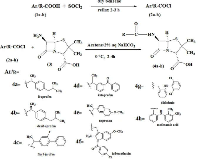
A solution of NSAIDs having carboxylic acid group(1a-h)(1mmol) in dry benzene (5–8mL) was refluxed with freshly distilled thionyl chloride (1.2mmol) for 2–3 h. After the completion of reaction, excess of thionyl chloride was removed under reduce pressure to afford the acid chlorides(2a-h)which were dissolved in anhydrous acetone for further use. The acid chlorides
(2a-h)were then treated with a solution of (+)-6-aminopenicillanic acid (6-APA, 1mmol) in 2% NaHCO3(40mL) diluted with acetone (30 mL). The reaction mixture was stirred for 2–4h
at room temperature and then concentrated under reduced pressure and washed with ethyl acetate (25mL). The aqueous layer was then acidified with HCl (0.1M), extracted with ethyl acetate and then washed with distilled water dried over anhydrous Na2SO4. The ethyl acetate
was rotary evaporated and triturated with n-hexane to afford the title compounds(4a-h).
(2S,5R,6S)-6-(3'-(4'-isobutylphenyl)propanamido)-3,3-dimethyl-7-oxo-4-thia-1-azabi-cyclo[3.2.0]heptane-2-carboxylic acid (4a). Yield 78%; m.p. 115–117°C; FTIR (KBr,υmax
cm-1): 1732 (C = Oβ-lactam), 1667 (C = O Amide), 2954 (sp3C-H), 1508 (C = C);1H-NMR
(DMSO-d6,δppm): 0.93 (6H, d,J= 5.8 Hz, H-13’,14’), 1.20 (3H, d,J= 6.0 Hz, H-4’), 1.70 (3H,
s, H-3”), 1.58 (3H, s, H-2”), 1.75 (1H, m, H-12), 2.36 (2H, d,J= 6.0, H-11), 3.43 (1H, d,J= 9.0, H-5), 4.3 (1H, d,J= 9.0, H-6), 7.15 (2H, d,J= 6.5 Hz, H-6’, 8’), 7.20 (2H, d,J= 6.5 Hz, H-5’,9’);
13C-NMR (DMSO-d
6)δ18 (C-13’, 14’), 22 (C-12’), 27 (C-4’), 30 (C-3’), 44 (C-11’), 70 (C-5),
72.5 (C-6), 130 (C-6’,10’), 133 (C-7’,9’), 141 (C-5’), 164 (C = O, acid), 167 (C-2’), 173 (C-7); ESI-MS:m/z427 [M + 23] (M + Na)+.
(2S,5R,6S)-6-((R)-3’-(4’ -isobutylphenyl)propanamido)-3,3-dimethyl-7-oxo-4-thia-1-azabicyclo[3.2.0]heptane-2-carboxylic acid (4b). Yield 63%; m.p. 110–112°C; FTIR (KBr,
υmaxcm-1): 1726 (C = Oβ-lactam), 1656 (C = O Amide), 2953.71 (sp3C-H stretch), 1510.74
(C = C) aromatic;1H-NMR (DMSO-d
6,δppm): 0.91(6H, d,J6.0 Hz, H-13’,14’), 1.28 (3H, d,
Hz, H-3’), 4.72(1H, s, H-2HHhhhh), 4.86 (1H, d,J= 9 Hz, H-5), 5.91(1H, d, J = 9 Hz, H-6), 7.0 (2H, d, J = 7.5 Hz, H-7’, 9’), 7.24 (2H, d,J= 7.5 Hz, H-6’, 10’);13C-NMR (DMSO-d6,δppm):
15(C-4’), 22(C-13’,14’), 27.1(C-2”), 28(C-12’), 31.2(C-3”), 41(C-3’), 44(C-11’), 60(C-6), 64(C-2), 72(C-5), 78(C-3), 127 (C-6’, C-10’), 160(C = O, acid), 165 (C-2’), 170(C-7); ESI-MS:m/z 427 [M + 23] (M + Na)+.
(2S,5R,6S)-6-(3'-(7'-fluoro-[11',8']-biphenyl]-5'-yl)propanamido)-3,3-dimethyl-7-oxo-4-thia-1-azabicyclo[3.2.0]heptane-2-carboxylic acid (4c). Yield 65%; m.p. 85–87°C; FTIR (KBr,υmaxcm-1): 1721 (C = Oβ-lactam), 1648 (C = O Amide), 1220 (C-F stretch);1H-NMR
(DMSO-d6,δppm): 1.20 (3H, d,J= 6.8 Hz, H-4’), 1.61 (3H, s, H-2”), 1.71 (3H, s, H-3”), 6.89
(1H, s, H-6’), 3.50 (1H, q,J= 6.8 Hz H-3’), 4.80 (1H, d,J= 9 Hz, H-5), 5.1 (1H, d,J= 9 Hz, H-6), 7.20 (1H, m, H-10’), 7.30 (1H, m, H-14’), 7.49 (2H, m, H-13’,15’), 7.50 (2H, m, H-12’16’), 7.72 (1H, m, H-9’);13C-NMR (DMSO-d6δppm): 14 (C-4’), 28 (C-2”), 30 (C-3”), 60 (C-6), 62
(C-2), 70 (C-5), 78 (C-3), 120 (C-6’), 126–128 (C-12’-16’), 130–135 (C-8’-10’), 160 (C-7’), 165 (C-5’), 168 (C = O, acid), 170 (C-2’), 171 (C-7); ESI-MS:m/z465 [M + 23] (M + Na)+.
(2S,5R,6S)-6-(3'-(7'-benzoylphenyl)propanamido)-3,3-dimethyl-7-oxo-4-thia-1-azabicy-clo[3.2.0]heptane-2-carboxylic acid (4d). Yield 61%; m.p. 64°C; FT-IR (KBr,υmaxcm-1):
1733 (C = Oβ-lactam), 1655 (C = O Amide), 1595 (Ar-C = O), 2973 (sp3C-H stretch); 1HNMR
(DMSO-d6,δppm): 1.20 (3H, d,J= 6.0 Hz, H-4’), 1.61 (3H, s, H-2”), 1.71 (3H, s,
H-3”), 3.43 (1H, q, H-3’), 4.70 (1H, s, C-2), 4.74 (1H, d,J= 8.50 Hz, H-5), 5.0 (1H, d,J= 8.50 Hz, H-6), 7.50 (2H, m, H-14’, H-16’), 7.55 (1H, s, H-6’), 7.60 (1H, m, H-15’), 7.80 (2H, d,J= 6.5 Hz, H-13’, 17’);13C-NMR (DMSO-d
6,δppm): 15(C-4’), 28(C-3”), 30(C-2”), 40(C-3’), 60(C-6),
64(C-2), 70(C-5), 128–140(Aromatic), 165(C-acid), 170(C-2’), 173(C-7), 190(C-11’); ESI-MS: m/z475 [M + 23] (M + Na)+.
(2S,5R,6S)-6-(3'-(11'-methoxynaphthalen-5'-yl)propanamido)-3,3-dimethyl-7-oxo-4-thia-1-azabicyclo[3.2.0]heptane-2-carboxylic acid (4e). Yield 75%; m.p. 150–152°C; FT-IR (KBr,υmaxcm-1):1720 (C = Oβ-lactam), 1653 (C = O Amide), 1069 (C-O), 1401 (C-N); 1HNMR (DMSO-d
6,δppm): 1.53(3H, s, H-4’), 1.61 (1H, s, H-2”), 1.71 (1H, s, H-3”), 3.50(1H,
q, H-3’), 3.80 (3H, s, H-16’), 4.50(1H, d,J= 8.90 Hz, H-5), 4.70(1H, s, H-2), 5.00(1H, d, J= 8.90, H-6), 7.20(1H, m, H-7’), 7.22(1H, m, H-11’), 7.35–7.37(2H, m, H-6’10’), 7.85(2H, m, H-8’, 13’);13C-NMR (DMSO-d6,δppm): 14(C-4’), 28(C-3”), 30(C-2”), 40(C-3’), 60(C-6), 62
(C-2), 70(C-5), 78(C-3), 105(C-10’), 115(C-12’), 125–130(C-6’, 7’8’, 13, 14’), 133 (C-5’), 157 (C-11’), 168(C-acid), 170 (C-2’), 172(C-7); ESI-MS:m/z451 [M + 23] (M + Na)+.
(2S,5R,6S)-6-(3'-(4'-(20'-chlorobenzoyl)-10'-methoxy-5'-methyl-1H-indol-6'-yl) aceta-mido)-3,3-dimethyl-7-oxo-4-thia-1-azabicyclo[3.2.0]heptane-2-carboxylic acid (4f). Yield 58%; m.p. 165–167°C; FT-IR (KBr,υmaxcm-1): 1723 (C = Oβ-lactam), 1637 (C = O Amide),
1400 (C-O stretch), 3413 (N-H stretch), 1174 (C-N stretch);1HNMR (DMSO-d6,δppm): 1.59
(3H, s, H-3”), 1.65 (3H, s, H-2”), 1.68 (3H, s, H-2”), 1.71 (3H, s, H-3”), 2.20 (3H, s, H-13’), 3.20 (2H, s, H-3’), 3.80 (3H, s, H-15’), 4.70 (1H, s, H-2), 4.80 (1H, d,J= 9.0 Hz, H-5), 5.00 (1H, d, J= 9.0 Hz, H-6), 6.30 (1H, s, H-11’), 6.60 (2H, d,J= 7.1 Hz, H- 19’, 21’), 7.79 (1H, m, H-9’), 7.82 (2H, d,J= 7.1 Hz, H-18’, 22’);13C-NMR (DMSO-d6,δppm): 15 (C-13’), 27(C-3”),), 30
(C-3’), 32 (C-2”), 53 (C-15’), 60 (C-6), 63 (C-2), 79 (C-3), 99 (C-11’), 110 (C-9’), 130 (C-5’), 140 (C-12’), 160 (C-10’), 162 (C-7), 167 (C-16’), 168 (C = O, acid), 170 (C-5), 172 (C-2’); ESI-MS:m/z581 [M + 23] (M + Na)+.
(2S,5R,6S)-6-(2-(2-((2,6-dichlorophenyl)amino)phenyl)acetamido)-3,3-dimethyl-7-oxo-4-thia-1-azabicyclo[3.2.0]heptane-2-carboxylic acid (4g). Yield 63%; m.p 142–145°C; FT-IR (KBr,υmaxcm-1): 1729 (C = Oβ-lactam), 1650 (C = O Amide), 1173 (C-Cl stretch);1HNMR
(DMSO-d6,δppm): 1.44 (1H, s, H-3”), 1.53 (1H, s, H-2”), 3.30 (2H, s, H-3’), 4.68 (1H, s, H-5),
4.80 (1H, s, H-6), 4.82 (1H, s, H-2), 6.50 (1H, m, H-4’), 6.70–7.00 (3H, m, H-5’6’, 7’), 7.07 (1H, m, H-14’),7.20 (2H, d,J= 6.8 Hz, H-13’, 15’),;13C-NMR (DMSO-d
(C-3”), 60 (C-6), 70 (C-5), 78 (C-3), 120 (C-14’), 123–125 (C-4’, 7’9’), 128 (C-13’, 15’), 135 (C-11’), 140 (C-12’, 16’), 163 (C-2’), 167 (C = O, acid), 172 (C-7); ESI-MS:m/z517 [M + 23] (M + Na)+.
(2S,5R,6S)-6-(2-((2,3-dimethylphenyl)amino)benzamido)-3,3-dimethyl-7-oxo-4-thia-1-azabicyclo[3.2.0]heptane-2-carboxylic acid (4h). Yield 51%; m.p 205–210°C; FT-IR (KBr,
υmaxcm-1): 3472 (O-H), 1753 (C = Oβ-lactam), 1690 (C = O Amide), 1614 (C = C) aromatic; 1HNMR (DMSO-d
6,δppm): 1.58 (3H, s, H-3”), 1.68 (3H, s, H-2”), 2.10 (3H, s, H-17’), 2.30
(3H, s, H-16’), 4.70 (1H, s, H-3), 4.86 (1H, d,J= 9.0 Hz, H-5), 5.15 (1H, d,J= 9.0 Hz, 6-CH), 6.30 (1H, m, H-15’), 6.70 (1H, m, H-13’), 6.80 (1H, m, H-14’), 7.01 (1H, m, H-7’), 8.40 (1H, m, H-5’);13C-NMR (DMSO-d6,δppm): 20 (C-17’), 28 (C-3”), 30 (C-2”), 60 (C-6), 65 (C-3), 70
(C-5), 118–120 (7’, 5’, 13’, 3’) 125–129 (C-14’15’8’), 148 (C-4’), 168 (C-2’), 170 (C = O, acid), 172 (C-7); ESI-MS:m/z462 [M + 23] (M + Na)+.
Antimicrobial Study
The synthesized compounds (4a-h)were screened for antimicrobial activity by using agar well method against three Gram positive bacteriaMicrococcus luteus,Staphylococcus aureusATCC No. 29213,Staphylococcus epidermidusATTC No. 29232 and two Gram negative bacteria Escherichia coliATCC No.25922,Salmonilla typhae[23]. Antibacterial activity was determined by using the Mueller Hinton Agar (MHA). The fresh inoculums of these bacteria were pre-pared and diluted by sterilized normal saline. The turbidity of these cultures was adjusted by using 0.5Mc-Farland. A homogeneous bacterial lawn was developed by sterile cotton swabs. The inoculated plates were bored by 6 mm sized borer to make the wells. The sample dilutions were prepared by dissolving each sample (1.0mg) in 1.0 mL of DMSO used as negative control in this bioassay. The equimolar concentration of Amoxicillin (1.0mg/mL), a broad spectrum antibiotic (positive control) was prepared. These plates were incubated at 37°C for 24 hours. Antibacterial activity of penicillin derivatives(4a-h)was determined by measuring the diame-ter of zone of inhibition (mm, ± standard deviation) and presented by subtracting the activity of the negative control. The percent zone of inhibition is calculated as;
%zone of inhibition ¼ zone of inhibition by compound
zone of inhibition by standard drug x100
Enzymatic Assay
The enzyme inhibition assay was performed by using 6.2 nM E. coli MurC (UDP-N-acetyl-muramic acid:L-alanine ligase) and 196 AM ATP, 75 AM UNAM, and 120 AM L-alanine. For IC50determinations, Compound4ewas dissolved and serially diluted in dimethyl sulfoxide
(DMSO) and 2μl added to each reaction to span a concentration range from 200 to 0.4μM.
AMP-PCP was dissolved in water and added to each reaction to span a concentration range of 2 mM to 4μM. Reactions were incubated for 20 min at room temperature and then quenched
by addition of 150μl Malachite green phosphate detection reagent [24]. After 5 min, microtiter
plates were read for absorbance at 650 nm using a Spectramax 384 Plus reader (Molecular Devices). IC50values were calculated by fitting to the two-parameter equation for inhibition in
In-Silico
Docking
Ligand preparation. The two and three dimensional models of the synthesized com-pounds were generated using ChemBio Ultra 12 and saved as PDB format. These models may not accurately represent the atom’s location in the actual molecule and possess high energy strain at various bonds or conformational strain between atoms. To correct the models, the sketched structures were energy minimized using MM2 force field method which is an applica-tion of ChemBio 3D Ultra. This applicaapplica-tion calculates a new posiapplica-tion of each atom so that the cumulative potential energy for the models is minimized. PDBQT files can be generated (inter-actively or in batch mode) and viewed using Autoduck tool (ADT) to add charges to the ligands which also automatically merged the non-polar hydrogen’s. AutoDock Vina 1.1.2: La Jolla, CA, U.S.A. uses the same PDBQT file format for molecular docking studies.
Accession of Target Protein
Protein Data Bank (PDB) is a structural repository for biological macromolecules such as pro-teins and their complexes (www.rcsb.org/pdb). A serine-based penicillin binding protein (PDB entry 1CEF) with known active binding sites complexed with the drug Cefotaxime is used in this study [26]. The three dimensional structure of the target protein was retrieved from PDB by giving the PDB ID in the database. Protein Data Bank (PDB) files may have a variety of problems that need to be corrected before they can be used for docking. Before docking, the entire water molecules were removed from the protein molecule. Polar hydrogen’s were added as they are needed in the input structures to correctly type heavy atoms as hydrogen bond donor. Swiss pdb viewer (SPDV) 4.1.0 was used to minimize energy of the receptor model to eliminate unreasonable features which uses algorithm from a modeling program called GRO-MOS to find the nearest low energy conformation of the selected groups. The modified recep-tor file was then saved in the PDBQT format for docking studies.
Lipinski’s rule of five. Rule of five is beneficial to assess in vivo absorption abilities of the designed compounds. The newly synthesized compounds if fulfill the rule of five then it possess good oral absorption. A ligand have molar mass less than 500, hydrogen bond donors (-OH, NH) less than five, hydrogen bond acceptors (N, O) less than ten and calculated logPis less than five satisfy the rule of five. In the field of drug designing now a day’s rule of five has been widely applied on newly synthesized compounds for their further use as drug candidates. All of the synthesized compounds in the present study satisfy the rule of five except compound4f
which has one violation i.e its molecular mass is greater than 500.
Results and Discussion
Chemistry
Penicillin derivatives(4a-h)have been synthesized by following the preciously described method [27] with slight modification shown inFig 1. The acid chlorides(2a-h)of NSAIDs have been synthesized in the first step which in the second step was then condensed with the 6-aminopenicillinic acid to afford the final products. The title compounds(4a-h)were synthe-sized by simple nucleophilic substitution of halogen in acid chlorides by amino group of 6-aminopenicillinic acid. The structures of all of the synthesized penicillines derivatives were confirmed by FTIR,1H NMR and13C NMR spectroscopic data. The carbonyl of the beta lac-tam appeared between 1720–1753cm-1is more deshielded than the carbonyl of the amide
func-tionality which appeared at 1637–1690 cm-1in FTIR spectrum. This is because of the strain of the beta lactam ring. In the13C NMR spectral data of compounds(4a-h)the beta lactam
car-bonyl appeared at 170–175ppm more down field than the amide carbonyl which appeared at 160–168ppm.
Fig 1. Synthesis of Penicillin derivatives (4a-h).
Antimicrobial Activity
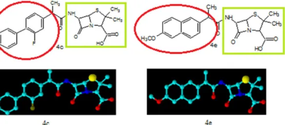
The synthesized penicillin’s derivatives(4a-h)were screened for their antibacterial activity against five different bacteria,Salmonilla typhaeandMicrococcus luteusare clinical isolates, rest of three are pathogenic and their ATTC numbers are provided. The bioactivity of synthe-sized compounds is presented in zone of inhibition of bacterial growth in millimeter (mm). The Amoxicillin a penicillin derivative was used as positive control to compare the antibacterial potential of our synthesized penicillins derivatives. The antibacterial activity results indicated that compounds4a, 4fand4hexhibited 60%, 72% and 64% bacterial zone inhibition againstE. colirespectively. On the other hand4a, 4band4hdisplayed 56%, 60% and 68% inhibitory activity againstS.aureusrespectively. The excellent zone inhibition was shown by the com-pound4cand4e. The central penam nucleus in all of these compounds is same but the side chain functionality is different as highlighted inFig 2which is responsible for difference in bio-activity among these compounds.
The compound4eexhibited 100% growth inhibition againstStaphylococcus aureuswhile compound4cshowed 76% of zone inhibition when compared with the standard. The com-pound4eshowed 80% and 76% of zone inhibition againstStaphylococcus epidermidusand Escherichia colirespectively but compound4cdisplayed 80% of zone inhibition against both Staphylococcus epidermidusandEscherichia coli. The compound4epossess methoxy substi-tuted naphthyl moiety as side chain functionality which may play very significant role in anti-bacterial activity. In case of compound4cthe fluorine substituted biphenyl ring system is present which is responsible for its high antibacterial potential. We found thatMicrococcus luteusis more resistant among the tested bacteria in this study against all of the synthesized compounds and the standard drugTable 1. The percent zone of inhibition was calculated on the basis of activity of the reference drug (see experimental part for exact formula). The central penam nucleus is common in both the compounds(4c)and(4e)but the side chain amide R group play significant role in antibacterial potential. Both these compounds also differ in degree of hydrophobicity because of different functionalities. The side chain R group contains two phenyl rings in both these derivatives but in case of compound(4e)phenyl rings are fused while in compound(4c)two independent phenyl rings are present. These structural features explain the difference in the antibacterial potential among the two compounds.
Fig 2. Chemical structure and three dimensional view of compounds 4c and 4e with highlighted green show the main penicillin nucleus and red show the designed moiety to increase the bioactivity.
Enzyme Inhibition Assay. TheE.coliMurC (UDP-N-acetylmuramic acid:L-alanine ligase) is an essential enzyme in bacteria for peptidoglycan biosynthesis. It catalyzes the conver-sion of UDP-MurNAc to UDP-MurNAc-Ala in the assembly of the disaccharide-peptide unit required for peptidoglycan. Thus inhibitors of MurC are the potential candidates for the devel-opment of potent antibacterials. The title compound4ewas selected to performE.coliMurC inhibitory activity using the Malachite assay for phosphate detection. Compound4eshowed an IC50of 12.5μM compared to 17μM for AMP-PCP, a non-hydrolyzable ATP analogue used
as a control inhibitor.
In-Silico
Docking Studies
Molecular docking is a virtual substitute of the x-ray crystallographic study of the drug binding to the target protein/DNA. In X-rays crystallography, the crystals of the enzyme is placed in the solution of the drug for binding to the active site and the resultant complex is then analyzed by crystallographic method to explore the structure activity relationship. The same result can be obtained by Molecular docking which involve the virtual complexing of the drug candidates in the active site of the crystallographic structure of target protein to predict the structure activ-ity relationship. First of all the active binding sites in penicillins binding protein (1CEF) were identified, these sites are important in molecular docking and de novo drug design. The volume of the binding pocket was identified using Pocket Finder (http://www.modelling.leeds.ac.uk/ qsitefinder/) and the binding site volume was found to be 240 Cubic Angstroms while the total volume of the protien was 31978 Cubic Angstroms. The binding pocket obtained from this study is essentially the same as that seen in the crystal structure of serine-based D-alanyl-D-ala-nine carboxypeptidase/transpeptidase (PDB1CEF). The active binding site located in a cleft between the five-stranded anti-parallelβsheet and the largeαhelical clusterS1 Fig. TheS1 Tablerepresents the various types of interactions between compounds and penicillin binding protein (1CEF) functionalities.
The conserved amino acid residues also termed as super-sites were then identified as it is functionally more important for kinetics and thermodynamics of protein folding than the non-conserved amino acid residues. Earlier studies have highlighted that Ser62 present in the active site binds directly to penicillin like drugs. This residue is conserved among all twenty one homo-log’s identified indicating the importance of the Ser62.S2 Figshowed the conserved amino acid
Table 1. Antibacterial activity result of penicillin derivatives (4a-h).
Zone of inhibition(mm±standard deviation)
Codes E.c. S.t. S.e. S.a. M.l.
4a 30±0.13 - 12±0.09 28±0.25
-4b - - - 30±0.19
-4c 40±0.16 - 24±0.06 38±0.08
-4d 30±0.20 - 14±0.17 0
-4e 38±0.18 - 24±0.21 50±0.18
-4f 36±0.09 - 2±0.12 4±0.05
-4g 10±0.11 - 14±0.08 16±0.15
-4h 32±0.16 - 2±0.22 34±0.13
-Amoxicillin 50±0.06 36±0.16 30±0.08 50±0.18 8±0.05
Data given are mean of three replicates±standard error. Activity presented in millimeters (mm). (-) = No activity measured.Escharichia coli(E.c.), Salmonilla typhae(S.t.),Staphylococcus epidermidus(S.e.),Staphylococcus aureus(S.a.),Micrococcus luteus(M.l.)
residue in the receptor protein. Residues Asn161, Ser 130, His 298, Thr 299 and Lys 65 which lies within the binding site are highly conserved and may play a major role in substrate binding or catalysis. Asn161 is conserved among eighteen of the twenty one homologs identified. The hallmark of all serine based penicillin interacting proteins is the presence of three well conserved motifs in the active site, the SXXK, (S/Y)XN and KT(S)G triads. All the Penicillin analogues docked with PBP using AutoDock vina gives lowest energy complexes stabilized by intermolec-ular hydrogen bonds andπ-πstacking interactions. The interactions in these complexes vary
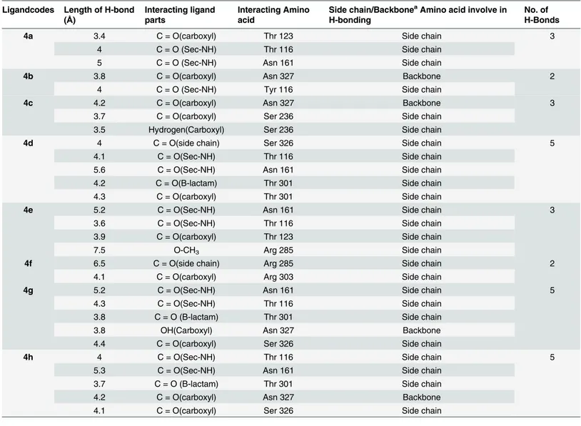
depending on the size, linkage and the functional groups. These stacking interactions have been proposed as the reason for the increased binding affinities of these larger inhibitors. Hydrogen bonding contributes most to the binding affinities of all penicillin derivatives with the receptor protein.Table 2presented the interacting parts of the ligands and the amino acids of receptors along with number and bond length of hydrogen bonds.
The carboxyl oxygen in compound4cinvolved in hydrogen bonding with amino acids ASN327 and SER236 with bond length 4.2Ǻand 3.7Ǻrespectively (Fig 3). The carboxyl
Table 2. Hydrogen bonding between penicillin derivatives (4a-h) and receptor.
Ligandcodes Length of H-bond (Å)
Interacting ligand parts
Interacting Amino acid
Side chain/BackboneaAmino acid involve in H-bonding
No. of H-Bonds
4a 3.4 C = O(carboxyl) Thr 123 Side chain 3
4 C = O (Sec-NH) Thr 116 Side chain
5 C = O (Sec-NH) Asn 161 Side chain
4b 3.8 C = O(carboxyl) Asn 327 Backbone 2
4 C = O (Sec-NH) Tyr 116 Side chain
4c 4.2 C = O(carboxyl) Asn 327 Backbone 3
3.7 C = O(carboxyl) Ser 236 Side chain
3.5 Hydrogen(Carboxyl) Ser 236 Side chain
4d 4 C = O(side chain) Ser 326 Side chain 5
4.1 C = O(Sec-NH) Thr 116 Side chain
5.6 C = O(Sec-NH) Asn 161 Side chain
4.2 C = O(B-lactam) Thr 301 Side chain
4.3 C = O(carboxyl) Thr 301 Side chain
4e 5.2 C = O(Sec-NH) Asn 161 Side chain 3
3.6 C = O(Sec-NH) Thr 116 Side chain
3.9 C = O(carboxyl) Thr 123 Side chain
7.5 O-CH3 Arg 285 Side chain
4f 6.5 C = O(side chain) Arg 285 Side chain 2
4.1 C = O(carboxyl) Arg 303 Side chain
4g 5.2 C = O(Sec-NH) Asn 161 Side chain 5
4.3 C = O(Sec-NH) Thr 116 Side chain
3.8 C = O (B-lactam) Thr 301 Side chain
3.8 OH(Carboxyl) Asn 327 Backbone
4.4 C = O(carboxyl) Ser 326 Side chain
4h 4 C = O(Sec-NH) Thr 116 Side chain 5
5.3 C = O(Sec-NH) Asn 161 Side chain
3.7 C = O (B-lactam) Thr 301 Side chain
4.2 C = O(carboxyl) Asn 327 Backbone
4.1 C = O(carboxyl) Ser 326 Side chain
aAmino acid main chain comprising of NH
2-CH2-COOH.
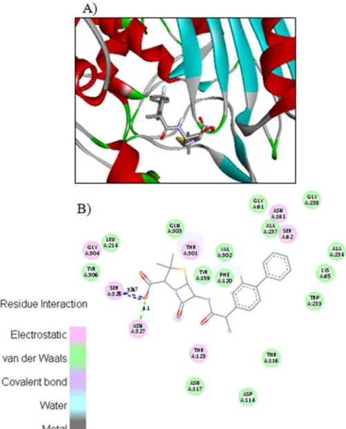
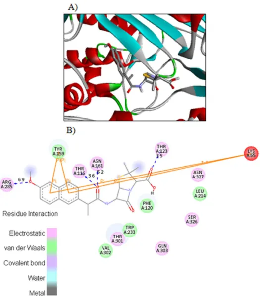
hydrogen in compound4calso interacts with side chain SER236 through hydrogen bond hav-ing bond length 3.5Ǻ.Fig 4displayed the two and three dimensional ligand-protein interac-tions of compound4ewith the active binding sites of penicillins binding protein. It was found from the figure 8 that compound4eformedπ-πstacks between naphthyl ring of the inhibitor
and TYR159 and LYS65 of PBP. The carboxyl oxygen involved in the hydrogen bonding with THR123 having bond length 3.9Ǻ. The methoxy oxygen in the same compound interacts with ARG285 and secondary amide oxygen bind with side chain amino acids ASN161 and THR116 with bond length 5.2 and 3.6Ǻrespectively. The binding affinities calculated for compounds
4cand4eare 8.8 and 8.9Kcal/mol respectively. The most potent penicillin’s derivatives also showed good docking scores.
Fig 3. The potential ligand-protein interactions of compound 4c with the active site of Penicillin binding protein (PDB ID 1CEF) generated by using Discovery Studio 4.0.A) The three-dimensional docking of the compound4cin the binding pocket. B) The two dimensional interactions of4cwith amino acid residues are shown as balls colored by the type of interaction.
The side chain indole moiety in case of compound4fshowπ-πstacks interactions with
TYR159 and LYS65. On the other hand carboxylic oxygen and amide carbonyl oxygen in com-pound4fforms hydrogen bonds with GLN303 and ARG285 having bond length respectively. Among all of the synthesized penicillin derivatives compound4fgives the lowest energy com-plex with binding energy of -9.2kcal/mol. Theβ-lactam carbonyl carbon of4d, 4g, and4halso forms H-bonds at the interacting distances of 4.2, 3.8 and 3.7Ǻrespectively. Two and three dimensional potential ligand-protein interactions of synthesized penicillin derivatives(4a, 4b, 4d, 4f, 4g and 4h)with the active site of Penicillin binding protein (PDB ID 1CEF) are shown inS3toS8Figs respectively as supporting information. All of the synthesized compounds eval-uated for their docking orientation to PBP exhibited reasonable affinity with good dock score. The binding energies of all these penicillin derivatives in the most favorable conformation are given inTable 3.
Fig 4. The potential ligand-protein interactions of compound 4e with the active site of Penicillin binding protein (PDB ID 1CEF) generated by using Discovery Studio 4.0.A) The three-dimensional docking of the compound4ein the binding pocket. B) The two dimensional interactions of4ewith amino acid residues are shown as balls colored by the type of interaction.
Lipinski
’
s Rule of Five
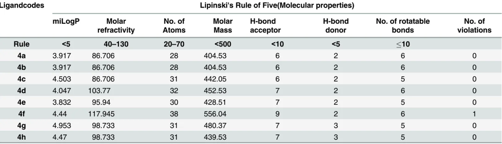
The synthesized compounds were tested for Lipinski’s Rule of 5 using the Molinspiration server (http://www.molinspiration.com). The inputs were given as a SMILES string. The calcu-latedlogPvalues and other structural properties of synthesized penicillin analogues(4a-h)are shown inTable 4. The results of the calculations for the molecules designed in this study show that all molecules have a potential for good in vivo absorption, since all the compounds shows zero voilation of the rule, except for single one in case of4fwhose molecular weight exceed the allowed range.
Conclusions
In conclusion we describe the synthesis and antimicrobial screening of novel penicillin ana-logues incorporating NSAIDs moiety as potent antibacterial agents.In-silicodocking of these derivatives with Penicillin binding protein (PDBID 1CEF) was performed in order to predict their binding affinity. The title compounds4cand4eshowed remarkable activity againstE. coli,S.epidermidusandS.aureuswith good docking scores. The title compound4ealso showed good enzyme inhibitory activity againstE.coliMurC with IC5012.5μM. All of the synthesized
derivatives(4a-h)exhibited high binding affinity with binding energies between -7.8 to 9.2kcal/mol. The carbonyl oxygen of carboxyl functionality in all molecules was involved in H-bonding with active site residues of target with the bond length ranges from 3.4 to 4.4Ǻ. Simi-larly, the carbonyl oxygen of the secondary amide forms H-bonds in all molecules with the exception of4f. Among the compounds tested for docking study,4fshowed high affinity with
Table 3. The binding Affinities of the penicillin derivatives (4a-h).
Compounds Mode Binding Affinity(kcal/mol)
4a 1 -7.8
4b 1 -7.8
4c 1 -8.8
4d 1 -8.8
4e 1 -8.9
4f 1 -9.2
4g 1 -8.2
4h 1 -9.0
doi:10.1371/journal.pone.0135293.t003
Table 4. Lipinski’s Rule of Five screening data for penicillin derivatives (4a-h).
Ligandcodes Lipinski's Rule of Five(Molecular properties)
miLogP Molar
refractivity
No. of Atoms
Molar Mass
H-bond acceptor
H-bond donor
No. of rotatable bonds
No. of violations
Rule <5 40–130 20–70 <500 <10 <5 10
4a 3.917 86.706 28 404.53 6 2 6 0
4b 3.917 86.706 28 404.53 6 2 6 0
4c 4.503 86.706 31 442.05 6 2 5 0
4d 4.047 103.77 32 452.53 7 2 6 0
4e 3.832 95.94 30 428.51 7 2 5 0
4f 4.44 117.945 38 556.04 9 2 6 1
4g 4.953 98.733 31 480.37 7 3 5 0
4h 4.47 98.733 31 439.53 7 3 5 0
low energy of -9.2kcal/mol with employed protein. The clinical isolatesMicrococcus luteusand Salmonilla typhaewere found to be most resistant against all of synthesized compounds and standard also. Our results endorse us that compounds4cand4emay serve as a structural tem-plate for the design and development of highly potent antimicrobial agents.
Supporting Information
S1 Fig. Image showing the position of the active site occupied between five-stranded anti-parallelβ-sheet and the largeα-helical cluster.
(TIF)
S2 Fig. The conserved amino acids residues of the receptor protein.
(TIF)
S3 Fig. The potential ligand-protein interactions of compound 4a with the active site of Pen-icillin binding protein (PDB ID 1CEF) generated by using Discovery Studio 4.0.A) The three-dimensional docking of the compound4ain the binding pocket. B) The two dimensional interactions of4awith amino acid residues are shown as balls colored by the type of interaction. (TIF)
S4 Fig. The potential ligand-protein interactions of compound 4b with the active site of Pen-icillin binding protein (PDB ID 1CEF) generated by using Discovery Studio 4.0.A) The three-dimensional docking of the compound4bin the binding pocket. B) The two dimensional interactions of4bwith amino acid residues are shown as balls colored by the type of interaction. (TIF)
S5 Fig. The potential ligand-protein interactions of compound 4d with the active site of Penicillin binding protein (PDB ID 1CEF) generated by using Discovery Studio 4.0.A) The three-dimensional docking of the compound4din the binding pocket. B) The two dimensional interactions of4dwith amino acid residues are shown as balls colored by the type of interac-tion.
(TIF)
S6 Fig. The potential ligand-protein interactions of compound 4f with the active site of Penicillin binding protein (PDB ID 1CEF) generated by using Discovery Studio 4.0.A) The three-dimensional docking of the compound4fin the binding pocket. B) The two dimensional interactions of4fwith amino acid residues are shown as balls colored by the type of interac-tion.
(TIF)
S7 Fig. The potential ligand-protein interactions of compound 4g with the active site of Penicillin binding protein (PDB ID 1CEF) generated by using Discovery Studio 4.0.A) The three-dimensional docking of the compound4gin the binding pocket. B) The two dimensional interactions of4gwith amino acid residues are shown as balls colored by the type of interac-tion.
(TIF)
S8 Fig. The potential ligand-protein interactions of compound 4h with the active site of Penicillin binding protein (PDB ID 1CEF) generated by using Discovery Studio 4.0.A) The three-dimensional docking of the compound4hin the binding pocket. B) The two dimensional interactions of4hwith amino acid residues are shown as balls colored by the type of interac-tion.
S1 Table. Descriptions of the various types of interactions between inhibitors and target protein functionalities shown in figures.
(DOCX)
Acknowledgments
The authors are thankful to Dr. Zaheer Ahmad Assistant Professor, AIOU, for providing the lab facilities for initial screening of the synthesized compounds. The authors are also grateful to National Institute of Health for providing some bacterial strain used in the present studies.
Author Contributions
Conceived and designed the experiments: ZA AB MM UN. Performed the experiments: ZA AB. Analyzed the data: ZA AB. Contributed reagents/materials/analysis tools: ZA AB UN. Wrote the paper: ZA AB MM.
References
1. Babington R, Matas S, Macros MP, Galve R (2012) Current bioanalytical methods for detection of peni-cillins. Anal Bioanal Chem., 403: 1549–1556. doi:10.1007/s00216-012-5960-4PMID:22488111 2. Jones RN, Barry AL, Thornsberry C (1989) In-vitro studies of meropenem. J. Antimicrob. Chemother.,
24: (Suppl. A), 9–29. PMID:2808218
3. Chu DTW, Plattner JJ, Katz L (1996) New Directions in Antibacterial Research. J. Med. Chem., 39: 3853–3874. PMID:8831751
4. Page MI. The Chemistry ofβ-Lactams; Blackie Academic & Professional: (ed. Page M. I.) London 1992, 79.
5. Kumar S, Basha SKN, Kumarnallasivan P, Vijanianand PR, Pradeepchandran R, Jayaveera KN, et al. (2010) Computational design and docking studies on Escherichia coliβ-Ketoacyl-Acyl carrier protein synthesis III using auto dock. J. Pharm. Res., 7: 1460–1462.
6. Brulé C, Brynaert MJ. Penicillins. In: Katritziy AR(ed.) Comprehensive Heterocyclic chemistry III, 3rd Edn. Elsevier, Belgium, 2008, 173–273.
7. Reid AJ, Simpson IN, Harper PB, Amyes SG (1987) Ampicillin resistance in Haemophilus influenzae: identification of resistance mechanisms. J. Antimicrob. Chemother., 20: 645–465. PMID:3501421 8. Jorgensen JH (1992,) Update on mechanisms and prevalence of antimicrobial resistance
inHaemophi-lus influenzae. Clin. Infect. Dis., 14: 1119–1123. PMID:1600014
9. Macheboeuf P, Contreras-Martel C, Job V, Dideberg O, Dessen A (2006) Penicillin bindingproteins: key players in bacterial cell cycle and drug resistance processes.FEMS Microbiol.Rev., 30: 673–691.
10. Lovering AL, deCastro LH, Lim D, Strynadka NC (2007) Structural insight into the transglycosylation step of bacterial cell-wall biosynthesis. Science, 315: 1402–1405. PMID:17347437
11. Navratna V, Nadig S, Sood V, Prasad K, Arakere G, Gopal B (2010) Molecular Basis for the Role of Staphylococcus aureusPenicillin Binding Protein 4 in Antimicrobial Resistance. J. Bacteriol., 192: 134–144. doi:10.1128/JB.00822-09PMID:19854906
12. Luengo JM, Iriso JL, López-Nieto MJ (1986) Direct enzymatic synthesis of natural penicillins using phe-nylacetyl-CoA: 6-APA phenylacetyl transferase of Penicillium chrysogenum: minimal and maximal side chain length requirements.J.Antibiot., 39: 1754–1759.
13. Trott O, Olson AJ (2010) "AutoDock Vina: improving the speed and accuracy of docking with a new scoring function, efficient optimization, and multithreading." J. Comput. Chem. 31: 455–461. doi:10. 1002/jcc.21334PMID:19499576
14. Koska J, Spassov VZ, Maynard AJ, Yan L, Austin N, Flook PK, et al.(2008) Fully automated molecular mechanics based induced fit protein-ligand docking method. J. Chem. Inf. Model 48: 1965–1973. doi:
10.1021/ci800081sPMID:18816046
15. Yusuf D, Davis AM, Kleywegt GJ, Schmitt S (2008) An alternative method for the evaluation of docking performance: RSR vs RMSD. J. Chem. Inf. Model 48: 1411–1422. doi:10.1021/ci800084xPMID:
18598022
17. Sousa SF, Fernandes PA, Ramos MJ (2006) Protein–ligand docking: Current status and future chal-lenges. Protien-Struct Funci Bioinformatics 65: 15–26.
18. Lipinski CA, Lombardo F, Dominy PW, Feeney PJ (1997) Experimental and computational approaches to estimate solubility and permeability in drug discovery and development setting. Adv. Drug Deliv. Rev. 23: 3–25.
19. Albert PLi (2005) Preclinical in vitro screening assays for drug-like properties. Drug Discov. Today Technol. 2: 179–185. doi:10.1016/j.ddtec.2005.05.024PMID:24981846
20. Ashraf Z, Saeed A, Nadeem H (2014) Design, synthesis and docking studies of some novel isocou-marin analogues as antimicrobial agents. RSC Adv. 4: 53842–53853.
21. Saeed A, Abbas N, Ashraf Z, Bolte M (2014) Novel 5-Acetyl-3-aryl-2-thioxo-dihydropyrimidine-4,6 (1H,5H)-diones: One Pot Three-Component Synthesis, Characterization and Antibacterial Activity. J. Heterocycl. Chem. 51: 398–403.
22. Marmor S, Petersen CP, Reck F, Yang W, Gao N, Fisher SL (2001) Biochemical characterization of a phosphinate inhibitor of Escherichia coli MurC, Biochemistry 40: 12207–12214. PMID:11580296 23. Baseer M, Ansari FL, Ashraf Z (2013) Synthesis, in vitro antibacterial and antifungal activity of some
n-acetylated and non-n-acetylated pyrazolines. Pak. J. Pharm. Sci. 26: 67–73.
24. Lanzetta PA, Alvarez LJ, Reinach PS, Candia OA (1979) An improved assay for nanomole amounts of inorganic phosphate, Anal. Biochem. 100: 95–97.
25. Leatherbarrow RJ. GraFit 4.0.12, Erithacus Software, Staines, 1998.
26. Kelly JA, Dideberg O, Charlier P, Wery JP, Libert M, Moews PC, et al. (1986) On the origin of bacterial resistance to penicillin: comparison of a beta-lactamase and a penicillin target. Science 231: 1429– 1431. PMID:3082007
27. Faridoon, Hussein MW, Islam NUl, Guddat LW, Schenk G, McGeary RP (2012) Penicillin inhibitors of purple acid phosphatase. Bioorg. Med. Chem. Lett. 22: 2555–2559. doi:10.1016/j.bmcl.2012.01.123