Diversity of Aquatic
Pseudomonas
Species
and Their Activity against the Fish
Pathogenic Oomycete
Saprolegnia
Yiying Liu1,2, Elzbieta Rzeszutek3, Menno van der Voort2, Cheng-Hsuan Wu4,5, Even Thoen6,7, Ida Skaar6, Vincent Bulone3, Pieter C. Dorrestein4,8,9,10, Jos M. Raaijmakers1, Irene de Bruijn1*
1Department of Microbial Ecology, Netherlands Institute of Ecology (NIOO-KNAW), Wageningen, The Netherlands,2Laboratory of Phytopathology, Wageningen University, Wageningen, The Netherlands, 3Division of Glycoscience, School of Biotechnology, Royal Institute of Technology, Stockholm, Sweden, 4Department of Chemistry and Biochemistry, University of California San Diego, La Jolla, California, United States of America,5Department of Chemistry, Boston University, Boston, United States of America, 6Norwegian Veterinary Institute, Oslo, Norway,7Norwegian University of Life Sciences, Oslo, Norway, 8Skaggs School of Pharmacy and Pharmaceutical Sciences, University of California San Diego, La Jolla, California, United States of America,9Center for Marine Biotechnology and Biomedicine, Scripps Institution of Oceanography, University of California San Diego, La Jolla, California, United States of America, 10Department of Pharmacology, University of California San Diego, La Jolla, California, United States of America
*i.debruijn@nioo.knaw.nl
Abstract
Emerging fungal and oomycete pathogens are increasingly threatening animals and plants globally. Amongst oomycetes,Saprolegniaspecies adversely affect wild and cultivated populations of amphibians and fish, leading to substantial reductions in biodiversity and food productivity. With the ban of several chemical control measures, new sustainable methods are needed to mitigateSaprolegniainfections in aquaculture. Here, PhyloChip-based community analyses showed that the Pseudomonadales, particularlyPseudomonas species, represent one of the largest bacterial orders associated with salmon eggs from a commercial hatchery. Among thePseudomonasspecies isolated from salmon eggs, significantly more biosurfactant producers were retrieved from healthy salmon eggs than fromSaprolegnia-infected eggs. Subsequentin vivoactivity bioassays showed that Pseu-domonasisolate H6 significantly reduced salmon egg mortality caused bySaprolegnia diclina. Live colony mass spectrometry showed that strain H6 produces a viscosin-like lipopeptide surfactant. This biosurfactant inhibited growth ofSaprolegnia in vitro, but no sig-nificant protection of salmon eggs against Saprolegniosis was observed. These results indi-cate that live inocula of aquaticPseudomonasstrains, instead of their bioactive compound, can provide new (micro)biological and sustainable means to mitigate oomycete diseases in aquaculture.
OPEN ACCESS
Citation:Liu Y, Rzeszutek E, van der Voort M, Wu C-H, Thoen E, Skaar I, et al. (2015) Diversity of Aquatic
PseudomonasSpecies and Their Activity against the Fish Pathogenic OomyceteSaprolegnia. PLoS ONE 10(8): e0136241. doi:10.1371/journal.pone.0136241
Editor:Adelaide Almeida, University of Aveiro, PORTUGAL
Received:May 18, 2015
Accepted:July 30, 2015
Published:August 28, 2015
Copyright:© 2015 Liu et al. This is an open access article distributed under the terms of theCreative Commons Attribution License, which permits unrestricted use, distribution, and reproduction in any medium, provided the original author and source are credited.
Data Availability Statement:Data have been deposited in GenBank under accession numbers KP890304-KP890314 and KT223371-KT223386, respectively.
Introduction
Emerging fungal and fungal-like diseases are causing severe ecological disruptions and are rec-ognised as a global threat to biodiversity and food security [1,2]. For example,Fusarium solani
is involved in mass mortality of eggs of the endangered sea turtles in Cape Verde [3], and
Batrachochytrium dendrobatidisandB.salmandrivoransare causing major amphibian declines globally [4,5]. Amongst oomycetes,AphanomycesandSaprolegniaspecies are causing signifi-cant declines in crayfish, fish and amphibian populations [1,6–11].Saprolegniaspecies are the causative agents of Saprolegniosis, a disease characterized by fluffy and filamentous white or grey mycelial patches on fish, fish eggs or amphibians [11]. In aquaculture,Saprolegniaspecies regularly infect freshwater cultured salmonids, including Atlantic salmon and rainbow trout, and non-salmonids like eel, perch, carp and catfish [7,12]. In Japan, at least 50% annual mor-tality in Coho salmon due to Saprolegniosis was reported [7,13,14]. Also the‘winter kill’by
Saprolegniaspecies in channel catfish in the USA resulted in a substantial financial loss of approximately $40 million [7].
Formalin is now commonly used to control Saprolegniosis, but is expected to be banned soon due to adverse environmental effects [7]. Several treatments have been tested to prevent Saprolegniosis, such as hydrogen cyanide, Pyceze (bronopol), sea water flushes and NaCl, but none of these measures exerted control to a level similar as obtained with malachite green, a chemical banned due to its carcinogenic properties [7]. Currently, no vaccination is available for Saprolegniosis [8]. Therefore, new sustainable measures are urgently needed. A potential approach to control Saprolegniosis and other emerging diseases involves the application of beneficial microbes. A limited number of bacterial genera and species, includingAeromonas
andPseudomonas, have been reported as potential anti-pathogen agents in aquaculture, but a comprehensive understanding of the microbiome composition of fish eggs and their protective potential is still limited [15–21].
In previous work, we detected 31,281 bacterial and archaeal operational taxonomic units (OTUs) on salmon eggs from a hatchery by PhyloChip metataxonomic analysis. The highest number of OTUs belonged to Proteobacteria [22]. Based on this large-scale metataxonomic analysis, the diversity of salmon egg-associated Proteobacteria and their functional potential to protect fish eggs againstSaprolegniawere investigated in this study. We focused specifically on the Pseudomonadales and isolated severalPseudomonasstrains, assessed their genotypic diver-sity and tested their inhibitory activity againstSaprolegniaspecies bothin vitroandin vivo. Given the zoosporicidal activity of biosurfactants produced byPseudomonasspecies [23–25], the isolates obtained from salmon eggs were also phenotypically screened for biosurfactant production. For thePseudomonasisolate that provided the best protection against Saprolegnio-sis on salmon eggs, chemical profiling was performed by Nanospray Desorption ElectroSpray Ionization (NanoDESI) live colony mass spectrometry followed by MS/MS analysis for partial identification of the biosurfactant.
Materials and Methods
Isolation of bacteria associated with salmon eggs
Healthy andSaprolegnia-infected salmon eggs (N= 6 for healthy andN= 6 for diseased eggs) and their corresponding incubation water was collected from a commercial hatchery [22]. To release bacteria from the surface of eggs, approximately 30 eggs and 20 ml of incubation water of each sample was transferred into a glass tube, vortexed for one minute, sonicated for one minute and vortexed again for one minute. The total culturable bacteria were isolated and enumerated by plating on 1/10thstrength tryptone soya broth (Oxoid) with 15–20 gl-1agar
Commission under HORIZON 2020; and BE-Basic Foundation (project number F07.003.01). The funders had no role in study design, data collection and analysis, decision to publish, or preparation of the manuscript.
(1/10TSA) supplemented with 100μg ml-1Delvocid (DSM, Delft, Netherlands) to inhibit
fun-gal growth.Pseudomonasstrains were isolated and enumerated on semi-selectivePseudomonas
agar F (PSA, Difco) supplemented with 100μg ml-1Delvocid, 12.5μg ml-1chloramphenicol
and 50μg ml-1ampicillin. Both media were incubated at 25°C for 4 days. From each replicate sample and each growth medium, approximately 40 bacterial isolates were randomly selected and stored, which resulted in a total of approximately 900 random bacterial isolates.
In vitro
activity and biosurfactant production by the bacterial isolates
All bacterial isolates were tested for activity againstSaprolegnia diclinastrain VS20 and Sapro-legnia parasiticastrain CBS 223.65 (C65) according to Liuet al. [22]. Bacteria were spot-inocu-lated at the edge of plates of 1/5thstrength potato dextrose broth (Difco) with 15
–20 g l-1agar
(1/5PDA) and incubated for 2–4 days at 25°C prior to inoculation of a plug ofSaprolegniain the centre of a 1/5PDA plate. Hyphal growth inhibition was monitored for all bacterial isolates during incubation for 4–5 days at 18°C. All bacterial isolates were also screened for biosurfac-tant production by the drop collapse assay according to the method described by de Bruijn
et al. [23].
Identification and phylogeny of bacterial isolates
Pseudomonasisolates inhibitingSaprolegniahyphal growth and/or producing biosurfactants
were subjected to BOX-PCR fingerprinting using primer BOX A1R [26,27]. Representative isolates from the BOX groups with at least 4 isolates for diseased or for healthy salmon egg samples were chosen for phylogenetic analysis. Therefore, a total of 27 representative isolates (S1 Table) were selected and identified by 16S rRNA sequencing. Phylogenetic analyses, including the 16S rRNA sequences of 29 known reference strains [28], was performed accord-ing to Liuet al. [22]. The evolutionary distances were calculated using the Kimura 2-parameter
method [29].
In vivo
bioassays to test disease suppression by Proteobacteria
The representative isolates were chosen for activity testing in salmon egg bioassays (S1 Table). From the shared representative isolates,Pseudomonasisolates S1 and S2, which belonged to the largest shared BOX group and originated from both healthy and diseased salmon eggs were selected. Collectively, a total of 11 representative isolates were selected for activity testing in salmon egg bioassays (S1 Table).
The experimental set-up of thein vivobioassays was similar to that described by Liuet al. [22]. For bioassay 1 conducted in Norway in 2012, salmon eggs used in this bioassay were 385 degree-days at the day of shipment. Each treatment was conducted in a separate incubation unit containing three perforated cups with 30 live salmon eggs per cup. The 11 representative isolates described above were pre-grown on PSA for 2 days at 25°C, washed with sterile de-mineralized water and added to each salmon egg incubation unit to a final cell density of 108 CFU ml-1.S.diclina765F3 and bacteria were added to the incubation units with 2.5 litre of
For bioassay 2 conducted in The Netherlands in 2014, the experimental design was adjusted to monitor the disease progress over a longer period of time. Therefore, the salmon eggs were
‘younger’than those in bioassay 1, i.e. 321 degree-days at the day of shipment. Each treatment was conducted in two separate incubation units; each incubation unit contained three perfo-rated cups with 51±2 live salmon eggs per cup. Spontaneous rifampicin resistant mutants of thePseudomonasisolates were generated to allow monitoring of their population dynamics. ThePseudomonasisolates were added to each salmon egg incubation unit filled with 2 litres of well water [22] to the final cell density of 107CFU ml-1.S.diclina1152F4 [22] was used in bioassay 2. Salmon eggs not treated with the bacterial isolates or exposed toS.diclinaonly served as the controls. The egg incubation water of each incubation unit was sampled on day 1, day 4 (0 dpi) and day 24 (20 dpi) and dilution-plated on PSA+rifampicin to determine the density of the introduced isolates in the water. Colonization of the appliedPseudomonas iso-lates on the salmon egg surface was determined by rolling one to two eggs from each cup on PSA+rifampicin at 0 and 20 dpi as described by Liuet al. [22]. The percentage of salmon eggs to which hyphae ofS.diclinawere attached was determined at 20 dpi according to Liuet al. [22].
S.diclina765F3 from the healthy sample andS.diclina1152F4 from the diseased sample were used as pathogen source in the bioassay 1 in 2012 and bioassay 2 in 2014, respectively. These twoS.diclinaisolates showed similar pathogenicity on salmon eggs in 2012 [22].
Nucleotide sequence accession numbers
The 16S rRNA sequences ofPseudomonasstrains D1, D2, D3, S1, S2, H1, H2, H3, H4, H5, H6 and S3-S18 have been deposited in GenBank under accession numbers KP890304-KP890314 and KT223371-KT223386, respectively.
Live colony NanoDESI MS/MS data acquisition and molecular
networking
The most antagonisticPseudomonasisolate H6 was subjected to chemical profiling by
Nanos-pray Desorption ElectroSNanos-pray Ionization (NanoDESI) live colony mass spectrometry as described previously [30,31].P.fluorescensSS101 andP.fluorescensSBW25 [30] were used as references.Pseudomonasstrains were cultured in Luria-Bertani broth by shaking overnight at 28°C. ForPseudomonasH6, four cultures (0.5μl each) were spot-inoculated on ISP2 agar
and incubated for 48 hours at 30°C [30].P.fluorescensSS101 andP.fluorescensSBW25 were streaked on 1/5thstrength NBY agar (1 gl-1glucose, 1.6 gl-1nutrient broth, 0.4 gl-1yeast extract
and 15 gl-1agar) and cultured for 48 hours at 25°C. Data collection with a data-dependent MS/
MS method was conducted on a hybrid 6.4T LTQ-FT (Thermo Electron) mass spectrometer according to Nguyenet al. [30].
The MS/MS data of strainsPseudomonasH6,P.fluorescensSS101 andP.fluorescensSBW25
were combined with data obtained by Nguyenet al. (2013) [30] for 18 other Pseudomonads and 42 Bacilli. Metabolic networks were generated by clustering as described previously [32] using the GNPS website (http://gnps.ucsd.edu/). Algorithms were the same as described by Nguyenet al. [30]. Networks were visualized using Cytoscape (v 3.1.1). The two plugins used
for aiding data visualization and sequence tagging were described previously by Nguyenet al. [30].
HPLC analysis
After lyophilization, the biosurfactant was dissolved in Milli-Q water. Reversed-Phase High Performance Liquid Chromatography (RP-HPLC) analysis was performed by injection of 100μl sample on a Waters 996 HPLC equipped with a Symmetry C18 Column (100 Å, 5μm, 3.9 mm X 150 mm, Waters) as described previously [33]. Samples were analysed at a flow rate of 0.5 ml/min for 50 min in an isocratic mobile phase of 45:40:15 acetonitrile:methanol:Milli-Q water with 0.1% (v/v) trifluoroacetic acid.
In vitro
inhibitory activity of biosurfactants against
Saprolegnia
hyphal
growth
The biosurfactant ofPseudomonasH6 was tested for activity against hyphae ofS.diclina
1152F4 andS.parasiticaC65 by adding the biosurfactant to 1 ml 1/5thstrength potato dextrose broth (1/5PDB) in 24-well cell-culture plates (Greiner Bio-One, Kremsmünster, Austria) to final concentrations of 15, 40, 100, 200μg ml-1, respectively. The lipopeptide surfactant masse-tolide A produced byP.fluorescensSS101 was used as a standard. An agar plug of pre-grown
S.diclina1152F4 orS.parasiticaC65 of approximately 0.2 cm2was added to each well and incubated at 25°C. Hyphal growth inhibition was scored after 3 days forS.diclina1152F4 or 2 days forS.parasiticaC65. Morphological abnormalities were monitored under an Olympus SZX12 stereomicroscope and image capture was accomplished using a Zeiss AxioCam MRc 5 camera with Zeiss AxioVision software (AxioVs40 V 4.8.2.0). ImageJ 1.47v [34] was used to measure the diameter of 10 hyphae in each treatment.
In vivo
effects of biosurfactants on Saprolegniosis
The salmon eggs had been incubated at 0.92°C on average at AquaGen AS (Trondheim, Nor-way) and their age was 135 degree-days at the day of shipment. The set-up of the salmon egg bioassay was similar to that of bioassay 2 described above. The biosurfactants from Pseudomo-nasH6 and SS101 were tested at concentrations of 15±3μg ml-1and 40±8μg ml-1. Malachite
green (2.5±0.5μg ml-1(ppm)) was used as a chemical reference. All treatments were performed
with three separate incubation units (biological replicates), except for the treatments without
S.diclina1152F4 where one incubation unit with three incubation cups (technical reps) was used; each cup contained 51±2 live salmon eggs. Application of biosurfactants or malachite green was conducted every 2–3 days by reducing the water level to 600±100 ml and exposing the salmon eggs to the corresponding chemicals for 90–120 min with aeration. Afterwards, the
treated water was removed. Each incubation unit was rinsed by 100–200 ml fresh well water and finally 2 litres of fresh well water was added. The percentage of hyphal attachment of
S.diclinato the salmon eggs was determined on 18 dpi.
Results and Discussion
PhyloChip-based community profiling
Previously we analysed the bacterial community compositions of healthy salmon eggs and
diseased and healthy salmon eggs. Given thatPseudomonasspecies (Pseudomonadales) are considered as potential aquaculture probiotics [35], a more in-depth analysis was conducted here to unravel the genotypic and functional diversity ofPseudomonasspecies associated with healthy and diseased salmon eggs (S1 Fig).
Isolation,
in vitro
activity and phylogeny of aquatic
Pseudomonas
The bacteria attached to the salmon eggs were released by vortexing and sonication in the incu-bation water from the hatchery. The total count of culturable aerobic bacteria was approxi-mately 3X10⁷CFU ml-1for both healthy and diseased salmon eggs (S2 Fig). Amongst 440 randomly selected isolates, 7% and 13% inhibited hyphal growth ofSaprolegnia diclinaVS20 andSaprolegnia parasiticaC65, respectively. Drop collapse assays further revealed that 3% of the 1/10TSA isolates produced biosurfactants under the experimental conditions tested. For both the healthy and diseased salmon egg samples, the putativePseudomonaspopulation den-sity enumerated on semi-selective PSA medium was approximately 106CFU ml-1(S2 Fig).
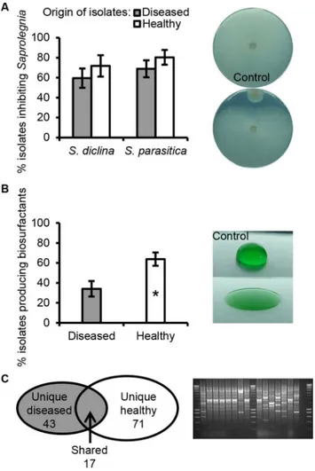
Amongst 465 randomly selectedPseudomonasisolates, 60–69% of the isolates from the dis-eased salmon eggs and 72–80% of the isolates from the healthy salmon eggs inhibited hyphal growth ofS.diclinaVS20 and/orS.parasiticaC65. No statistically significant difference was observed between diseased and healthy salmon eggs in the percentage of isolates within vitro
growth-inhibiting activities (Fig 2A). However, based on the results obtained in drop collapse assays, healthy eggs harboured a significantly higher frequency (64±7%) of biosurfactant-pro-ducingPseudomonasisolates than diseased salmon eggs (34±8%) (Fig 2B). A marine biosurfac-tant-producingLactobacillus pentosusprovided protection for the crustaceanArtemiaagainst pathogenicVibrio alginolyticus, suggesting a potential role of biosurfactants in disease suppres-sion [36].
Genotypic profiling by BOX-PCR of thePseudomonasisolates that inhibitedSaprolegnia
hyphal growthin vitroand/or produced biosurfactants, resulted in 131 BOX groups with 43 and 71 unique groups from diseased (D) or healthy (H) salmon egg samples, respectively, and 17 groups with isolates found in both diseased and healthy samples (referred to as‘shared’ iso-lates (S)) (Fig 2C). Representative isolates from BOX groups that consisted of at least 4 isolates
Fig 1. Proteobacteria (A) and Gammaproteobacteria (B) community analyses of diseased and healthy salmon eggs by PhyloChip-based analyses.Salmon eggs were sampled from a commercial hatchery [22]. Indicated is the average number of OTUs at different taxonomic levels: the phylum Proteobacteria (A) and the class Gammaproteobacteria (B). Error bars represent S.E.M. (N= 6).
unique for diseased (D) or healthy (H) samples, as well as 18 isolates from the 9‘shared’(S)
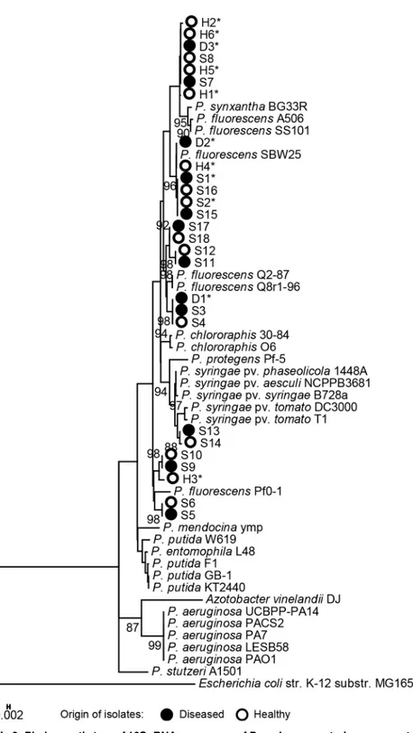
BOX groups (S1 Table) were subjected to phylogenetic analyses. No distinct differences were observed in the 16S rRNA-based phylogenetic delineation ofPseudomonasisolates from
dis-eased or healthy salmon eggs. Most of the isolates belonged to theP.fluorescensclade, includ-ing the isolates from the shared BOX groups, except isolate S13 and S14 that clustered with
P.syringae(Fig 3).
Bioactivity of aquatic
Pseudomonas in vivo
WhenPseudomonasisolates were applied at an initial density of 107CFU ml-1to the incuba-tion waterin vivo, seven out of elevenPseudomonasisolates significantly reduced hyphal attachment ofS.diclinato salmon eggs (Fig 4). Strains originally isolated from healthy salmon
Fig 2. Phenotypic and genotypic analysis ofPseudomonasisolates.The isolates were obtained from healthy salmon egg samples and salmon eggs infected withSaprolegnia. (A) Mean percentage of the number of bacterial isolates that were inhibitory to hyphal growth ofSaprolegnia diclinaVS20 andSaprolegnia parasiticaCBS 223.65, tested by observation of an inhibition halo around the bacterial colony (right panel insert). (B) Mean percentage of the number of bacterial isolates that produce biosurfactants based on the drop collapse assay (right panel insert). Error bars represent the S.E.M. (N= 6). The asterisk indicates a statistically significant difference (P<0.05, Student’st-test). (C) Genotypic BOX-PCR grouping of bacterial isolates that inhibitingSaprolegniahyphal growth and/or producing biosurfactants. The Venn diagram (left panel insert) shows the total number of BOX groups obtained for isolates from either diseased, healthy or both samples. An example of the DNA profiles obtained by BOX-PCR is shown in agarose gel (right panel insert).
Fig 3. Phylogenetic tree of 16S rRNA sequences ofPseudomonasstrains representative of 18 BOX-PCR groups.The 16S rRNA sequences were approximately 960 bp. The BOX-PCR groups were identified among thePseudomonasisolates from healthy and diseased salmon eggs (S1 Table). A total of 29 referencePseudomonasspecies/strains were included to delineate the 27 aquaticPseudomonasstrains obtained in this study. Bootstrap values at the nodes are based on 1000 replications. Only those branch values higher than 80% are shown. Asterisks indicate the isolates selected for salmon egg bioassays.
egg samples showed a better control efficacy than those originally isolated from diseased salmon egg samples (Fig 4), suggesting that the healthy salmon eggs harbourPseudomonas
strains with stronger activity againstSaprolegnia. The bacterial density in the incubation water
decreased from 106−10
7CFU ml-1on 0 dpi to 103
−10
5CFU ml-1on 20 dpi. When tested
against anotherS.diclinaisolate andS.parasiticaisolate under different temperatures and
bacterial densities, onlyPseudomonasstrain H6 showed the most consistent activity against
Saprolegniain all cases (S3andS4AFigs). Hence, strain H6 was selected for further
characterization.
Chemical profiling of aquatic
Pseudomonas
strain H6
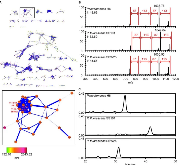
To elucidate which compoundsPseudomonasstrain H6 produces, live colony mass spectrome-try and MS/MS analyses were performed with two phylogenetically related strainsP. fluores-censSS101 and SBW25 as references. A Cytoscape network with spectra of in total 21 Pseudomonads and 42 Bacilli strains [30] showed thatPseudomonasH6 produces a predomi-nant compound with a parent mass-to-charge ratio ofm/z1148.65, which clustered together with the lipopeptides massetolide A (m/z1162.69) and viscosin (m/z1148.67) produced by strains SS101 and SBW25, respectively (Fig 5A). Further examination of the raw MS/MS data showed that mass shifts could be linked to specific amino acids, which created identical sequence tags of 87-113-87-113 Da (Ser-Leu/Ile-Ser-Leu/Ile) for the lipopeptide surfactants produced byPseudomonasH6,P.fluorescensSS101 and SBW25 (Fig 5B) [30]. These results suggest thatPseudomonasH6 produces a lipopeptide surfactant with a peptide moiety that is, most likely, structurally similar to that of massetolide A and viscosin. RP-HPLC analysis of the lipopeptide surfactants extracted from these three strains revealed a difference in retention time of the biosurfactants fromPseudomonasH6,P.fluorescensSS101 and SBW25 (Fig 5C), suggesting that the lipopeptide biosurfactant produced byPseudomonasH6 is structurally not identical to massetolide A or viscosin. The small shift in retention could also be due to a struc-tural difference in the lipid moiety.
Fig 4.In vivoactivity of 11Pseudomonasstrains againstS.diclina1152F4.In bioassay 2, the mean percentage of eggs to which ofS.diclina1152F4 hyphae attached was determined at 20 days post inoculation (dpi) [22]. Bacterial strains were introduced at an initial cell density of 107CFU ml-1. The
incubation temperature was 5–7°C. Error bars represent S.E.M. (N= 6). Asterisks indicate statistically significant differences with the control,S.diclinaonly, based on a one-way analysis of variance andpost hoc
LSD analysis (P<0.05).
Activity profiling of lipopeptide surfactant from aquatic
Pseudomonas
strain H6
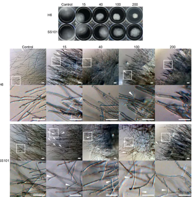
We tested the effect of the purified lipopeptide surfactants ofPseudomonasH6 andP. fluores-censSS101 on hyphal growth ofS.diclina1152F4 (Fig 6A). Both surfactants showed growth-inhibitory activity at 15μg ml-1, the lowest concentration tested. Hyphal growth was almost completely inhibited at 100μg ml-1for the biosurfactant ofPseudomonasH6 and at 200μg
ml-1for massetolide A ofP.fluorescensSS101 (Fig 6A). The biosurfactant of H6 also showed a
Fig 5. Live colony mass spectrometry and MS/MS networking.(A) Molecular network ofPseudomonasH6,P.fluorescensSS101,P.fluorescensSBW25 and 18 other Pseudomonads and 42 Bacilli [30]. The viscosin-like cluster was boxed and enlarged. (B) Selection of nodes ofPseudomonasH6,P.fluorescens
SS101 and SBW25 for MS/MS raw spectra analyses and generation of amino acid sequence tag. The sequence tag is created by analysing the mass shifts between adjacent ions in the MS/MS spectra, which are corresponding to the mass of an amino acid. The value of the parent mass ions forPseudomonasH6 andP.fluorescensSBW25 in Fig 5B is not completely matching to the parent mass ion as indicated in Fig 5A, because during network analyses a mass tolerance setting of 0.3 Da was used. (C) Reversed-phase HPLC chromatograms of cell-free culture extracts of lipopeptide surfactants fromPseudomonasH6,
P.fluorescensSS101 and SBW25.
stronger activity compared to massetolide A against hyphal growth ofS.parasitica(Fig 7A). Lipopeptide surfactants are known to cause hyphal swelling, hyphal branching, zoospore lysis and inhibition of cyst germination of plant pathogenic oomycetes [23,37,38]. When we inves-tigated the effect of the lipopeptide surfactants of strains H6 and SS101 microscopically, the diameter ofS.parasiticahyphae grown in the presence of biosurfactants was larger (S2 Table) and exhibited a higher number of branches compared to the control. This phenotypic effect intensified with increasing biosurfactant concentrations; furthermore, massetolide A induces more hyphal branching than thePseudomonasH6 biosurfactant (Fig 7B).
The lipopeptide surfactants fromPseudomonasH6 andP.fluorescensSS101 were tested in anin vivobioassay at concentrations of 15±3μg ml-1and 40±8μg ml-1of incubation water.
Because the set-up of thein vivobioassays involved large volumes and the yield of biosurfac-tants was relatively low, higher biosurfactant concentrations were not tested. At the concentra-tions tested, no significant reducconcentra-tions in attachment ofS.diclinahyphae to salmon eggs were found, whereas the chemical control (malachite green) did significantly reduce hyphal attach-ment (Fig 6B). Although the lipopeptide surfactants were applied every 2–3 days for a duration of 90–120 min each time, instability (e.g. degradation) and/or a too short exposure time may
have caused a lack ofin vivoactivity. Biosurfactants have been shown to be degraded in aquatic systems and non-sterile soil [39–41] and the same may have occurred in the salmon egg
Fig 6. Effect of biosurfactants fromPseudomonasH6 andP.fluorescensSS101 onS.diclina1152F4.(A)
S.diclina1152F4 hyphal plugs were incubated for 3 days in 1/5PDB supplemented with biosurfactants from
PseudomonasH6 orP.fluorescensSS101 (massetolide A) at concentrations ranging from 15 to 200μg ml-1.
(B) The activity of the biosurfactants againstS.diclinaunderin vivocondition was assessed by determining the mean percentage of salmon eggs to whichS.diclina1152F4 hyphae were attached at 18 dpi. Salmon eggs were treated with biosurfactants fromPseudomonasH6 andP.fluorescensSS101 at 15±3μg ml-1and 40±8μg
ml-1. Eggs exposed toS.diclinaonly were used as negative control. Eggs treated with 2.5±0.5
μg ml-1(ppm)
malachite green andS.diclinawere used as the chemical reference. Error bars represent S.E.M. (N= 9). The asterisk indicates a statistically significant difference from the negative control (S.diclinaonly) based on one-way analysis of variance andpost hocLSD analysis (P<0.05).
incubation units. The fate of the biosurfactants could not be analysed by RP-HPLC analysis, since the applied concentrations were below the detection limit. Although the biosurfactants alone did not affect hyphal attachment bySaprolegnia in vivo, this does not exclude that these compounds may play a role in the activity of the producing bacterial strain, an aspect that remains to be further investigated. Lipopeptide surfactants are well-known for their role in bio-film formation [23,37] and may have enabledPseudomonasH6 to colonize the salmon egg sur-face to form a protective biofilm that avoids hyphal attachment bySaprolegnia. Also the
biosurfactant produced byLactobacillus pentosuswas suggested to facilitate the adhesion to the
Fig 7. Effect of biosurfactants fromPseudomonasH6 andP.fluorescensSS101 onS.parasiticaCBS 223.65.The effect was according to hyphal growth (A) and hyphal morphology (B).S.parasiticahyphal plugs were grown for 48 hours in 1/5PDB supplemented with biosurfactants fromPseudomonas
H6 orP.fluorescensSS101 (massetolide A) at concentrations ranging from 15 to 200μg ml-1. The bottom row shows the enlargement of the area indicated in
the top row. White arrows indicate branching hyphae. The scale bars in the pictures of panel B represent 100μm.
Artemiagut, thereby excluding the colonization of pathogenicVibrio alginolyticus[36]. The bacterial biofilm provides an ecological niche in which the bacterial cells produce a protective extracellular matrix that is beneficial to their growth and development by excluding other microbes [35]. Whether the biosurfactants are actually produced in the egg incubation units or on the egg surfaces by the introducedPseudomonasstrains is not known. Recently, Song and colleagues [42] showed that massetolide A production byP.fluorescensSS101 increased at lower temperatures, indicating that the low temperatures (5–10°C) used in the salmon egg assays may be favourable for biosurfactant production. To provide more evidence for a role of the lipopeptide surfactant ofPseudomonasH6 inin situproduction and protection against Saprolegniosis, site-directed mutagenesis of the biosynthetic genes should be performed in future studies, followed byin vivobioassays where gene transcriptional analyses are conducted and activities of surfactant-deficient mutants are compared to those of the wild type strain H6. Additionally, even thoughP.fluorescensis recognized as a beneficial microbe against Saproleg-nia[20], is it also known to be pathogenic to a wide range of fish species, mostly to carps [43] but also to salmonids like rainbow trout (Oncorhynchus mykiss) [43] and Chinook salmon (Oncorhynchus tshawytscha) [44]. For some of thePseudomonasisolates tested here, except H6, we indeed observed adverse effects on the salmon eggs (S4B Fig). Further studies should also look into effects ofP.fluorescensH6 on the life-cycle of salmon post hatching.
Conclusions
Aquaculture is one of the fastest growing animal food sectors [45], partly as a response to the increasing demand for fish protein and regulations to prevent overfishing from wild popula-tions [7]. Considering the long-term importance of aquaculture for food production and eco-nomic development, sustainable measures are urgently needed to mitigate emerging diseases including Saprolegniosis. Although the biosurfactant fromPseudomonasH6 did not show activityin vivo, the bacterial strain itself did provide promising antagonistic activity against
Saprolegniainfections of salmon eggs. Our research provides a framework for selecting benefi-cial bacteria that can suppress Saprolegniosis and possibly other emerging diseases in
aquaculture.
Supporting Information
S1 Fig. Overall strategy used to decipher diversity of aquaticPseudomonasspecies and their activity against the fish pathogenic oomyceteSaprolegnia.
(TIF)
S2 Fig. Colony count of salmon egg incubation water dilution-plated on 1/10TSA and PSA.
Error bars represent S.E.M. (N= 6). (TIF)
S3 Fig.In vivoactivity of the 11 aquaticPseudomonasstrains againstS.diclinaon salmon eggs.The mean percentage of egg mortality caused byS.diclina765F3 was determined at 6 days post inoculation (dpi) of this oomycete pathogen.Pseudomonasstrains D1-D3 and S1 originated from diseased salmon eggs, whereas strains S2 and H1-H6 originated from healthy salmon eggs. All strains were introduced at an initial cell density of 108CFU ml-1. The incuba-tion temperature was 10±1°C. Error bars represent S.E.M. (N= 3). The asterisk indicates a sta-tistically significant difference from the control (S.diclinaonly) based on a one-way analysis of variance andpost hocLSD analysis (P<0.05).
S4 Fig.In vivoactivity of 11Pseudomonasisolates againstS.parasiticaand their effect on salmon egg mortality.In bioassay 1, the initial bacterial density was 108CFU ml-1and incuba-tion temperature was 10±1°C. (A) Mean percentage of egg mortality was determined at 6 dpi ofS.parasitica762F4 [22].PseudomonasH6 reduced (P= 0.062) mortality compared to the control withS.parasiticaonly. (B) Mean percentage of egg mortality inoculated with cell sus-pensions of 11Pseudomonasstrains only. Error bars represent S.E.M. (N= 3). Asterisks indi-cate statistically significant differences from the controls,S.parasiticaonly (A) or non-treated (B) based on one-way analysis of variance andpost hocLSD analysis on ArcSin square root transformed data (P<0.05).
(TIF)
S1 Table. BOX-PCR genotypic grouping of bacteria isolated from diseased and healthy salmon eggs by plating incubation water on thePseudomonassemi-selective medium PSA.
Only the BOX groups that consisted of at least 4 isolates from either diseased or healthy salmon egg samples are shown. One representative isolate from each BOX group was selected for activ-ity testing in salmon egg bioassays. From the shared representative isolates,Pseudomonas iso-lates S1 and S2, which belonged to the largest shared BOX group and originated from both healthy and diseased salmon eggs were selected.aIsolates H3 and S2 were obtained from 1/
10TSA, not from PSA. (PDF)
S2 Table. Effect of biosurfactants fromPseudomonasH6 andP.fluorescensSS101 on hyphal diameter ofS.parasiticaCBS 223.65.Diameter of 10S.parasiticahyphae of each treatment was measured by ImageJ 1.47v. Mean diameter and standard error of the mean are shown. Asterisks indicate statistically significant differences compared to the controls, based on a one-way analysis of variance andpost hocLSD analysis (P<0.05).
(PDF)
Acknowledgments
We appreciate the help and valuable advices from Menno ter Veld and Geert Wiegertjes (Ani-mal Sciences, Wageningen University, The Netherlands) for thein vivoexperiments. This manuscript is publication number 5912 of Netherlands Institute of Ecology (NIOO-KNAW).
Author Contributions
Conceived and designed the experiments: YL ER IdB MvdV VB JMR. Performed the experi-ments: YL ER MvdV CHW ET. Analyzed the data: YL ER CHW MvdV IdB. Wrote the paper: YL ER IdB MvdV VB JMR. Performed the strain isolations and characterization, genomic fin-gerprinting, phylogenetic analyses, in vitro and in vivo experiments: YL. Performed tests of massetolide A against S. parasitica: YL ER. Performed live colony mass spectrometry analysis: MvdV CHW PCD. Created figures: YL ER CHW MvdV IdB. Contributed to HPLC analysis: MvdV. Contributed to design of bioassays: ET IS. Contributed to performance of bioassays: ET. Contributed to review of the manuscript: all authors.
References
2. Gozlan RE, Marshall W, Lilje O, Jessop C, Gleason FH, Andreou D. Current ecological understanding of fungal-like pathogens of fish: what lies beneath? Frontiers in Microbiology. 2014; 5. doi:10.3389/ fmicb.2014.00062
3. Sarmiento-Ramírez JM, Abella-Pérez E, Phillott AD, Sim J, van West P, Martín MP, et al. Global Distri-bution of Two Fungal Pathogens Threatening Endangered Sea Turtles. PLoS ONE. 2014; 9(1): e85853. doi:10.1371/journal.pone.0085853PMID:24465748
4. Martel A, Spitzen-van der Sluijs A, Blooi M, Bert W, Ducatelle R, Fisher MC, et al.Batrachochytrium sal-amandrivoranssp. nov. causes lethal chytridiomycosis in amphibians. Proceedings of the National Academy of Sciences of the United States of America. 2013; 110(38):15325–9. doi:10.1073/pnas. 1307356110PMID:WOS:000324495300049.
5. Woodhams DC, Bosch J, Briggs CJ, Cashins S, Davis LR, Lauer A, et al. Mitigating amphibian disease: strategies to maintain wild populations and control chytridiomycosis. Frontiers in Zoology. 2011; 8. doi: 10.1186/1742-9994-8-8PMID:WOS:000290856400001.
6. Phillips AJ, Anderson VL, Robertson EJ, Secombes CJ, van West P. New insights into animal patho-genic oomycetes. Trends in Microbiology. 2008; 16(1):13–9. doi:http://dx.doi.org/10.1016/j.tim.2007. 10.013PMID:18096392
7. Bruno D, van West P, Beakes G.Saprolegniaand other oomycetes. In: Woo P, Bruno D, editors. Fish Diseases and Disorders, Viral, Bacterial and Fungal Infections. 3. 2nd ed. Wallingford, UK: CABI: Wallingford, UK; 2011. p. 669–720.
8. van den Berg AH, McLaggan D, Diéguez-Uribeondo J, van West P. The impact of the water moulds
Saprolegnia diclinaandSaprolegnia parasiticaon natural ecosystems and the aquaculture industry. Fungal Biology Reviews. 2013; 27(2):33–42. doi:http://dx.doi.org/10.1016/j.fbr.2013.05.001
9. Fernández-Benéitez MJ, Ortiz-Santaliestra ME, Lizana M, Diéguez-Uribeondo J.Saprolegnia diclina: another species responsible for the emergent disease‘Saprolegniainfections’in amphibians. FEMS Microbiology Letters. 2008; 279(1):23–9. doi:10.1111/j.1574-6968.2007.01002.xPMID:18177304
10. Krugner-Higby L, Haak D, Johnson P, Shields J, Jones WI, Reece K, et al. Ulcerative disease outbreak in crayfishOrconectes propinquuslinked toSaprolegnia australisin Big Muskellunge Lake, Wisconsin. Diseases of Aquatic Organisms. 2010; 91(1):57–66. doi:10.3354/dao02237PMID:20853742
11. van West P.Saprolegnia parasitica, an oomycete pathogen with a fishy appetite: new challenges for an old problem. Mycologist. 2006; 20(3):99–104. doi:http://dx.doi.org/10.1016/j.mycol.2006.06.004
12. Das SK, Murmu K, Das A, Shakuntala I, Das RK, Ngachan SV, et al. Studies on the identification and control of pathogenSaprolegniain selected Indian major carp fingerlings at mid hill altitude. Journal of environmental biology / Academy of Environmental Biology, India. 2012; 33(3):545–9. Epub 2012/10/ 04. PMID:23029901.
13. Hatai K, Hoshiai G. Mass mortality in cultured coho salmon (Oncorhynchus kisutch) due toSaprolegnia parasiticacoker. Journal of Wildlife Diseases. 1992; 28(4):532–6. doi:10.7589/0090-3558-28.4.532 PMID:1474649
14. Hatai K, Hoshiai G-I. Pathogenicity ofSaprolegnia parasiticaCoker. In: Mueller GJ, editor. Salmon Saprolegniasis. Portland, Oregon: U.S. Department of Energy, Bonneville Power Administration, Port-land, Oregon; 1994.
15. Lategan MJ, Gibson LF. Antagonistic activity ofAeromonas mediastrain A199 againstSaprolegniasp., an opportunistic pathogen of the eel,Anguilla australisRichardson. Journal of Fish Diseases. 2003; 26 (3):147–53. doi:10.1046/j.1365-2761.2003.00443.xPMID:WOS:000181372700003.
16. Lategan MJ, Torpy FR, Gibson LF. Biocontrol of saprolegniosis in silver perchBidyanus bidyanus
(Mitchell) byAeromonas mediastrain A199. Aquaculture. 2004; 235(1–4):77–88. doi:10.1016/j. aquaculture.2003.09.014PMID:WOS:000221547500007.
17. Lategan MJ, Torpy FR, Gibson LF. Control of saprolegniosis in the eelAnguilla australisRichardson, by
Aeromonas mediastrain A199. Aquaculture. 2004; 240(1–4):19–27. doi:10.1016/j.aquaculture.2004. 04.009PMID:WOS:000224814900002.
18. Hatai K, Willoughby LG.Saprolegnia parasiticafrom rainbow trout inhibited by the bacterium Pseudo-monas fluorescens. Bull Eur Ass Fish Pathol. 1988; 8(2):27–9.
19. Hussein MMA, Hatai K.In vitroinhibition ofSaprolegniaby bacteria isolated from lesions of salmonids with saprolegniasis. Fish Pathology. 2001; 36(2):73–8. PMID:WOS:000169441400004.
20. Bly JE, Quiniou SMA, Lawson LA, Clem LW. Inhibition ofSaprolegniapathogenic for fish by Pseudo-monas fluorescens. Journal of Fish Diseases. 1997; 20(1):35–40. doi: 10.1046/j.1365-2761.1997.d01-104.xPMID:WOS:A1997WB58100005.
brown and rainbow trout. Diseases of Aquatic Organisms. 2011; 96(2):125–35. doi:10.3354/dao02391 PMID:WOS:000294732900005.
22. Liu Y, de Bruijn I, Jack ALH, Drynan K, van den Berg AH, Thoen E, et al. Deciphering microbial land-scapes of fish eggs to mitigate emerging diseases. ISME J. 2014; 8(10):2002–14. doi:10.1038/ismej. 2014.44PMID:24671087
23. de Bruijn I, de Kock MJD, Yang M, de Waard P, van Beek TA, Raaijmakers JM. Genome-based discov-ery, structure prediction and functional analysis of cyclic lipopeptide antibiotics inPseudomonas spe-cies. Molecular Microbiology. 2007; 63(2):417–28. doi:10.1111/j.1365-2958.2006.05525.xPMID: 17241198
24. De Souza JT, De Boer M, De Waard P, Van Beek TA, Raaijmakers JM. Biochemical, genetic, and zoos-poricidal properties of cyclic lipopeptide surfactants produced byPseudomonas fluorescens. Applied and environmental microbiology. 2003; 69(12):7161–72. Epub 2003/12/09. PMID:14660362; PubMed Central PMCID: PMCPmc309978.
25. Raaijmakers JM, de Bruijn I, de Kock MJD. Cyclic lipopeptide production by plant-associated Pseudo-monasspp.: diversity, activity, biosynthesis, and regulation. Molecular Plant-Microbe Interactions. 2006; 19(7):699–710. doi:10.1094/MPMI-19-0699PMID:16838783
26. Rademaker JLW, Louws FJ, de Bruijn FJ. Characterization of the diversity of ecologically important microbes by rep-PCR genomic fingerprinting. In: Akkermans ADL, van Elsas JD, de Bruijn FJ, editors. Molecular Microbial Ecology Manual. Dordrecht: Kluwer; 1998. p. 1–26.
27. Versalovic J, Schneider M, de Bruijn FJ, Lupski JR. Genomic fingerprinting of bacteria using repetitive sequence based PCR (rep-PCR). Meth Cell Mol Biol. 1994; 5:25–40.
28. Loper JE, Hassan KA, Mavrodi DV, Davis EW II, Lim CK, Shaffer BT, et al. Comparative genomics of plant-associatedPseudomonasspp.: insights into diversity and inheritance of traits involved in multi-trophic interactions. PLoS Genet. 2012; 8(7):e1002784. doi:10.1371/journal.pgen.1002784PMID: 22792073
29. Kimura M. A simple method for estimating evolutionary rates of base substitutions through comparative studies of nucleotide sequences. J Mol Evol. 1980; 16(2):111–20. Epub 1980/12/01. PMID:7463489. 30. Nguyen DD, Wu C-H, Moree WJ, Lamsa A, Medema MH, Zhao X, et al. MS/MS networking guided
analysis of molecule and gene cluster families. Proceedings of the National Academy of Sciences. 2013. doi:10.1073/pnas.1303471110
31. Watrous J, Roach P, Alexandrov T, Heath BS, Yang JY, Kersten RD, et al. Mass spectral molecular networking of living microbial colonies. Proceedings of the National Academy of Sciences. 2012. doi: 10.1073/pnas.1203689109
32. Pierce CY, Barr JR, Cody RB, Massung RF, Woolfitt AR, Moura H, et al. Ambient generation of fatty acid methyl ester ions from bacterial whole cells by direct analysis in real time (DART) mass spectrome-try. Chemical Communications. 2007;(8: ):807–9. doi:10.1039/B613200FPMID:17308638
33. Cheng X, van der Voort M, Raaijmakers JM. Gac-mediated changes in pyrroloquinoline quinone bio-synthesis enhance the antimicrobial activity ofPseudomonas fluorescensSBW25. Environmental Microbiology Reports. 2015:n/a-n/a. doi:10.1111/1758-2229.12231
34. Schneider CA, Rasband WS, Eliceiri KW. NIH Image to ImageJ: 25 years of image analysis. Nat Meth. 2012; 9(7):671–5.
35. Verschuere L, Rombaut G, Sorgeloos P, Verstraete W. Probiotic bacteria as biological control agents in aquaculture. Microbiology and Molecular Biology Reviews. 2000; 64(4):655–+. doi:10.1128/mmbr.64. 4.655–671.2000PMID:WOS:000167056400001.
36. Garces ME, Sequeiros C, Olivera NL. MarineLactobacillus pentosusH16 protectsArtemia franciscana
fromVibrio alginolyticuspathogenic effects. Diseases of Aquatic Organisms. 2015; 113(1):41–50. doi: 10.3354/dao02815PMID:WOS:000349301900005.
37. de Bruijn I, de Kock MJD, de Waard P, van Beek TA, Raaijmakers JM. Massetolide a biosynthesis in
Pseudomonas fluorescens. Journal of Bacteriology. 2008; 190(8):2777–89. doi:10.1128/jb.01563-07 PMID:WOS:000254773200015.
38. van de Mortel JE, Ha T, Govers F, Raaijmakers JM. Cellular Responses of the Late Blight Pathogen
Phytophthora infestansto Cyclic Lipopeptide Surfactants and Their Dependence on G Proteins. Applied and environmental microbiology. 2009; 75(15):4950–7. doi:10.1128/aem.00241-09PMID: WOS:000268311600003.
39. Nielsen TH, Sørensen J. Production of cyclic lipopeptides byPseudomonas fluorescensstrains in bulk soil and in the sugar beet rhizosphere. Applied and environmental microbiology. 2003; 69(2):861–8. doi:10.1128/aem.69.2.861–868.2003PMID:WOS:000180927100018.
41. Abu-Ghunmi L, Badawi M, Fayyad M. Fate of Triton X-100 Applications on Water and Soil Environ-ments: A Review. J Surfact Deterg. 2014; 17(5):833–8. doi:10.1007/s11743-014-1584-3
42. Song C, Aundy K, van de Mortel J, Raaijmakers JM. Discovery of new regulatory genes of lipopeptide biosynthesis inPseudomonas fluorescens. FEMS Microbiology Letters. 2014; 356(2):166–75. doi:10. 1111/1574-6968.12404PMID:25202778
43. Austin B, Austin DA. Bacterial fish pathogens: disease of farmed and wild fish: Springer Science & Business Media; 2007.
44. Loch TP, Scribner K, Tempelman R, Whelan G, Faisal M. Bacterial infections of Chinook salmon,